Embed Size (px)
Citation preview

Linköping University Medical Dissertations
No. 1078
ALTERATIONS IN THE PI3K/AKT SIGNALING PATHWAY AND
RESPONSE TO ADJUVANT TREATMENT IN BREAST CANCER
Gizeh Pérez-Tenorio
Division of Oncology
Department of Clinical and Experimental Medicine
Faculty of Health Sciences, SE-58185 Linköping, Sweden
Linköping 2008

Cover illustration: “Light shining through the intertwined branches of a
signaling pathway”. Picture reproduced with permission of Massimiliano
Gentile.
© 2008 Gizeh Pérez-Tenorio
Permission was obtained to reprint papers I-III.
ISBN 978-91-7393-810-5
ISSN 0345-0082
Printed in Sweden by LiU-Tryck, Linköping 2008

To the patients who made possible these studies
To my mother

SUPERVISOR
Olle Stål, Professor
Division of Oncology
Faculty of Health Sciences
Linköping University
OPPONENT
Anne Lykkesfeldt, Senior Scientist
Department of Tumor Endocrinology
Institute of Cancer Biology
Copenhagen
EXAMINATION BOARD
Ingemar Rundquist, Associate Professor
Division of Cellbiology
Faculty of Health Sciences
Linköping University
Stig Holmberg, Associate Professor
Department of Surgery
Sahlgrenska University Hospital
Göteborg
Jan-Ingvar Jönsson, Associate Professor
Division of Cellbiology
Faculty of Health Sciences
Linköping University

ABSTRACT ............................................................................................................7 SAMMANFATTNING........................................................................................9 ABBREVIATIONS.............................................................................................11 LIST OF PAPERS...............................................................................................14 INTRODUCTION .............................................................................................15 THE NORMAL BREAST .................................................................................16
1 HORMONES AND RECEPTORS ...................................................19 2 BREAST CANCER ...............................................................................21
2.1 Epidemiology .................................................................................21 2.2 Etiology ...........................................................................................22 2.3 Heterogeneity .................................................................................23
3 ONCOGENES AND TUMOR SUPPRESSOR GENES .............26 3.1 Oncogenes ......................................................................................26
3.1.1 Amplification in the 11q13..................................................27 3.1.2 Amplification in 17q12 and 17q23.....................................28
3.2 Tumor suppressor genes ..............................................................28 4 PROGNOSTIC AND PREDICTIVE FACTORS..........................29 5 TREATMENT .......................................................................................31 6 TAMOXIFEN AND TAMOXIFEN RESISTANCE ....................33 7 THE PI3K/AKT PATHWAY IN CANCER...................................35
7.1 HER-2 .............................................................................................35 7.2 PI3K ................................................................................................36 7.3 PTEN ..............................................................................................39 7.4 AKT.................................................................................................40
7.4.1 AKT activation and signaling downstream.......................41 7.4.1.1 p70S6K1 and p70S6K2 .................................................45
AIMS OF THE STUDY.....................................................................................47 ETHICAL CONSIDERATIONS ....................................................................48 MATERIALS AND METHODS .....................................................................49
1 Patient material........................................................................................49 2 Cell lines ...................................................................................................52
METHODS ..........................................................................................................52 1 Flow cytometry .......................................................................................53
1.1 S phase fraction..............................................................................53 1.2 HER-2 content...............................................................................54 1.3 Apoptosis ........................................................................................55
2 Immunohistochemistry..........................................................................55 3 Western blotting......................................................................................57 4 Polymerase chain reaction (PCR) .........................................................58 5 Real-time PCR.........................................................................................59 6 Single-strand conformation analysis ....................................................61

7 Sequence analysis ....................................................................................62 8 STATISTICAL METHODS................................................................63
RESULTS AND DISCUSSION .......................................................................66 CONCLUSIONS.................................................................................................79 CLINICAL RELEVANCE................................................................................81 FUTURE PERSPECTIVES ..............................................................................82 ACKNOWLEDGMENTS ................................................................................83 REFERENCE LIST............................................................................................87
ABSTRACT
7
ABSTRACT
Crosstalk between ERs, HER-2 and the phosphatidylinositol 3’ kinase
(PI3K)/AKT signaling pathway could be a cause of therapeutic resistance in
breast cancer. The PI3K/AKT pathway controls cell proliferation, cell growth and
survival, and its members include oncogenes and tumor suppressor genes.
Alterations in this pathway are frequent in cancer. In this thesis, we aimed to study
the biological significance of some of these alterations in a tumor context as well
as their clinical value. PIK3CA gene, encoding the PI3K catalytic subunit, was
examined for mutations. The tumor suppressor PTEN, that counteracts PI3K-
mediated effects, was studied at the protein level whereas amplification of
RPS6KB1 (S6K1) and RPS6KB2 (S6K2) genes, encoding two substrates of the
mammalian target of rapamycin (mTOR) acting downstream PI3K/AKT, was also
inspected. AKT phosphorylation or activation (pAKT) was determined by
immunohistochemistry. Other factors related with this pathway, such as HER-2,
heregulin (HRG) β1, the cell cycle inhibitor p21WAF1/CIP1, the pro-apoptotic factor
Bcl-2, and cyclin D1, were also considered. These studies were perfomed in two
patient materials consisting of premenopausal patients that received endocrine
treatment (paper I) and postmenopausal patients randomized to receive
radiotherapy (RT) or chemotherapy (CMF) in combination with tamoxifen (Tam)
or no endocrine treatment (papers II-IV). In the first material, we found that
pAKT indicated higher risk of distant recurrence among endocrine treated
patients. In the second material HRGβ1 induced accumulation cytoplasmic p21 in
vitro and pAKT was associated with cytoplasmic p21 in the tumors. In addition,
p21 cellular location identified subgroups of ER+ patients with different
responses to tamoxifen. Other alterations such as PIK3CA mutations and PTEN
loss were positively associated in this material. PIK3CA mutations lowered the risk

ABSTRACT
8
for local recurrences while PTEN loss conferred radiosensitivity as a single
variable or combined with mutated PIK3CA. PIK3CA mutations and/or PTEN
loss was associated with lower S-phase (SPF). Nevertheless, among patients with
low proliferating tumors, these alterations predicted higher risk of recurrence in
contrast to those with high proliferating tumors. Finally, we found amplification of
the S6K1 and S6K2 genes. S6K2 amplification was associated with cyclin D1 gene
amplification, predicted poor recurrence-free survival and breast cancer death, and
indicated benefit from tamoxifen. On the other hand, S6K1 amplification was
associated with HER-2 amplification/overexpression, indicated higher risk of
recurrence and was a predictor of poor response to radiotherapy. These results
indicate the potential of this pathway as therapeutic source.

SAMMANFATTNING
9
SAMMANFATTNING
Bröstcancer är en vanlig sjukdom och dödsorsak bland kvinnor i Sverige.
Könshormonet östrogen tillsammas med cellernas receptorer för hormonet spelar
en viktig roll för bröstcancerutvecklingen. Därför behandlas denna sjukdom med
anti-hormonella substanser inriktade mot hämning av östrogensyntes/östrogen
receptorn. Tamoxifen är den vanligaste formen av anti-östrogenbehandling som
används efter operation. Tamoxifenbehandling förbättrar betydligt 5-
årsöverlevnaden hos patienter med östrogenreceptorpositiva tumörer. Emellertid
finns det patienter som återkommer med metastaser efter en tid. I det här
projektet studerar vi andra receptorer samt deras signalvägar som kan aktivera
östrogenreceptorn och därmed orsaka tamoxifenresistens.
En sådan receptor är HER-2 vilken överuttrycks i 15-20% vid bröstumörer. HER-
2 receptorn kan rekrytera proteiner med enzymatisk aktivitet, till exempel PI3K.
PI3K aktiverar ett annat enzym, AKT, vilket är inblandat i en kaskad som leder till
tumörtillväxt och tumöröverlevnad (genom till exempel aktivering av
östrogenreceptorn). Våra resultat hitills visar att patienter med aktiverat AKT
(pAKT) har större risk att få metastaser och därmed sämre överlevnad än patienter
utan pAKT, detta trots hormonell behandling. I större material där HER-2
proteinuttrycket korrelerar med pAKT har vi också funnit att patienter med AKT-
negativa tumörer kunde dra nytta av både tamoxifen och strålbehandling. Vi har
även undersökt PIK3CA genen (som kodar för en del av PI3K) och hittat
mutationer i 24% av bröstumörerna. Det är dock ännu oklart hur dessa mutationer
ska tas hänsyn till för att kunna bestämma en effektiv behandling. PTEN är ett
annat enzym som motverkar PI3K-aktivitet. Bortfall av PTEN förekommer ofta i
bröstcancer och har associerats med PI3K/AKT aktivering. I vårt material var
PTEN-förlust frekvent (37%) och associerades med PIK3CA mutationer. PTEN
förlust som ensam faktor eller tillsammans med PIK3CA mutationer ökade
strålkänslighet. Andra proteiner som är inblandade i PI3K signalvägen är S6K1

SAMMANFATTNING
10
och S6K2 och dessa har betydelse för cellens proteinsyntes. Nyligen har vi kunnat
visa att generna för både S6K1/2 finns i många kopior (genamplifering) I
tumörcellerna hos bröstcancerpatienter. Dessutom fanns det ett positivt samband
mellan S6K1/2 amplifiering och amplifiering av andra kända cancergener (som t.
ex HER-2 och cyclin D1) men förhållandet till PIK3CA-mutationer var det
omvända. Patienter med antigen S6K1 eller HER-2 amplifierade tumörer svarade
dåligt på strålbehandling men skulle möjligen kunna behandlas med en specifik
substans riktad mot S6K1 eller HER-2. Ett ökat antal kopior av S6K2 indikerade
dålig prognos men bra nytta av tamoxifen. Våra resultat visar att PI3K/AKT
signalvägen ofta är aktiverad vid bröstcancer och skulle kunna vara en viktig
måltavla för behandling.

ABBREVIATIONS
11
ABBREVIATIONS
AKT v-akt murine thymoma viral oncogene homolog
pAKT Phospho AKT or activated AKT
ATM Ataxia telangiectasia mutated
ASK1 Apoptosis signal-regulating kinase 1
BAD BCL2-antagonist of cell death
Bax BCL2-associated X protein
Bcl-2 B-cell CLL/lymphoma 2
BRCA1,2 Breast cancer 1 and 2, early onset
CCND1 Gene encoding cyclin D1
CDK Cyclin dependent kinases
CHK CHK checkpoint homolog
c-Myc v-myc myelocytomatosis viral oncogene homolog
CTTN Cortactin
4EBP-1 Transcription factor 4E binding protein 1
EGF Epidermal growth factor
EGFR Epidermal growth factor-receptor
eIF4 Eukaryotic translation initiation factor 4A
ER Estrogen receptor
FAK Focal adhesion kinase
FKHR Forkhead member of transcription factors
GnRH Gonadotropin releasing hormone
GSK3 Glycogen synthase kinase 3
HER-2 v-erb-b2 erythroblastic leukemia viral oncogene homolog
2, neuro/glioblastoma derived oncogene homolog
HRG Heregulin
ABBREVIATIONS
12
(17β)HSD1 17β-hydroxysteroid dehydrogenase
I-κB Inhibitor of nuclear factor κB
IGF Insulin-like growth factor
IKK I-κB kinase
ILK Integrin-linked kinase
IRS-1 Insulin-receptor substrate 1
JUN Jun oncogene
LRRC32/GARP Leucine rich repeat containing 32
MAPK Mitogen-activated protein kinase
MEK Mitogen-activated and extracelular signal-regulated-kinase
Mdm2 Murine double minute 2
MKKK4 Mitogen activated protein kinase kinase-4
mTOR Mammalian target of rapamycin
NF-κB Nuclear factor κB
p21WAF1/CIP1 Cell cycle inhibitor
PAK1 p21/Cdc42/Rac1-activated kinase 1
PCNA Proliferating cell nuclear antigen
PDGFR Platelet derived growth factor receptor
PDK Protein dependent kinase
PgR Progesterone receptor
PHLPP PH domain and leucine rich repeat protein phosphatase
PI3K Phosphatidylinositol 3’ kinase
PIK3CA Gene encoding the PI3K catalytic subunit p110α
PIK3CR Gene encoding PI3K p85α subunit
PKB Protein kinase B
PKC Protein kinase C
PPARg Peroxisome proliferator activated receptor g

ABBREVIATIONS
13
PPM1D Human wild type p53-induced phosphatase 1
PTEN Phosphatase and tensin homolog deleted on
chromosome 10
P70S6K 40S ribosomal protein S6 kinase
RAD51 RAD51 homolog (RecA homolog, E. coli)
Ras Rat sarcoma viral oncogene homolog
RB Retinoblastoma protein
Rheb GTPase Ras homolog enriched in brain
RPS6KB Gene encoding p70S6K
SPRY2 Human sprouty homolog 2
TBX-2 T box transcription factor-2
TGF Transforming growth factor
TSC1/2 Tuberous Sclerosis Complex proteins

LIST OF PAPERS
14
LIST OF PAPERS
This thesis includes the following papers:
I. Pérez-Tenorio G, Stål O; Southeast Sweden Breast Cancer Group.
(2002). Activation of AKT/PKB in breast cancer predicts a worse
outcome among endocrine treated patients. Br. J. Cancer 86:540-5.
II. Pérez-Tenorio G, Berglund F, Esguerra Merca A, Nordenskjöld B,
Rutqvist LE, Skoog L, Stål O. (2006). Cytoplasmic p21WAF1/CIP1
correlates with AKT activation and poor response to tamoxifen in
breast cancer. Int. J. Oncol 28:1031-42
III. Pérez-Tenorio G, Alkhori L, Olsson B, Waltersson MA,
Nordenskjöld B, Rutqvist LE, Skoog L, Stål O. (2007). PIK3CA
mutations and PTEN loss correlate with similar prognostic factors
and are not mutually exclusive in breast cancer. Clin. Cancer Res
13:3577-84.
IV. Pérez-Tenorio G, Karlsson E, Waltersson MA, Olsson B, Holmlund
B, Nordenskjöld B, Fornander T, Skoog L and Stål O. (2008). Clinical
value of RPS6KB1 and RPS6KB2 gene amplification in
postmenopausal breast cancer. Submitted

INTRODUCTION
15
INTRODUCTION
The female breast develops progressively stimulated by estrogens,
progesterone, and other growth and inhibitory factors. A delicate interplay
of all of these stimuli guarantees that some cells proliferate; others rest,
while others die in a concerted way. Despite this, at some point, breast
cancer may arise. The disease is difficult to define because of its
heterogeneity, unknown timing, different primary target cells, as well as the
multitude of genes, and signaling pathways involved. Endocrine therapy,
especially tamoxifen, remains the most used systemic treatment in breast
cancer, with estrogen receptor (ER) expression as the guide for the
therapeutic decision. Tamoxifen inhibits ER-mediated gene transcription,
leading to cell cycle arrest and apoptosis. However, in spite of a high
response rate, tumor resistance may develop over time affecting patient’s
survival. Antiestrogen resistance has been explained by several mechanisms,
including interactions between growth factor receptors and ER cascades.
Especially interesting for us has been the crosstalk between ERs, HER-2
and the phosphatidylinositol 3’ kinase (PI3K)/AKT signaling pathway. The
PI3K/AKT pathway controls biological functions such as cell proliferation,
cell growth and survival, and its members include oncogenes and tumor
suppressor genes. Alterations in this pathway are frequent in cancer,
providing the tumor cells with survival and proliferative advantages.

INTRODUCTION
16
THE NORMAL BREAST
Changes in the female breast are more notorious at puberty when the
glandular and the connective tissue develop to ensure milk production. The
mammary glandular tissue is composed of a network of ducts that end in
the functional units of the breast: the lobules (Figure 1). Each lobule
consists of around 20 small glandular structures called acini, alveoli, or
ductules (Robert B. Clarke, 2002) which open into the terminal duct called
terminal duct lobular unit (TDLU). When an average of 11 acini cluster
around the terminal duct, they are called lobule type 1 (lob 1) which
become lob 2 and 3 by branching and differentiation. Lob 4 structures
appear only after pregnancy.
Figure 1. Diagram of the normal breast representing branches of the ductal
network ending in lobules. The putative stem cells appear as a black dot in the
lobule 1 structures at the terminal ductal lobular units.
TDLU
PutativeStem cells
Lobule 3
Lobule 1
Lobule 2
Terminal ductSubsegmental
duct
Segmental duct
Sinus
Nipple
Ductal tree2 alveoli
Lactiferousduct
TDLU
PutativeStem cells
Lobule 3
Lobule 1
Lobule 2
Terminal ductSubsegmental
duct
Segmental duct
Sinus
Nipple
Ductal tree2 alveoli
Lactiferousduct
INTRODUCTION
17
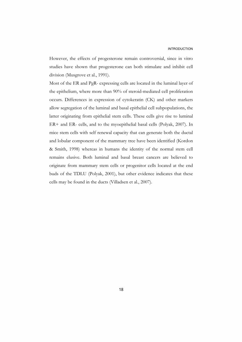
Acini, like ducts, are ring-shaped structures with a layer of epithelial cells
lining the lumen. In the adult lactating gland, the acini enlarge and the
cytoplasm of the epithelial cells fills with milk-containing vacuoles. Each
lobule has a lactiferous duct that allows the passage of milk toward the
nipple, where it collects in a widening of the ducts called sinuses. The entire
ductal network is called a ductal tree and is composed of three cell lineages:
luminal or alveolar epithelial cells lining the lumen in the TDLU, that
produce milk, ductal epithelial cells, and a more external layer of contractile
myoepithelial cells in contact with a basal membrane, which facilitates milk
passage (Figure 2).
Besides the glandular structures, the breast also contains connective tissue
with blood and lymphatic vessels, adipose, and nervous tissue, which
provide nutrition and support. The mammary gland is a hormone
responsive organ, and its development requires estrogen and progesterone,
two ovarian hormones acting on their receptors: estrogen receptor and
progesterone receptor (PgR). In the normal gland both estradiol and
progesterone regulate cell growth in a paracrine fashion (Anderson et al.,
1998), by stimulating the local production of growth factors such as
transforming growth factor α (TGFα), epidermal growth factor (EGF),
insulin-like growth factor (IGF), amphiregulins, and heregulins (HRG).
Many of these growth factors share common signaling pathways such as
mitogen activated protein kinase (MAPK) and PI3K/AKT, whose
activation ultimately leads to cell growth by induction of cell cycle
regulators such as cyclin D1 and ER-activation. The action of estradiol is
manifested in ductal growth and dichotomous branching. On the other
hand, progesterone seems to be more important during pregnancy, when it
stimulates epithelial cell proliferation, branching, and lobular differentiation.
INTRODUCTION
18
However, the effects of progesterone remain controversial, since in vitro
studies have shown that progesterone can both stimulate and inhibit cell
division (Musgrove et al., 1991).
Most of the ER and PgR- expressing cells are located in the luminal layer of
the epithelium, where more than 90% of steroid-mediated cell proliferation
occurs. Differences in expression of cytokeratin (CK) and other markers
allow segregation of the luminal and basal epithelial cell subpopulations, the
latter originating from epithelial stem cells. These cells give rise to luminal
ER+ and ER- cells, and to the myoepithelial basal cells (Polyak, 2007). In
mice stem cells with self renewal capacity that can generate both the ductal
and lobular component of the mammary tree have been identified (Kordon
& Smith, 1998) whereas in humans the identity of the normal stem cell
remains elusive. Both luminal and basal breast cancers are believed to
originate from mammary stem cells or progenitor cells located at the end
buds of the TDLU (Polyak, 2001), but other evidence indicates that these
cells may be found in the ducts (Villadsen et al., 2007).
INTRODUCTION
19
Figure 2. Diagram of the TDLU. Arrows indicate the three cell lineages present
in the ductal network and the basal membrane. Modified from (Polyak, 2007)
.1 HORMONES AND RECEPTORS
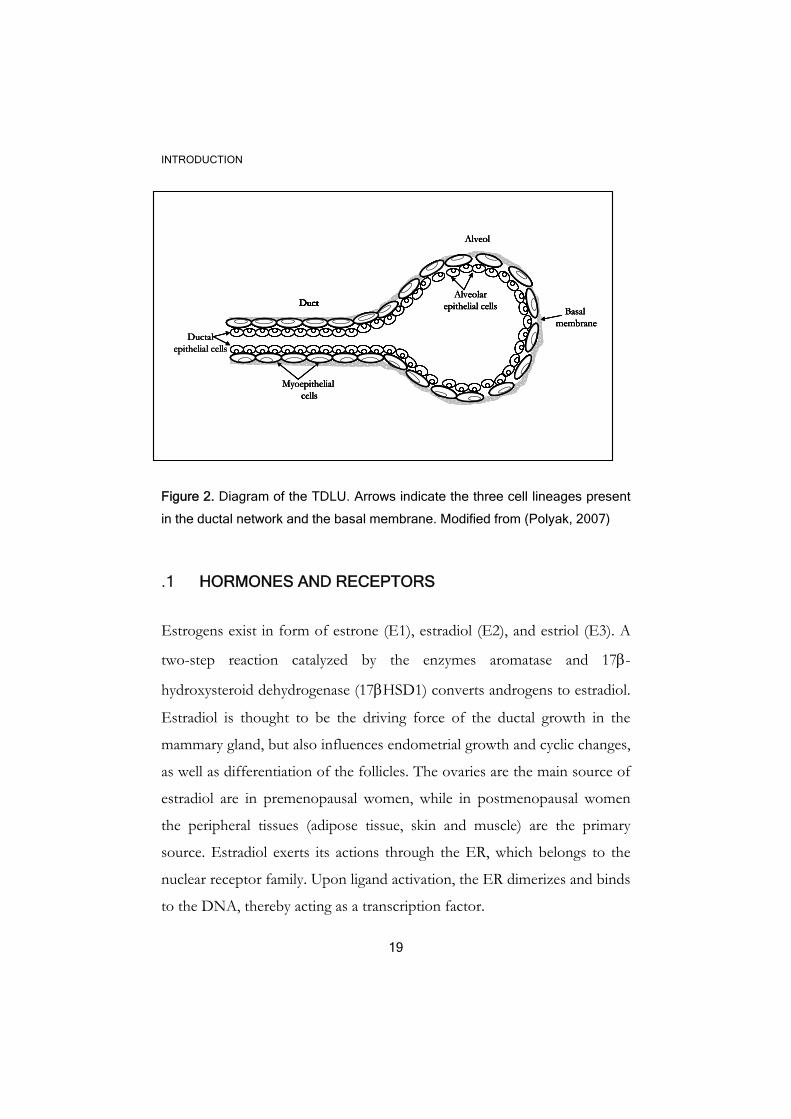
Estrogens exist in form of estrone (E1), estradiol (E2), and estriol (E3). A
two-step reaction catalyzed by the enzymes aromatase and 17β-
hydroxysteroid dehydrogenase (17βHSD1) converts androgens to estradiol.
Estradiol is thought to be the driving force of the ductal growth in the
mammary gland, but also influences endometrial growth and cyclic changes,
as well as differentiation of the follicles. The ovaries are the main source of
estradiol are in premenopausal women, while in postmenopausal women
the peripheral tissues (adipose tissue, skin and muscle) are the primary
source. Estradiol exerts its actions through the ER, which belongs to the
nuclear receptor family. Upon ligand activation, the ER dimerizes and binds
to the DNA, thereby acting as a transcription factor.
Ductalepithelial cells
Duct
Alveol
DuctAlveolar
epithelial cells
Myoepithelialcells
Basal membrane
Ductalepithelial cells
Duct
Alveol
DuctAlveolar
epithelial cells
Myoepithelialcells
Basal membrane
Ductalepithelial cells
Duct
Alveol
DuctAlveolar
epithelial cells
Myoepithelialcells
Basal membrane
Duct
Alveol
DuctAlveolar
epithelial cells
Myoepithelialcells
Basal membrane

INTRODUCTION
20
Figure 3. Different pathways leading to ER activation. ERE (estrogen
responsive element), TF (transcription factor), GF (growth factor), GFR (growth
factor receptor), pi (phosphorylation). The bent arrows indicate transcriptional
activation. Modified from (Heldring et al., 2007).
In the classical pathway, the ER is directly coupled to the sequence of
estrogen response elements (ERE)-containing genes. In the non-classical
pathways the ER can also interact with the DNA by means of other
transcription factors or be involved in non-genomic actions arising from
the cell membrane (Pappas et al., 1995). Moreover, the ER can be activated
by phosphorylation in a ligand-independent way (Figure 3)
ER+ ER ERE
ER ER
CLASSICAL PATHWAY
E2
NONCLASSICAL PATHWAYS
ER+ ER ERE
ER ER
E2 TF TF
DIR
EC
TIN
DIR
EC
TN
ON
GE
NO
MIC CELL MEMBRANE
CYTOPLASM
ERE2
ER
?
?
ER
ERSecond
messengers
GR
OW
TH
FA
CT
OR
M
ED
IAT
ED
GFR
GF
KINASE ER
pi
ERE
ER ER
pi pi
ER+ ER ERE
ER ER
CLASSICAL PATHWAY
E2
NONCLASSICAL PATHWAYS
ER+ ER ERE
ER ER
E2 TF TF
DIR
EC
TIN
DIR
EC
TN
ON
GE
NO
MIC CELL MEMBRANE
CYTOPLASM
ERE2
ER
?
?
ER
ERSecond
messengers
GR
OW
TH
FA
CT
OR
M
ED
IAT
ED
GFR
GF
KINASE ER
pi
ERE
ER ER
pi pi
INTRODUCTION
21
ER was renamed to ERα after the discovery of the ERβ, cloned in 1996
(Kuiper et al., 1996). Both hormone receptors bind to estradiol but are
expressed in different tissues, and seem to have opposite biological effects.
In mammals, both ERs are expressed in the luminal cells of the normal
breast, but the ERβ can be also found in myoepithelial cells and the
surrounding stroma (Speirs et al., 2002). Functionally, ERα regulates
normal and malignant cell growth in a paracrine or autocrine fashion,
respectively; while the ERβ has been considered a tumor suppressor due to
its ability to suppress the transcriptional effect of the ERα, and to its anti-
proliferative and pro-apoptotic effects during carcinogenesis (Saji et al.,
2005). The progesterone receptor is an estrogen-regulated gene used to
indicate the functionality of the ER, for example, patients with ER+/PgR+
tumors receive more benefit from tamoxifen when compared to the
ER+/PgR- group (Ravdin et al., 1992). PgR has been detected in some ER-
cases indicating a false negative result or poor assay sensitivity. The
predictive value of PgR in absence of ER is still under discussion
(EBCTCG, 1998).
.2 BREAST CANCER
.2.1 Epidemiology
According to data published in 2006 by the National Board of Health and
Welfare (http://www.socialstyrelsen.se/en/), breast cancer represents
29.4% of all female cancers and it is the most common malignancy among

INTRODUCTION
22
women in Sweden. Only in 2006, 7059 new cases were diagnosed, 60% of
them were ≥ 60 years old women and only 3.8% were < 40 years at the
time of diagnosis, indicating that breast cancer risk increases with age. More
than 82 000 women live with the disease (diagnosed between 1958 and
2006) and approximately 1500 die every year, with breast cancer as the
cause of death. Breast cancer in men has also been reported, though it is
infrequent. Only 36 men received this diagnosis during year 2006. Despite
the high incidence in Sweden among women (a 1.3%/year increase over a
20 year period), the mortality rate has decreased in western countries due to
improvements in mass screening, increased use of adjuvant systemic
treatment, and introduction of new drugs.
.2.2 Etiology
Breast cancer is a heterogeneous disease that develops under a long period
following several yet uncharacterized steps. Neither the identity of the first
malignant cell nor the decisive genetic alterations that lead to breast cancer
are known, which makes it difficult to reach a consensus about the etiology
of the disease. In some cases of hereditary cancer the family history plays a
decisive role for development of the disease, but mutations of the BRCA1,
BRCA2 (breast cancer 1 and 2 respectively), and TP53 genes only accounts
for the minority of breast cancers which indicates the existence of some
other factors. It is speculated that the likelihood of breast cancer
occurrence depends on the number of stem cells at risk, which is
determined from the time in the uterus or early in life. In adult life, the
interplay of growth factors, hormones, and their receptors stimulate
survival and proliferation of mutated cells, whereas pregnancy may cause

INTRODUCTION
23
breast cancer progression by either disruption of the normal cell
microenvironment during the phase of breast involution, or by promoting
expansion of the already initiated cells (Bissell, 2007). An etiologic model,
including both epidemiological and experimental data, brings some
understanding of the causative agents and their timing (Trichopoulos et al.,
2005). For example, high mammografic density, high mammary gland
mass, big size of the breast, adult height, and birth size are likely to reflect
the total number of mammary stem cells present and associated with higher
risk. Other factors, such as earlier menarche, late menopause,
postmenopausal-overweight, hormone replacement treatment, and alcohol
intake are related to the influence of hormonal and growth factors.
The risk of breast cancer increases otherwise with age and depends on the
lifestyle and environmental conditions.
.2.3 Heterogeneity
Breast cancer cells diverge, both genotypically and phenotypically,
depending on the molecular alteration that originated the tumor, the cell
type that originated the tumor, and the fact that mammary cells have
different susceptibility to malignant transformation. Breast cancer is
thought to originate after a multistep carcinogenesis. The multistep model
of breast cancer proposes a linear development of the disease from
hyperplasia to carcinoma in situ and then to invasive and metastatic
carcinoma (Figure 4). Progression occurs under the control of
hormones/hormone receptors, growth factor/growth factor receptors, and
oncogenes/tumor suppressor genes (Beckmann et al., 1997) which upon
INTRODUCTION
24
genetical alterations equip the epithelial cells with proliferative and survival
advantages.
Recently, the disease has been classified into five different entities with
specific gene expression profiles (Andre et al., 2007; Perou et al., 2000). The
groups, luminal A and B, basal cell-like, HER-2+ and normal-like, have
been matched to the known clinical variables (Calza et al., 2006). Luminal A
tumors express ER and seem to have a better prognosis compared to the
other subtypes. Basal cell-like tumors are negative for ER, PgR, and HER-
2, frequently present p53 mutations, are positive for the epidermal growth
factor-receptor (EGFR) and express CK 5 and 17. BRCA1 mutated cancers
typically represent this group. HER-2+ types overexpress HER-2 and are
clearly a distinct subgroup that receives advantage from some therapeutic
modalities. The characteristics of the luminal B and the normal-like types
are not well defined. The five types are already present at the ductal
carcinoma in situ stage (DCIS) (Yu et al., 2004) which may suggest different
tumor progression pathways for each of them (Polyak, 2007).

INTRODUCTION
25
Figure 4. Multi-step model of breast cancer initiation, progression and
metastasis. The process is influenced by genetic, epigenetic or environmental
alterations. Arrow-heads indicate that the identity of the target genes is still
unknown. Modified from (Polyak, 2001).
Moreover, molecular subtypes that differ in their degrees of proliferation
and differentiation have been reported, which support once more the idea
of breast cancer heterogeneity (Bertucci & Birnbaum, 2008). Differences at
the intratumoral levels, based on different primary target cells have been
explained by two theories, the clonal evolution and the stem cell hypothesis,
which agree on the monoclonal origin of breast cancer and disagree on the
identity of the primary target cell. According to the stem cell theory, breast
cancer originates in a small stem cell population that persists in the tumor
during its initiation, progression, and recurrence. These cells have the ability
of cell renewal and differentiation giving rise to all the cells in the tumor
and to tumor heterogeneity. On the other hand, the clonal evolution model
Normal HyperplasiaAtypical hyperplasia
In situcarcinoma
Invasivecarcinoma
Metastaticcarcinoma
Genetic-epigenetic-environmental changes?
? ? ? ?
1 Luminal A2 Luminal B3 Basal-like4 HER2+5 Normal-like
Normal HyperplasiaAtypical hyperplasia
In situcarcinoma
Invasivecarcinoma
Metastaticcarcinoma
Genetic-epigenetic-environmental changes?
? ? ? ?
1 Luminal A2 Luminal B3 Basal-like4 HER2+5 Normal-like
INTRODUCTION
26
supports the idea that breast cancer originates from a normal cell that
undergoes multiple mutations, which confer the most aggressive and
tumor-driving phenotype to malignant cells. However, newer experimental
data support a new model, in which tumor cells could originate from a
normal mammary cell or progenitor cell, and then self-renew or undergo a
combination of differentiation and clonal selection due to the natural
pressure of the environment and mutations. In this way the tumor would
be composed of a combination of differentiated and less proliferative cells
as well as self-renewing cells with proliferative advantages acquired from
the mutations (Campbell & Polyak, 2007).
.3 ONCOGENES AND TUMOR SUPPRESSOR GENES
.3.1 Oncogenes
Oncogenes were first identified in a virus as altered forms of cellular genes
(proto-oncogenes) able to transform normal cells by altering their
phenotype and conferring tumorigenic properties. Oncogenes can encode
growth factors, growth factor receptors, Ser/Thr protein kinases, nuclear
transcription factors, GTPases, and other factors related with growth and
differentiation. Therefore, these genes are tightly regulated, and when this
control fails cancer may arise. Proto-oncogenes can be activated by
different mechanisms such as mutations, chromosome rearrangements,
increased gene expression, and epigenetic mechanisms, which taken
together lead to increased protein expression or constitutive activation of
the gene product. A common mechanism in breast cancer is gene
amplification, which associates with increased copy number of a certain

INTRODUCTION
27
gene relatively to the rest of the genome. Examples of chromosome areas
affected in breast cancer by amplification are the chromosomal regions
8p12, 8q24, 11q13, 17q12, 17q23 and 20q13 (Letessier et al., 2006; Sinclair
et al., 2003).
.3.1.1 Amplification in the 11q13
The chromosome locus 11q13 is amplified in up to 15% of breast cancers
(Ormandy et al., 2003). This region harbors four distinct cores of
amplification. Some of the genes found in these cores are LRRC32 or
GARP (leucine rich repeat containing 32) and PAK1 (p21/Cdc42/Rac1-
activated kinase 1) in core 1 (Bostner et al., 2007), CCND1 (cyclin D1) in
core 3, and EMS1 or CTTN (cortactin) in core 4. High frequency of
amplification in some of these regions indicates that important oncogenes
may be contained within them. One of the most promising candidates is
cyclin D1. Cyclin D1 is a cell cycle regulator that binds cyclin dependent
kinases (CDK) 4/6 to drive G1-S progression. Cyclin D1 is frequently
amplified and overexpressed in breast cancer (Dickson et al., 1995), and
often associated with ER expression. In vitro studies have shown that
cyclin D1 promotes ER-activation (Zwijsen et al., 1997). Cyclin D1
overexpression in breast cancer is associated with growth factor
independency (Musgrove et al., 1994) and tumorigenesis in transgenic mice
(Wang et al., 1994) and its clinical value to predict both disease-free/overall
survival (Bieche et al., 2002) and response to therapy (Ahnstrom et al.,
2005; Jirstrom et al., 2005; Musgrove et al., 1994; Rudas et al., 2008; Wang
et al., 1994) has been reported.
INTRODUCTION
28
.3.1.2 Amplification in 17q12 and 17q23
Chromosomal region 17q12-21 is often amplified in breast cancer and the
major oncogene candidate in this area is the HER-2 gene (Yokota et al.,
1986). Gains in the 17q22-24 area were first reported in primary breast
cancers in 1994 (Kallioniemi et al., 1994) and thereafter in other studies
(Sinclair et al., 2003). Gains in the 17q23 area have also been reported in
other tumors, but the higher level of amplification predominates in breast
cancer. In vitro studies with the breast cancer cell line BT-474 detected two
peaks of amplification, the first containing the HER-2 gene and the other
that was distally located to the 17q22-24 region (Barlund et al., 1997).
Further analysis identified RPS6KB1, T box transcription factor-2 (TBX-2)
gene, and the human wild type p53-induced phosphatase 1 (PPM1D) as
possible oncogene candidates in the 17q22-24 area due to its amplification
and overexpression in MCF-7 cells (Couch et al., 1999; Sinclair et al., 2003).
The role of each gene in this amplicon is obscured due to co-amplifications
with or without protein overexpression and different cellular contexts.
.3.2 Tumor suppressor genes
Tumor suppressor genes (TSGs) are those genes that cause malignancy by
loss of its function. TSGs often hinder malignant transformation due to
negative regulatory effect on cell growth or by participating in DNA repair
and apoptosis. In a minority of breast cancers, these genes are affected by
germline mutations and inherited (present in all the cells of the body) but in
sporadic cases, which are the commonest manifestation of breast cancer,
the same genes can harbor sporadic somatic mutations (in some cells of the
INTRODUCTION
29
body). Opposite to oncogenes, TSG can be inactivated by allelic loss or loss
of heterozygosity (LOH) where the part of one chromosome containing the
TSG is lost while the other chromosome is unaffected. According to the
“two hits” hypothesis, proposed by Knudson (Knudson, 1971), TSGs
unmask the malignancy usually by alterations of the two alleles, which may
occur by inherited mutation of one allele followed by somatic mutation or
loss of the other. This hypothesis, proved to be true for retinoblastoma
disease resulting the susceptibility gene (RB1) in the first TSG to be
reported. In some cases there are other mechanisms involved in the
inactivation of the gene product such as promoter methylation (impairs
transcription), increased proteasomal degradation, increase in some other
proteins that interferes with its function or cell delocalization and
microRNAs. Some examples of TSG in breast cancer are: RB, p16, TP53,
BRCA1 and BRCA2, CHK2 (CHK checkpoint homolog 2), ATM (Ataxia
telangiectasia mutated) and PTEN (Phosphatase and tensin homolog
deleted on chromosome 10) that will be discussed below (Osborne et al.,
2004).
.4 PROGNOSTIC AND PREDICTIVE FACTORS
Breast cancer prognosis is generally good and many patients live longer
without relapses. However, for some patients the relapses appear already
within the first 5 years after the diagnosis but a recurrence may occur even
after 10 years or more. Therefore, it is important to divide the patients into
different risks groups to treat them efficiently. With help of prognostic
factors, it is possible to envisage the natural course of the disease while the
predictive factors provide information on the likelihood of the treatment
INTRODUCTION
30
response. At present, the most important prognostic factor in the clinic is
the TNM system (Table I), which allows tumor classification according to
the size of the tumor (T), the nodal infiltration (N) and the presence of
metastasis (M). Another useful prognostic indicator is the Nottingham
grade including the degree of nuclear atypia, the degree of tubular
formations, and mitotic activity. In Sweden, and other countries, this
grading system is used (Elston & Ellis, 1991). Other factors predicting
breast cancer survival and response to treatment are those related with cell
proliferation (thymidine labeling index, mitotic index, Ki-67, PCNA, and
bromodeoxyuridine labeling). Among them, the S phase fraction (SPF) is a
valuable prognostic factor (Stal et al., 1993).
Table I. TNM system.
STAGE CRITERIA
0 Carcinoma in situ
I Tumor ≤ 2cm, axilary lymph nodes not involved
II Tumor 2-5 cm and/or involved but mobile axilary lymph
nodes
III Tumor > 5 cm and /or fixed axilary lymph nodes; includes
inflammatory breast cancer
IV Distant metastases beyond ipsilateral axillary lymph nodes
More than 70% of breast cancers express ER which is used to predict
patient outcome and response to tamoxifen (Bezwoda et al., 1991; Clark &
McGuire, 1988; Clark et al., 1984; Heel et al., 1978; Osborne et al., 1980).
However, 30% of the ER+ tumors are non-responsive to the treatment (de
novo resistance) and many others become refractory (acquired resistance)
INTRODUCTION
31
in presence of the receptor, indicating that the mere presence of ER is not
the ideal predictive marker and other factors are needed (Payne et al., 2008).
In an effort to satisfy the individual needs of the patients, newer array
based-analysis have been developed. Examples of these are the 70 gene-
signature (van 't Veer et al., 2002) that predict short interval to metastasis
among the node negative patients, the 231 gene-signature associated with
survival (van de Vijver et al., 2002), and the 93 gene-signature (Sotiriou et
al., 2003). Many of the genes involved in these signatures are related to cell
cycle regulation, invasion, metastasis, angiogenesis, DNA-replication or
chromosomal stability. However in order to design the most optimal
experiment to be able to choose the appropriate prognostic or predictive
marker, among thousands of factors, it is vital to know the genes, proteins
and pathways that lie behind the resistance.
.5 TREATMENT
The most common treatments in breast cancer are surgery, radiotherapy,
chemotherapy, endocrine treatment and antibodies. Surgery can conserve
part of the breast (breast-conserving) or remove the whole gland
(mastectomy). Radical mastectomy is preferred in case of large tumors,
several tumors, and inflammatory or difuse cancer among other requisites.
In order to control tumor spreading a biopsy is taken from the first nodes
that receive lymph from the tumor (sentinel nodes). This technique often
replaces the axillary dissection in patients without evident lymphonode
infiltration. Surgery is often the initial treatment followed by other auxiliary
or adjuvant treatment. Radiotherapy, for example, is often recommended
after breast-conserving surgery or to patients with lymphonodal infiltration.
The main purpose of this treatment is to reduce the risk for local

INTRODUCTION
32
recurrences. Patients in this thesis received 46 Gy with 2 Gy/fraction 5
days a week for a total of 4.5 weeks. The standard treatment in South-East
Sweden is 50 Gy in 25 fractions. Another adjuvant treatment is
chemotherapy also called CMF in this thesis due to the three components
comprised in this regime (cyclophosphamide or chlorambucil, methotrexate
and fluorouracil). CMF are cytostatic substanses mostly affecting the
proliferative fraction of tumor cells. The risk for breast cancer-death is
reduced when CMF followed by 5 years tamoxifen (see below) is applied
directly after surgery compared to surgery alone (Bergh J et al., 2007).
Tamoxifen, a selective estrogen receptor modulator (SERM), is the most
common therapy used in the ER+ breast cancers. Tamoxifen (ICI 46,474)
(Harper & Walpole, 1967), that had been a failure as a contraceptive agent,
was first used in 1971 to treat breast cancer (Clarke et al., 2001; Jordan,
2003). This compound binds to the ligand-binding domain (LBD) of the
ER antagonizing the actions of estradiol and the receptor association with
co-activators. In addition to these effects, tamoxifen has also agonist
properties in other tissues such as heart and bone and is associated with
increased risk of endometrial cancer (Riggins et al., 2007). Another class of
compounds in use are the aromatase inhibitors (AI) which target the
enzyme that converts androgens to estrogens. Both substances are used in
postmenopausal women where the principal sources of the hormone are
the peripheral tissues. The treatment of choice for premenopausal women,
besides tamoxifen, are the gonadotropin releasing-hormone (GnRH)
analogs like goserelin (Zoladex). Production of estrogen by the ovaries is
stimulated by luteinising hormone (LH) and follicle stimulating hormone
(FSH), produced by the pituitary gland. Goserelin stops the production of
LH from the pituitary gland, which leads to a reduction of oestrogen. Thus,
tamoxifen, AIs and GnRH analogs act through different mechanisms to
INTRODUCTION
33
deprive the cells of estrogens stimulatory actions. Finally, those tumors that
overexpress HER-2 are treated with trastuzumab, a monoclonal antibody,
often given in combination with cytostatics.
.6 TAMOXIFEN AND TAMOXIFEN RESISTANCE
Antiestrogen resistance may be explained by several mechanisms, including
loss or mutation of ER, increased estradiol level, alterations in antiestrogen
metabolism or interactions between growth factor receptors and ER
cascades (Clarke et al., 2001; Riggins et al., 2007). These mechanisms,
mainly involved in cell proliferation (Doisneau-Sixou et al., 2003), may
coexist with those affecting cell death. Increasing amounts of evidence
indicates that the mechanisms whereby drugs such as the GnRH analogues,
AI and tamoxifen exert the cytotoxic action also include apoptosis (Imai &
Tamaya, 2000; Perry et al., 1995; Riggins et al., 2005). Therefore, factors
involved in the apoptotic failure may also contribute to the antiestrogen
resistance. Among the effects of tamoxifen are reduction in expression of
c-myc and cyclin D1, accumulation of hypophosphorylated RB protein,
nuclear induction of the cell cycle inhibitors p21WAF1/CIP1 and p27Kip1,
inhibition of Bcl-2 and induction of Bax expression. The question is how
cancer cells circumvent these effects to survive and proliferate.
One answer could be the crosstalk between the signaling pathways
emerging from the ER and other growth factor receptors (Figure 5). For
example, EGFR, HER-2 and IGF-1R are often elevated in unresponsive
tumors (Johnston et al., 2003; Nicholson et al., 1999). Several other studies
have suggested that overexpression of HER-2 in ER+ cell lines confers
resistance to the endocrine treatment, being the PI3K/AKT pathway often
INTRODUCTION
34
in the same picture (to be discussed below) (Kurokawa & Arteaga, 2003;
Nelson & Fry, 2001; Zhou et al., 2001).
For some years ago, the PI3K/AKT cascade, which is the major survival
pathway for many cell types, was shown to activate the ER protecting the
cells from tamoxifen-induced apoptosis (Campbell et al., 2001). Since then
the amount of experimental evidence has increased. Overexpression of
activated AKT in breast cancer cells induces estrogen independence and
resistance to the endocrine treatment while its inhibition causes the
opposite effect. Cell lines selected against tamoxifen relay on AKT
activation to conserve this phenotype (Frogne et al., 2005). Moreover, the
mammalian target of rapamycin (mTOR), is activated by AKT in
tamoxifen-resistant cells, that upon rapamycin treatment recover response
to tamoxifen. AKT can also sequester p21WAF1 and p27KIP1 (Zhou et
al., 2001) in the cytoplasm where these proteins are unable to mediate the
cytostatic effects of tamoxifen. On the other side, AKT can also induce the
transcriptional activity of ERβ (Duong et al., 2006) indicating that the
effects of this signaling pathway may hide some surprises.

INTRODUCTION
35
Figure 5. Crosstalk between ER and growth factor receptor pathways that
activate AKT and ultimately lead to cell proliferation and survival. Modified from
(Riggins et al., 2007).
.7 THE PI3K/AKT PATHWAY IN CANCER
.7.1 HER-2
HER-2/c-erbB2 belongs to a family of tyrosine-kinase receptors (TKR)
together with EGFR (HER-1), HER-3 and HER-4. HER-2 contributes to
malignant growth by activating and recruiting signaling cascades involved in
cell proliferation and survival, like for example MAPK and PI3K/AKT
AKT
RTK (EGFR,HER-2,IGF-IR)
Pi PI3K
Bcl-2
Bad/Bcl-xl
mTOR
ERpi
C-MycCyclin D1Cyclin E
ERpi
Pak1
Cytoplasmicp21p27
Ras
ERK1/2
PROLIFERATION SURVIVAL
ENDOCRINE THERAPY RESISTANCE
NU
CLE
US
CYTO
PLA
SM
MITOCHONDRIA
AKT
RTK (EGFR,HER-2,IGF-IR)
Pi PI3K
Bcl-2
Bad/Bcl-xl
mTOR
ERpi
C-MycCyclin D1Cyclin E
ERpi
Pak1
Cytoplasmicp21p27
Ras
ERK1/2
PROLIFERATION SURVIVAL
ENDOCRINE THERAPY RESISTANCE
NU
CLE
US
CYTO
PLA
SM
MITOCHONDRIA

INTRODUCTION
36
pathways (Grant et al., 2002). The HER-2 gene has been found
amplified/overexpressed in 10-30% of breast tumors (Lofts & Gullick,
1992; Singleton & Strickler, 1992; Slamon et al., 1987). This is often
associated with more aggressive tumors and poor treatment response
(Carlomagno et al., 1996; Slamon et al., 1989; Stal et al., 1995). In
ER+/HER-2+ cancers the response rate to tamoxifen is reduced in
comparison with ER+ tumors with normal HER-2 expression (Nicholson
et al., 1990). However, HER-2 is more accepted as a predictor of
trastuzumab (Herceptin®) treatment while its predictive value for
endocrine treatment is still under discussion (Arpino et al., 2004; Elledge et
al., 1998). Although HER-2 does not directly belong to the PI3K/AKT
pathway, it is frequently involved in PI3K activation and in breast cancer.
.7.2 PI3K
Phosphatidylinositol 3 kinase is a dual kinase that phosphorylates
phosphoinositides and serine/threonine residues on proteins. The main
substrates are phosphatidylinositol 4P and phosphatidylinositol 4,5P2
(PIP2) that become phosphatidylinositol 3,4P2 or phosphatidylinositol
3,4,5P3 (PIP3) after phosphorylation at the 3’ position of the inositol ring
(Whitman et al., 1988) and the p85 regulatory subunit (Dhand et al., 1994).
The kinase is a heterodimer with a regulatory and a catalytic subunit
composed of five structural domains. In its basal state the p110 subunit is
bound to and inhibited by the p85 regulatory subunit, whose structure
consists mainly of bindings sites for adaptor proteins, PI3K catalytic
subunits or TKR. PI3K activation occurs at the cell membrane when the
p110 catalytic subunit is in close proximity to its lipid substrates and the

INTRODUCTION
37
p85-inhibitory effect is released. At the cell membrane, the p85 regulatory
subunit can interact directly with phosphotyrosine residues present in
activated growth factor receptors or indirectly with the insulin receptor
substrates 1 and 2 (IRS-1 and IRS-2). The catalytic p110 subunit can also
interact with Ras through its Ras-binding domain (RBD).
The PI3K family is organized in several classes and subclasses based on
differences in tissue distribution, structure, substrate affinity, activation and
function. The class IA comprises the catalytic subunits p110α, β and δ that
can heterodimerize with one of the regulatory subunits p85α/p55α/ p50α
or p85β/p55β or p55γ. In this thesis we will concentrate on class IA
because this class seems to be more important in carcinogenesis (Denley et
al., 2008; Vivanco & Sawyers, 2002).
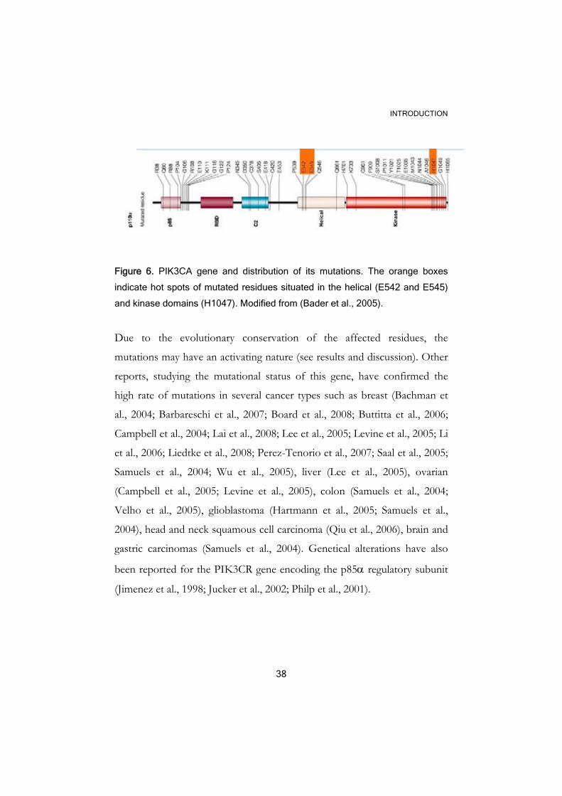
The PIK3CA gene, situated on chromosome 3q26.3, encodes the p110α
catalytic subunit. The protein (110 kD) is composed of five structural
domains: a p85 binding domain situated at the N terminal end, a Ras
binding domain, a domain called C2 (protein kinase C homology domain 2)
proposed to bind cellular membranes, a helical domain of unknown
function and the catalytical domain to the C terminal end (Huang et al.,
2007). The gene consists of 20 exons and was originally found amplified in
cancer (Hui et al., 2001; Ma et al., 2000; Shayesteh et al., 1999). But in 2004,
Samuel and collaborators revealed high frequency of mutations in this gene
(Samuels et al., 2004). The mutations clustered in >85% of the cases to the
exons 9 (helical domain) and 20 (catalytical domain) thereby defined as “hot
spots”. The most affected codons were 542, 545 (exon 9) and 1047 (exon
20) (Figure 6).
INTRODUCTION
38
Figure 6. PIK3CA gene and distribution of its mutations. The orange boxes
indicate hot spots of mutated residues situated in the helical (E542 and E545)
and kinase domains (H1047). Modified from (Bader et al., 2005).
Due to the evolutionary conservation of the affected residues, the
mutations may have an activating nature (see results and discussion). Other
reports, studying the mutational status of this gene, have confirmed the
high rate of mutations in several cancer types such as breast (Bachman et
al., 2004; Barbareschi et al., 2007; Board et al., 2008; Buttitta et al., 2006;
Campbell et al., 2004; Lai et al., 2008; Lee et al., 2005; Levine et al., 2005; Li
et al., 2006; Liedtke et al., 2008; Perez-Tenorio et al., 2007; Saal et al., 2005;
Samuels et al., 2004; Wu et al., 2005), liver (Lee et al., 2005), ovarian
(Campbell et al., 2005; Levine et al., 2005), colon (Samuels et al., 2004;
Velho et al., 2005), glioblastoma (Hartmann et al., 2005; Samuels et al.,
2004), head and neck squamous cell carcinoma (Qiu et al., 2006), brain and
gastric carcinomas (Samuels et al., 2004). Genetical alterations have also
been reported for the PIK3CR gene encoding the p85α regulatory subunit
(Jimenez et al., 1998; Jucker et al., 2002; Philp et al., 2001).
INTRODUCTION
39
.7.3 PTEN
PTEN, situated on chromosome 10q23.3, was first reported as a protein
tyrosine phosphatase and as a tumor suppressor gene mutated in several
cancers (Li et al., 1997; Steck et al., 1997) and germline mutations of this
gene are associated with hereditary cancer syndromes like Bannayan-
Zonana and Cowden’s disease. PTEN has indeed double phosphatase
activity on lipids and proteins. The main lipid substrates are the products of
PI3K (Maehama et al., 2001) while the protein phosphatase activity is
associated with inactivation of focal adhesion kinase (FAK), Src homology
2 domain containing-protein (Shc), platelet derived growth factor receptor
(PDGFR) and PTEN itself (Suzuki et al., 2008). PTEN regulation can
occur at transcriptional and post-translational levels, by interaction with
other proteins or by relocation to different cell compartments. At
transcriptional level, PTEN is positively regulated by EGFR, p53, resistin,
peroxisome proliferator activated receptor γ (PPARγ), human sprouty
homolog 2 (SPRY2) and phytoestrogens. It is negatively regulated by
mitogen activated protein kinase kinase-4 (MKKK4), transforming growth
factor β (TGFβ) and recently reported, by the proto-oncogenic
transcription factor JUN (Suzuki et al., 2008). Post-translational
mechanisms include phosphorylation, acetylation or oxidation, all of them
leading to PTEN inactivation. Moreover, PTEN interactions with other
proteins either stabilize PTEN (Wu et al., 2000), target it for degradation
(Tang & Eng, 2006) or decide PTEN location in the cell. PTEN can be
recruited to the cell membrane to access its substrates or shuttle between
the cytoplasm and the nucleus. The role of PTEN in the nucleus was
deduced from the presence of PIP3 in this cell compartment (Caramelli et
INTRODUCTION
40
al., 1996). Nuclear PTEN seems to be engaged in down regulation of cyclin
D1 and phosphoMAPK, which is crucial for cell cycle arrest, whereas
cytoplasmic PTEN is required to decrease phospho AKT (pAKT) levels,
up regulate the cell cycle inhibitor p27Kip1 and induce apoptosis (Chung &
Eng, 2005; Chung et al., 2006). Lost nuclear PTEN has been associated
with tumor formation (Perren et al., 1999). Other PI3K-independent
functions of PTEN have been found: p53 acetylation in response to DNA
damage (Li et al., 2006) and restriction of cell migration. PTEN alterations
in cancer manifest in form of loss of heterozygosity (LOH), protein loss,
mutations and epigenetic alterations (Ali et al., 1999; Aveyard et al., 1999;
Dahia, 2000; Dreher et al., 2004; Forgacs et al., 1998; Li et al., 1997).
Hence, low frequency of mutations has been reported in breast cancers.
With the introduction of a new technique high frequency of gross PTEN
mutations among BRCA1 mutated cancers (Saal et al., 2008), can be found.
.7.4 AKT
AKT (v-AKT murine thymoma viral oncogene homolog) also known as
protein kinase B (PKB) is the human homolog of the v-AKT oncogene
(Bellacosa et al., 1991; Burgering & Coffer, 1995; Jones et al., 1991; Staal,
1987). There are three AKT isoforms encoded by three different genes:
AKT1, AKT2 and AKT3. The structure of all three isoforms is conserved
through evolution and consists of an amino terminal pleckstrin homology
(PH) domain, a kinase domain and a carboxy terminal regulatory domain
with certain similarity to this found in AGC kinases (cyclic AMP
dependent-protein kinase, cyclic GMP-dependent protein kinase and
protein kinase C). All AKT isoforms are distributed ubiquitously in human

INTRODUCTION
41
tissues and their functions have been deduced in part from knockout
studies. For example, AKT1 and AKT3 knockout mice exhibit decreased
body size and impaired brain development respectively while AKT2 null
mice develop type II diabetes.
All the AKT isoforms are found altered in cancer either by amplification
like in the case of AKT1 (Staal, 1987) and AKT2 (Nakayama et al., 2006;
Ruggeri et al., 1998), protein overexpression (AKT1, AKT2, AKT3) or
activation (AKT1, AKT2) (Ermoian et al., 2002; Gupta et al., 2002;
Horiguchi et al., 2003; Hsu et al., 2001; Kanamori et al., 2001; Kreisberg et
al., 2004; Kurose et al., 2001; Malik et al., 2002; Min et al., 2004; Nakayama
et al., 2001; Nam et al., 2003; Roy et al., 2002; Schlieman et al., 2003; Sun et
al., 2001; Terakawa et al., 2003; Tokunaga et al., 2006; Yuan et al., 2000).
AKT1 has been also found mutated in breast, colorectal and ovarian
cancers (Brugge et al., 2007; Carpten et al., 2007). These alterations have
prognostic significance in cancer (Dai et al., 2005; Ermoian et al., 2002;
Kreisberg et al., 2004; Min et al., 2004; Nakanishi et al., 2005; Nam et al.,
2003; Schlieman et al., 2003; Terakawa et al., 2003; Tsurutani et al., 2006)
indicating the potential of AKT as a therapeutic target.
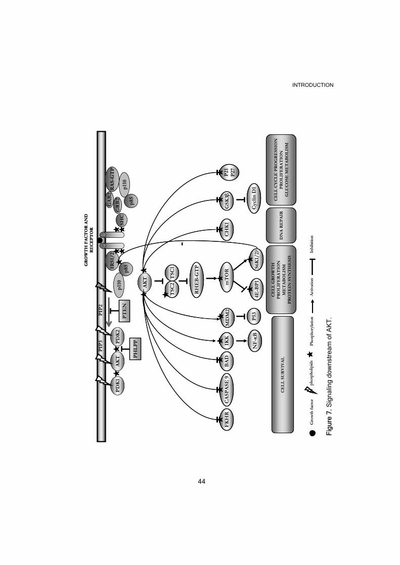
.7.4.1 AKT activation and signaling downstream
Ligand-mediated activation of a plethora of TKR and other receptors leads
to AKT activation (Hanada et al., 2004). AKT was early reported as a PI3K
target (Burgering & Coffer, 1995; Franke et al., 1995) since the PH domain
of AKT interacts with the PI3K substrates to be recruited to the cell
membrane where it can be activated by phosphorylation (Figure 7). The
phosphorylation sites crucial for the full activation of AKT are T308, in the

INTRODUCTION
42
activation loop, and S473 in the hydrophobic motif (Alessi et al., 1996). The
kinase responsible for T308 phosphorylation is protein dependent kinase 1
(PDK1) (Alessi et al., 1996) whereas the identity of a PDK2, responsible
for S473 phosphorylation, is not so well defined. Among the PDK2
candidates are integrin-linked kinase (ILK) (Persad et al., 2001), mTOR
complex 2 (mTORC2) (Sarbassov et al., 2005) and AKT itself (Toker &
Newton, 2000). After activation AKT can be transferred to the cytoplasm
or the nucleus where it can phosphorylate its targets. AKT can be
negatively regulated by PTEN (Stambolic et al., 1998) and the PH domain
and the leucine-rich repeat protein phosphatase (PHLPP) (Gao et al., 2005).
AKT activation triggers many biological processes that may be relevant to
cancer. For instance, cell survival, through phosphorylation and inactivation
of many pro-apoptotic factors such as the BCL2-antagonist of cell death
(BAD), which sequesters the apoptotic factors BCL-Xl and BCL-2 in a
non-functional complex, caspase-9, and a forkhead member of
transcription factors (FKHR), involved in transcription of several pro-
apoptotic genes. Indirectly, AKT can also exert a positive effect on the pro-
survival nuclear factor κB (NF-κB), by activating the I-κB kinase (IKK)
that causes degradation of the NF-κB inhibitor (I-κB). Moreover, AKT can
phosphorylate the murine double minute 2 (Mdm2), a negative regulator of
the pro-apoptotic tumor suppressor p53, leading to Mdm2 nuclear
translocation and better access to p53. Another effect of AKT activation is
to increase cell proliferation by inhibiting glycogen synthase kinase 3
(GSK3)-induced cyclin D1 degradation. AKT has also been shown to
delocalize p21 and p27 to the cytoplasm inhibiting their function as cell
cycle inhibitors. In addition to its role in survival and proliferation, AKT
activation is also associated with genetic instability through the DNA
INTRODUCTION
43
damage checkpoint gene 1 (CHK1) inhibition and increased cell growth, a
process mainly controlled by mTOR. AKT activates mTOR indirectly by
phosphorylating and inducing degradation of the Tuberous Sclerosis
Complex proteins 1/2 (TSC1/2). Normally, the tumor suppressor TSC1/2
is able to drive the GTPase Ras homolog enriched in brain (Rheb) into a
GDP-bound inactive state that is not able to phosphorylate and activate
mTORC1. Because of mTOR activation, two main substrates are
phosphorylated: the 40S ribosomal protein S6 kinase (p70S6 kinase 1 or
S6K1) (discussed below) and the eukaryotic translation initiation factor 4E
binding protein 1 (4EBP-1) initiating transcription of genes involved in cell
proliferation, participating in ribosome biogenesis or regulating cellular
metabolism.
INTRODUCTION
44
Fig
ure
7. S
igna
ling
dow
nstr
eam
of A
KT
.
GR
OW
TH
FA
CT
OR
AN
DR
EC
EP
TO
R
AK
TP
DK
1P
DK
2IR
S1/
2
p85
p11
0
PIP
2P
IP3
RA
S-G
TP
p85
p11
0P
TE
NSH
CG
RB
2
GA
B2
PH
LP
P
GSK
3βB
AD
CA
SPA
SE 9
FK
HR
IKK
NF
-κB
P53
MD
M2
Cyc
lin D
1
P21
P27
CH
K1
AK
T
mT
OR
TSC
2T
SC1
RH
EB
-GT
P S6K
1/2?
4E-B
P1
-
CE
LL
SU
RV
IVA
L
CE
LL
GR
OW
TH
PR
OL
IFE
RA
TIO
NM
ET
AB
OL
ISM
PR
OT
EIN
SY
NT
HE
SIS
DN
A R
EP
AIR
CE
LL
CY
CL
E P
RO
GR
ESS
ION
PR
OL
IFE
RA
TIO
NG
LU
CO
SE M
ET
AB
OL
ISM
phos
phol
ipid
sA
ctiv
atio
nIn
hibi
tion
Phos
phor
ylat
ion
Gro
wth
fact
or
GR
OW
TH
FA
CT
OR
AN
DR
EC
EP
TO
R
AK
TP
DK
1P
DK
2IR
S1/
2
p85
p11
0
PIP
2P
IP3
RA
S-G
TP
p85
p11
0P
TE
NSH
CG
RB
2
GA
B2
PH
LP
P
GSK
3βB
AD
CA
SPA
SE 9
FK
HR
IKK
NF
-κB
P53
MD
M2
Cyc
lin D
1
P21
P27
CH
K1
AK
T
mT
OR
TSC
2T
SC1
RH
EB
-GT
P S6K
1/2?
4E-B
P1
-
CE
LL
SU
RV
IVA
L
CE
LL
GR
OW
TH
PR
OL
IFE
RA
TIO
NM
ET
AB
OL
ISM
PR
OT
EIN
SY
NT
HE
SIS
DN
A R
EP
AIR
CE
LL
CY
CL
E P
RO
GR
ESS
ION
PR
OL
IFE
RA
TIO
NG
LU
CO
SE M
ET
AB
OL
ISM
phos
phol
ipid
sA
ctiv
atio
nIn
hibi
tion
Phos
phor
ylat
ion
Gro
wth
fact
or
INTRODUCTION
45
.7.4.1.1 p70S6K1 and p70S6K2
Besides S6K1, there is another kinase S6K2, encoded by a different gene.
S6K1 and S6K2 are serine/threonine protein kinases that belong to the
family of AGC protein kinases. S6K1 and 2 are able to phosphorylate the
40S ribosomal protein S6 thereby enhancing protein biosynthesis
(Jastrzebski et al., 2007), cell growth and cell cycle progression. S6K1 is
believed to regulate G1-S transition while S6K2 seems to be more
important during G2/M phase (Boyer et al., 2008). Besides these actions,
S6K1 and perhaps S6K2 participate in a negative feedback loop where
overactivation of the proteins leads to AKT inhibition (Figure 7).
Alternative splicing of the S6K1 (RPS6KB1) and S6K2 (RPS6KB2) genes
give rise to the p70 (α II) and p85 (α I) isoforms of S6K1 or to the p54 (β
II) and p60 (β I) isoforms of S6K2. Both kinases can be found in the
nucleus and the cytoplasm and recently S6K2 has been located to the
centrosome (Rossi et al., 2007). S6K1 and 2 share 70% homology with
>83% in the catalytic domain alone (Gout et al., 1998; Koh et al., 1999)
which may suggest redundant biological functions. However knock out
models indicated that S6K1 and S6K2 control body size and metabolism
through different mechanisms (Jastrzebski et al., 2007; Pende et al., 2004).
Moreover, a closer structural inspection reveals that S6K2 contains a C-
terminal proline-rich region, absent in S6K1, that may be involved in SH3
protein-protein interactions (Lee-Fruman et al., 1999).
S6K1 activity increases due to sequential phosphorylation upon nutrient or
growth factor stimulation. S6K1 can be activated in a PI3K/AKT
dependent manner by mTORC1 or independently of PI3K and even
mTOR (Jastrzebski et al., 2007). S6K2 and S6K1 activation shares many

INTRODUCTION
46
features but additionally, PKC and MEK signaling pathways seems to play a
more important role in S6K2 activation compared to S6K1 (Jastrzebski et
al.,2007).
The genes encoding these kinases are situated relatively close to well known
amplicons (17q12-q21 and 11q13) containing the HER-2 and the CCND1
oncogenes respectively. The RPS6KB1 gene has been found amplified in
about 9 % of breast cancers (Barlund et al., 2000).
AIMS OF THE STUDY
47
AIMS OF THE STUDY
Paper I
1- To determine the frequency of AKT-1 expression and activation in
breast cancer.
2- To study the associations of AKT-1 expression and activation with
other variables and patient survival after endocrine treatment.
Paper II
1- To study the in vitro effects of heregulin ß1 upon AKT activation, p21
cellular location and response to tamoxifen.
2- To analyze the expression and localization of p21 in breast tumors, its
association with other clinico-pathological variables and AKT activation, as
well as its clinical relevance.
Paper III
To determine the frequency of PIK3CA mutations and PTEN loss. To
explore whether PIK3CA mutations and PTEN loss are mutually exclusive
mechanisms, correlate with other known clinico-pathological markers or
have clinical implication in breast cancer.
Paper IV
To study the frequency of S6K 1/2 amplification in breast cancer. Looking
for coamplifications with the HER-2 and CCND1 genes, associations with
other clinical variables and members of the PI3K pathway. To determine
the clinical value of S6K 1/2 amplification.

ETHICAL CONSIDERATIONS
48
ETHICAL CONSIDERATIONS
There are a plethora of guidelines and regulations that cover the potential
conflict between research interests, patient integrity, autonomy, and the
preservation of public trust in biomedical research (Helgesson et al., 2007).
Some of the ethical issues that we can identify in our particular studies
include the collection of patient samples without explicit consent, the
patient’s right to receive feedback of the results of the study, the secrecy in
the management of personal data as well as the repetitive use of biological
material (tumor tissue or cell lines) from deceased. The patient materials
included in these studies are sample collections from the Biobanks at the
Karolinska and Linköping University hospitals. Since the patient identity
has been protected by a code, it is unavailable for public knowledge and
approval from the local ethics committees has been obtained before
carrying out the corresponding investigations.
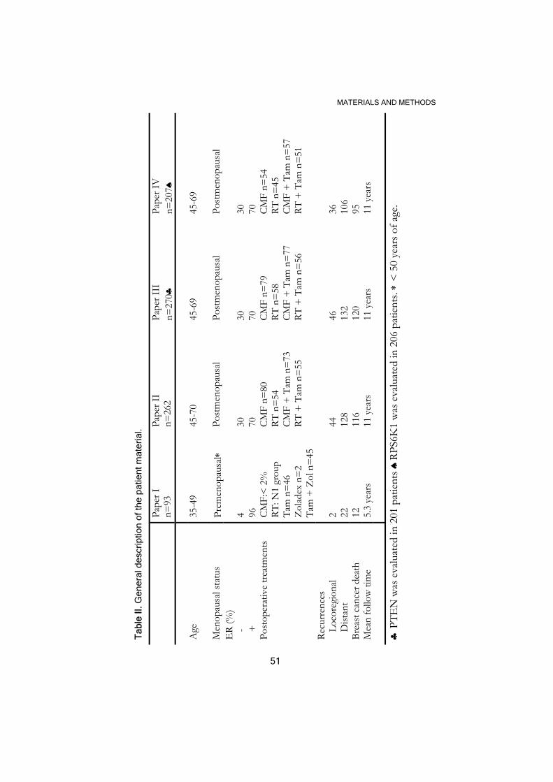
MATERIALS AND METHODS
49
MATERIALS AND METHODS
.1 Patient material
In these papers we used frozen sections or DNA from tumors still available
after hormone receptor assays. The material was labeled and stored at
-70 °C until used.
The 93 premenopausal women included in paper I participated in two
different trials (Baum et al., 2006; Ryden et al., 2005). All the patients
included in the original trials had invasive breast cancer stage II (pT2 N0
M0, pT1 N1 M0 and pT2 N1 M0) and underwent radical surgery in the
form of a modified radical mastectomy or breast-conserving surgery with
axillary lymph node dissection. The patients with lymphonode infiltration
(N1) or breast-conserving surgery received radiotherapy. All the patients
were also treated postoperatively with tamoxifen, goserelin (introduced
after 1990) or both endocrine modalities (see Table II for details).
The postmenopausal patients included in papers II-IV (Table II) had
unilateral, operable breast cancer and were required to have either
histologically verified lymph node metastases or a tumor diameter,
measured on the surgical specimen, exceeding 30 mm. They were
randomized to four treatment groups: adjuvant chemotherapy, adjuvant
chemotherapy plus tamoxifen, radiotherapy, and radiotherapy plus
tamoxifen. Chemotherapy treatment consisted of 12 courses of CMF.
Surgery consisted of modified radical mastectomy. Only those tumors
judged to have more than 50% of malignant cells in the tumor sections
were included in paper IV and the subsets of patients included in the

MATERIALS AND METHODS
50
different studies showed not bias in comparison with all the 679
postmenopausal patients in the whole trial in terms of tumor characteristics
and treatment.
MATERIALS AND METHODS
51
Tab
le II
. Gen
eral
des
crip
tion
of th
e pa
tient
mat
eria
l.
Pa
per I
n=
93
Pape
r II
n=26
2 Pa
per I
II
n=27
0♣
Pape
r IV
n=
207♠
Age
35
-49
45
-70
45-6
9 45
-69
M
enop
ausa
l sta
tus
Prem
enop
ausa
l∗
Post
men
opau
sal
Post
men
opau
sal
Post
men
opau
sal
ER
(%)
-
+
4 96
30
70
30
70
30
70
Post
oper
ativ
e tre
atm
ents
CM
F:<
2%
RT: N
1 gr
oup
CMF
n=80
RT
n=
54
CMF
n=79
RT
n=
58
CMF
n=54
RT
n=
45
Ta
m n
=46
Z
olad
ex n
=2
Tam
+ Z
ol n
=45
CMF
+ T
am n
=73
RT
+ T
am n
=55
CM
F +
Tam
n=
77
RT +
Tam
n=
56
CMF
+ T
am n
=57
RT
+ T
am n
=51
Recu
rren
ces
Lo
core
gion
al
Dist
ant
2 22
44
128
46
132
36
106
Brea
st c
ance
r dea
th
12
116
120
95
Mea
n fo
llow
tim
e 5.
3 ye
ars
11 y
ears
11
yea
rs
11 y
ears
♣ P
TEN
was
eva
luat
ed in
201
pat
ients
♠RP
S6K
1 w
as e
valu
ated
in 2
06 p
atien
ts. ∗
< 5
0 ye
ars o
f age
.

MATERIALS AND METHODS
52
.2 Cell lines
Attempts to culture breast cancer cell lines started in 1937 but it was not
until 1958 that Lasfargues and Ozzello succeded for the first time with the
long-term culture of a breast cancer cell line, BT-20 (Lasfargues & Ozzello,
1958). Other well known breast cancer cells were isolated in the 1970s: SK-
Br3 (Trempe & Fogh, 1973), MDA-MB-231 (Cailleau et al., 1974), T-47D
(Keydar et al., 1979), ZR-75-1 (Engel et al., 1978), MDA-MB-468 (Cailleau
et al., 1978) and BT-483 (Engel & Young, 1978). Among the most famous
breast cancer cells are MCF-7 (Soule et al., 1973), where “M” stands for
Michigan, “C” for Cancer and “F” for Foundation. The number 7 refers to
the number of attempts that were required to perpetuate this cell line from
a patient. Another cell line mentioned in paper III is MCF-10A (Soule et
al., 1990), isolated in 1990 and derived from a patient with fibrocystic breast
disease.
METHODS
This section explains the fundaments of each technique providing a general
description of the methods included in this thesis. The details will be found
in the Materials and Methods section of each particular paper.

MATERIALS AND METHODS
53
.1 Flow cytometry
Flow cytometry is a powerful technique that allows analysis of multiple
parameters in complex cell populations in a very short time. A flow
cytometer is divided into different parts: the fluidic system that deliver the
samples into the machine, the optical system composed by a laser, which is
the light source, objects that direct the light and detectors that receive the
emitted light and finally, the electronic and peripheral computer systems
which are used to translate the impulses into digital data. The cell samples
or particles are delivered in a stream of sheath fluid in a way that only one
cell at a time reaches the laser beam. This facilitates the gathering of
information related to the size, complexity, cell phenotype, DNA content,
viability, etc. While the cell size and complexity are deduced from the
forward and side scattered light respectively, the other parameters can be
studied by labeling the cells with different fluorescent-conjugates as we will
describe below. This technique has a broad spectrum of applications from
immunophenotyping to cell sorting. In this thesis we have employed flow
cytometry to quantify the fraction of cells in S phase (papers I-IV), to
determine the expression of HER-2 (paper I-IV) and to measure apoptosis
(paper II).
.1.1 S phase fraction
To quantify the DNA content of single cells different methods have been
described. In our lab we have chosen a method that employs the NP-40
detergent in combination with the proteolytic enzyme trypsin to isolate the
nucleus (Vindelov et al., 1983). The advantages of this method are based on

MATERIALS AND METHODS
54
the simplicity of the technique, the high reproducibility and the versatility of
the material that can be used. The DNA in the cells is visualized by staining
with different fluorescent dyes like propidium iodide. Due to the direct
relation between the number of incorporated dye molecules and the DNA
content (stoichiometry) it is possible to display the DNA content in a
frequency distribution graph (histogram) of fluorescent intensity v.s
number of cells, where the fluorescent intensity is proportional to the
amount of incorporated dye. Since the DNA content is related to the phase
of the cell cycle, it is possible to deduce the proliferative status of the cells
from the DNA histogram. We have employed the ModiFit software to
quantify the S-phase fraction.
.1.2 HER-2 content
Determination of HER-2 by flow cytometry combines the high sensitivity
of immunofluorescence with the high speed of the flow cytometer. After a
rather quick protocol, a primary anti-HER-2 antibody or an unspecific
immunoglobulin (negative control) followed by a secondary antibody
coupled to a fluorescent dye could indirectly detect the antigen. The HER-2
positive tumors could be determined by a ratio between the fluorescence
associated with the specific antibody and the negative control. At the same
time, the histogram for PI provided information about the cell cycle
distribution of the cells. The clear advantages of this technique, including
speed, multiparametric analysis, and quantitative measurements are
somehow overshadowed by the fact that the visual inspection of the cells is
missing.
MATERIALS AND METHODS
55
.1.3 Apoptosis
Apoptosis and necrosis are two forms of cell death that differentiate in
many aspects and can be detected by several techniques. Apoptotic cells
can shrink, altering the form and composition of the cell membrane, but
keep membrane integrity whereas necrotic cells explode with leakage of the
intracellular components causing inflammation. Another feature of
apoptotic cells is the non-random DNA fragmentation that can be detected
as a ladder pattern in agarose gels, the increased permeability of the
mitochondria due to formation of pores and the involvement of proteolytic
enzymes that are in charge of degradation of many of the intracellular
proteins. Different techniques have been develop for flow cytometry. For
instance, these assays can measure DNA fragmentation, alterations in
membrane symmetry, release of mitochondrial content and activation of
cysteinyl-aspartate-specific proteinases (caspases). Caspases, play a central
role in apoptosis and the targets of their cleavage constitute a key to
differentiate necrotic from apoptotic cells. The M30-cytodeath antibody is
directed against the CK18-NE antigen exposed in CK18 only after caspase
cleavage. This event is specific for apoptotic cells and has broad detection
windows from the early to the late stages of apoptosis. Several caspases
seem to cleave CK18 in vivo which may compensate for the absence of a
specific caspase in certain cells.
.2 Immunohistochemistry
Immunohistochemistry (IHC) is a technique used to detect proteins in
tissues by combining immunological detection with morphological analysis.

MATERIALS AND METHODS
56
The success of this technique relays on: the section type, fixatives used,
procedure to retrieve the antigen and the detection method and evaluation.
An essential part of the technique is the preservation of the cells and tissues
in a life-like manner. To achieve this tissues are incubated with a fixative.
Fixatives are substances used to preserve the antigen structure and
conformation from the rigors of the technique and the staining procedure.
Among the fixatives used are acetone (paper I-II) that denaturates the
proteins by coagulation, and formaldehyde (paper III) that forms
crosslinkages. To improve the immunological reaction in the fixed tissue,
the antigens can be retrieved by enzymatic digestion, microwave irradiation,
autoclaving or pressure-cooking in the appropriate buffer. A common
buffer solution is 10 mM citrate buffer at pH 6.0 as described in paper III.
However to assure a good quality of the material it is also necessary that the
sections are representatives of the whole tumor. The antibodies also play an
essential role in this procedure since they link the specific antigens with a
dye allowing further visualization in a light or electron microscopy. The
antibodies are raised in animals previously immunized with the protein of
interest or a part of it (antigen). Depending on their clonal origin the
antibodies are classified as polyclonals (secreted by several cell clones) or
monoclonals (secreted by a single plasma cell clone). While the polyclonal
antibodies are immunochemically dissimilar and react with several epitopes
on the antigen, the monoclonals are secreted from a single plasma cell
clone, are immunochemically identical and recognize a single epitope on the
antigen. Though monoclonal antibodies are expected to be the primary
choice in IHC due to their specificity some problems can arise. For
example crossreactivity can occur when several antigens share the same
epitope or false negative responses if the epitope is lost. To avoid some of
the problems related to antibody specificity it is necessary to control the
MATERIALS AND METHODS
57
reaction. The primary antibodies can be replaced by either another
irrelevant antibody of the same class and subclass, by affinity-absorbed
antiserum or by preimmune serum from the same animal that produced the
antibody.
Additionally negative, positive or internal tissue controls can be used. The
positive control is the tissue that expresses the antigen in question, the
negative control does not contain the antigen and the internal control
contains the antigen in the tumor cells and adjacent normal structures.
In these papers we have used an avidin/streptavidin method (paper I-II)
using a biotin labeled secondary antibody followed by streptavidin
conjugated with horseradish peroxidase (HRP) or a two-step method based
on an HRP labeled polymer which is conjugated to the secondary antibody
(paper III)
.3 Western blotting
The western blot (WB) was described for the first time in 1979 by Towbin
et al. (Towbin et al., 1979) and received this name as the alternative for
proteins when there was a “Southern” for DNA and a “Northern” for
RNA. The WB discriminates the antigenicity and molecular weight of the
proteins. The disadvantage compared to IHC is that the antigens are not
visualized in the original tissue. By this method, the protein mixture is run
through a sodium dodecyl sulfate polyacrylamide gel electrophoresis (SDS-
PAGE) and then transferred to a membrane. In further steps the
membrane is blocked in buffers with animal serum, bovine serum albumin
(BSA) or milk, incubated with primary antibodies, secondary- enzyme
labeled antibodies and finally developed by autoradiography or enhanced
MATERIALS AND METHODS
58
chemiluminescence (ECL). The technique is not truly quantitative though
some attemps have been done to quantify the intensity of the bands by
using different computer programs.
.4 Polymerase chain reaction (PCR)
The PCR method generated a biotechnological revolution. It was conceived
in 1983 by Kary Mullis (Mullis et al., 1986). The applications are many
ranging from detection of mutations to presence of unwanted genetic
material, in forensic medicine to archeology.
The PCR is a chain reaction of a thermostable polymerase (Taq) that occurs
in vitro leading to exponentially amplification of a certain DNA sequence.
In the test tube, seven essential components are needed: the DNA
polymerase, a primer-pair, deoxynucleoside triphosphates (dNTPs),
divalent cations such as Mg2+, buffer to maintain pH, monovalent cations
and template DNA. Thereby the reaction goes through three basic iterative
steps that occur at different temperatures. The first step is denaturation of
the template to unzip the two DNA strands. It occurs at 94-95°C for 20
seconds. Then, primer annealing at a temperature usually 3-5° C lower than
the calculated melting temperature at which the oligonucleotide primers
dissociate from their template. Finally, the extension reaction at 72°C,
where the dNTPs are incorporated to the growing DNA strand. The
number of cycles required to achieve a detectable PCR product varies
depending on the number of copies of template DNA and the efficiency of
the Taq polymerase but generally fluctuates between 30-35 cycles. As the
reaction proceeds the amount of DNA sequence between primers double
MATERIALS AND METHODS
59
and in a matter of few hours the number of DNA copies can reach a
billion.
In paper III we performed an alternative PCR called “touchdown PCR”
(Don et al., 1991) which is done by using progressively descending
annealing temperatures in a consecutive few cycles. The aim of this PCR
variant was to avoid contamination of our sequence target with another
nearly identical sequence. At the end of the reaction an aliquot is withdrawn
and run in an agarose gel to control the PCR products.
.5 Real-time PCR
The standard PCR is not a quantitative method. The introduction of the
real-time PCR technique made possible to monitor and quantify the PCR-
products as the reaction occurs. Fluorescent dyes, coupled to a probe or to
DNA, are used to detect the PCR products as they accumulate. Briefly, the
specific primers are added to the PCR reaction together with the sample
DNA and the other reagents. The probes are usually coupled to a reporter
and a quencher dye where the reporter can be 5’FAMTM and the quencher
either a TAMRATM fluoreccent or a non fluorencent minor groove binder
(MGB) at the 3’ end. The MGB probes (paper IV) are shorter and easier to
detach from the DNA sequence as the polymerase extend the primed
sequence. Once the probe detaches from the DNA and is cleaved by the
polymerase, the quencher dye is no longer able to quench the reporter and
the emitted fluorescence, proportional to the amount of DNA, is detected
(Figure 8). Because the target sequences are amplified and detected in the
same instrument there is no need of post PCR steps. A program plots the
fluorescence intensity over the number of cycles. Since the PCR reaction is
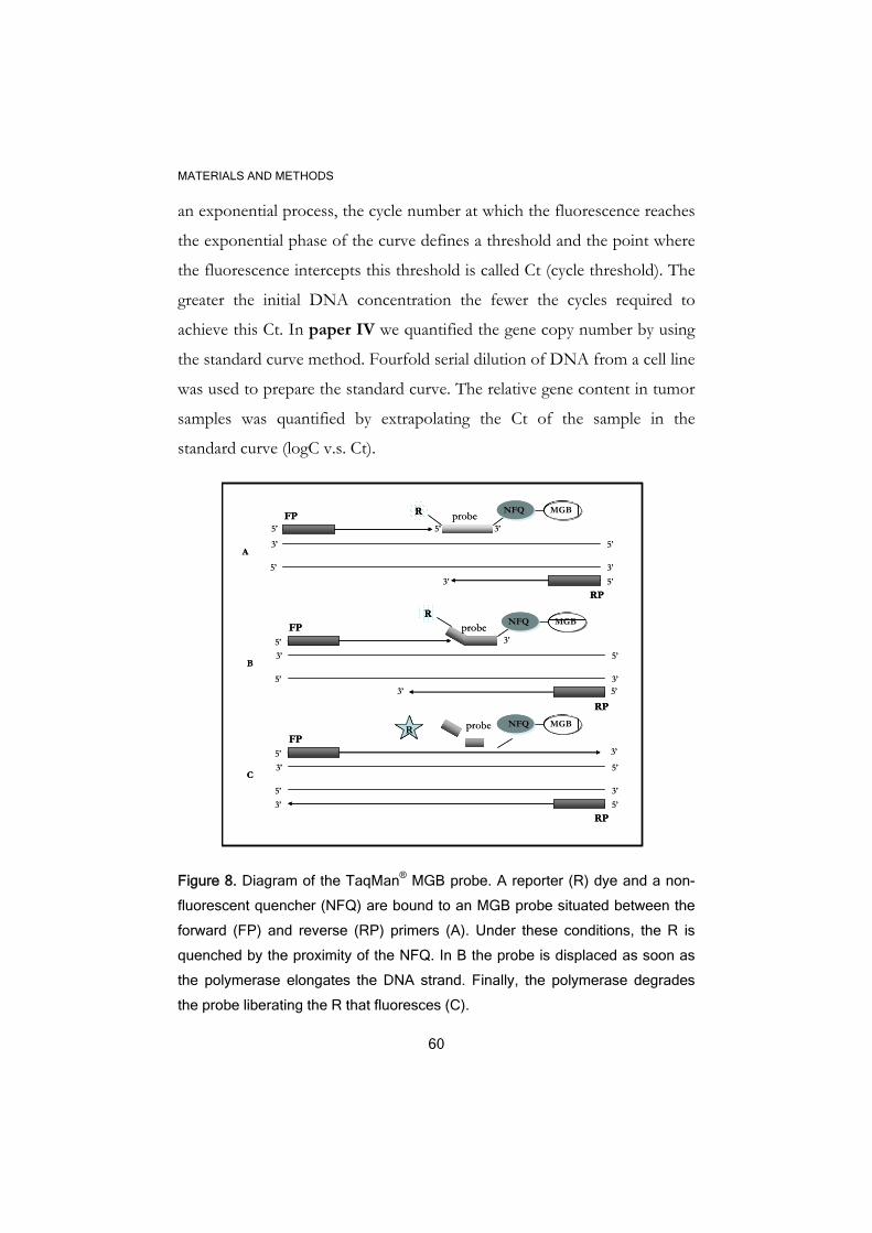
MATERIALS AND METHODS
60
an exponential process, the cycle number at which the fluorescence reaches
the exponential phase of the curve defines a threshold and the point where
the fluorescence intercepts this threshold is called Ct (cycle threshold). The
greater the initial DNA concentration the fewer the cycles required to
achieve this Ct. In paper IV we quantified the gene copy number by using
the standard curve method. Fourfold serial dilution of DNA from a cell line
was used to prepare the standard curve. The relative gene content in tumor
samples was quantified by extrapolating the Ct of the sample in the
standard curve (logC v.s. Ct).
Figure 8. Diagram of the TaqMan® MGB probe. A reporter (R) dye and a non-
fluorescent quencher (NFQ) are bound to an MGB probe situated between the
forward (FP) and reverse (RP) primers (A). Under these conditions, the R is
quenched by the proximity of the NFQ. In B the probe is displaced as soon as
the polymerase elongates the DNA strand. Finally, the polymerase degrades
the probe liberating the R that fluoresces (C).
A3’
3’5’
5’
R probe NFQ MGBFP
RP
3’
3’5’
5’
probe NFQ MGB
3’FP
RP
B
3’
3’5’
5’
probe NFQ MGB
3’FP
RP
C
5’3’
3’ 5’
5’3’
R
R
5’ 3’5’
5’
5’
A3’
3’5’
5’
R probe NFQ MGBFP
RP
3’
3’5’
5’
probe NFQ MGB
3’FP
RP
B
3’
3’5’
5’
probe NFQ MGB
3’FP
RP
C
5’3’
3’ 5’
5’3’
RR
R
5’ 3’5’
5’
5’

MATERIALS AND METHODS
61
.6 Single-strand conformation analysis
A method to detect single-strand conformation polymorphisms (SSCP) was
first described by Orita and collaborators in 1989 (Orita et al., 1989). With
the novel technique, it was possible to detect DNA polymorphisms and
point mutations at different positions in DNA fragments. The SSCP takes
advantage of the mobility shifts caused by small nucleotide alterations that
lead to conformational changes of the single-stranded DNA (Figure 9).
Each single strand will adopt a certain conformation dependent of the
primary structure. Small variations such as mutations, deletions or gains will
produce conformational alterations leading to different patterns of
migration in a gel compared to the normal sequence. To detect the aberrant
bands, the sequence is radiolabeled, denatured and run through a gel. The
bands are then visualized by exposing the dried gels to X-rays and detected
by autoradiography. The shifted bands can be cut from the gel and
sequenced.
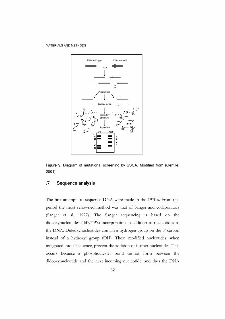
MATERIALS AND METHODS
62
Figure 9. Diagram of mutational screening by SSCA. Modified from (Gentile,
2001).
.7 Sequence analysis
The first attempts to sequence DNA were made in the 1970’s. From this
period the most renowned method was that of Sanger and collaborators
(Sanger et al., 1977). The Sanger sequencing is based on the
dideoxynucleotides (ddNTP’s) incorporation in addition to nucleotides to
the DNA. Dideoxynucleotides contain a hydrogen group on the 3’ carbon
instead of a hydroxyl group (OH). These modified nucleotides, when
integrated into a sequence, prevent the addition of further nucleotides. This
occurs because a phosphodiester bond cannot form between the
dideoxynucleotide and the next incoming nucleotide, and thus the DNA
PCR
DNA mutatedDNA wild type
Denaturation
Cooling down
Secondarystructures
B
B
CC
D
D
CC
B
BX
X
AA
AB
CD
B
X
A
C
-
+
Separation
WT MutA
A
PCR
DNA mutatedDNA wild type
Denaturation
Cooling down
Secondarystructures
B
B
CC
D
D
CC
B
BX
X
AA
AB
CD
B
X
A
C
-
+
Separation
WT MutA
A
MATERIALS AND METHODS
63
chain is terminated. Four tubes are prepared each containing one labeled
ddNTP (either ddATP, ddGTP, ddCTP or ddTTP); four dNTPs, and the
DNA polymerase in the corresponding PCR buffer. As the DNA
elongation proceeds in each tube the dNTPs are going to be incorporated.
The reactions will stop with the random incorporation of one ddNTP
instead of a dNTP to the growing chains. Since all the reactions start at the
same nucleotide but ends at specific bases at the end of the reactions there
will be a mixture of different length DNA strands that can be run trough a
polyacrylamide gel. The resolved bands can be detected by exposing the
dried gel to UV light or X rays depending on the labeling method (Figure
10).
In paper III the mutations were confirmed by automated sequencing based
on a modification of the traditional Sanger method in which ddNTPs are
labeled with a fluorescent dye for detection. In this case the ddNTPs are
labeled with two fluorescent dyes: fluorescein and four different
rhodamines. Fluorescein acts as a donor dye, absorbing energy from the
incident laser light and transferring it to the acceptor dye (rhodamine)
situated on the same ddNTP (Ju et al., 1995). Each acceptor dye then emits
light at its characteristic wavelength for detection, allowing the
identification of the nucleotide that terminated the reaction. This method
produces a more accurate sequence due to its sensitivity.
.8 STATISTICAL METHODS
In the clinical material, the relationships between different variables were
assessed by the Chi square test or the Chi square test for trend (Spearman’s
rank correlation), when the variables included more than two categories

MATERIALS AND METHODS
64
(papers I-IV). To estimate the role of one (univariate analysis) or a group
of variables (multivariate analysis) in survival analysis the Cox’s
proportional hazard regression model was used. The Cox’s proportional
hazard regression was also used in the interaction analysis between a certain
variable and treatment. The multiple sample test which is a generalization
of Gehan’s generalized Wilcoxon test was also used to assess the
differences between groups in the survival analysis. The Product-limit
method was employed to estimate the cumulative probabilities of
recurrence free survival and the differences in recurrence free survival
between groups was explored by the Log-rank test
For the in vitro study (paper II) the differences between two groups of
treatments was calculated by the Student’s t-test for independent samples.
The criterion for statistical significance was P< 0.05 and all the statistical
procedures are comprised in STATISTICA (data analysis software system),
version 8.0. www.statsoft.com.
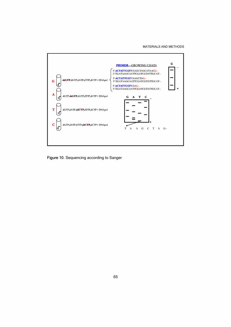
MATERIALS AND METHODS
65
G
A
T
C
ddGTP,dGTP,dATP,dTTP,dCTP+ DNApol
dGTP,ddATP,dATP,dTTP,dCTP+ DNApol
dGTP,dATP,ddTTP,dTTP,dCTP+ DNApol
dGTP,dATP,dTTP,ddCTP,dCTP+ DNApol
5’-ACTATTCGTTAAG--3’-TGATAAGCAATTCGATCGTATTGCAT--
5’-ACTATTCGTTAAGCTAG--3’-TGATAAGCAATTCGATCGTATTGCAT--
5’-ACTATTCGTTAAGCTAGCATAACG--3’-TGATAAGCAATTCGATCGTATTGCAT--
G_
+
G A T C
PRIMER---GROWING CHAIN
_
+
T A A G C T A G--
Figure 10. Sequencing according to Sanger
RESULTS AND DISCUSSION
66
RESULTS AND DISCUSSION
Most of the findings in this thesis are based on observations from tumor
specimens preserved after surgical treatment. Different properties of the
tumors were revealed with help of immunological methods to detect
protein expression and DNA analysis to determine the copy number and
mutational status of different genes. Some of the variables studied in the
papers were already known prognostic or predictive markers giving
information about the course of the disease or the treatment response.
Other variables, including those that belong to the PI3K/AKT pathway
were intriguing due to their novelty and almost unknown biological
function in the tumor context.
We found correlations between these variables that may be of clinical
significance. In order to explore cause–effect relationships we also tested
our hypotheses using in vitro methodologies. While in vitro models may not
mirror in vivo situations, they do inspire new ideas that can be validated in
vivo.
In paper I we explored the expression and activation of AKT in a clinical
material consisting of 93 tumors from premenopausal breast cancer patients
by immunohistochemistry. From the three AKT isoforms, we chose to
study the expression of AKT1 because it was thought to play the most
prominent role in breast tumorigenesis compared to AKT2 and AKT3.
AKT1 was expressed in 46% of the tumors, with predominant cytoplasmic
location, while activated AKT (pAKT) was positive in 54% of the cases.
Despite the positive correlation between AKT1 expresion and activation
(P=0.04), some AKT1 negative/pAKT positive cases were observed,
suggesting the presence of another activated isoform being recognized by

RESULTS AND DISCUSSION
67
the pAKT antibody. The structural homology of these proteins could
facilitate a cross-reaction of the antibody with a different isoform as it was
suggested later (Stal et al., 2003). In this study there was a positive
correlation between AKT1, AKT2 and pAKT though stronger between
pAKT and AKT1. In order to increase the statistical power a new variable
was conceived (tAKT). tAKT positive patients were those showing strong
tumor staining with at least one of the three antibodies and the frequency
of tAKT positive patients (55%) was similar to that found with pAKT in
paper I.
The high frequency of AKT activation found in breast cancer suggests an
important role for this pathway in the tumor. For example, pAKT has been
detected in 22-64% of breast cancers (Schmitz et al., 2004; Shi et al., 2003),
employing different antibodies and scoring systems. Recent studies confirm
the role of AKT1 in breast cancer progression (Ju et al., 2007) but its
transforming activity is only evident together with other alterations, such as
HER-2 overexpression/activation or PTEN loss (Dummler & Hemmings,
2007). In our material (paper I), AKT activation was not correlated to
HER-2 expression, which is partly explained by the low frequency of HER-
2+ cases (n=6, 7%), as expected in a sample mostly conformed by ER or
PgR positive tumours. However, in a larger material consisting of 280
postmenopausal patients, we did find this association (Stal et al., 2003).
Interestingly, HRGβ1, the most potent ligand for the activation of HER-2,
3, and 4 receptors, was found associated to pAKT when expressed in
stromal cells (P=0.017), which suggests a possible paracrine or juxtacrine
mechanism of AKT activation. Tumor-associated fibroblasts have been
found to confer morphogenic and mitogenic induction of epithelial cells
(Shekhar et al., 2001) and to induce acceleration of epithelial tumor growth
RESULTS AND DISCUSSION
68
in vivo (Camps et al., 1990). Conversely to what we expected, pAKT was
inversely correlated to SPF (P=0.001) (Table I, paper I). Since our article
was published, additional elements have come to explain the divergent roles
of AKT kinases in cancer. The three isoforms induce transformation in
vitro, however AKT1 and AKT2 seem to have opposing functions in cell
proliferation. While AKT1 induces cell proliferation, AKT2 promotes cell
cycle exit in nontransformed mammalian cells (Heron-Milhavet et al.,
2006). The effects of AKT2 have been explained in part by interactions
with the cell cycle inhibitor p21 CIP1 in the nucleus. Moreover, AKT has
been reported to decrease the growth rate in cells transfected with mutant
PTEN in comparison with the empty vector (McCubrey et al., 2008), and
its being found by others to cause ERβ induction (Duong et al., 2006). Still,
we found that pAKT was associated to higher risk for distant metastasis
among the ER+/endocrine-treated patients, results later confirmed by
others (Kirkegaard et al., 2005; Tokunaga et al., 2006(b); Tokunaga et al.,
2006(a)) suggesting an interaction between AKT and ER signaling
pathways.
In univariate analysis, pAKT positivity predicted a higher risk to develop
metastasis with borderline significance (RR=2.4, 95% CI, 1.0-6.2) (Figure
2A, paper I), while multivariate analysis including pAKT and traditional
prognostic factors, indicated that pAKT was an independent predictor of
distant recurrence (P=0.004) (Table II, paper I). The analysis of the
interaction between pAKT and SPF revealed that a low SPF was a
favorable feature only if the tumor was in addition pAKT negative. This
suggests that distant relapses could arise, probably due to malignant cell
survival rather than cell proliferation considering the low proliferative state
of the tumor (Figure 2B, paper I).
RESULTS AND DISCUSSION
69
Nodal status and pAKT had independent prognostic value, with all patients
in the group node-/pAKT- having a better 6 years-survival than the
node+/pAkt+ group (Figure 2C, paper I).
In an attempt to elucidate the effects of HRGβ1 on cell survival,
proliferation, and AKT activation upon endocrine treatment, we used the
breast cancer cell line MCF-7. The ER+ MCF-7 cells are hormone
dependent and tamoxifen-responsive. In these cells, AKT phosphorylation
lasted at least 72h after addition of HRGβ1, which indicates a possible role
for HRGβ1 (Figure 1A, paper II). HRGβ1 also stimulated p21 protein
expression in the same time-window that AKT activation occurred. Since
the small decrease in the levels of p21 observed upon treatment with Tam
and E2 disappeared in the presence of HRGβ1 (Figure 1B, paper II), we
speculated that HRGβ1 could also stabilize p21. Moreover, upon HRGβ1-
treatment, p21 levels increased both in the nucleus and in the cytoplasm as
demonstrated by Western blot (Figure 2, paper II) and
immunofluorescence (Figure 3, paper II). This modest accumulation of
cytoplasmic p21 suggests that HRGβ1 can also mediate p21 cellular
allocation, recently attributed to AKT (Zhou et al., 2001). HRGβ1 also
induced cell proliferation and counteract the action of tamoxifen upon cell
cycle progression (measured as percentage of cells in S-phase) and cell
death (apoptosis), (Figures 4 and 5, paper II). Interestingly, a recent
report shows that HRGβ1 is able to induce breast cancer cell proliferation
by induction of p21 and cyclin D1 (Yang et al., 2008), but the authors did
not specify the cellular location of p21. Other authors found that HRG-
transfected cells accumulate p21 in the nucleus, as discussed in paper II.
The role of the induction/delocalization of p21 by HRGß1 during
development of tamoxifen resistance is not documented in the literature. In
RESULTS AND DISCUSSION
70
a 3-D model with MCF-7-derived cells, where tamoxifen inhibited ER-
mediated transcription and cell proliferation, the proportion of p21-positive
cells decreased upon tamoxifen treatment (Truchet et al., 2008). More in
accordance to what is expected, another study showed that p21 loss leads to
the tamoxifen growth-induced phenotype. These facts suggest that p21 is
an essential player to consider during tamoxifen treatment. Our results
obtained in clinical samples from 280 postmenopausal women revealed that
p21, in addition to its classical nuclear location, can also be found in the
cytoplasm of malignant cells at a higher frequency (26.7%) than in the
nucleus (14.5%). When we evaluated the effect of tamoxifen among
patients with ER+ tumors we found that patients whose tumors classified
as p21 N+/C- benefited significantly from tamoxifen (P=0.0082), as did
the patients that had either undetectable p21 levels or double positive
phenotype (P=0.034). In contrast the p21 N-/C+ group appeared not to
respond to tamoxifen (P= 0.7). This group however contained a small
number of patients (Figure 8 and Table II, paper II). It is still unclear
how cytoplasmic p21 could affect the response to tamoxifen, but it is
known that the cytoplasmic protein can mediate cell survival through
binding and inactivation of the apoptosis signal-regulating kinase 1
(ASK1)(Asada et al., 1999). On the other hand, nuclear p21 has been
already associated with better survival under antiestrogen treatment
(discussed in paper II). Why the p21N+/C- patients presented the worst
outcome in comparison with the other groups in absence of the drug is
speculated in paper II. A recent report suggests that the favorable
prognosis of ER+ breast cancer can be explained by the antiproliferative
function of the ER by its physical interaction with p21 in the absence of
estrogens. Consequently, this interaction could be favored if tamoxifen is
present as the ER cannot bind E2, but not in its absence where the ER-E2

RESULTS AND DISCUSSION
71
complex could provoke cell proliferation (Maynadier et al., 2008). In
general, accumulation of p21 was neither beneficial in the cytoplasm nor in
the nucleus. The patients included in the p21 N+/C- group also registered
the worst distant recurrence-free survival compared to the other p21
phenotypes (P=0.034) (Figure 7, paper II). Since the patients comprised
in this study were randomized to different treatments, we cannot conclude
which therapeutic regime failed in the presence of nuclear p21. Poor disease
free-survival has been observed among p21+ patients that received CMF,
as discussed in paper II, but we could not detect any obvious interaction
between p21 and the benefit from CMF versus radiotherapy.
Moreover, we could not find a significant association between p21, nodal
status, or tumor size. Cytoplasmic p21 was often registered among HER-
2+ tumors, but this association was not significant. However, there was an
association between cytoplasmic (P=0.00001) and nuclear p21 (P=0.022)
with AKT phosphorylation and it was in the same material we previously
reported a significant association between pAKT and HER-2+ status (Stal
et al., 2003).
Overexpression or constitutive activation of growth factors and growth
factor receptors, such as HRGβ1 and HER-2, contribute to AKT
activation, and these may coexist with other cellular alterations. Mutations
of the PIK3CA gene and PTEN loss are two that we discussed in this
thesis. The PI3K is conformed by a regulatory p85 and a catalytic p110
subunit. Most of the PI3K mutations, reported so far in cancer, occur in
the gene encoding the p110α subunit. Among the 21 exons that compose
this gene, more than 85% of the mutations are concentrated in exons 9,
encoding part of the helical domain, and exon 20, encoding part of the
kinase domain. In our material (see paper III) we found missense

RESULTS AND DISCUSSION
72
mutations in 24% of the tumors, in agreement with the literature, where the
mean percentage of mutations is 25%. The most frequent mutations were
E545K, followed by E542K in exon 9; and H1047R in exon 20 (Table I,
paper III). The higher frequency of mutations (67%) reported for cell lines
could be partly explained by the more challenging conditions of the in vitro
culture, increasing the risk of the cells for more genetical alterations. But
these mutations must confer some advantages to the tumor cells since
MCF-10A, a non tumorigenic epithelial breast cell line, did not present
mutations. In fact, the most frequent mutations described so far (E545K,
E542K, and H1047R) are associated in vitro with increased lipid kinase
activity, cell growth, growth factor independency, transforming activity and
invasive potential (Carson et al., 2008). They are also related to increased
AKT activation(Zhang et al., 2007). In vivo studies have also shown that
alterations in the p110α helical or catalytic domains results in a more active
PI3K pathway, and could raise tumors in animal models (Bader et al., 2006;
Zhao et al., 2005). However these results might not be comparable with
others using natural mutant cells (Morrow et al., 2005). Those authors
suggested that PIK3CA mutations in the helical domain may even lead to
reduced rate of PI3K activation.
Associations with other clinical markers could also help to decipher the role
of these mutations in vivo. In our material PIK3CA was often related to
indicators of good prognosis like ER expression (P= 0.052) and negative
HER-2 status (protein, P= 0.013 and gene amplification, P=0.083), which
can also be observed in our panel of cell lines often sharing an ER+/HER-
2- phenotype when mutated. In addition the PIK3CA mutations were
related to small tumor size (P= 0.057). However, PIK3CA mutations are
also associated to other factors like PTEN loss (P=0.0024), AKT1+ (P=
0.032), and high expression of cyclin D1 (P= 0.031) which in turn could
RESULTS AND DISCUSSION
73
indicate a bad course for the disease. PIK3CA mutations did not associate
with node status, AKT2 expression, or pAKT. However, pAKT+
(P=0.0033), tAKT positive (P=0.0019), and cyclin D1++ (P= 0.031)
phenotypes were significantly associated with mutated PIK3CA (PIK3CA
mut) and/or HER-2+) in a combined variable.
Other authors have reported association with ER+ tumors but also with
presence of normal PTEN and high HER-2 expression (Saal et al., 2005).
These authors conclude that PIK3CA and PTEN alterations were
redundant at the same time that they found some PIK3CA/PTEN mutants
and HER-2 negative cases. In line with our results a recent report also
found PIK3CA mutations frequently concordant with PTEN loss in breast
cancer (Stemke-Hale et al., 2008).
Why would it be advantageous for a malignant cell to have this “redundant”
phenotype? As demonstrated in endometrial carcinoma, where PIK3CA
and PTEN mutations coexist, PTEN knockdowns/PIK3CA mutants
additively enhanced AKT activity compared with a single alteration (Oda et
al., 2005). PTEN protein phosphatase activity, that operates independently
of the PI3K pathway, could be another reason to support the non-
redundant role of PTEN (Freeman et al., 2003; Okumura et al., 2005) and
finally the complexity of this signaling pathway may require more than one
input for its full activation .
PTEN protein expression was also studied by immunohistochemistry and
considered positive, in 126/201 tumors (63%), and weak or negative in 75
tumors (37%). PTEN was mainly located in the cytoplasm of tumor cells
and in some cases in the nucleus (Figure 3, paper III). These results are in
agreement with those of other authors that reported loss of PTEN in 27%-
50% of invasive cancers (Depowski et al., 2001; Tsutsui et al., 2005). In our
material, PTEN loss correlated with ER+ status (P=0.0015), small tumor
RESULTS AND DISCUSSION
74
size (P=0.022), and low HER-2 expression (P=0.011). Likewise a combined
variable PTEN/PIK3CA, was associated with ER+ (P=0.0017), small
tumor size (P=0.0075), non-amplified HER-2 (P=0.054), low HER-2
protein expression (P=0.00080), and low SPF (P=0.014) (Table II, paper
III). In some studies PTEN loss has been correlated to clinical parameters
such as node metastasis, shorter disease free survival, tumor grade, or
aneuploidy, but in others it has been found irrelevant to the clinical
outcome (Panigrahi et al., 2004).
Based on these clinical correlations it was difficult to predict the prognostic
role of PIK3CA mutations and PTEN loss in breast cancer. We found that
the risk to relapse with a local recurrence was significantly lower among
patients with mutated PIK3CA in comparison with those who carried wild
type PIK3CA in a univariate analysis (P= 0.023) (Figure 2A, paper III)
and a trend was observed in a multivariate analysis (P=0.07).
PI3K has been proposed to cause cell death mediated by hypoxia, glucose
deprivation, or serum withdrawal (see discussion of paper III) which in
part challenges the general view of PI3K as a survival pathway. PI3K
mutations indicated good prognosis in Japanese women (Maruyama et al.,
2007) and have been related to decreased rate of node positive disease
among ER+ tumors (Liedtke et al., 2008). Moreover, exon 9 and exon 20
mutations seems to have different functions as demonstrated in vitro and in
vivo. Exon 20 has been associated to good prognosis in the same material
that exon 9 mutation predicted for worse outcome (Barbareschi et al.,
2007), whereas others have found exon 20 mutations coupled to poor
prognosis for patients with invasive carcinomas (Lai et al., 2008) or both
mutations associated to poor patient survival (Li et al., 2006). We also
found that among the tumors with low SPF (SPF<5%), the PIK3CA
mutated and/or PTEN negative type predicted for worse recurrence-free
RESULTS AND DISCUSSION
75
survival (P=0.020) while it indicated better survival among the group with
higher S-phase (SPF>10%) (P=0.0073) (Figure 4, paper III).
PTEN alone did not provide clinical information regarding distant or local
recurrences, or breast cancer survival. The patients in the PTEN negative
group tended to benefit more from radiotherapy than from chemotherapy
(P=0.02) compared to those in the PTEN + group (P=0.29) and so did the
patients with mutated PIK3CA and/or PTEN - type.
The most accepted idea behind PI3K/PTEN alterations is that this
phenotype associates with AKT activation and survival of radiation-induced
apoptosis. Indeed lack of PTEN sequesters CHK1 and initiates genetic
instability (Puc et al., 2005), facilitating other genetic alterations that may
confer survival and growth advantages to the malignant cells. Are the
PIK3CA mutations some of them? Another possibility could be that the
unstable cells will not survive the mutagenic effects of radiation.
Interestingly, PIK3CA mutated and PTEN deficient tumors present a low
percentage of cells in S-phase. The effect of cell cycle arrest in response to
radiation is also a controversial issue. Cell cycle arrest upon radiation can
give time to the cells to repair the major damages and survive the effect of
radiation (Lees-Miller, 2008) but at the same time it contributes to
radiosensitization (Albert et al., 2006). Moreover, PTEN can enhance
transcription of RAD51 reducing the incidence of spontaneous double
strand breaks, but in PTEN deficient cells RAD51 might be suppressed. A
recent report, showing that patients with low expression of
BRCA1/BRCA2/RAD51 complex respond well to radiation (Soderlund et
al., 2007), allowing us to suggest that PTEN deficient cells are more
sensitive to radiation.
Another member of the PI3K/AKT pathway that has been involved in
therapy resistance is mTOR. Experimental and clinical evidence indicate

RESULTS AND DISCUSSION
76
that mTOR inhibitors not only improve the therapeutic effect of tamoxifen
and letrozole (Chollet et al., 2006), but are also effective in combination
with the dual HER-1/HER-2 inhibitor lapatinib and the HER-2 inhibitor
trastuzumab (Vazquez-Martin et al., 2008). Among all the compounds that
have been developed to target the PI3K pathway, mTOR inhibitors
predominate in clinical trials (LoPiccolo et al., 2008). Taken together, these
findings indicate that mTOR level and its downstream effectors could be
interesting targets to explore for new alterations with therapeutic impact.
For example, RPS6KB1, a gene encoding the mTOR substrate p70S6
kinase 1, has been found amplified in 8-10% of breast tumors (Barlund et
al., 2000; Barlund et al., 1997; Sinclair et al., 2003), while very little has been
published in cancer about the other gene (RPS6KB2) encoding the other S6
kinase. This prompted us to explore our material looking for RPS6KB1/2
gene amplification. We found RPS6KB1 and RPS6KB2 amplification in
22/206 cases (10.7%) and in 9/207 cases (4.3%), respectively, but also in
cell lines (75% and 50%) (supplement, paper IV). Even though the
frequency of amplification in tumors was rather low in comparison with
other alterations in this pathway, amplified S6K1 and S6K2 were
independent prognostic factors indicating higher risk to develop recurrence.
The S6K2 gene was also an independent predictor for breast cancer-related
death (Table II, paper IV). Statistical analysis revealed that S6K1
amplification was significantly associated with HER-2 gene amplification
and protein expression, while S6K2 was associated with ER+ status and
CCND1 amplification (Table I, paper IV).
Interestingly S6K1 and/or S6K2 amplification and mutations in the
PIK3CA gene were inversely correlated, (P=0.004), while no association
was found with PTEN deficiency or AKT activation. This supports the
idea of inter-tumor heterogeneity where some of the tumors are driven by
RESULTS AND DISCUSSION
77
PIK3CA mutations or PTEN deficiency, where others take advantage from
HER-2 and/or S6K1/2 amplification. From those alterations, AKT
activation was correlated only with HER-2, indicating the existence of
AKT- independent mechanisms in the PTEN negative/PIK3CA mutated
tumors. However, the simplest explanation would be that we have only
detected the phospho-Ser 473 residue leaving the Trh 308 unexplored.
The possibility of HER-2 and S6K1 co-amplification has been discussed
before due to the physical proximity of the 17q12-21 and the 17q22-24
regions, and S6K1 was identified as the first candidate oncogene in the
17q23 region (Couch et al., 1999). In our material, we found that, in terms
of locoregional control, the patients without S6K1 gene amplification
benefited more from radiotherapy than from chemotherapy (P=0.0038) in
comparison with the patients carrying this genetic alteration (P=0.39). 17q
amplicon, including S6K1 and/or HER-2 gene amplification, also indicated
poor response to radiotherapy (Figure 2, paper IV). Both 17q and S6K1
amplification status interacted significantly with the benefit from
radiotherapy (Table III, paper IV).
Other authors have found stimulation of the HER-2/PI3K/AKT signaling
pathway to be involved in resistance to radiation-induced apoptosis
(Soderlund et al., 2005), and trastuzumab to be a radiosensitizer (Liang et
al., 2003). In a previous study (Stal et al., 2003) AKT activation was also
associated with poor response to radiotherapy in comparison with
chemotherapy. The S6K1/ 2 proteins share common as well as distinct
cellular substrates and functions (Martin et al., 2001; Shima et al., 1998), and
both may be involved in a feedback loop leading to AKT inactivation
(Manning et al., 2005), suggesting that in absence of AKT these kinases
might take over the signaling pathway. In fact, mTOR inhibitors have been
introduced in the clinical practice and sometimes the disappointing results

RESULTS AND DISCUSSION
78
are coupled to a possible AKT activation as a side effect (LoPiccolo et al.,
2008). However, in vitro studies have shown that by blocking mTOR it is
possible to impair ER-mediated cell proliferation (Chang et al., 2007) and to
restore response to tamoxifen in breast cancer cells with aberrant AKT
activity (deGraffenried et al., 2004). Interestingly, among the ER+ patients
included in this study, an increased number of the S6K2 gene copies
indicated better response to tamoxifen in comparison with the S6K2
negative group (Table IV, paper IV).

CONCLUSIONS
79
CONCLUSIONS
Paper I
AKT-1 protein expression and activation are frequent events in
premenopausal breast cancer. AKT-1 activation is related with low cell
proliferation however, pAKT positive patients also register shorter distant
recurrence-free survival suggesting that this factor might be used to predict
the response to endocrine therapy in premenopausal patients.
Paper II
HRGß1 stimulates AKT activation in MCF-7 cells. It can also induce p21
expression and may induce cytoplasmic accumulation. HRGβ1 can impair
the cytostatic and cytotoxic effect of tamoxifen but seems to be more
important as a survival factor.
p21 is present in the cytoplasm of the malignant cells more frequently than
in the nucleus. Cytoplasmic p21 is associated with pAKT, in line with the
hypothesis that AKT induces p21 cytoplasmic delocalization.
Cellular location of p21 may help to identify subgroups with different
therapeutic response among ER+ patients.
Paper III
PIK3CA mutations and PTEN loss are common alterations in breast
cancer and coexist in a proportion of tumors. These alterations are more
frequent among ER+, small and low proliferative tumors and this
phenotype may differ from that of HER-2 positive tumors. PIK3CA
mutations and/or PTEN loss predicts better recurrence-free survival

CONCLUSIONS
80
among patients with high proliferating tumors while among those with low
proliferating disease it indicates a poor outcome.
PIK3CA mutations indicate a lower risk to relapse with local recurrences
while low PTEN, as a single variable or combined with PIK3CA mutations
tended to confer radiosensitivity.
Paper IV
The S6K1 and 2 genes are amplified in breast cancer and coamplified with
HER-2 and CCND1 respectively. S6K1 and/or S6K2 amplifications are
inversely correlated with PIK3CA mutations.
S6K2 amplification may be used to predict development of recurrences and
higher risk for breast cancer death.
Patients with S6K2-increased gene copy number will benefit from
tamoxifen treatment.
S6K1 amplification indicates higher risk to develop recurrences and poor
response to radiotherapy.
CLINICAL RELEVANCE
81
CLINICAL RELEVANCE
Our results support the notion of the high frequency of PI3K/AKT
pathway alterations in breast cancer. Taking into consideration HER-2
overexpression/amplification, PIK3CA mutations, low PTEN expression
or RPS6KB1/RPS6KB2 amplification, the frequency of alterations was
more than 75% (76.9%). However, this combined variable did not have an
obvious clinical value, which could be explained by the complexity of this
signaling pathway, where the hierarchy and biological function of its
members is not fully understood. Based on the different clinical
associations, two different paths emerged from these studies. One, that
seems to be govern by HER-2 amplification or overexpression and
RPS6KB1 amplification and the other driven by PIK3CA mutations and
PTEN loss. While the first group is related to poor response to
radiotherapy in comparison with chemotherapy the second phenotype
conferred radiosensitivity. Another aspect to consider is how to stratify the
patients in order to improve the accuracy of the diagnosis and treatment.
PIK3CA mutated/low PTEN phenotype associated to ER+, small and low
proliferating tumors. However, the patients with low proliferative tumors
carrying this phenotype often had a shorter recurrence-free survival in
comparison with the wild type counterpart while the group with highly
proliferating tumors exhibited a longer recurrence-free survival in presence
of these alterations. Likewise p21 identified subgroups of ER+ patients
with different responses to the endocrine treatment. Regarding AKT
activation, we can conclude that it seemed to be involved in response to the
endocrine treatment among the premenopausal women while it was linked
more to radiotherapy resistance in the postmenopausal patient material.
FUTURE PERSPECTIVES
82
FUTURE PERSPECTIVES
1- It would be interesting to elucidate the individual role of the three AKT
isoforms in a larger patient material from a randomised trial not forgetting
to explore the expression of the pThr308 AKT.
2- Whether HRGß1 induces p21 expression and delocalization through the
PI3K/AKT signaling pathway and how this could impair the effect of
tamoxifen deserves a major study.
3- To characterize the impact of the different PIK3CA mutants in a tumor
context may help to understand the role of PIK3CA mutations in breast
cancer.
4- Immunohistochemical detection of p70S6K1 and p70S6K2 should be
performed in order to look for associations between gene amplification and
protein expression. Furthermore, in vitro studies may help to verify the role
of these proteins in breast cancer.

ACKNOWLEDGMENTS
83
ACKNOWLEDGMENTS
My interest in cancer research began at a lecture about monoclonal
antibodies. The man who spoke knew how to catch the attention of young
students with color slides of the immunological struggle. Jorge Gavilondo,
the professor at that time, is unaware that his name has a place in my
acknowledgments.
But here I am, in our lab, in Sweden. There are many persons that I would
like to thank, like professor Bo Nordenskjöld, you welcomed me so kindly
to this country and changed my life. Olle Stål, I feel so lucky having you as
my supervisor. You know the secrets of every signaling pathway but I
admire you most for your kindness, your sense of justice and generosity.
You guided me wisely and gave me so much freedom that sometimes I
thought I was alone. However, whenever I needed you I could find you
there, firm as a rock. Birgit Olsson, you probably suspect how much I like
you. You have opened so many doors for me: to your home, to the lab, to
the Swedish language. You have taught me the essentials of Christmas
decoration, advanced cooking and Real-Time PCR. I wish I had your
energy and a little part of your big heart. To the Ph.Ds. that create such a
good atmosphere in the lab. We can talk about EVERYTHING and
nothing at the same time. We have seen each other celebrate birthdays and
publications, engage and marry, build houses and families. We can meet to
discuss the latest article or to see SATC. We can make diet and exercises or
jump to a “semla” all with the same enthusiasm. We have shared so much
during these years…Daniella Pfeiffer, you that always have time to listen
and to help. Åsa Wallin, Little Miss Sunshine, I am sure you will be fine.
Karin Söderlund or Leifler, I will buy your first book …perhaps that

ACKNOWLEDGEMENTS
84
about cell-death? Josefine Bostner, I admire your strength. Marie
Ahnström Waltersson, you are almost there! Cissi Bivik, you infiltrated
this group bringing with you a good portion of humor and common sense,
Agneta Jansson, thanks for good advice in the lab and in the kitchen.
Cecilia Gunnarsson and Marie Askmalm, the medical “gurus” in our
group, thanks for answering all my questions about medical and non-
medical issues. Jingfang Gao, did you know that the keyword of my PC
was DUMPLING? 祝你好運, Piiha Lotta, you have something to teach
about discipline and determination, Andreas Lewander, good luck with
your research! Tove you came later but have already found a place in this
group. Pia Wegman and Sten Wingren once were you also part of this
group.
To Xiao-Feng Sun, such a gentle person, thanks for our conversations and
all these times that you care about my doings and health.
To Liliane Ferraud and Birgitta Holmlund, the hidden force behind the
ER and PgR, my eternal providers of the Vindelov fluids (without
complains). Thanks for welcoming me. You are such nice persons.
To Anna Esguerra Merca, Lisa Alkhori and Elin Karlsson, my students
and coauthors. It has been a pleasure working with you. Keep curious!
To Håkan W, Kerstin, Irene, Pia, life as a Ph.D. student becomes less
miserable and a lot more enjoyable having your help. To all the friendly
souls at KEF, for fikas, relaxing conversations, and help.
Almighty Chatarina Malm, thank you so much for your help with all the
administrative issues, for your kindness and care. Anette Wiklund, for
your endless patience with all my “föräldraledighetsblanketter” and other
“spiritual” issues.

ACKNOWLEDGMENTS
85
Florence Sjögren, once upon a time you unveiled for me the secrets of the
flow cytometer and now you corrected my English. Thanks! Anette
Molbaek, you helped me to decipher the mystery of the sequences.
Till pedagogerna på Vitra förskolan som har vartit så extra-förståndiga
med en glömsk, springande och sliten mamma.
To Massimiliano Gentile, when I came all confused and scared I found a
friend in you. Thanks for your generosity. I am still missing your
extraordinary sense of humor and our good chats about films, books,
technology and life.
To Anneli Karlsson, Camilla Gårdman, Malin Bergman, we have
experienced together some “ups and downs”. Fortunately, we have each
other to laugh. I hope we will keep in touch.
To Ainhoa, Marta, Anke and Daniel, my dear friends, I cannot imagine
life without our private jokes, gatherings, small surprises and long phone
calls. I always feel that we have each other. Christianne, Jesús, Ana y
Fermín, muchas gracias por haberme acogido, a mí y a mi familia bajo sus
alas. Siempre serán bienvenidos.
To Rosaura, Rafa y Virginia, mis amigos cubanos, que sería de mi sin
nuestros encuentros, sin sus sabios consejos. Con ustedes me siento como
en casa.
Alexandra (Alex), without your help I could not have finished this thesis.
I’m happy that we became friends.
Hanna Olausson med familj, jag vet inte riktigt hur stort ditt hjärta är
Hanna, du som har delat din familj med mig och som har varit där och
lyssnat och uppmuntrat. Jag känner mig rik med er som vänner.
Maria Elena Faxas, you welcomed me to your lab in Cuba, when there
was no lab and then guided me through the scientific and cultural etiquette.
We shared the passion for French language and more than one conspiracy.

ACKNOWLEDGEMENTS
86
We even won a price for finding the coupling between immunology and
witchcraft. Carlos García Santana, you introduced me to the basics of the
immune-swamp and showed me its beauty. It was at your lab in Cuba that I
heard for the first time about Sweden. I admire you for your tremendous
knowledge “que abarca desde lo sublime hasta la timba”.
Mis amigos de Cuba, Ofelia Sevy, Eladio Iglesias, Zaima Mazorra,
Dmitri Prieto, Karina Mendoza, L. Orlando Pardo… que me
acompañaron durante la carrera y ahora estan desperdigados por el mundo
haciendo las mas diversas cosas. Les deseo éxito en todo lo que emprendan!
Ana Maria Barral, mi hermana y querida amiga, you found a mountain for
me and encouraged me all the way up. I trust you and very much know
how good teacher you are. Your students, like your child, will be prepared
to leave you but will always come back…
Familjen Hammarberg, ni är alltid välkomna och efterlängtade.
Familjen Johansson, till Kumla vill man alltid åka för där finns det gott
om plats och kärlek för en stor familj.
Mi papa, por tu confianza, cariño y ejemplo de trabajo.
Håkan, Sebastian, Beatrice och Mattias, ni är min trygghet och min
bästa inspirationskälla... och med er är jag hemma.
Special thanks to the Swedish Cancer Foundation and the Lion
Foundation that supported this work.

REFERENCE LIST
87
REFERENCE LIST
Ahnstrom M, Nordenskjold B, Rutqvist LE, Skoog L and Stal O. (2005).
Role of cyclin D1 in ErbB2-positive breast cancer and tamoxifen resistance. Breast Cancer Res Treat 91:145-51.
Albert JM, Kim KW, Cao C and Lu B. (2006). Targeting the Akt/mammalian target of rapamycin pathway for radiosensitization of breast cancer. Mol Cancer Ther 5:1183-9.
Alessi DR, Andjelkovic M, Caudwell B, Cron P, Morrice N, Cohen P et al. (1996). Mechanism of activation of protein kinase B by insulin and IGF-1. Embo J 15:6541-51.
Ali IU, Schriml LM and Dean M. (1999). Mutational spectra of PTEN/MMAC1 gene: a tumor suppressor with lipid phosphatase activity. J Natl Cancer Inst 91:1922-32.
Anderson E, Clarke RB and Howell A. (1998). Estrogen responsiveness and control of normal human breast proliferation. J Mammary Gland Biol Neoplasia 3:23-35.
Andre F, Domont J and Delaloge S. (2007). What can breast cancer molecular sub-classification add to conventional diagnostic tools? Ann Oncol 18 Suppl 9:ix33-6.
Arpino G, Green SJ, Allred DC, Lew D, Martino S, Osborne CK et al. (2004). HER-2 amplification, HER-1 expression, and tamoxifen response in estrogen receptor-positive metastatic breast cancer: a southwest oncology group study. Clin Cancer Res 10:5670-6.
Asada M, Yamada T, Ichijo H, Delia D, Miyazono K, Fukumuro K et al. (1999). Apoptosis inhibitory activity of cytoplasmic p21(Cip1/WAF1) in monocytic differentiation. EMBO J 18:1223-34.
Aveyard JS, Skilleter A, Habuchi T and Knowles MA. (1999). Somatic mutation of PTEN in bladder carcinoma. Br J Cancer 80:904-8.
Bachman KE, Argani P, Samuels Y, Silliman N, Ptak J, Szabo S et al. (2004). The PIK3CA gene is mutated with high frequency in human breast cancers. Cancer Biol Ther 3:772-5. Epub 2004 Aug 26.
Bader AG, Kang S and Vogt PK. (2006). Cancer-specific mutations in PIK3CA are oncogenic in vivo. Proc Natl Acad Sci U S A 103:1475-9. Epub 2006 Jan 23.
Bader AG, Kang S, Zhao L and Vogt PK. (2005). Oncogenic PI3K deregulates transcription and translation. Nat Rev Cancer 5:921-9.

REFERENCE LIST
88
Barbareschi M, Buttitta F, Felicioni L, Cotrupi S, Barassi F, Del Grammastro M et al. (2007). Different prognostic roles of mutations in the helical and kinase domains of the PIK3CA gene in breast carcinomas. Clin Cancer Res 13:6064-9.
Barlund M, Forozan F, Kononen J, Bubendorf L, Chen Y, Bittner ML et al. (2000). Detecting activation of ribosomal protein S6 kinase by complementary DNA and tissue microarray analysis. J Natl Cancer Inst 92:1252-9.
Barlund M, Tirkkonen M, Forozan F, Tanner MM, Kallioniemi O and Kallioniemi A. (1997). Increased copy number at 17q22-q24 by CGH in breast cancer is due to high-level amplification of two separate regions. Genes Chromosomes Cancer 20:372-6.
Baum M, Hackshaw A, Houghton J, Rutqvist, Fornander T, Nordenskjold B et al. (2006). Adjuvant goserelin in pre-menopausal patients with early breast cancer: Results from the ZIPP study. Eur J Cancer 42:895-904. Epub 2006 Mar 20.
Beckmann MW, Niederacher D, Schnurch HG, Gusterson BA and Bender HG. (1997). Multistep carcinogenesis of breast cancer and tumour heterogeneity. J Mol Med 75:429-39.
Bellacosa A, Testa JR, Staal SP and Tsichlis PN. (1991). A retroviral oncogene, akt, encoding a serine-threonine kinase containing an SH2-like region. Science 254:274-7.
Bergh J, Brandberg Y, Ernberg I, Frisell J, Furst CJ, Hall P. (2007). Bröstcancer. Karolinska Institutet University Press.
Bertucci F and Birnbaum D. (2008). Reasons for breast cancer heterogeneity. J Biol 7:6.
Bezwoda WR, Esser JD, Dansey R, Kessel I and Lange M. (1991). The value of estrogen and progesterone receptor determinations in advanced breast cancer. Estrogen receptor level but not progesterone receptor level correlates with response to tamoxifen. Cancer 68:867-72.
Bieche I, Olivi M, Nogues C, Vidaud M and Lidereau R. (2002). Prognostic value of CCND1 gene status in sporadic breast tumours, as determined by real-time quantitative PCR assays. Br J Cancer 86:580-6.
Bissell MJ. (2007). Modelling molecular mechanisms of breast cancer and invasion: lessons from the normal gland. Biochem Soc Trans 35:18-22.
Board RE, Thelwell NJ, Ravetto PF, Little S, Ranson M, Dive C et al. (2008). Multiplexed assays for detection of mutations in PIK3CA. Clin Chem 54:757-60.

REFERENCE LIST
89
Bostner J, Ahnstrom Waltersson M, Fornander T, Skoog L, Nordenskjold B and Stal O. (2007). Amplification of CCND1 and PAK1 as predictors of recurrence and tamoxifen resistance in postmenopausal breast cancer. Oncogene 26:6997-7005.
Boyer D, Quintanilla R and Lee-Fruman KK. (2008). Regulation of catalytic activity of S6 kinase 2 during cell cycle. Mol Cell Biochem 307:59-64.
Brugge J, Hung MC and Mills GB. (2007). A new mutational AKTivation in the PI3K pathway. Cancer Cell 12:104-7.
Burgering BM and Coffer PJ. (1995). Protein kinase B (c-Akt) in phosphatidylinositol-3-OH kinase signal transduction. Nature 376:599-602.
Buttitta F, Felicioni L, Barassi F, Martella C, Paolizzi D, Fresu G et al. (2006). PIK3CA mutation and histological type in breast carcinoma: high frequency of mutations in lobular carcinoma. J Pathol 208:350-5.
Cailleau R, Olive M and Cruciger QV. (1978). Long-term human breast carcinoma cell lines of metastatic origin: preliminary characterization. In Vitro 14:911-5.
Cailleau R, Young R, Olive M and Reeves WJ, Jr. (1974). Breast tumor cell lines from pleural effusions. J Natl Cancer Inst 53:661-74.
Calza S, Hall P, Auer G, Bjohle J, Klaar S, Kronenwett U et al. (2006). Intrinsic molecular signature of breast cancer in a population-based cohort of 412 patients. Breast Cancer Res 8:R34.
Campbell IG, Russell SE, Choong DY, Montgomery KG, Ciavarella ML, Hooi CS et al. (2004). Mutation of the PIK3CA gene in ovarian and breast cancer. Cancer Res 64:7678-81.
Campbell IG, Russell SE and Phillips WA. (2005). PIK3CA mutations in ovarian cancer. Clin Cancer Res 11:7042; author reply 7042-3.
Campbell LL and Polyak K. (2007). Breast tumor heterogeneity: cancer stem cells or clonal evolution? Cell Cycle 6:2332-8.
Campbell RA, Bhat-Nakshatri P, Patel NM, Constantinidou D, Ali S and Nakshatri H. (2001). Phosphatidylinositol 3-kinase/AKT-mediated activation of estrogen receptor alpha: a new model for anti-estrogen resistance. J Biol Chem 276:9817-24.
Camps JL, Chang SM, Hsu TC, Freeman MR, Hong SJ, Zhau HE et al. (1990). Fibroblast-mediated acceleration of human epithelial tumor growth in vivo. Proc Natl Acad Sci U S A 87:75-9.
Caramelli E, Matteucci A, Zini N, Carini C, Guidotti L, Ricci D et al. (1996). Nuclear phosphoinositide-specific phospholipase C, phosphatidylinositol 4,5-bisphosphate and protein kinase C during rat spermatogenesis. Eur J Cell Biol 71:154-64.
REFERENCE LIST
90
Carlomagno C, Perrone F, Gallo C, De Laurentiis M, Lauria R, Morabito A et al. (1996). c-erb B2 overexpression decreases the benefit of adjuvant tamoxifen in early-stage breast cancer without axillary lymph node metastases. J Clin Oncol 14:2702-8.
Carpten JD, Faber AL, Horn C, Donoho GP, Briggs SL, Robbins CM et al. (2007). A transforming mutation in the pleckstrin homology domain of AKT1 in cancer. Nature 448:439-44.
Carson JD, Van Aller G, Lehr R, Sinnamon RH, Kirkpatrick RB, Auger KR et al. (2008). Effects of oncogenic p110alpha subunit mutations on the lipid kinase activity of phosphoinositide 3-kinase. Biochem J 409:519-24.
Chang SB, Miron P, Miron A and Iglehart JD. (2007). Rapamycin inhibits proliferation of estrogen-receptor-positive breast cancer cells. J Surg Res 138:37-44.
Chollet P, Abrial C, Tacca O, Mouret-Reynier MA, Leheurteur M, Durando X et al. (2006). Mammalian target of rapamycin inhibitors in combination with letrozole in breast cancer. Clin Breast Cancer 7:336-8.
Chung JH and Eng C. (2005). Nuclear-cytoplasmic partitioning of phosphatase and tensin homologue deleted on chromosome 10 (PTEN) differentially regulates the cell cycle and apoptosis. Cancer Res 65:8096-100.
Chung JH, Ostrowski MC, Romigh T, Minaguchi T, Waite KA and Eng C. (2006). The ERK1/2 pathway modulates nuclear PTEN-mediated cell cycle arrest by cyclin D1 transcriptional regulation. Hum Mol Genet 15:2553-9.
Clark GM and McGuire WL. (1988). Steroid receptors and other prognostic factors in primary breast cancer. Semin Oncol 15:20-5.
Clark GM, Osborne CK and McGuire WL. (1984). Correlations between estrogen receptor, progesterone receptor, and patient characteristics in human breast cancer. J Clin Oncol 2:1102-9.
Clarke R, Leonessa F, Welch JN and Skaar TC. (2001). Cellular and molecular pharmacology of antiestrogen action and resistance. Pharmacol Rev 53:25-71.
Couch FJ, Wang XY, Wu GJ, Qian J, Jenkins RB and James CD. (1999). Localization of PS6K to chromosomal region 17q23 and determination of its amplification in breast cancer. Cancer Res 59:1408-11.
Dahia PL. (2000). PTEN, a unique tumor suppressor gene. Endocr Relat Cancer 7:115-29.

REFERENCE LIST
91
Dai DL, Martinka M and Li G. (2005). Prognostic significance of activated Akt expression in melanoma: a clinicopathologic study of 292 cases. J Clin Oncol 23:1473-82.
deGraffenried LA, Friedrichs WE, Russell DH, Donzis EJ, Middleton AK, Silva JM et al. (2004). Inhibition of mTOR activity restores tamoxifen response in breast cancer cells with aberrant Akt Activity. Clin Cancer Res 10:8059-67.
Denley A, Kang S, Karst U and Vogt PK. (2008). Oncogenic signaling of class I PI3K isoforms. Oncogene 27:2561-74.
Depowski PL, Rosenthal SI and Ross JS. (2001). Loss of expression of the PTEN gene protein product is associated with poor outcome in breast cancer. Mod Pathol 14:672-6.
Dhand R, Hiles I, Panayotou G, Roche S, Fry MJ, Gout I et al. (1994). PI 3-kinase is a dual specificity enzyme: autoregulation by an intrinsic protein-serine kinase activity. EMBO J 13:522-33.
Dickson C, Fantl V, Gillett C, Brookes S, Bartek J, Smith R et al. (1995). Amplification of chromosome band 11q13 and a role for cyclin D1 in human breast cancer. Cancer Lett 90:43-50.
Doisneau-Sixou SF, Sergio CM, Carroll JS, Hui R, Musgrove EA and Sutherland RL. (2003). Estrogen and antiestrogen regulation of cell cycle progression in breast cancer cells. Endocr Relat Cancer 10:179-86.
Don RH, Cox PT, Wainwright BJ, Baker K and Mattick JS. (1991). 'Touchdown' PCR to circumvent spurious priming during gene amplification. Nucleic Acids Res 19:4008.
Dreher T, Zentgraf H, Abel U, Kappeler A, Michel MS, Bleyl U et al. (2004). Reduction of PTEN and p27kip1 expression correlates with tumor grade in prostate cancer. Analysis in radical prostatectomy specimens and needle biopsies. Virchows Arch 444:509-17.
Dummler B and Hemmings BA. (2007). Physiological roles of PKB/Akt isoforms in development and disease. Biochem Soc Trans 35:231-5.
Duong BN, Elliott S, Frigo DE, Melnik LI, Vanhoy L, Tomchuck S et al. (2006). AKT regulation of estrogen receptor beta transcriptional activity in breast cancer. Cancer Res 66:8373-81.
Early Breast Cancer Trialists' Collaborative Group (EBCTCG) EBCTCG. (1998). Lancet, Vol. 351. pp 1451-67.
Elledge RM, Green S, Ciocca D, Pugh R, Allred DC, Clark GM et al. (1998). HER-2 expression and response to tamoxifen in estrogen receptor-positive breast cancer: a Southwest Oncology Group Study. Clin Cancer Res 4:7-12.

REFERENCE LIST
92
Elston CW and Ellis IO. (1991). Pathological prognostic factors in breast cancer. I. The value of histological grade in breast cancer: experience from a large study with long-term follow-up. Histopathology 19:403-10.
Engel LW and Young NA. (1978). Human breast carcinoma cells in continuous culture: a review. Cancer Res 38:4327-39.
Engel LW, Young NA, Tralka TS, Lippman ME, O'Brien SJ and Joyce MJ. (1978). Establishment and characterization of three new continuous cell lines derived from human breast carcinomas. Cancer Res 38:3352-64.
Ermoian RP, Furniss CS, Lamborn KR, Basila D, Berger MS, Gottschalk AR et al. (2002). Dysregulation of PTEN and protein kinase B is associated with glioma histology and patient survival. Clin Cancer Res 8:1100-6.
Forgacs E, Biesterveld EJ, Sekido Y, Fong K, Muneer S, Wistuba, II et al. (1998). Mutation analysis of the PTEN/MMAC1 gene in lung cancer. Oncogene 17:1557-65.
Franke TF, Yang SI, Chan TO, Datta K, Kazlauskas A, Morrison DK et al. (1995). The protein kinase encoded by the Akt proto-oncogene is a target of the PDGF-activated phosphatidylinositol 3-kinase. Cell 81:727-36.
Freeman DJ, Li AG, Wei G, Li HH, Kertesz N, Lesche R et al. (2003). PTEN tumor suppressor regulates p53 protein levels and activity through phosphatase-dependent and -independent mechanisms. Cancer Cell 3:117-30.
Frogne T, Jepsen JS, Larsen SS, Fog CK, Brockdorff BL and Lykkesfeldt AE. (2005). Antiestrogen-resistant human breast cancer cells require activated protein kinase B/Akt for growth. Endocr Relat Cancer 12:599-614.
Gao T, Furnari F and Newton AC. (2005). PHLPP: a phosphatase that directly dephosphorylates Akt, promotes apoptosis, and suppresses tumor growth. Mol Cell 18:13-24.
Gentile M. (2001). Department of Biomedicine and surgery, Vol. PhD. Linköping University: Linköping.
Gout I, Minami T, Hara K, Tsujishita Y, Filonenko V, Waterfield MD et al. (1998). Molecular cloning and characterization of a novel p70 S6 kinase, p70 S6 kinase beta containing a proline-rich region. J Biol Chem 273:30061-4.
Grant S, Qiao L and Dent P. (2002). Roles of ERBB family receptor tyrosine kinases, and downstream signaling pathways, in the control of cell growth and survival. Front Biosci 7:d376-89.

REFERENCE LIST
93
Gupta AK, McKenna WG, Weber CN, Feldman MD, Goldsmith JD, Mick R et al. (2002). Local recurrence in head and neck cancer: relationship to radiation resistance and signal transduction. Clin Cancer Res 8:885-92.
Hanada M, Feng J and Hemmings BA. (2004). Structure, regulation and function of PKB/AKT--a major therapeutic target. Biochim Biophys Acta 1697:3-16.
Harper MJ and Walpole AL. (1967). A new derivative of triphenylethylene: effect on implantation and mode of action in rats. J Reprod Fertil 13:101-19.
Hartmann C, Bartels G, Gehlhaar C, Holtkamp N and von Deimling A. (2005). PIK3CA mutations in glioblastoma multiforme. Acta Neuropathol 109:639-42. Epub 2005 May 28.
Heel RC, Brogden RN, Speight TM and Avery GS. (1978). Tamoxifen: a review of its pharmacological properties and therapeutic use in the treatment of breast cancer. Drugs 16:1-24.
Heldring N, Pike A, Andersson S, Matthews J, Cheng G, Hartman J et al. (2007). Estrogen receptors: how do they signal and what are their targets. Physiol Rev 87:905-31.
Helgesson G, Dillner J, Carlson J, Bartram CR and Hansson MG. (2007). Ethical framework for previously collected biobank samples. Nat Biotechnol 25:973-6.
Heron-Milhavet L, Franckhauser C, Rana V, Berthenet C, Fisher D, Hemmings BA et al. (2006). Only Akt1 is required for proliferation, while Akt2 promotes cell cycle exit through p21 binding. Mol Cell Biol 26:8267-80. Epub 2006 Sep 18.
Horiguchi A, Oya M, Uchida A, Marumo K and Murai M. (2003). Elevated Akt activation and its impact on clinicopathological features of renal cell carcinoma. J Urol 169:710-3.
Hsu J, Shi Y, Krajewski S, Renner S, Fisher M, Reed JC et al. (2001). The AKT kinase is activated in multiple myeloma tumor cells. Blood 98:2853-5.
Huang CH, Mandelker D, Schmidt-Kittler O, Samuels Y, Velculescu VE, Kinzler KW et al. (2007). The structure of a human p110alpha/p85alpha complex elucidates the effects of oncogenic PI3Kalpha mutations. Science 318:1744-8.
Hui AB, Lo KW, Yin XL, Poon WS and Ng HK. (2001). Detection of multiple gene amplifications in glioblastoma multiforme using array-based comparative genomic hybridization. Lab Invest 81:717-23.
Imai A and Tamaya T. (2000). GnRH receptor and apoptotic signaling. Vitam Horm 59:1-33.

REFERENCE LIST
94
Jastrzebski K, Hannan KM, Tchoubrieva EB, Hannan RD and Pearson RB. (2007). Coordinate regulation of ribosome biogenesis and function by the ribosomal protein S6 kinase, a key mediator of mTOR function. Growth Factors 25:209-26.
Jimenez C, Jones DR, Rodriguez-Viciana P, Gonzalez-Garcia A, Leonardo E, Wennstrom S et al. (1998). Identification and characterization of a new oncogene derived from the regulatory subunit of phosphoinositide 3-kinase. Embo J 17:743-53.
Jirstrom K, Stendahl M, Ryden L, Kronblad A, Bendahl PO, Stal O et al. (2005). Adverse effect of adjuvant tamoxifen in premenopausal breast cancer with cyclin D1 gene amplification. Cancer Res 65:8009-16.
Johnston SR, Head J, Pancholi S, Detre S, Martin LA, Smith IE et al. (2003). Integration of signal transduction inhibitors with endocrine therapy: an approach to overcoming hormone resistance in breast cancer. Clin Cancer Res 9:524S-32S.
Jones PF, Jakubowicz T, Pitossi FJ, Maurer F and Hemmings BA. (1991). Molecular cloning and identification of a serine/threonine protein kinase of the second-messenger subfamily. Proc Natl Acad Sci U S A 88:4171-5.
Jordan VC. (2003). Is tamoxifen the Rosetta stone for breast cancer? J Natl Cancer Inst 95:338-40.
Ju J, Ruan C, Fuller CW, Glazer AN and Mathies RA. (1995). Fluorescence energy transfer dye-labeled primers for DNA sequencing and analysis. Proc Natl Acad Sci U S A 92:4347-51.
Ju X, Katiyar S, Wang C, Liu M, Jiao X, Li S et al. (2007). Akt1 governs breast cancer progression in vivo. Proc Natl Acad Sci U S A 104:7438-43.
Jucker M, Sudel K, Horn S, Sickel M, Wegner W, Fiedler W et al. (2002). Expression of a mutated form of the p85alpha regulatory subunit of phosphatidylinositol 3-kinase in a Hodgkin's lymphoma-derived cell line (CO). Leukemia 16:894-901.
Kallioniemi A, Kallioniemi OP, Piper J, Tanner M, Stokke T, Chen L et al. (1994). Detection and mapping of amplified DNA sequences in breast cancer by comparative genomic hybridization. Proc Natl Acad Sci U S A 91:2156-60.
Kanamori Y, Kigawa J, Itamochi H, Shimada M, Takahashi M, Kamazawa S et al. (2001). Correlation between loss of PTEN expression and Akt phosphorylation in endometrial carcinoma. Clin Cancer Res 7:892-5.

REFERENCE LIST
95
Keydar I, Chen L, Karby S, Weiss FR, Delarea J, Radu M et al. (1979). Establishment and characterization of a cell line of human breast carcinoma origin. Eur J Cancer 15:659-70.
Kirkegaard T, Witton CJ, McGlynn LM, Tovey SM, Dunne B, Lyon A et al. (2005). AKT activation predicts outcome in breast cancer patients treated with tamoxifen. J Pathol 207:139-46.
Knudson AG, Jr. (1971). Mutation and cancer: statistical study of retinoblastoma. Proc Natl Acad Sci U S A 68:820-3.
Koh H, Jee K, Lee B, Kim J, Kim D, Yun YH et al. (1999). Cloning and characterization of a nuclear S6 kinase, S6 kinase-related kinase (SRK); a novel nuclear target of Akt. Oncogene 18:5115-9.
Kordon EC and Smith GH. (1998). An entire functional mammary gland may comprise the progeny from a single cell. Development 125:1921-30.
Kreisberg JI, Malik SN, Prihoda TJ, Bedolla RG, Troyer DA, Kreisberg S et al. (2004). Phosphorylation of Akt (Ser473) is an excellent predictor of poor clinical outcome in prostate cancer. Cancer Res 64:5232-6.
Kuiper GG, Enmark E, Pelto-Huikko M, Nilsson S and Gustafsson JA. (1996). Cloning of a novel receptor expressed in rat prostate and ovary. Proc Natl Acad Sci U S A 93:5925-30.
Kurokawa H and Arteaga CL. (2003). ErbB (HER) receptors can abrogate antiestrogen action in human breast cancer by multiple signaling mechanisms. Clin Cancer Res 9:511S-5S.
Kurose K, Zhou XP, Araki T, Cannistra SA, Maher ER and Eng C. (2001). Frequent loss of PTEN expression is linked to elevated phosphorylated Akt levels, but not associated with p27 and cyclin D1 expression, in primary epithelial ovarian carcinomas. Am J Pathol 158:2097-106.
Lai YL, Mau BL, Cheng WH, Chen HM, Chiu HH and Tzen CY. (2008). PIK3CA exon 20 mutation is independently associated with a poor prognosis in breast cancer patients. Ann Surg Oncol 15:1064-9. Epub 2008 Jan 9.
Lasfargues EY and Ozzello L. (1958). Cultivation of human breast carcinomas. J Natl Cancer Inst 21:1131-47.
Lee-Fruman KK, Kuo CJ, Lippincott J, Terada N and Blenis J. (1999). Characterization of S6K2, a novel kinase homologous to S6K1. Oncogene 18:5108-14.
Lee JW, Soung YH, Kim SY, Lee HW, Park WS, Nam SW et al. (2005). PIK3CA gene is frequently mutated in breast carcinomas and hepatocellular carcinomas. Oncogene 24:1477-80.

REFERENCE LIST
96
Lees-Miller SP. (2008). PIKK-ing a new partner: a new role for PKB in the DNA damage response. Cancer Cell 13:379-80.
Letessier A, Sircoulomb F, Ginestier C, Cervera N, Monville F, Gelsi-Boyer V et al. (2006). Frequency, prognostic impact, and subtype association of 8p12, 8q24, 11q13, 12p13, 17q12, and 20q13 amplifications in breast cancers. BMC Cancer 6:245.
Levine DA, Bogomolniy F, Yee CJ, Lash A, Barakat RR, Borgen PI et al. (2005). Frequent mutation of the PIK3CA gene in ovarian and breast cancers. Clin Cancer Res 11:2875-8.
Li AG, Piluso LG, Cai X, Wei G, Sellers WR and Liu X. (2006). Mechanistic insights into maintenance of high p53 acetylation by PTEN. Mol Cell 23:575-87.
Li J, Yen C, Liaw D, Podsypanina K, Bose S, Wang SI et al. (1997). PTEN, a putative protein tyrosine phosphatase gene mutated in human brain, breast, and prostate cancer. Science 275:1943-7.
Li SY, Rong M, Grieu F and Iacopetta B. (2006). PIK3CA mutations in breast cancer are associated with poor outcome. Breast Cancer Res Treat 96:91-5.
Liang K, Lu Y, Jin W, Ang KK, Milas L and Fan Z. (2003). Sensitization of breast cancer cells to radiation by trastuzumab. Mol Cancer Ther 2:1113-20.
Liedtke C, Cardone L, Tordai A, Yan K, Gomez HL, Figureoa LJ et al. (2008). PIK3CA-activating mutations and chemotherapy sensitivity in stage II-III breast cancer. Breast Cancer Res 10:R27.
Lofts FJ and Gullick WJ. (1992). c-erbB2 amplification and overexpression in human tumors. Cancer Treat Res 61:161-79.
LoPiccolo J, Blumenthal GM, Bernstein WB and Dennis PA. (2008). Targeting the PI3K/Akt/mTOR pathway: effective combinations and clinical considerations. Drug Resist Updat 11:32-50. Epub 2007 Dec 31.
Ma YY, Wei SJ, Lin YC, Lung JC, Chang TC, Whang-Peng J et al. (2000). PIK3CA as an oncogene in cervical cancer. Oncogene 19:2739-44.
Maehama T, Taylor GS and Dixon JE. (2001). PTEN and myotubularin: novel phosphoinositide phosphatases. Annu Rev Biochem 70:247-79.
Malik SN, Brattain M, Ghosh PM, Troyer DA, Prihoda T, Bedolla R et al. (2002). Immunohistochemical demonstration of phospho-Akt in high Gleason grade prostate cancer. Clin Cancer Res 8:1168-71.
Manning BD, Logsdon MN, Lipovsky AI, Abbott D, Kwiatkowski DJ and Cantley LC. (2005). Feedback inhibition of Akt signaling limits the growth of tumors lacking Tsc2. Genes Dev 19:1773-8.

REFERENCE LIST
97
Martin KA, Schalm SS, Richardson C, Romanelli A, Keon KL and Blenis J. (2001). Regulation of ribosomal S6 kinase 2 by effectors of the phosphoinositide 3-kinase pathway. J Biol Chem 276:7884-91.
Maruyama N, Miyoshi Y, Taguchi T, Tamaki Y, Monden M and Noguchi S. (2007). Clinicopathologic analysis of breast cancers with PIK3CA mutations in Japanese women. Clin Cancer Res 13:408-14.
Maynadier M, Ramirez JM, Cathiard AM, Platet N, Gras D, Gleizes M et al. (2008). Unliganded estrogen receptor alpha inhibits breast cancer cell growth through interaction with a cyclin-dependent kinase inhibitor (p21(WAF1)). FASEB J 22:671-81. Epub 2007 Oct 2.
McCubrey JA, Sokolosky ML, Lehmann BD, Taylor JR, Navolanic PM, Chappell WH et al. (2008). Alteration of Akt activity increases chemotherapeutic drug and hormonal resistance in breast cancer yet confers an achilles heel by sensitization to targeted therapy. Adv Enzyme Regul.
Min YH, Cheong JW, Kim JY, Eom JI, Lee ST, Hahn JS et al. (2004). Cytoplasmic mislocalization of p27Kip1 protein is associated with constitutive phosphorylation of Akt or protein kinase B and poor prognosis in acute myelogenous leukemia. Cancer Res 64:5225-31.
Morrow CJ, Gray A and Dive C. (2005). Comparison of phosphatidylinositol-3-kinase signalling within a panel of human colorectal cancer cell lines with mutant or wild-type PIK3CA. FEBS Lett 579:5123-8.
Mullis K, Faloona F, Scharf S, Saiki R, Horn G and Erlich H. (1986). Specific enzymatic amplification of DNA in vitro: the polymerase chain reaction. Cold Spring Harb Symp Quant Biol 51:263-73.
Musgrove EA, Lee CS, Buckley MF and Sutherland RL. (1994). Cyclin D1 induction in breast cancer cells shortens G1 and is sufficient for cells arrested in G1 to complete the cell cycle. Proc Natl Acad Sci U S A 91:8022-6.
Musgrove EA, Lee CS and Sutherland RL. (1991). Progestins both stimulate and inhibit breast cancer cell cycle progression while increasing expression of transforming growth factor alpha, epidermal growth factor receptor, c-fos, and c-myc genes. Mol Cell Biol 11:5032-43.
Nakanishi K, Sakamoto M, Yamasaki S, Todo S and Hirohashi S. (2005). Akt phosphorylation is a risk factor for early disease recurrence and poor prognosis in hepatocellular carcinoma. Cancer 103:307-12.
Nakayama H, Ikebe T, Beppu M and Shirasuna K. (2001). High expression levels of nuclear factor kappaB, IkappaB kinase alpha and Akt

REFERENCE LIST
98
kinase in squamous cell carcinoma of the oral cavity. Cancer 92:3037-44.
Nakayama K, Nakayama N, Kurman RJ, Cope L, Pohl G, Samuels Y et al. (2006). Sequence mutations and amplification of PIK3CA and AKT2 genes in purified ovarian serous neoplasms. Cancer Biol Ther 5:779-85. Epub 2006 Jul 26.
Nam SY, Lee HS, Jung GA, Choi J, Cho SJ, Kim MK et al. (2003). Akt/PKB activation in gastric carcinomas correlates with clinicopathologic variables and prognosis. Apmis 111:1105-13.
Nelson JM and Fry DW. (2001). Akt, MAPK (Erk1/2), and p38 act in concert to promote apoptosis in response to ErbB receptor family inhibition. J Biol Chem 276:14842-7.
Nicholson RI, McClelland RA, Robertson JF and Gee JM. (1999). Involvement of steroid hormone and growth factor cross-talk in endocrine response in breast cancer. Endocr Relat Cancer 6:373-87.
Nicholson S, Wright C, Sainsbury JR, Halcrow P, Kelly P, Angus B et al. (1990). Epidermal growth factor receptor (EGFr) as a marker for poor prognosis in node-negative breast cancer patients: neu and tamoxifen failure. J Steroid Biochem Mol Biol 37:811-4.
Oda K, Stokoe D, Taketani Y and McCormick F. (2005). High frequency of coexistent mutations of PIK3CA and PTEN genes in endometrial carcinoma. Cancer Res 65:10669-73.
Okumura K, Zhao M, Depinho RA, Furnari FB and Cavenee WK. (2005). Cellular transformation by the MSP58 oncogene is inhibited by its physical interaction with the PTEN tumor suppressor. Proc Natl Acad Sci U S A 102:2703-6. Epub 2005 Jan 19.
Orita M, Iwahana H, Kanazawa H, Hayashi K and Sekiya T. (1989). Detection of polymorphisms of human DNA by gel electrophoresis as single-strand conformation polymorphisms. Proc Natl Acad Sci U S A 86:2766-70.
Ormandy CJ, Musgrove EA, Hui R, Daly RJ and Sutherland RL. (2003). Cyclin D1, EMS1 and 11q13 amplification in breast cancer. Breast Cancer Res Treat 78:323-35.
Osborne C, Wilson P and Tripathy D. (2004). Oncogenes and tumor suppressor genes in breast cancer: potential diagnostic and therapeutic applications. Oncologist 9:361-77.
Osborne CK, Yochmowitz MG, Knight WA, 3rd and McGuire WL. (1980). The value of estrogen and progesterone receptors in the treatment of breast cancer. Cancer 46:2884-8.
Panigrahi AR, Pinder SE, Chan SY, Paish EC, Robertson JF and Ellis IO. (2004). The role of PTEN and its signalling pathways, including

REFERENCE LIST
99
AKT, in breast cancer; an assessment of relationships with other prognostic factors and with outcome. J Pathol 204:93-100.
Pappas TC, Gametchu B and Watson CS. (1995). Membrane estrogen receptors identified by multiple antibody labeling and impeded-ligand binding. FASEB J 9:404-10.
Payne SJ, Bowen RL, Jones JL and Wells CA. (2008). Predictive markers in breast cancer--the present. Histopathology 52:82-90.
Pende M, Um SH, Mieulet V, Sticker M, Goss VL, Mestan J et al. (2004). S6K1(-/-)/S6K2(-/-) mice exhibit perinatal lethality and rapamycin-sensitive 5'-terminal oligopyrimidine mRNA translation and reveal a mitogen-activated protein kinase-dependent S6 kinase pathway. Mol Cell Biol 24:3112-24.
Perez-Tenorio G, Alkhori L, Olsson B, Waltersson MA, Nordenskjold B, Rutqvist LE et al. (2007). PIK3CA mutations and PTEN loss correlate with similar prognostic factors and are not mutually exclusive in breast cancer. Clin Cancer Res 13:3577-84.
Perou CM, Sorlie T, Eisen MB, van de Rijn M, Jeffrey SS, Rees CA et al. (2000). Molecular portraits of human breast tumours. Nature 406:747-52.
Perren A, Weng LP, Boag AH, Ziebold U, Thakore K, Dahia PL et al. (1999). Immunohistochemical evidence of loss of PTEN expression in primary ductal adenocarcinomas of the breast. Am J Pathol 155:1253-60.
Perry RR, Kang Y and Greaves B. (1995). Effects of tamoxifen on growth and apoptosis of estrogen-dependent and -independent human breast cancer cells. Ann Surg Oncol 2:238-45.
Persad S, Attwell S, Gray V, Mawji N, Deng JT, Leung D et al. (2001). Regulation of protein kinase B/Akt-serine 473 phosphorylation by integrin-linked kinase: critical roles for kinase activity and amino acids arginine 211 and serine 343. J Biol Chem 276:27462-9.
Philp AJ, Campbell IG, Leet C, Vincan E, Rockman SP, Whitehead RH et al. (2001). The phosphatidylinositol 3'-kinase p85alpha gene is an oncogene in human ovarian and colon tumors. Cancer Res 61:7426-9.
Polyak K. (2001). On the birth of breast cancer. Biochim Biophys Acta 1552:1-13.
Polyak K. (2007). Breast cancer: origins and evolution. J Clin Invest 117:3155-63.
Puc J, Keniry M, Li HS, Pandita TK, Choudhury AD, Memeo L et al. (2005). Lack of PTEN sequesters CHK1 and initiates genetic instability. Cancer Cell 7:193-204.

REFERENCE LIST
100
Qiu W, Schonleben F, Li X, Ho DJ, Close LG, Manolidis S et al. (2006). PIK3CA mutations in head and neck squamous cell carcinoma. Clin Cancer Res 12:1441-6.
Ravdin PM, Green S, Dorr TM, McGuire WL, Fabian C, Pugh RP et al. (1992). Prognostic significance of progesterone receptor levels in estrogen receptor-positive patients with metastatic breast cancer treated with tamoxifen: results of a prospective Southwest Oncology Group study. J Clin Oncol 10:1284-91.
Riggins RB, Bouton AH, Liu MC and Clarke R. (2005). Antiestrogens, aromatase inhibitors, and apoptosis in breast cancer. Vitam Horm 71:201-37.
Riggins RB, Schrecengost RS, Guerrero MS and Bouton AH. (2007). Pathways to tamoxifen resistance. Cancer Lett 256:1-24.
Robert B. Clarke AHaEA. (2002). Breast cancer Prognosis, treatment and prevention. Jr P (ed.), pp 73-91.
Rossi R, Pester JM, McDowell M, Soza S, Montecucco A and Lee-Fruman KK. (2007). Identification of S6K2 as a centrosome-located kinase. FEBS Lett 581:4058-64.
Roy HK, Olusola BF, Clemens DL, Karolski WJ, Ratashak A, Lynch HT et al. (2002). AKT proto-oncogene overexpression is an early event during sporadic colon carcinogenesis. Carcinogenesis 23:201-5.
Rudas M, Lehnert M, Huynh A, Jakesz R, Singer C, Lax S et al. (2008). Cyclin D1 Expression in Breast Cancer Patients Receiving Adjuvant Tamoxifen-Based Therapy. Clin Cancer Res 14:1767-1774.
Ruggeri BA, Huang L, Wood M, Cheng JQ and Testa JR. (1998). Amplification and overexpression of the AKT2 oncogene in a subset of human pancreatic ductal adenocarcinomas. Mol Carcinog 21:81-6.
Ryden L, Jonsson PE, Chebil G, Dufmats M, Ferno M, Jirstrom K et al. (2005). Two years of adjuvant tamoxifen in premenopausal patients with breast cancer: a randomised, controlled trial with long-term follow-up. Eur J Cancer 41:256-64.
Saal LH, Gruvberger-Saal SK, Persson C, Lovgren K, Jumppanen M, Staaf J et al. (2008). Recurrent gross mutations of the PTEN tumor suppressor gene in breast cancers with deficient DSB repair. Nat Genet 40:102-7. Epub 2007 Dec 9.
Saal LH, Holm K, Maurer M, Memeo L, Su T, Wang X et al. (2005). PIK3CA mutations correlate with hormone receptors, node metastasis, and ERBB2, and are mutually exclusive with PTEN loss in human breast carcinoma. Cancer Res 65:2554-9.

REFERENCE LIST
101
Saji S, Hirose M and Toi M. (2005). Clinical significance of estrogen receptor beta in breast cancer. Cancer Chemother Pharmacol 56 Suppl 1:21-6.
Samuels Y, Wang Z, Bardelli A, Silliman N, Ptak J, Szabo S et al. (2004). High frequency of mutations of the PIK3CA gene in human cancers. Science 304:554. Epub 2004 Mar 11.
Sanger F, Nicklen S and Coulson AR. (1977). DNA sequencing with chain-terminating inhibitors. Proc Natl Acad Sci U S A 74:5463-7.
Sarbassov DD, Guertin DA, Ali SM and Sabatini DM. (2005). Phosphorylation and regulation of Akt/PKB by the rictor-mTOR complex. Science 307:1098-101.
Schlieman MG, Fahy BN, Ramsamooj R, Beckett L and Bold RJ. (2003). Incidence, mechanism and prognostic value of activated AKT in pancreas cancer. Br J Cancer 89:2110-5.
Schmitz KJ, Otterbach F, Callies R, Levkau B, Holscher M, Hoffmann O et al. (2004). Prognostic relevance of activated Akt kinase in node-negative breast cancer: a clinicopathological study of 99 cases. Mod Pathol 17:15-21.
Shayesteh L, Lu Y, Kuo WL, Baldocchi R, Godfrey T, Collins C et al. (1999). PIK3CA is implicated as an oncogene in ovarian cancer. Nat Genet 21:99-102.
Shekhar MP, Werdell J, Santner SJ, Pauley RJ and Tait L. (2001). Breast stroma plays a dominant regulatory role in breast epithelial growth and differentiation: implications for tumor development and progression. Cancer Res 61:1320-6.
Shi W, Zhang X, Pintilie M, Ma N, Miller N, Banerjee D et al. (2003). Dysregulated PTEN-PKB and negative receptor status in human breast cancer. Int J Cancer 104:195-203.
Shima H, Pende M, Chen Y, Fumagalli S, Thomas G and Kozma SC. (1998). Disruption of the p70(s6k)/p85(s6k) gene reveals a small mouse phenotype and a new functional S6 kinase. Embo J 17:6649-59.
Sinclair CS, Rowley M, Naderi A and Couch FJ. (2003). The 17q23 amplicon and breast cancer. Breast Cancer Res Treat 78:313-22.
Singleton TP and Strickler JG. (1992). Clinical and pathologic significance of the c-erbB-2 (HER-2/neu) oncogene. Pathol Annu 27:165-90.
Slamon DJ, Clark GM, Wong SG, Levin WJ, Ullrich A and McGuire WL. (1987). Human breast cancer: correlation of relapse and survival with amplification of the HER-2/neu oncogene. Science 235:177-82.

REFERENCE LIST
102
Slamon DJ, Godolphin W, Jones LA, Holt JA, Wong SG, Keith DE et al. (1989). Studies of the HER-2/neu proto-oncogene in human breast and ovarian cancer. Science 244:707-12.
Soderlund K, Perez-Tenorio G and Stal O. (2005). Activation of the phosphatidylinositol 3-kinase/Akt pathway prevents radiation-induced apoptosis in breast cancer cells. Int J Oncol 26:25-32.
Soderlund K, Skoog L, Fornander T and Askmalm MS. (2007). The BRCA1/BRCA2/Rad51 complex is a prognostic and predictive factor in early breast cancer. Radiother Oncol 84:242-51. Epub 2007 Aug 17.
Sotiriou C, Neo SY, McShane LM, Korn EL, Long PM, Jazaeri A et al. (2003). Breast cancer classification and prognosis based on gene expression profiles from a population-based study. Proc Natl Acad Sci U S A 100:10393-8. Epub 2003 Aug 13.
Soule HD, Maloney TM, Wolman SR, Peterson WD, Jr., Brenz R, McGrath CM et al. (1990). Isolation and characterization of a spontaneously immortalized human breast epithelial cell line, MCF-10. Cancer Res 50:6075-86.
Soule HD, Vazguez J, Long A, Albert S and Brennan M. (1973). A human cell line from a pleural effusion derived from a breast carcinoma. J Natl Cancer Inst 51:1409-16.
Speirs V, Skliris GP, Burdall SE and Carder PJ. (2002). Distinct expression patterns of ER alpha and ER beta in normal human mammary gland. J Clin Pathol 55:371-4.
Staal SP. (1987). Molecular cloning of the akt oncogene and its human homologues AKT1 and AKT2: amplification of AKT1 in a primary human gastric adenocarcinoma. Proc Natl Acad Sci U S A 84:5034-7.
Stal O, Dufmats M, Hatschek T, Carstensen J, Klintenberg C, Rutqvist LE et al. (1993). S-phase fraction is a prognostic factor in stage I breast carcinoma. J Clin Oncol 11:1717-22.
Stal O, Perez-Tenorio G, Akerberg L, Olsson B, Nordenskjold B, Skoog L et al. (2003). Akt kinases in breast cancer and the results of adjuvant therapy. Breast Cancer Res 5:R37-44.
Stal O, Sullivan S, Wingren S, Skoog L, Rutqvist LE, Carstensen JM et al. (1995). c-erbB-2 expression and benefit from adjuvant chemotherapy and radiotherapy of breast cancer. Eur J Cancer 31A:2185-90.
Stambolic V, Suzuki A, de la Pompa JL, Brothers GM, Mirtsos C, Sasaki T et al. (1998). Negative regulation of PKB/Akt-dependent cell survival by the tumor suppressor PTEN. Cell 95:29-39.

REFERENCE LIST
103
Steck PA, Pershouse MA, Jasser SA, Yung WK, Lin H, Ligon AH et al. (1997). Identification of a candidate tumour suppressor gene, MMAC1, at chromosome 10q23.3 that is mutated in multiple advanced cancers. Nat Genet 15:356-62.
Stemke-Hale K, Gonzalez-Angulo AM, Lluch A, Neve RM, Kuo WL, Davies M et al. (2008). An integrative genomic and proteomic analysis of PIK3CA, PTEN, and AKT mutations in breast cancer. Cancer Res 68:6084-91.
Sun M, Wang G, Paciga JE, Feldman RI, Yuan ZQ, Ma XL et al. (2001). AKT1/PKBalpha kinase is frequently elevated in human cancers and its constitutive activation is required for oncogenic transformation in NIH3T3 cells. Am J Pathol 159:431-7.
Suzuki A, Nakano T, Mak TW and Sasaki T. (2008). Portrait of PTEN: messages from mutant mice. Cancer Sci 99:209-13.
Tang Y and Eng C. (2006). PTEN autoregulates its expression by stabilization of p53 in a phosphatase-independent manner. Cancer Res 66:736-42.
Terakawa N, Kanamori Y and Yoshida S. (2003). Loss of PTEN expression followed by Akt phosphorylation is a poor prognostic factor for patients with endometrial cancer. Endocr Relat Cancer 10:203-8.
Toker A and Newton AC. (2000). Akt/protein kinase B is regulated by autophosphorylation at the hypothetical PDK-2 site. J Biol Chem 275:8271-4.
Tokunaga E, Kataoka A, Kimura Y, Oki E, Mashino K, Nishida K et al. (2006(b)). The association between Akt activation and resistance to hormone therapy in metastatic breast cancer. Eur J Cancer 42:629-35. Epub 2006 Feb 7.
Tokunaga E, Kimura Y, Mashino K, Oki E, Kataoka A, Ohno S et al. (2006(a)). Activation of PI3K/Akt signaling and hormone resistance in breast cancer. Breast Cancer 13:137-44.
Towbin H, Staehelin T and Gordon J. (1979). Electrophoretic transfer of proteins from polyacrylamide gels to nitrocellulose sheets: procedure and some applications. Proc Natl Acad Sci U S A 76:4350-4.
Trempe G and Fogh J. (1973). Variation in characteristics of human tumor cell lines derived from similar tumors. In Vitro 8:433.
Trichopoulos D, Lagiou P and Adami HO. (2005). Towards an integrated model for breast cancer etiology: the crucial role of the number of mammary tissue-specific stem cells. Breast Cancer Res 7:13-7.
Truchet I, Jozan S, Baron S, Frongia C, Balaguer P, Richard-Foy H et al. (2008). Estrogen and antiestrogen-dependent regulation of breast

REFERENCE LIST
104
cancer cell proliferation in multicellular spheroids: Influence of cell microenvironment. Int J Oncol 32:1033-9.
Tsurutani J, Fukuoka J, Tsurutani H, Shih JH, Hewitt SM, Travis WD et al. (2006). Evaluation of two phosphorylation sites improves the prognostic significance of Akt activation in non-small-cell lung cancer tumors. J Clin Oncol 24:306-14.
Tsutsui S, Inoue H, Yasuda K, Suzuki K, Higashi H, Era S et al. (2005). Reduced expression of PTEN protein and its prognostic implications in invasive ductal carcinoma of the breast. Oncology 68:398-404. Epub 2005 Jul 12.
van 't Veer LJ, Dai H, van de Vijver MJ, He YD, Hart AA, Mao M et al. (2002). Gene expression profiling predicts clinical outcome of breast cancer. Nature 415:530-6.
van de Vijver MJ, He YD, van't Veer LJ, Dai H, Hart AA, Voskuil DW et al. (2002). A gene-expression signature as a predictor of survival in breast cancer. N Engl J Med 347:1999-2009.
Vazquez-Martin A, Oliveras-Ferraros C, Colomer R, Brunet J and Menendez JA. (2008). Low-scale phosphoproteome analyses identify the mTOR effector p70 S6 kinase 1 as a specific biomarker of the dual-HER1/HER2 tyrosine kinase inhibitor lapatinib (Tykerb) in human breast carcinoma cells. Ann Oncol 19:1097-109. Epub 2008 Feb 17.
Velho S, Oliveira C, Ferreira A, Ferreira AC, Suriano G, Schwartz S, Jr. et al. (2005). The prevalence of PIK3CA mutations in gastric and colon cancer. Eur J Cancer 41:1649-54.
Villadsen R, Fridriksdottir AJ, Ronnov-Jessen L, Gudjonsson T, Rank F, LaBarge MA et al. (2007). Evidence for a stem cell hierarchy in the adult human breast. J Cell Biol 177:87-101.
Vindelov LL, Christensen IJ and Nissen NI. (1983). A detergent-trypsin method for the preparation of nuclei for flow cytometric DNA analysis. Cytometry 3:323-7.
Vivanco I and Sawyers CL. (2002). The phosphatidylinositol 3-Kinase AKT pathway in human cancer. Nat Rev Cancer 2:489-501.
Wang TC, Cardiff RD, Zukerberg L, Lees E, Arnold A and Schmidt EV. (1994). Mammary hyperplasia and carcinoma in MMTV-cyclin D1 transgenic mice. Nature 369:669-71.
Whitman M, Downes CP, Keeler M, Keller T and Cantley L. (1988). Type I phosphatidylinositol kinase makes a novel inositol phospholipid, phosphatidylinositol-3-phosphate. Nature 332:644-6.

REFERENCE LIST
105
Wu G, Xing M, Mambo E, Huang X, Liu J, Guo Z et al. (2005). Somatic mutation and gain of copy number of PIK3CA in human breast cancer. Breast Cancer Res 7:R609-16. Epub 2005 May 31.
Wu Y, Dowbenko D, Spencer S, Laura R, Lee J, Gu Q et al. (2000). Interaction of the tumor suppressor PTEN/MMAC with a PDZ domain of MAGI3, a novel membrane-associated guanylate kinase. J Biol Chem 275:21477-85.
Yang C, Klein EA, Assoian RK and Kazanietz MG. (2008). Heregulin beta1 promotes breast cancer cell proliferation through Rac/ERK-dependent induction of cyclin D1 and p21Cip1. Biochem J 410:167-75.
Yokota J, Yamamoto T, Toyoshima K, Terada M, Sugimura T, Battifora H et al. (1986). Amplification of c-erbB-2 oncogene in human adenocarcinomas in vivo. Lancet 1:765-7.
Yu K, Lee CH, Tan PH and Tan P. (2004). Conservation of breast cancer molecular subtypes and transcriptional patterns of tumor progression across distinct ethnic populations. Clin Cancer Res 10:5508-17.
Yuan ZQ, Sun M, Feldman RI, Wang G, Ma X, Jiang C et al. (2000). Frequent activation of AKT2 and induction of apoptosis by inhibition of phosphoinositide-3-OH kinase/Akt pathway in human ovarian cancer. Oncogene 19:2324-30.
Zhang H, Liu G, Dziubinski M, Yang Z, Ethier SP and Wu G. (2007). Comprehensive analysis of oncogenic effects of PIK3CA mutations in human mammary epithelial cells. [Epub ahead of print]
Zhao JJ, Liu Z, Wang L, Shin E, Loda MF and Roberts TM. (2005). The oncogenic properties of mutant p110alpha and p110beta phosphatidylinositol 3-kinases in human mammary epithelial cells. Proc Natl Acad Sci U S A 102:18443-8. Epub 2005 Dec 8.
Zhou BP, Liao Y, Xia W, Spohn B, Lee MH and Hung MC. (2001). Cytoplasmic localization of p21Cip1/WAF1 by Akt-induced phosphorylation in HER-2/neu-overexpressing cells. Nat Cell Biol 3:245-52.
Zwijsen RM, Wientjens E, Klompmaker R, van der Sman J, Bernards R and Michalides RJ. (1997). CDK-independent activation of estrogen receptor by cyclin D1. Cell 88:405-15.