Embed Size (px)
Citation preview

HAL Id: hal-03014038https://hal-cnrs.archives-ouvertes.fr/hal-03014038
Submitted on 19 Nov 2020
HAL is a multi-disciplinary open accessarchive for the deposit and dissemination of sci-entific research documents, whether they are pub-lished or not. The documents may come fromteaching and research institutions in France orabroad, or from public or private research centers.
L’archive ouverte pluridisciplinaire HAL, estdestinée au dépôt et à la diffusion de documentsscientifiques de niveau recherche, publiés ou non,émanant des établissements d’enseignement et derecherche français ou étrangers, des laboratoirespublics ou privés.
Link between steroidogenesis, the cell cycle, and PKA inadrenocortical tumor cells
Marthe Rizk-Rabin, Sabrina Chaoui-Ibadioune, Anna Vaczlavik, ChristopherRibes, Michel Polak, Bruno Ragazzon, Jerôme Bertherat
To cite this version:Marthe Rizk-Rabin, Sabrina Chaoui-Ibadioune, Anna Vaczlavik, Christopher Ribes, Michel Polak, etal.. Link between steroidogenesis, the cell cycle, and PKA in adrenocortical tumor cells. Molecularand Cellular Endocrinology, Elsevier, 2020, 500, �10.1016/j.mce.2019.110636�. �hal-03014038�
Link between steroidogenesis, the cell cycle, and PKA in adrenocortical tumor cells 1
2
Marthe Rizk-Rabin1, Sabrina Chaoui- Ibadioune1, Anna Vaczlavik1, Christopher Ribes1, 3
Michel Polak1.3, Bruno Ragazzon1, and Jerôme Bertherat1,2. 4
5
1Institut Cochin, U1016, CNRS (UMR 8104), Université Paris Descartes, Paris, France, 6
2Hôpital Cochin, Department of Endocrinology. Center for Rare Adrenal Diseases, Paris, 7
France. 8
3Hopital Necker Enfants maladies , Department of Endocrinology , Paris France. 9
10
Corresponding author: Marthe Rizk-Rabin, [email protected] 11
Institut Cochin, Faculté Cochin Département Endocrinologie Métabolisme et Diabète. 24 Rue 12
du Fbg St Jacques. 75014 Paris. 13
Tel: 01 53 73 27 31, Fax: 01 44 41 24 21 14
Short title: Cell cycle and PKA control steroidogenesis 15
Key words: cell-cycle phases, PKA activity, MAP kinase, steroidogenesis genes, Star, PKA 16
R1A and CA. 17
18
19
20
21
22
23
24
25

2
Abstract 26
Adrenocortical tumors (ACTs) frequently cause steroid excess and present cell-cycle 27
dysregulation. cAMP/PKA signaling is involved in steroid synthesis and play a role in cell-28
cycle regulation. We investigated, by cell synchronization in the different phases of the cell-29
cycle, the control of steroidogenesis and the contribution of PKA in adrenocortical cells 30
(H295R and culture of primary pigmented nodular adrenocortical disease cells). Cells showed 31
increased steroidogenesis and a maximal PKA activity at G2 phase, and a reduction at G1 32
phase. PRKACA overexpression, or cAMP stimulation, enhanced PKA activity and induced 33
steroidogenesis in all synchronized groups but is not sufficient to drive cell-cycle progression. 34
PRKAR1A inactivation enhanced PKA activity and induced STAR gene expression, only in 35
cells in G1, and triggered cell-cycle progression in all groups. 36
These findings provide evidence for a tight association between steroidogenesis and cell-cycle 37
in ACTs. Moreover, PRKAR1A is essential for mediating the function of PKA activity on both 38
steroidogenesis and cell-cycle progression in adrenocortical cells. 39
40
41
42
43
44
45
46
47
48
49
50
3
1. Introduction 51
Adrenocortical tumors (ACTs) are often associated with increased steroid secretion, which 52
contributes to morbidity. This has been observed for benign tumors, such as those of primary 53
pigmented nodular adrenocortical disease (PPNAD) (Espiard et al., 2014), macronodular 54
hyperplasia (Louiset et al., 2013), adrenocortical adenomas (Bassett et al., 2005), and 55
malignant adrenal cortical carcinoma (ACC) (Nakamura et al., 2015;Abiven et al., 2006). 56
Even ACCs, which are classified as endocrine-inactive based on routine hormonal 57
assessment, are often responsible for abnormal steroid secretion, as demonstrated by mass 58
spectrometry analysis (Arlt et al., 2011). ACT cells also frequently display cell-cycle control 59
defects (Ragazzon et al., 2011;Stratakis, 2009;Assie et al., 2014). Consistent with this 60
observation, amplifications and deletions of genes encoding key regulators of the cell cycle 61
(CDK4, CCNE1, RB1, CDKN2A) have been described in several studies and are more 62
frequent in ACCs (Assie et al., 2014;De Martino et al., 2013;Ross et al., 2014;Pereira et al., 63
2018). 64
It is well established that the effects of steroid hormones, such as glucocorticoids, androgens, 65
and estrogens, on cell proliferation and differentiation are mediated by the regulation of key 66
events in the control of cell-cycle progression or arrest (Kullmann et al., 2013;Heisler et al., 67
1997;Amanatullah et al., 2002), but little is known concerning steroid-hormone secretion 68
during the cell cycle in the adrenal gland. The adrenolytic drug mitotane (op’DDD), used in 69
ACC treatment, decreases cortisol secretion and induces the destruction of the zona 70
fasciculata and zona reticularis of the adrenal cortex (Cerquetti et al., 2008). Its use has been 71
shown to be associated with cell-cycle arrest at the G2/M phase and apoptosis (Doghman et 72
al., 2013;Hescot et al., 2013). 73

4
Genetic alterations leading to constitutive activation of the cAMP/PKA pathway in various 74
types of ACTs, such as cortisol secreting adenomas (ACA) or PPNAD, result in steroid over-75
secretion (Lodish et al., 2016). The best examples are inactivating mutations of the regulatory 76
subunit R1A of PKA (PRKAR1A), which is mutated in the germline DNA of patients with 77
PPNAD and Carney complex or tumor DNA in a subset of secreting ACAs and ACCs 78
(Kirschner et al., 2000;Bertherat et al., 2003;Zheng et al., 2016). In benign adrenal tumors 79
associated with Cushing syndrome, PRKACA somatic mutations also result in constitutive 80
PKA activity by disrupting the interaction between PRKACA and the regulatory subunits of 81
PKA, including PRKAR1A (Beuschlein et al., 2014;Goh et al., 2014;Calebiro et al., 2014;Cao 82
et al., 2014;Sato et al., 2014). Changes in cAMP signal transduction also play a role in 83
regulating the cell cycle (Massimi et al., 2017;Gupta et al., 2017;Sun et al., 2009). Activation 84
of the cAMP/PKA pathway in many cell types inhibits proliferation by preventing cells from 85
entering the S phase and arresting cells in G1 (Duncan et al., 2006;Hewer et al., 2011). In 86
contrast, stimulation of the cAMP/PKA pathway may facilitate cell proliferation, such as in 87
thyroid and pituitary tumors (Medri et al., 1994;Spada et al., 1992). 88
Cyclic AMP exerts its actions primarily through the activation of protein kinase A (PKA). 89
PKA is composed of two types of regulatory and four types of catalytic subunits. The PKA 90
regulatory subunits have been studied for their role in tumor formation (Taylor et al., 2013). 91
In vivo and in vitro studies have shown that inactivating PRKAR1A leads to tumorigenesis 92
(Kirschner et al., 2005;Nadella et al., 2005). These studies showed that the lack of Prkar1a 93
protein in primary mouse cells in vitro causes constitutive PKA activation and 94
immortalization of primary mouse embryonic fibroblasts (MEFs). At the molecular level, 95
knocking out Prkar1a led to the upregulation of D-type cyclins (Kirschner et al., 2005). 96
Similarly, we showed that inactivation of PRKAR1A confers resistance to apoptosis and 97
enhances proliferation in the adrenocortical cell line H295R (Basso et al., 2014). PRKAR1A 98

5
inactivation disrupted the cell-cycle checkpoint and cells accumulated at the G2 phase with an 99
increase in cyclin D and PKA CA activity. These results suggested that there may be a link 100
between the increase in steroid secretion and cell-cycle dysregulation, which are 101
concomitantly observed in adrenal tumors. 102
Here, we investigated the cell-cycle-dependent control of adrenal-steroid oversecretion and 103
the contribution of cAMP/PKA in two cell lines synchronized by pharmacological drugs: 104
H295R, a human adrenal cortex cell line derived from an ACC (Gazdar et al., 1990) that does 105
not harbor any mutations in the cAMP/PKA pathway, and primary cultured PPNAD cells, 106
with inactivating mutations of PKAR1A. We mimicked the constitutive activation of PKA that 107
is observed in adrenal tumors with PRKAR1A inactivating (Kirschner et al., 2000;Bertherat et 108
al., 2003) or PRKACA activating (Beuschlein et al., 2014;Calebiro et al., 2014;Weigand et al., 109
2017) mutations using either cAMP treatment, transient silencing of PRKAR1A by 110
transfection with an siRNA, or overexpression of one of the catalytic subunits of PKA, 111
concomitant with the pharmacological drugs. 112
Overall, our data show that the hypersecretion of steroids by the H295R ACC cell line and 113
PPNAD is controlled by the phase of the cell cycle and that PKA activity plays a role in such 114
coordination. 115
116

6
2. Materials and methods 117
2.1 Cell culture 118
Human H295R adrenocortical carcinoma cells, purchased from the ATCC, were grown as 119
previously described (Bouizar et al., 2010;Ragazzon et al., 2009;Groussin et al., 2000), in 120
DMEM Ham/F12 medium (Sigma, St. Quentin Fallavier France) supplemented with 50 121
units/ml penicillin, 50 mg/ml streptomycin, 2 mmol/L glutamine, 2% Ultroser G2 (Biosepra, 122
Fremont, CA), and ITS (5 µg/ml insulin, 5 µg/ml transferrin, and 5 ng/ml selenium) (Sigma) 123
at 37°C in an atmosphere of 5% carbon dioxide/95% air. Cells were allowed to attach for 24 h 124
before use. 125
2.2 Primary cell culture of PPNAD 126
Human adrenals were obtained after informed consent from two CNC patients undergoing 127
surgery for PPNAD. Adrenal tissue collection was approved by the ethics committee of 128
Cochin Hospital. PPNAD adrenal tissues were obtained during surgery and immediately 129
immersed in culture medium. Briefly, adrenocortical explants were enzymatically dispersed 130
as previously described (Cazabat et al., 2014;Bram et al., 2016). Adrenocortical cells were 131
cultured at 37°C in 5% CO2 and allowed to attach for four days before use. Cells were 132
incubated with various pharmacological inhibitors to synchronize the cell-cycle for 24 h with 133
fresh DMEM medium, as for the H295R cell line. 134
2.3 Western blotting 135
Whole-cell protein lysates were obtained by extraction with RIPA lysis buffer containing 50 136
mM Tris-HCl, pH 7.5, 1 mM EDTA, 150 mM NaCl, 0.1% v/v Nonidet P-40 (NP40), anti-137
phosphatase, and anti-protease (Roche, Boulogne Billancourt, France) as previously described 138
(Basso et al., 2014). Western blotting was performed with primary mouse antibodies for R1A 139
(1/500, BD Laboratories, Rungis, France), R2B (1/1000), PKA catalytic subunits (1/1000) 140

7
(Santa Cruz Biotechnology, Nanterre, France), and Star (1/1000) (Cell Signaling, St Quentin 141
en Yvelines, France).We used anti- -actin (1/2500) (Sigma) or GAPDH antibodies (1/1000) 142
(Santa Cruz Biotechnology) as loading controls. The antigen-antibody complexes were 143
visualized with appropriate secondary antibodies (Santa Cruz Biotechnology) by 144
chemiluminescence detection (ECLTM kit, Thermo scientific, Saint Herblan France). The 145
signals were digitized with the Syngene/Gene Tool analysis system. (Ozyme, Montigny Le 146
Bretonneux, France) 147
2.4 Cell-cycle distribution 148
Cell cultures were harvested, rinsed with PBS, fixed in 500 l 70% ethanol, and frozen (-149
20°C). Fixed cells were centrifuged, rinsed in PBS, suspended in 500 l PBS containing 50 150
g/ml propidium iodide (Sigma) and 100 g/ml RNase A (Sigma), and examined by flow 151
cytometry (FACscan, EPICS XL Coulter, Villepinte France). Data were analyzed using 152
Multicycle software. All experiments were carried out three times. Each histogram was 153
constructed from at least 10,000 cells (Rizk-Rabin et al., 2008). 154
2.5 Cell-cycle synchronization 155
We used three different inhibitors, each specific for a cell-cycle check point: L mimosine for 156
G1 (Vackova et al., 2003), aphidicolin for G1/S (Sorscher et al., 1991), and nocodazole for 157
G2 (Ho et al., 2001) (Sigma). The cell-cycle phases of H295R cells arrested at a specific point 158
in the cell cycle were well defined in a previous report (Bouizar et al., 2010). Cells were 159
cultured in 12-well plates at a density of 150 x 103 cells/well and then treated for 24 h with L-160
mimosine (400 µM) or aphidicolin (10 µM) or for 24 h or 48 h with nocodazole (10 µM). For 161
cell-cycle release of the H295R cells, following 24 or 48 h of synchronization, cells were 162
washed three times with PBS and cultured in medium without drugs for 6, 12, 24, 36, or 48 h. 163
For PKA activation or inhibition, cells were transiently transfected with an siRNA for 164

8
PRKAR1A, the control scramble RNA (siS), PKACA (in the PCDNA3+ vector, a gift from Dr 165
D. Calebiro), or the empty vector, or incubated with the PKA inhibitor PKI 14-22 166
(Calbiochem, Fontenay sous-Bois France ) and cultured with the different drugs for 24 h. 167
2.6 Transfection and co-transfection 168
H295R cells cultured in six-well plates were transfected with 100 pmol siRNA PRKAR1A 169
(UGAAUGGGCAACCAGUGUUdTdT), the siRNA negative control (siS) 170
(CAGUCGCGUUUGCGACUGGdTdT), 500 ng PKACA or PCDNA3+, as empty vector, 171
using the Effectene reagent (Promega, Charbonnières les Bains, France). Cells were analyzed 172
24 h after transfection. For luciferase assays, 24-well plates were used. Cells were co-173
transfected with the siRNAs or vectors and a luciferase reporter gene driven by Star 174
promoters. Firefly and Renilla luciferase activities (Promega) were measured as previously 175
described (Basso et al., 2014;Ragazzon et al., 2009). 176
2.7 Steroid measurement. 177
Cortisol and aldosterone levels in cell medium were measured by competitive immunoassays 178
and revealed by chemiluminescence assays (Laboratory of Hormonology at Cochin Hospital, 179
France ). 180
2.8 Protein kinase A assay 181
The PepTag nonradioactive protein kinase assay kit (Promega) was used to measure PKA 182
activity, according to the manufacturer’s instructions, as described in (Rizk-Rabin et al., 183
2008) 184
2.9 Analysis of RNA by quantitative PCR 185
Total RNA, extracted from the cell lines, was treated with DNase and further purified using 186
the RNeasy Mini kit and RNase-free DNase Set (Promega), according to the manufacturer’s 187
instructions. Purified RNA was reverse transcribed with high-capacity cDNA reverse 188

9
transcriptase (Applied Biosystems, Illkirch France) and expression levels of the target genes 189
analyzed by quantitative PCR using a LightCycler Fast Start SyberGreen kit (Roche 190
Diagnostics, Meylan France), according to the manufacturer’s instructions. The PCR 191
conditions for all target steroidogenic and cyclin genes are shown in Table 1. Relative 192
quantification of target cDNA was determined by calculating the difference in cross-threshold 193
(CT) values after normalization to PPIA (CYCLO) signals (DDCT method). 194
2.10 Statistical analysis 195
Data were analysed by ANOVA, Student’s t-test, o regression analysis with Fisher’s projected 196
least significant difference or Scheffe’s F test for comparison of the means (StatView 5.0, 197
SAS Institute, Cary, NC, USA). Data are expressed as the means ± S.E.M. Values of P = 0.05 198
were considered significant. The number of experiments is indicated in each figure legend. 199
Results of all data for which statistical analysis was performed represent the quantification of 200
at least three duplicate experiments. Control conditions were set as 1. Significance was set at 201
p < 0.05(*), p < 0.01 (**), and p < 0.001 (***). 202
203

10
3. Results 204
3.1 Cell-cycle synchronization of H295R and PPNAD cells 205
We synchronized the cells by treatment with L-mimosine, aphidicolin, or nocodazole, 206
assessed by propidium iodide staining and cytometry analysis (Figure 1). We obtained similar 207
results for both cell types. Asynchronous cells treated with vehicle (DMSO) displayed a 208
typical profile, with a higher percentage of cells in G1 (Figure 1A I,II) . Treatment of the cells 209
with L-mimosine resulted in the accumulation of cells in G1 and decreased the percentage of 210
cells in G2 (Figure 1B I,II). Aphidicolin treatment resulted in the accumulation of cells in late 211
G1, with fewer cells in S and G2 than for the control (Figure 1C I,II) as demonstrated in 212
(Bouizar et al., 2010). Nocodazole treatment resulted in a decrease in the number of cells in 213
G1 and the accumulation of cells in G2 (Figure1D I,II) as assessed in (Bouizar et al., 2010). 214
All phases of the cell cycle were verified by analyzing the expression of various cyclins to 215
distinguish between the different check points of the cell cycle (Supplemental Figure 1). 216
Cyclin D1 mRNA levels increase substantially in cells blocked with L-mimosine and this 217
increase serves as a marker of initiation of the G1 phase, the level of Cyclin E increases in 218
Aphidicolin-treated cells and serves as a marker of late G1/S, and increases in cyclin B and D 219
levels serve as markers of the G2 Phase (Pereira et al., 2018). 220
3.2 Steroidogenesis is higher in the G2 phase in synchronized H295R and PPNAD cells 221
H295R cells secrete both glucocorticoids and mineralocorticoids, whereas PPNAD secretes 222
only glucocorticoids. The analysis of steroid secretion showed significantly greater cortisol 223
secretion by both H295R and PPNAD cells at G2 (Figure 1D III, IV). In H295R cells, 224
aldosterone secretion was also higher at G2 (Figure 1D III). In contrast, cortisol and 225
aldosterone secretion was lower in H295R at G1 (p < 0.01), whereas there was no difference 226
for PPNAD (Figure 1A III, IV). Finally, we observed no differences for either cell type at 227
G1/S (Figure 1C III, IV). 228

11
We analyzed the impact of cell-cycle synchronization on the expression of genes responsible 229
for steroidogenesis by real-time RT-PCR. Cells synchronized with nocodazole accumulated at 230
G2 and showed a significant increase in mRNA levels for all studied steroidogenic enzyme 231
genes, both in H295R and PPNAD, except for CYP11A1 in H295R and CYP17A in PPNAD 232
(Figure 2C I, II). In contrast, we only observed a significant increase in CYP17A (p < 0.05) 233
and decrease in CYP21A2 mRNA levels in H295R in cells synchronized in G1 (Figure 2B I), 234
whereas we observed a decrease in CYP11A1 and CYP21A2 mRNA levels in PPNAD cells 235
(Figure 2A II). H295R cells at the early G1/S transition also showed a significant decrease in 236
CYP11A1 mRNA levels (Figure 2B I). Star protein expression only increased in cells at G2 in 237
H295R (Figure 2 III). The increase in Star protein levels at G2 correlated with an increase in 238
mRNA expression in cultured the H295R and PPNAD cells (Supplemental Figure 2). 239
We verified that the changes in steroidogenesis during the cell cycle resulted from 240
physiological regulation and were not due to side effects of the drugs by performing similar 241
analyses on H295R cells that were released from synchronization and cultured in normal 242
medium to follow their progression through the cell cycle from 0 to 6, 12, 24, 36, and 48 h 243
after release. The release of cells synchronized at G1 (53.2% G1, 37% S, and 9.7% G2) by L-244
mimosine treatment, resulted in an increase in the percentage of cells in S at 12 h after release, 245
followed by an increase in cells in G2 at 36 and 48 h (Figure 3A I). The expression of 246
steroidogenic enzyme genes by RT/PCR at various times after release is presented as the fold 247
change with respect to asynchronous cells at time 0, set to one. There was a progressive 248
increase of CYP11B1, CYP11B2, CYP11A1, NR5A1 (SF1), and STAR mRNA levels from 6 to 249
12 h, followed by a peak of expression after 36 h, concomitant with entry into G2 (Figure 3B 250
I and C I ). 251

12
The release of cells blocked in G2 (11.7% G1, 16.8% S, and 71.5% G2) with nocodazole (48 252
h of treatment) resulted in their progression through the cell cycle (Figure 3A, right). They 253
then re-entered G2 phase from 36 to 48 h after release (Figure 3A II). There was a progressive 254
decrease in CYP11B2 mRNA levels from 0 to 24 h, coinciding with the decrease in the 255
percentage of cells in G2, followed by a distinct increase from 36 h that reached the initial 256
level observed at time 0 of the release (Figure 3B II). The expression of CYP11B1 and 257
CYP11A1 mRNA levels did not change from 0 to 24 h and then increased (Figure 3B II). 258
NR5A1 (SF1) and STAR mRNA levels increased at 36 h (Figure 3C II). 259
These data confirm that steroidogenic enzyme mRNA levels change during cell-cycle 260
progression. 261
3.3 PKA and MAP kinase activity in synchronized H295R cells 262
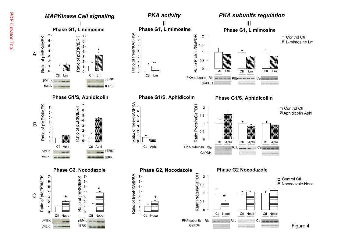
Cell-cycle progression is often associated with the modulation of PKA and MAP kinase 263
activity. Thus, we next assessed PKA and MAP kinase activity in synchronized cells. ERK1/2 264
phosphorylation, reflecting MAP kinase pathway activity, was significantly higher in all 265
synchronized groups than in asynchronous cells (Figure 4A-C I and Supplemental Figure 3). 266
MEK phosphorylation was also significantly higher in cells in G2 (Figure 4C I). Moreover, 267
there was significantly less PKA activity in cells in G1 (Figure 4A II), whereas it was 268
significantly higher in cells in G2 (Figure 4C II) than in asynchronous cells. 269
Under the same conditions, PKA R1A protein levels were markedly lower in cells at G2 270
(Figure 4C III), whereas those of PKA R2B and PKA CA were not significantly altered in any 271
of the studied groups (Figure 4A-C). 272
273
3.4 The effect of PKA activation on the control of the cell cycle and steroidogenesis in 274
synchronized H295R cells 275

13
The overexpression of steroidogenic enzyme genes at G2 is associated with an increase in the 276
level of PKA activity. We next investigated whether increased PKA activity effects the phases 277
of the cell cycle and steroidogenesis in synchronized cells. We thus increased PKA activity by 278
overexpressing PRKACA (Ca) or silencing PRKAR1A (Figure 5 I-II). PKA Ca was 279
overexpressed in transfected cells of the asynchronous control and all synchronized groups. 280
However, it was less highly overexpressed in the nocodazole group, for which the basal level 281
was much higher than that of the control asynchronous cells (Supplemental Figure 4A). The 282
expression of the PKA RI protein was reduced in all depleted PRKAR1A synchronized groups 283
(Supplemental Figure 4 B). However, the magnitude of the increase in Ca protein levels and 284
decrease in RIa protein levels depended on the phase of arrest. 285
Overexpression of PRKACA (Ca) for 24h in H295R cells had no effect on the cell cycle 286
(Figure 5A I) but increased both the activity of PKA and a luciferase reporter construct driven 287
by the STAR promoter (STAR-Luc) in all groups, independently of the phase of cell-cycle 288
arrest (Figure 5B and C I). STAR-Luc activity was the highest in cells in G2 (Figure 5C I). 289
Similarly, PKA activation by cAMP had no effect on the cell cycle (Supplemental Figure 5). 290
Transient depletion of PRKAR1A for 24 h was sufficient to increase the percentage/number of 291
cells in G2 in the asynchronous group and decreased the percentage of cells in G1 relative to 292
the control (siS, asynchronous cells; Figure 5A II). In L-mimosine treated cells, the 293
invalidation of PRKAR1A shifted the cells toward the S and G2 phases and decreased the 294
percentage of cells in G1 (p ≤ 0.02 siR1A vs siS). Simultaneous transfection with siR1A and 295
aphidicolin treatment for 24 h decreased the percentage/number of cells in G1 (p < 0.001 296
siR1A vs siS) and subsequently increased those in S (p < 0.05 siR1A vs siS). Simultaneous 297
nocodazole treatment and R1A silencing increased the percentage of cells in S and decreased 298
the percentage of those in G2 (p ≤ 0.05) (Figure 5A II). As expected, PRKAR1A silencing 299

14
enhanced PKA activity in all groups (Figure 5B II). Under the same conditions, PRKAR1A 300
silencing led to increased activity of the STAR-Luc reporter in the asynchronous group (p < 301
0.05) and L-mimosine treated cells (p < 0.05), whereas its activity remained unchanged in the 302
aphidicolin and nocodazole-treated cells (Figure 5C II). 303
3.5 Effect of PKA inhibition on control of the cell cycle in synchronized H295R cells 304
We confirmed the above data with the pharmacological inhibition of PKA activity by 305
treatment with the cell-permeable specific inhibitor Myr-PKI (14-22), which contains a PKA 306
substrate consensus sequence with the serine replaced by alanine (Wiley et al., 1999). The 307
major effect of PKA inhibition was observed in nocodazole treated cells (G2), for which there 308
was a decrease in the percentage of cells in G2 (p < 0.001) and an increase in the percentage 309
of those in S (p < 0.02) (Figure 6A). This effect was paralleled by a marked and significant 310
decrease in STAR-Luc activity in the nocodazole-treated cells (p < 0.001) (Figure 6B). 311
Overall, our results show that the PKA R1A subunit mediates the effect of PKA activity on 312
cell-cycle progression and the activation of steroidogenesis, depending on the phase of the 313
cell cycle. 314
315

15
4. Discussion. 316
Excess adrenal-steroid secretion can be explained by mutations of various key components of 317
the cAMP pathway that are also likely to be involved in adrenal tumorigenesis (Bertherat et 318
al., 2003;Beuschlein et al., 2014;Calebiro et al., 2014;Horvath et al., 2006;Horvath et al., 319
2008). Here, we demonstrate that steroidogenesis is modulated according to specific cell-320
cycle phases in both an established human adrenal cortical carcinoma cell line, H295R, and a 321
primary cell culture of PPNAD (harboring PKARIA inactivating mutations) from patients with 322
cortisol excess. 323
Major actors of steroidogenesis (NR5A, Star, and enzymes) were preferentially induced 324
during G2 (Figures 1, 2 and 3), leading to peak steroidogenesis during the same phase. 325
It is well known that cAMP/PKA and the MAP kinase activity vary during the cell cycle and 326
that MAP kinase activity is higher during the G2 phase in most cells (Philipova et al., 1998). 327
However, in most cells, cAMP levels and PKA activity are low during G2 and mitosis and 328
then become high in G1 (Bolte et al., 2003;Kurokawa et al., 1998;Yu et al., 2008). This 329
variation is cell-type specific, as the elevation of intracellular cyclic AMP (cAMP) is 330
considered to be a potent mitogenic signal for a number of cell types, including Swiss 3T3 331
cells, thyroid epithelial cells, and somatotroph cells of the anterior pituitary (Withers, 1997). 332
Here, we demonstrated that only the level of PKA activity varied notably according to the 333
stage of the cell cycle in the adrenocortical cell line H295R. PKA activity was high in G2 334
(Figure 4C II) and lower in G1 (Figure 4A II). Thus, both PKA and MAP kinase were 335
activated in G2 (Figure 4C, I, II). The change of PKA activity in H295R cells at G2 is a 336
molecular mechanism in which the cAMP/PKA pathway is specifically involved in the 337
control of mitosis, as it phosphorylates several substrates involved in its progression. The in 338
vivo dynamics of PKA activity during mitosis was highlighted by the study of Vandame P et 339

16
al. (Vandame et al., 2014) using FRET-based biosensors in single living Hela cells. They 340
showed that PKA activity is required for the maintenance of chromosome condensation and 341
that it may also be required during mitosis, because it phosphorylates histone H1.4, resulting 342
in the dissociation of this protein from the chromatin during mitosis. Moreover, Aurora A 343
kinase, a key protein for the progression of mitosis, is also a target of PKA. The high PKA 344
activity in G2 corresponded with a decrease in the level of the regulatory subunit R1A of 345
PKA (Figure 4C III). One explanation for the decrease in the level of PKA RIA protein is that 346
the increase in PKA activity at G2 liberates the PKA R1A protein from PKA CA, making it 347
more accessible to degradation. Another possibility is the consequence of the association of 348
PKA R1A with the cytoskeleton or microtubules through either low-affinity or highly 349
dynamic interactions, as shown in BW1J hepatoma cells (Imaizumi-Scherrer et al., 2001). 350
Mutations of PKA signaling components (GNAS, PRKAR1A, PRKACA, and 351
phosphodiesterase genes) in adrenal tumors activate the pathway (Nakajima et al., 2016;Mete 352
et al., 2018;Drougat et al., 2015). Although these mutations are associated with the 353
pathogenesis of benign tumors, we tested whether PKA may regulate the cell cycle and 354
steroidogenesis in a coordinated fashion by activating PKA in the adrenocortical cell line 355
H295R through overexpression of the PKA catalytic subunit alpha, stimulating the cells with 356
Bu2cAMP, or silencing PRKAR1A in combination with cell synchronization. The responses 357
of the cells to the activation of PKA were different depending on the PKA subunits used. 358
PRKACA overexpression stimulated the activity of a PKA-dependent construct (STAR-Luc) in 359
all groups but did not alter the phase of the cell cycle (Figure 5 A and C, I). The activation of 360
PKA by wild type PRKACA overexpression may not mimic the effect of PRKACA mutations, 361
as Bathon et al (Bathon et al., 2019) recently showed that transfection of HEK293 cells with 362
various PRKACA mutant constructs for 48 h highly increased PKA activity over that of wild 363
type levels and increased the phosphorylation of the specific PKA substrate histone H1.4, 364

17
involved in proliferation, relative to that by wild type PRKACA. Our result represents the 365
early effect of PKA activation (24-h transfection of H295R) by wild type PRKACA, which 366
indiscriminately activates substrates and not selected ones. Similarly, 24-h activation of PKA 367
by Bu2 cAMP did not alter the cell cycle profile, neither of asynchronous nor synchronized 368
cells. Our results are consistent with those of the study of Liu et al (Liu et al., 2004), in which 369
the effect of Bu2cAMP on proliferation and apoptosis was not observed at an early time point, 370
one to two days of incubation, but later, after three days to one week of incubation. 371
As expected and described by Basso et al. (Basso et al., 2014), inactivation of PRKAR1A in 372
asynchronous cells for 24 h increased both PKA and STAR-Luc reporter activity (Figure 5B 373
and C II) and led to the accumulation of the cells in G2 and a decrease in the number of cells 374
in G1 (Figure 5A II). In synchronized cells, PKA activity increased after PRKAR1A depletion. 375
The increase was greater in cells treated for G1 arrest than those treated for G1/S and G2 376
phase arrest. In contrast, the effects of the increase in PKA activity on steroidogenesis were 377
restricted to the cells treated for G1 arrest. In addition, the depletion of PRKAR1A induced 378
cell-cycle progression in all synchronized groups, as demonstrated by a significant decrease in 379
the percentage/number of cells in G1 reflecting progression to the S and then G2 phases 380
(Figure 5A II). 381
Moreover, PKA controlled progression of the cells through the cell cycle, as the inhibition of 382
PKA activity by PKI slowed the progression of cells with a high level of PKA activation and 383
those in G2 (Figure 6). 384
Thus, in our model, the essential and the early determinant of PKA activity as a regulator of 385
cell-cycle progression is the level of PKA R1A. However, our findings on malignant 386
adrenocortical cells that harbor different mutations (Tissier et al., 2005;Ragazzon et al., 2010) 387

18
might not apply to normal adrenocortical cells or benign adrenocortical tumors carrying 388
alterations in PKA signaling (Bathon et al., 2019). 389
PKA R1A can also interact directly with other specific proteins and modulates a number of 390
other signaling pathways. It has been demonstrated that PKA and PKA R1A are important for 391
regulation of the meiotic spindle in mammalian oocytes, in which increased PKA activity is 392
responsible for maintaining meiotic arrest. On the other hand, the knockdown of PRKAR1A, 393
as expected, increased PKA activity but the meiotic arrest of mouse oocytes was not 394
maintained (Duncan et al., 2006). PKA R1A is also associated with the second subunit of 395
replication factor C (RFC40) and functions as a nuclear transporter for RFC40, causing G1 396
arrest of MCF7 cells (Gupte et al., 2005). In the absence of PKA R1A, RFC40 may remain in 397
the cytoplasm and cells progress toward the G2 and M phases. 398
The role of PKA R1A on the control of cell-cycle progression can be explained by the spatial 399
regulation of PKA activity by A-kinase anchor proteins (AKAPs). AKAPs serve as scaffold 400
proteins to tether PKA in close proximity to its substrate, thereby locally regulating its 401
activity and PKA subunit specificity (Pidoux et al., 2010;Day et al., 2011). Recently, R1A 402
was shown to be associated with AKAPs, which form a complex with the ribosomal S6 403
protein kinase RSK1 kinase (Chaturvedi et al., 2009;Chaturvedi et al., 2006). Prkar1a 404
silencing in mouse lung fibroblasts (B82L) decreased the nuclear accumulation of active 405
RSK1 and increased its cytoplasmic localization. The resulting increase in active cytosolic 406
RSK1 levels is accompanied by increased phosphorylation of its cytosolic substrate tuberous 407
sclerosis complex 2 (TSC-2) and the anti-apoptotic factor BAD. As RSK1 shares common 408
phosphorylation sites with PKA on certain substrates, such as CREB, BAD, and Nur77, part 409
of the cell response in the absence of PKA R1A may not be due to increased PKA activity 410
(Chaturvedi et al., 2009) 411

19
In conclusion, we have provided evidence of a tight association between the phase of the cell 412
cycle and steroidogenesis in two adrenal tumors, PPNAD and the malignant H295R cell line. 413
Enhancing PKA activity clearly appears to be critical for steroid excess, whereas the PKA 414
R1A subunit is essential for triggering the cell cycle. 415
Declaration of interest: there is no conflict of interest that could be perceived to affect the 416
impartiality of the research reported. All authors declare that there is no conflict of interest. 417
Funding: this work was supported by the National Research Agency: grants ANR 14 CE12-418
000-02 and ANR 15 CE 12-0017 419
Acknowledgements: We thank the members of the FACS Core Facility CYBIO of the 420
Institute Cochin for their cooperation. The English of the manuscript has been corrected by a 421
native English-speaking scientific editor of Alex Edelman and Associates. 422
423

20
References 424 425 [1] Espiard, S., Ragazzon, B. and Bertherat, J., 2014. Protein kinase A alterations in 426
adrenocortical tumors, Horm Metab Res. 46, 869-75. 427 [2] Louiset, E., Duparc, C., Young, J., Renouf, S., Tetsi Nomigni, M., Boutelet, I., Libe, 428
R., Bram, Z., Groussin, L., Caron, P., Tabarin, A., Grunenberger, F., Christin-Maitre, 429 S., Bertagna, X., Kuhn, J.M., Anouar, Y., Bertherat, J. and Lefebvre, H., 2013. 430 Intraadrenal corticotropin in bilateral macronodular adrenal hyperplasia, N Engl J 431 Med. 369, 2115-25. 432
[3] Bassett, M.H., Mayhew, B., Rehman, K., White, P.C., Mantero, F., Arnaldi, G., 433 Stewart, P.M., Bujalska, I. and Rainey, W.E., 2005. Expression profiles for 434 steroidogenic enzymes in adrenocortical disease, J Clin Endocrinol Metab. 90, 5446-435 55. 436
[4] Nakamura, Y., Yamazaki, Y., Felizola, S.J., Ise, K., Morimoto, R., Satoh, F., Arai, Y. 437 and Sasano, H., 2015. Adrenocortical carcinoma: review of the pathologic features, 438 production of adrenal steroids, and molecular pathogenesis, Endocrinol Metab Clin 439 North Am. 44, 399-410. 440
[5] Abiven, G., Coste, J., Groussin, L., Anract, P., Tissier, F., Legmann, P., Dousset, B., 441 Bertagna, X. and Bertherat, J., 2006. Clinical and biological features in the prognosis 442 of adrenocortical cancer: poor outcome of cortisol-secreting tumors in a series of 202 443 consecutive patients, J Clin Endocrinol Metab. 91, 2650-5. 444
[6] Arlt, W., Biehl, M., Taylor, A.E., Hahner, S., Libe, R., Hughes, B.A., Schneider, P., 445 Smith, D.J., Stiekema, H., Krone, N., Porfiri, E., Opocher, G., Bertherat, J., Mantero, 446 F., Allolio, B., Terzolo, M., Nightingale, P., Shackleton, C.H., Bertagna, X., 447 Fassnacht, M. and Stewart, P.M., 2011. Urine steroid metabolomics as a biomarker 448 tool for detecting malignancy in adrenal tumors, J Clin Endocrinol Metab. 96, 3775-449 84. 450
[7] Ragazzon, B., Assie, G. and Bertherat, J., 2011. Transcriptome analysis of 451 adrenocortical cancers: from molecular classification to the identification of new 452 treatments, Endocr Relat Cancer. 18, R15-27. 453
[8] Stratakis, C.A., 2009. New genes and/or molecular pathways associated with adrenal 454 hyperplasias and related adrenocortical tumors, Mol Cell Endocrinol. 300, 152-7. 455
[9] Assie, G., Letouze, E., Fassnacht, M., Jouinot, A., Luscap, W., Barreau, O., Omeiri, 456 H., Rodriguez, S., Perlemoine, K., Rene-Corail, F., Elarouci, N., Sbiera, S., Kroiss, 457 M., Allolio, B., Waldmann, J., Quinkler, M., Mannelli, M., Mantero, F., Papathomas, 458 T., De Krijger, R., Tabarin, A., Kerlan, V., Baudin, E., Tissier, F., Dousset, B., 459 Groussin, L., Amar, L., Clauser, E., Bertagna, X., Ragazzon, B., Beuschlein, F., Libe, 460 R., de Reynies, A. and Bertherat, J., 2014. Integrated genomic characterization of 461 adrenocortical carcinoma, Nat Genet. 46, 607-12. 462
[10] De Martino, M.C., Al Ghuzlan, A., Aubert, S., Assie, G., Scoazec, J.Y., Leboulleux, 463 S., Do Cao, C., Libe, R., Nozieres, C., Lombes, M., Pattou, F., Borson-Chazot, F., 464 Hescot, S., Mazoyer, C., Young, J., Borget, I., Colao, A., Pivonello, R., Soria, J.C., 465 Bertherat, J., Schlumberger, M., Lacroix, L. and Baudin, E., 2013. Molecular 466 screening for a personalized treatment approach in advanced adrenocortical cancer, J 467 Clin Endocrinol Metab. 98, 4080-8. 468
[11] Ross, J.S., Wang, K., Rand, J.V., Gay, L., Presta, M.J., Sheehan, C.E., Ali, S.M., 469 Elvin, J.A., Labrecque, E., Hiemstra, C., Buell, J., Otto, G.A., Yelensky, R., Lipson, 470 D., Morosini, D., Chmielecki, J., Miller, V.A. and Stephens, P.J., 2014. Next-471 generation sequencing of adrenocortical carcinoma reveals new routes to targeted 472 therapies, J Clin Pathol. 67, 968-73. 473

21
[12] Pereira, S.S., Monteiro, M.P., Bourdeau, I., Lacroix, A. and Pignatelli, D., 2018. 474 MECHANISMS OF ENDOCRINOLOGY: Cell cycle regulation in adrenocortical 475 carcinoma, Eur J Endocrinol. 179, R95-R110. 476
[13] Kullmann, M.K., Grubbauer, C., Goetsch, K., Jakel, H., Podmirseg, S.R., 477 Trockenbacher, A., Ploner, C., Cato, A.C., Weiss, C., Kofler, R. and Hengst, L., 2013. 478 The p27-Skp2 axis mediates glucocorticoid-induced cell cycle arrest in T-lymphoma 479 cells, Cell Cycle. 12, 2625-35. 480
[14] Heisler, L.E., Evangelou, A., Lew, A.M., Trachtenberg, J., Elsholtz, H.P. and Brown, 481 T.J., 1997. Androgen-dependent cell cycle arrest and apoptotic death in PC-3 prostatic 482 cell cultures expressing a full-length human androgen receptor, Mol Cell Endocrinol. 483 126, 59-73. 484
[15] Amanatullah, D.F., Zafonte, B.T. and Pestell, R.G., 2002. The cell cycle in steroid 485 hormone regulated proliferation and differentiation, Minerva Endocrinol. 27, 7-20. 486
[16] Cerquetti, L., Bucci, B., Marchese, R., Misiti, S., De Paula, U., Miceli, R., Muleti, A., 487 Amendola, D., Piergrossi, P., Brunetti, E., Toscano, V. and Stigliano, A., 2008. 488 Mitotane increases the radiotherapy inhibitory effect and induces G2-arrest in 489 combined treatment on both H295R and SW13 adrenocortical cell lines, Endocr Relat 490 Cancer. 15, 623-34. 491
[17] Doghman, M. and Lalli, E., 2013. Lack of long-lasting effects of mitotane adjuvant 492 therapy in a mouse xenograft model of adrenocortical carcinoma, Mol Cell 493 Endocrinol. 381, 66-9. 494
[18] Hescot, S., Slama, A., Lombes, A., Paci, A., Remy, H., Leboulleux, S., Chadarevian, 495 R., Trabado, S., Amazit, L., Young, J., Baudin, E. and Lombes, M., 2013. Mitotane 496 alters mitochondrial respiratory chain activity by inducing cytochrome c oxidase 497 defect in human adrenocortical cells, Endocr Relat Cancer. 20, 371-81. 498
[19] Lodish, M. and Stratakis, C.A., 2016. A genetic and molecular update on 499 adrenocortical causes of Cushing syndrome, Nat Rev Endocrinol. 12, 255-62. 500
[20] Kirschner, L.S., Sandrini, F., Monbo, J., Lin, J.P., Carney, J.A. and Stratakis, C.A., 501 2000. Genetic heterogeneity and spectrum of mutations of the PRKAR1A gene in 502 patients with the carney complex, Hum Mol Genet. 9, 3037-46. 503
[21] Bertherat, J., Groussin, L., Sandrini, F., Matyakhina, L., Bei, T., Stergiopoulos, S., 504 Papageorgiou, T., Bourdeau, I., Kirschner, L.S., Vincent-Dejean, C., Perlemoine, K., 505 Gicquel, C., Bertagna, X. and Stratakis, C.A., 2003. Molecular and functional analysis 506 of PRKAR1A and its locus (17q22-24) in sporadic adrenocortical tumors: 17q losses, 507 somatic mutations, and protein kinase A expression and activity, Cancer Res. 63, 508 5308-19. 509
[22] Zheng, S., Cherniack, A.D., Dewal, N., Moffitt, R.A., Danilova, L., Murray, B.A., 510 Lerario, A.M., Else, T., Knijnenburg, T.A., Ciriello, G., Kim, S., Assie, G., Morozova, 511 O., Akbani, R., Shih, J., Hoadley, K.A., Choueiri, T.K., Waldmann, J., Mete, O., 512 Robertson, A.G., Wu, H.T., Raphael, B.J., Shao, L., Meyerson, M., Demeure, M.J., 513 Beuschlein, F., Gill, A.J., Sidhu, S.B., Almeida, M.Q., Fragoso, M., Cope, L.M., 514 Kebebew, E., Habra, M.A., Whitsett, T.G., Bussey, K.J., Rainey, W.E., Asa, S.L., 515 Bertherat, J., Fassnacht, M., Wheeler, D.A., Hammer, G.D., Giordano, T.J. and 516 Verhaak, R.G.W., 2016. Comprehensive Pan-Genomic Characterization of 517 Adrenocortical Carcinoma, Cancer Cell. 29, 723-736. 518
[23] Beuschlein, F., Fassnacht, M., Assie, G., Calebiro, D., Stratakis, C.A., Osswald, A., 519 Ronchi, C.L., Wieland, T., Sbiera, S., Faucz, F.R., Schaak, K., Schmittfull, A., 520 Schwarzmayr, T., Barreau, O., Vezzosi, D., Rizk-Rabin, M., Zabel, U., Szarek, E., 521 Salpea, P., Forlino, A., Vetro, A., Zuffardi, O., Kisker, C., Diener, S., Meitinger, T., 522 Lohse, M.J., Reincke, M., Bertherat, J., Strom, T.M. and Allolio, B., 2014. 523

22
Constitutive activation of PKA catalytic subunit in adrenal Cushing's syndrome, N 524 Engl J Med. 370, 1019-28. 525
[24] Goh, G., Scholl, U.I., Healy, J.M., Choi, M., Prasad, M.L., Nelson-Williams, C., 526 Kunstman, J.W., Korah, R., Suttorp, A.C., Dietrich, D., Haase, M., Willenberg, H.S., 527 Stalberg, P., Hellman, P., Akerstrom, G., Bjorklund, P., Carling, T. and Lifton, R.P., 528 2014. Recurrent activating mutation in PRKACA in cortisol-producing adrenal 529 tumors, Nat Genet. 46, 613-7. 530
[25] Calebiro, D., Hannawacker, A., Lyga, S., Bathon, K., Zabel, U., Ronchi, C., 531 Beuschlein, F., Reincke, M., Lorenz, K., Allolio, B., Kisker, C., Fassnacht, M. and 532 Lohse, M.J., 2014. PKA catalytic subunit mutations in adrenocortical Cushing's 533 adenoma impair association with the regulatory subunit, Nat Commun. 5, 5680. 534
[26] Cao, Y., He, M., Gao, Z., Peng, Y., Li, Y., Li, L., Zhou, W., Li, X., Zhong, X., Lei, 535 Y., Su, T., Wang, H., Jiang, Y., Yang, L., Wei, W., Yang, X., Jiang, X., Liu, L., He, J., 536 Ye, J., Wei, Q., Li, Y., Wang, W., Wang, J. and Ning, G., 2014. Activating hotspot 537 L205R mutation in PRKACA and adrenal Cushing's syndrome, Science. 344, 913-7. 538
[27] Sato, Y., Maekawa, S., Ishii, R., Sanada, M., Morikawa, T., Shiraishi, Y., Yoshida, K., 539 Nagata, Y., Sato-Otsubo, A., Yoshizato, T., Suzuki, H., Shiozawa, Y., Kataoka, K., 540 Kon, A., Aoki, K., Chiba, K., Tanaka, H., Kume, H., Miyano, S., Fukayama, M., 541 Nureki, O., Homma, Y. and Ogawa, S., 2014. Recurrent somatic mutations underlie 542 corticotropin-independent Cushing's syndrome, Science. 344, 917-20. 543
[28] Massimi, M., Cardarelli, S., Galli, F., Giardi, M.F., Ragusa, F., Panera, N., Cinque, B., 544 Cifone, M.G., Biagioni, S. and Giorgi, M., 2017. Increase of Intracellular Cyclic AMP 545 by PDE4 Inhibitors Affects HepG2 Cell Cycle Progression and Survival, J Cell 546 Biochem. 118, 1401-1411. 547
[29] Gupta, A., Tiwari, M., Prasad, S. and Chaube, S.K., 2017. Role of Cyclic Nucleotide 548 Phosphodiesterases During Meiotic Resumption From Diplotene Arrest in Mammalian 549 Oocytes, J Cell Biochem. 118, 446-452. 550
[30] Sun, Q.Y., Miao, Y.L. and Schatten, H., 2009. Towards a new understanding on the 551 regulation of mammalian oocyte meiosis resumption, Cell Cycle. 8, 2741-7. 552
[31] Duncan, F.E., Moss, S.B. and Williams, C.J., 2006. Knockdown of the cAMP-553 dependent protein kinase (PKA) Type Ialpha regulatory subunit in mouse oocytes 554 disrupts meiotic arrest and results in meiotic spindle defects, Dev Dyn. 235, 2961-8. 555
[32] Hewer, R.C., Sala-Newby, G.B., Wu, Y.J., Newby, A.C. and Bond, M., 2011. PKA 556 and Epac synergistically inhibit smooth muscle cell proliferation, J Mol Cell Cardiol. 557 50, 87-98. 558
[33] Medri, G., Sergi, I., Papandreou, M.J., Beck-Peccoz, P., Verrier, B. and Ronin, C., 559 1994. Dual activity of human pituitary thyrotrophin isoforms on thyroid cell growth, J 560 Mol Endocrinol. 13, 187-98. 561
[34] Spada, A., Vallar, L. and Faglia, G., 1992. G protein oncogenes in pituitary tumors, 562 Trends Endocrinol Metab. 3, 355-60. 563
[35] Taylor, S.S., Zhang, P., Steichen, J.M., Keshwani, M.M. and Kornev, A.P., 2013. 564 PKA: lessons learned after twenty years, Biochim Biophys Acta. 1834, 1271-8. 565
[36] Kirschner, L.S., Kusewitt, D.F., Matyakhina, L., Towns, W.H., 2nd, Carney, J.A., 566 Westphal, H. and Stratakis, C.A., 2005. A mouse model for the Carney complex tumor 567 syndrome develops neoplasia in cyclic AMP-responsive tissues, Cancer Res. 65, 568 4506-14. 569
[37] Nadella, K.S. and Kirschner, L.S., 2005. Disruption of protein kinase a regulation 570 causes immortalization and dysregulation of D-type cyclins, Cancer Res. 65, 10307-571 15. 572

23
[38] Basso, F., Rocchetti, F., Rodriguez, S., Nesterova, M., Cormier, F., Stratakis, C.A., 573 Ragazzon, B., Bertherat, J. and Rizk-Rabin, M., 2014. Comparison of the effects of 574 PRKAR1A and PRKAR2B depletion on signaling pathways, cell growth, and cell 575 cycle control of adrenocortical cells, Horm Metab Res. 46, 883-8. 576
[39] Gazdar, A.F., Oie, H.K., Shackleton, C.H., Chen, T.R., Triche, T.J., Myers, C.E., 577 Chrousos, G.P., Brennan, M.F., Stein, C.A. and La Rocca, R.V., 1990. Establishment 578 and characterization of a human adrenocortical carcinoma cell line that expresses 579 multiple pathways of steroid biosynthesis, Cancer Res. 50, 5488-96. 580
[40] Weigand, I., Ronchi, C.L., Rizk-Rabin, M., Dalmazi, G.D., Wild, V., Bathon, K., 581 Rubin, B., Calebiro, D., Beuschlein, F., Bertherat, J., Fassnacht, M. and Sbiera, S., 582 2017. Differential expression of the protein kinase A subunits in normal adrenal 583 glands and adrenocortical adenomas, Sci Rep. 7, 49. 584
[41] Bouizar, Z., Ragazzon, B., Viou, L., Hortane, M., Bertherat, J. and Rizk-Rabin, M., 585 2010. 8Cl-cAMP modifies the balance between PKAR1 and PKAR2 and modulates 586 the cell cycle, growth and apoptosis in human adrenocortical H295R cells, J Mol 587 Endocrinol. 44, 331-47. 588
[42] Ragazzon, B., Cazabat, L., Rizk-Rabin, M., Assie, G., Groussin, L., Fierrard, H., 589 Perlemoine, K., Martinez, A. and Bertherat, J., 2009. Inactivation of the Carney 590 complex gene 1 (protein kinase A regulatory subunit 1A) inhibits SMAD3 expression 591 and TGF beta-stimulated apoptosis in adrenocortical cells, Cancer Res. 69, 7278-84. 592
[43] Groussin, L., Massias, J.F., Bertagna, X. and Bertherat, J., 2000. Loss of expression of 593 the ubiquitous transcription factor cAMP response element-binding protein (CREB) 594 and compensatory overexpression of the activator CREMtau in the human 595 adrenocortical cancer cell line H295R, J Clin Endocrinol Metab. 85, 345-54. 596
[44] Cazabat, L., Ragazzon, B., Varin, A., Potier-Cartereau, M., Vandier, C., Vezzosi, D., 597 Risk-Rabin, M., Guellich, A., Schittl, J., Lechene, P., Richter, W., Nikolaev, V.O., 598 Zhang, J., Bertherat, J. and Vandecasteele, G., 2014. Inactivation of the Carney 599 complex gene 1 (PRKAR1A) alters spatiotemporal regulation of cAMP and cAMP-600 dependent protein kinase: a study using genetically encoded FRET-based reporters, 601 Hum Mol Genet. 23, 1163-74. 602
[45] Bram, Z., Louiset, E., Ragazzon, B., Renouf, S., Wils, J., Duparc, C., Boutelet, I., 603 Rizk-Rabin, M., Libe, R., Young, J., Carson, D., Vantyghem, M.C., Szarek, E., 604 Martinez, A., Stratakis, C.A., Bertherat, J. and Lefebvre, H., 2016. PKA regulatory 605 subunit 1A inactivating mutation induces serotonin signaling in primary pigmented 606 nodular adrenal disease, JCI Insight. 1, e87958. 607
[46] Rizk-Rabin, M., Assie, G., Rene-Corail, F., Perlemoine, K., Hamzaoui, H., Tissier, F., 608 Lieberherr, M., Bertagna, X., Bertherat, J. and Bouizar, Z., 2008. Differential 609 expression of parathyroid hormone-related protein in adrenocortical tumors: 610 autocrine/paracrine effects on the growth and signaling pathways in H295R cells, 611 Cancer Epidemiol Biomarkers Prev. 17, 2275-85. 612
[47] Vackova, I., Engelova, M., Marinov, I. and Tomanek, M., 2003. Cell cycle 613 synchronization of porcine granulosa cells in G1 stage with mimosine, Anim Reprod 614 Sci. 77, 235-45. 615
[48] Sorscher, D.H. and Cordeiro-Stone, M., 1991. Gene replication in the presence of 616 aphidicolin, Biochemistry. 30, 1086-90. 617
[49] Ho, Y.S., Duh, J.S., Jeng, J.H., Wang, Y.J., Liang, Y.C., Lin, C.H., Tseng, C.J., Yu, 618 C.F., Chen, R.J. and Lin, J.K., 2001. Griseofulvin potentiates antitumorigenesis effects 619 of nocodazole through induction of apoptosis and G2/M cell cycle arrest in human 620 colorectal cancer cells, Int J Cancer. 91, 393-401. 621

24
[50] Wiley, J.C., Wailes, L.A., Idzerda, R.L. and McKnight, G.S., 1999. Role of regulatory 622 subunits and protein kinase inhibitor (PKI) in determining nuclear localization and 623 activity of the catalytic subunit of protein kinase A, J Biol Chem. 274, 6381-7. 624
[51] Horvath, A., Boikos, S., Giatzakis, C., Robinson-White, A., Groussin, L., Griffin, K.J., 625 Stein, E., Levine, E., Delimpasi, G., Hsiao, H.P., Keil, M., Heyerdahl, S., Matyakhina, 626 L., Libe, R., Fratticci, A., Kirschner, L.S., Cramer, K., Gaillard, R.C., Bertagna, X., 627 Carney, J.A., Bertherat, J., Bossis, I. and Stratakis, C.A., 2006. A genome-wide scan 628 identifies mutations in the gene encoding phosphodiesterase 11A4 (PDE11A) in 629 individuals with adrenocortical hyperplasia, Nat Genet. 38, 794-800. 630
[52] Horvath, A., Giatzakis, C., Tsang, K., Greene, E., Osorio, P., Boikos, S., Libe, R., 631 Patronas, Y., Robinson-White, A., Remmers, E., Bertherat, J., Nesterova, M. and 632 Stratakis, C.A., 2008. A cAMP-specific phosphodiesterase (PDE8B) that is mutated in 633 adrenal hyperplasia is expressed widely in human and mouse tissues: a novel PDE8B 634 isoform in human adrenal cortex, Eur J Hum Genet. 16, 1245-53. 635
[53] Philipova, R. and Whitaker, M., 1998. MAP kinase activity increases during mitosis in 636 early sea urchin embryos, J Cell Sci. 111 (Pt 17), 2497-505. 637
[54] Bolte, M., Dieckhoff, P., Krause, C., Braus, G.H. and Irniger, S., 2003. Synergistic 638 inhibition of APC/C by glucose and activated Ras proteins can be mediated by each of 639 the Tpk1-3 proteins in Saccharomyces cerevisiae, Microbiology. 149, 1205-16. 640
[55] Kurokawa, K. and Kato, J., 1998. Cyclic AMP delays G2 progression and prevents 641 efficient accumulation of cyclin B1 proteins in mouse macrophage cells, Cell Struct 642 Funct. 23, 357-65. 643
[56] Yu, A., Zhang, Z., Bi, Q., Sun, B., Su, W., Guan, Y., Mu, R., Miao, C., Zhang, J. and 644 Yu, B., 2008. Regulation of cAMP on the first mitotic cell cycle of mouse embryos, 645 Mol Reprod Dev. 75, 489-95. 646
[57] Withers, D.J., 1997. Signalling pathways involved in the mitogenic effects of cAMP, 647 Clin Sci (Lond). 92, 445-51. 648
[58] Vandame, P., Spriet, C., Trinel, D., Gelaude, A., Caillau, K., Bompard, C., Biondi, E. 649 and Bodart, J.F., 2014. The spatio-temporal dynamics of PKA activity profile during 650 mitosis and its correlation to chromosome segregation, Cell Cycle. 13, 3232-40. 651
[59] Imaizumi-Scherrer, T., Faust, D.M., Barradeau, S., Hellio, R. and Weiss, M.C., 2001. 652 Type I protein kinase a is localized to interphase microtubules and strongly associated 653 with the mitotic spindle, Exp Cell Res. 264, 250-65. 654
[60] Nakajima, Y., Okamura, T., Horiguchi, K., Gohko, T., Miyamoto, T., Satoh, T., 655 Ozawa, A., Ishii, S., Yamada, E., Hashimoto, K., Okada, S., Takata, D., Horiguchi, J. 656 and Yamada, M., 2016. GNAS mutations in adrenal aldosterone-producing adenomas, 657 Endocr J. 63, 199-204. 658
[61] Mete, O. and Duan, K., 2018. The Many Faces of Primary Aldosteronism and Cushing 659 Syndrome: A Reflection of Adrenocortical Tumor Heterogeneity, Front Med 660 (Lausanne). 5, 54. 661
[62] Drougat, L., Omeiri, H., Lefevre, L. and Ragazzon, B., 2015. Novel Insights into the 662 Genetics and Pathophysiology of Adrenocortical Tumors, Front Endocrinol 663 (Lausanne). 6, 96. 664
[63] Bathon, K., Weigand, I., Vanselow, J.T., Ronchi, C.L., Sbiera, S., Schlosser, A., 665 Fassnacht, M. and Calebiro, D., 2019. Alterations in Protein Kinase A Substrate 666 Specificity as a Potential Cause of Cushing Syndrome, Endocrinology. 160, 447-459. 667
[64] Liu, J., Li, X.D., Ora, A., Heikkila, P., Vaheri, A. and Voutilainen, R., 2004. cAMP-668 dependent protein kinase activation inhibits proliferation and enhances apoptotic 669 effect of tumor necrosis factor-alpha in NCI-H295R adrenocortical cells, J Mol 670 Endocrinol. 33, 511-22. 671

25
[65] Tissier, F., Cavard, C., Groussin, L., Perlemoine, K., Fumey, G., Hagnere, A.M., 672 Rene-Corail, F., Jullian, E., Gicquel, C., Bertagna, X., Vacher-Lavenu, M.C., Perret, 673 C. and Bertherat, J., 2005. Mutations of beta-catenin in adrenocortical tumors: 674 activation of the Wnt signaling pathway is a frequent event in both benign and 675 malignant adrenocortical tumors, Cancer Res. 65, 7622-7. 676
[66] Ragazzon, B., Libe, R., Gaujoux, S., Assie, G., Fratticci, A., Launay, P., Clauser, E., 677 Bertagna, X., Tissier, F., de Reynies, A. and Bertherat, J., 2010. Transcriptome 678 analysis reveals that p53 and {beta}-catenin alterations occur in a group of aggressive 679 adrenocortical cancers, Cancer Res. 70, 8276-81. 680
[67] Gupte, R.S., Pozarowski, P., Grabarek, J., Traganos, F., Darzynkiewicz, Z. and Lee, 681 M.Y., 2005. RIalpha influences cellular proliferation in cancer cells by transporting 682 RFC40 into the nucleus, Cancer Biol Ther. 4, 429-37. 683
[68] Pidoux, G. and Tasken, K., 2010. Specificity and spatial dynamics of protein kinase A 684 signaling organized by A-kinase-anchoring proteins, J Mol Endocrinol. 44, 271-84. 685
[69] Day, M.E., Gaietta, G.M., Sastri, M., Koller, A., Mackey, M.R., Scott, J.D., Perkins, 686 G.A., Ellisman, M.H. and Taylor, S.S., 2011. Isoform-specific targeting of PKA to 687 multivesicular bodies, J Cell Biol. 193, 347-63. 688
[70] Chaturvedi, D., Cohen, M.S., Taunton, J. and Patel, T.B., 2009. The PKARIalpha 689 subunit of protein kinase A modulates the activation of p90RSK1 and its function, J 690 Biol Chem. 284, 23670-81. 691
[71] Chaturvedi, D., Poppleton, H.M., Stringfield, T., Barbier, A. and Patel, T.B., 2006. 692 Subcellular localization and biological actions of activated RSK1 are determined by 693 its interactions with subunits of cyclic AMP-dependent protein kinase, Mol Cell Biol. 694 26, 4586-600. 695
696 697 698
699
700
701
702
703
704
705
706
707
708
709

26
Figure legends 710
Figure 1. The effect of cell-cycle synchronization of the H295R cell line and primary PPNAD 711
cell culture for 24 h on the cell-cycle profile and steroid secretion. 712
Flowcytometry analysis of DNA content by propidium iodide incorporation in synchronized 713
H295R cells and PPNAD primary culture for A) control cultures without treatment and 714
cultures treated with B) L-mimosine, C) aphidicolin, or D) nocodazole. Histograms of cortisol 715
and aldosterone content secreted into the cell medium after cell-cycle synchronization are 716
shown to the right. The results are presented as the means ± SEM of three duplicate 717
independent experiments for H295R and one representative example of two duplicate 718
experiments for PPNAD. *p < 0.05, ** p< 0.01, ***p < 0.001. 719
Figure 2. The effect of cell synchronization on steroidogenic enzyme and Star protein 720
expression, mRNA expression by RT/PCR of steroidogenic enzymes in H295R cells and 721
PPNAD primary cultures in the A) G1, B) G1/S, and C) G2 phases of the cell cycle. Each 722
studied steroidogneic enzyme is normalized to the corresponding results in the control 723
asynchronous cells, which is set to one. For Star protein expression, the results are normalized 724
against those of control cells and a histogram of the western blot in synchronized H295R cells 725
and PPNAD primary cultures are shown on the right. The results are presented as the means ± 726
SEM of three duplicate independent experiments for H295R and one representative example 727
of two duplicate experiments for PPNAD. *p < 0.05, **p < 0.01, ***p < 0.001. 728
Figure 3. Cell-cycle profile and the kinetics of steroidogenic enzyme expression after cell-729
cycle arrest and release of H295R cells. 0 h represents the profile at the end of 730
synchronization after 24 h of treatment with L-mimosine (I) or 48 h with nocodazole (II). 731
Each time point is normalized to asynchronous control cells at 0 h of release. A) Histogram 732
showing the percentage of cells in each phase of the cell cycle at each time point, analyzed by 733

27
flow cytometry, showing the progression in the cell cycle. RT/PCR analysis of B) 734
steroidogenic enzyme and C) SF-1 and Star mRNA levels for each time point. The results are 735
presented as the means ± SEM of three duplicate independent experiments. The level of 736
significance is indicated at the top of each figure. 737
Figure 4. Effect of the different drugs on cell signaling pathways and PKA subunit regulation 738
in H295R cells. The effects are shown for A) L-mimosine, B) aphidicolin, and C) nocodazole. 739
MAP Kinase activity is shown by histograms of western blots of pERK/total ERK, and 740
pMEK/total MEK protein levels normalized to the control, set to one. We spliced the original 741
full gel images of ERK and MEK, presented in Supplementary Figure 3, to re-order the 742
samples in a more logical manner. PKA activity was measured by a nonradioactive PKA 743
assay and is presented as the ratio of free PKA/total PKA. Protein levels of the PKA subunits 744
R1A, R2B, and Ca in cells treated with the different drugs are shown by histograms of 745
western blots. The results of the histograms are expressed as the percentage relative band 746
density/the band density of GaPDH, normalized to the corresponding controls (arbitrarily set 747
to one) in H295R-treated cells. The results are presented as the means ± SEM of three 748
duplicate independent experiments. *p < 0.05, **p < 0.01. 749
Figure 5. Effect of PKA activation, by the over expression of PRKACA or PRKAR1A 750
depletion, on the regulation of the cell cycle, PKA activity, and steroidogenic response 751
(Star/luc-reporter gene activity) simultaneously with the synchronization of H295R cells. A) 752
Cell-cycle distribution analyzed by flow cytometry. B) PKA activity measured by a non-753
radioactive assay and presented as the ratio of free phosphorylated PKA/total PKA. C) STAR 754
reporter gene activity measured by Luciferase/Renilla. The results are presented as the means 755
± SEM of three duplicate independent experiments normalized to the control of each group, 756

28
set to one. *p < 0.05, **p < 0.01, ***p < 0.001, ****p < 0.0001 for the over expression of 757
PKA CA. 758
Figure 6. Inhibition of PKA activity by the PKA inhibitor PKI (5 µM) throughout cell cycle 759
synchronization (24 h) showing that mostly the nocodazole-treated cells were affected. A) 760
Cell-cycle phase distribution. B) Star/luc reporter gene activity. The measurement of the PKA 761
activity is not presented because PKA activity was inhibited and thus no PKA 762
phosphorylation. The results are presented as the means ± SEM of three duplicate 763
independent experiments. * p < 0.05, **p < 0.01, ***p < 0.001. 764
765
Supplementary Figures: 766
Supplementary Figure 1. Distribution of cyclin mRNA expression in in H295R cells arrested 767
at various phases of the cell cycle. A) Arrested in early G1 by L-mimosine treatment. B) 768
Arrested in late G1/S by aphidicolin treatment. C) Arrested in G2 by nocodazole treatment. 769
The results are presented as the means ± SEM of three duplicate independent experiments 770
normalized to the control of each group, set to one. *p < 0.05, **p < 0.01, ***p < 0.001. 771
Supplementary Figure 2. Star mRNA expression by RT/PCR in H295R cells and PPNAD 772
primary cultures in the G1, G1/S, and G2 phases of the cell cycle. The results are presented as 773
the means ± SEM of three duplicate independent experiments for H295R and one 774
representative example of two duplicate experiments for PPNAD. *p < 0.05, **p < 0.01,***p 775
< 0.001. 776
Supplementary Figure 3. Image of the full western blot of MAPKinase cell signaling used for 777
Figure 4. A) Image of the full western blot. B) The corresponding Histogram, as in Figure 4. 778
779

29
Supplementary Figure 4. PKA CA and PKA R1A protein expression in simultaneous A) 780
overexpression of wildtype PRKACA or B) depleted PRKAR1A and cell-cycle 781
synchronization. Results are normalized to the control vector or sis, set to one. The 782
comparison is inter group (Vector vs Ca or SiS vs siRI ) for each group. The results are 783
presented as the means ± SEM of three duplicate independent experiments normalized to the 784
control of each group, set to one. *p < 0.05, **p < 0.01, ***p < 0.001. 785
Supplementary Figure 5. Effects of cAMP stimulation on cell-cycle synchronization. The 786
distribution of the phases resulting from cell-cycle arrest were not affected by stimulation 787
with the cAMP analogue (dibutyryl adenosine 3’-5’ cyclic monophosphate). The 788
measurement of PKA activity and Star Luc reporter gene activity are not presented because 789
they are equally fullyl saturated in all of the synchronized groups. 790

Cell cycle synchronization
H295R
Aphidicolin
Cell
num
ber
Nocodazole
DNA content
L-mimosine
DNA content
Cell
num
ber
H295R
0
1
2
Cortisol Aldo
Phase G2
Cortisol Aldo
** **
Phase G1/S
0
1
2
0
1
2
Cortisol Aldo
Phase G1
** **
Steroids secretion
Phase G1
control
Lmimosine
Phase G1/S
Phase G2
*
Cort
isol nm
ol/ng
AR
N/µ
l
Cortisol
Cortisol
Cortisol
PPNAD
0
1
2
0
1
2
control
Aphidicolin
control
Nocodazole
0
1
2
Control
Cell
num
ber
DNA content
PPNAD
DNA content
Cell
num
ber
Figure 1
Control
DNA content
Nocodazole
DNA content
Aphidicolin
L-mimosine
Cell
num
ber
DNA content
Cell
num
ber
DNA content
Cell
num
ber
Cell
num
ber
A
B
C
D
0 0.2 0.4 0.6 0.8
G1 45 %
S 34%
G2 21%
0
10
0 2
00
30
0 4
00
50
0
G1 67%
S 27 %
G2 1%
0
10
0 2
00
30
0 4
00
50
0
0 0.2 0.4 0.6 0.8
0 0.2 0.4 0.6 0.8
G1 70 %
S 20 %
G2 10 %
0
10
0 2
00
30
0 4
00
50
0
G1: 34%
S :21%
G2: 45%
0
8
0 1
60
2
40
3
20
38
5
0 0.2 0.4 0.6 0.8
I II III IV
0 32 64 96 128 160
0 1
50 3
00
450
600
750 G1 51.2%
S 24.8 %
G2 23.8 %
0 32 64 96 128 160
0 1
10
22
0 3
30
44
0 5
50
G1 56%
S 37 %
G2 10%
0 32 64 96 128 160
0
11
0
22
0 3
30
44
0 5
50
G1 67.2%
S 13.5 %
G2 15 %
0
6
0 1
20
18
0 2
40
30
0 3
60
0 32 64 96 128 160
G1 25.3%
S 37 %
G2 37.7 %
G1 25.3%
S 37 %
G2 37.7 %

A
B
Steroidogenic Enzymes
Cyp0
1
2
3 Phase G1
11B1 11B211A1 17 21
*
Phase G1/S
11B1 11B211A1 17 210
1
2
3
Cyp
*
11B1 11B211A1 17 21Cyp
Phase G2
**
***
**
H295R PPNAD
PPNAD
Cyp
Phase G1
21 11B1 11B211A1 170
1
2
3
Cyp 11A1 170
1
2
3
21 11B1 11B2
Phase G1/S
Cyp
Phase G2
*
**
**
0
1
2
3
4
21 11B1 11B211A1 17
**
*
*
*Re
lative
mR
NA
/cyclo
ph
ilin
fold
chan
ge
/co
ntr
ol se
t to
one
C
Expression of Star
0
1
2
3
4
Star
b Actin
G1 G1/S G2Ctl0
0,5
1
1,5
2
2,5
3
**
**
*
Star/actin
Re
lative
le
ve
lof
Sta
r/actin
con
tro
l se
t to
one
Figure 2
I II IIIH295R
control
Lmimosine
Aphidicolin
Nocodazole
Star
b Actin
Ctl G1 G1/S G2
0
0,5
1
1,5
2
2,5Star/actin
**
**

Release from G1 L -mimosine synchronization
%G2
%S
%G153.2
37.1
9,7
57.4
37.8
4,8
42.5
44.8
12.7
33.5
34.7
31.6
38.3
29.3
32.5
39.7
50.7
9,5
0h 6h 12h 24h 36h 48h0%
20%
40%
60%
80%
100%
Re
lative
mR
NA
fold
chan
ge
/0h
le
ve
l
Release from G2 nocodazole synchronization
11.7
16.8
71.5
26.4
33.5
40.1
32.4
32.0
35.6
36.8
44.0
19.1
45.8
34.9
19.2
37.4
33.0
29.6
0%
20%
40%
60%
80%
100%
0h 6h 12h 24h 36h 48h
%G2
%S
%G1
Steroidogenic enzymes
A
B
C
Star and NR5A1 (SF-1)
CYP11A1
Cyp11B1
Cyp11B2
0
2
4
6
8
10
0h 6h 12h 24h 36h 48h
P=0.0001
ns
P=0.0001
P=0.001
ns
P=0.0001
P=0.0001
ns
P=0.002
P<0.0001
P<0.0001
P<0.0001
12h 24h 36h 48h
Cyp11A1Cyp11B1Cyp11B2
0h 6h 12h 24h 36h 48h0
1
2
3
4
5
SF-1
Star
12h 24h 36h 48h
ns
P=0.001P<0.0001
P<0.0001P=0.05
P=0.001
ns
P=0.001
SF-1
Star
0
1
2
3
4
5
0h 6h 12h 24h 36h 48h
SF-1
Star
P=0.01
P=0.05
ns
P=0.05
36h 48h
SF-1Star
02
4
6
8
10
12
14
0h 6h 12h 24h 36h 48h
CYP11A1
Cyp11B1
Cyp11B2
ns
ns
P=0.04
ns
ns
P=0.005
ns
ns
P=0.002
P=0.02
P=0.02
ns
P=0.05
P=0.001
ns
6h 12h 24h 36h 48h
Cyp11A1
Cyp11B1Cyp11B2
Figure 3
I II
Phase G1, L mimosine
Ctl Lm
*
0
1
2
3
4
5
6
7
0
1
2
3
4
5
6
**
Phase G1/S, Aphidicolin
0
1
2
3
4
5
6
7
**
0
1
2
3
4
5
6
7
*
tMEK
pMEK pERK
tERK
MAPKinase Cell signaling
tMEK
pMEK pERK
tERK
PKA subunits regulation
0
0,5
1
1,5
2
RIa RIIb Ca
Control Ctl
L-mimosine Lm
0
0,5
1
1,5
2
RIa RIIb Ca
Control Ctl
Aphidicolin Aphi
0
0,5
1
1,5
2
RIa RIIb Ca
Control Ctl
Nocodazole Noco
Phase G1, L mimosine
Phase G1/S, Aphidicollin
Phase G2 Nocodazole
GaPDH
GaPDH
PKA subunits
PKA subunits
PKA subunits
GaPDH
*
A
B
0
1
2
3
4
5
6
7
0
1
2
3
4
5
6R
atio o
f pM
EK
/tM
EK
Ratio o
f fr
eeP
KA
/tP
KA
0
1
2
3
4
5
6
7
0
1
2
3
4
5
6
Ratio o
f pE
RK
/tE
RK
Ratio o
f fr
eeP
KA
/tP
KA
0
1
2
3
4
5
6
7
0
1
2
3
4
5
6
Ratio o
f pE
RK
/tE
RK
0
1
2
3
4
5
6
7
0
1
2
3
4
5
6
Ratio o
f pM
EK
/tM
EK
0
1
2
3
4
5
6
7
0
1
2
3
4
5
6
7
Ratio o
f fr
eeP
KA
/tP
KA
Ctl Lm Ctl Lm
Ctl Aphi Ctl Aphi Ctl Aphi
Ctl Noco
**
Phase G2, Nocodazole
0
1
2
3
4
5
6
7
0
1
2
3
4
5
6
7
pERK
tERKtMEK
pMEK
C
0
1
2
3
4
5
6
7
0
1
2
3
4
5
6
7
Ratio o
f pM
EK
/tM
EK
Ratio o
f pE
RK
/tE
RK
Ctl Noco Ctl Noco
Ctl Aphi Ctl Aphi Ctl Aphi
Ctl Lm Ctl Lm Ctl Lm
Ratio P
rote
in/G
aP
DH
Ctl Noco Ctl Noco Ctl Noco
PKA activity
Phase G1, L mimosine
Phase G2, Nocodazole
Phase G1/S, Aphidicolin
Ratio P
rote
in/G
aP
DH
Ratio P
rote
in/G
aP
DH
Figure 4
I II III

Control NocodazoleL-mimosine Aphidicolin
Over expression of PRKACA
NocodazoleL-mimosineControl
******
*******
0
2
4
6
8
10
12
Vector Ca CaCa Ca
Aphidicolin
Vector Vector Vector
Sta
r/lu
cgene
report
er
activity
C
0
20
40
60
80
100
120
140
Vector Ca CaCa CaVector Vector Vector
****
**** ****
****
L-mimosine NocodazoleControl Aphidicolin
B
A
0
0,5
1
1,5
2
2,5
3
*
*
*
NocodazoleControl L-mimosine Aphidicolin
siS RIA siS RIA siS RIA siS RIASta
r /luc
gene
report
er
activity
%G2
0%
20%
40%
60%
80%
100%
46.9
23.2
34.6
42.2
20.1
42.5
58.3
23.3
20.1
47.5
29.4
23.5
64.3
16.4
19.4
49.2
24
19.5
26.8
27
48.4
24.2
32.6
40.2
RIA siS RIA siS RIA siS RIAsiS
Control NocodazoleL-mimosine Aphidicolin
%S
%G1
NocodazoleControl L-mimosine Aphidicolin
Depletion of PRKAR1A
0
0,5
1
1,5
2
2,5
siS RIA siS RIA siS RIA siS RIA
*****
**
SiRNA
SiRNA
SiRNA
*
*
*****
*
*
43
24
35
46.5
23.1
33.7
61.2
31
19.1
58.5
30.1
21
64
30.8
18
61.2
29
19.2
32.1
20.6
50
30.7
26.5
48.3
0%
20%
40%
60%
80%
100%
Vector Ca Vector Ca Vector Ca Vector Ca
%G2
%S
%G1
% C
ells
Figure 5
Ratio o
f fr
eeP
KA
/tP
KA
Ratio o
f fr
eeP
KA
/tP
KA
I II

50,8
21,3
35
49,1
26,5
35,4
62,7
32,9
22,6
62,7
31,2
20,7
70,4
26,4
18,6
71,1
21,6
17,3
30,8
33,1
50,9
32,5
37,1
45,7
0%
20%
40%
60%
80%
100%
%G2
%S
%G1
NocodazoleL-mimosineControl Aphidicolin
PKI - + - + - + - +
PKA inhibition by PKI
Nocodazole
Sta
r/lu
cgene
report
er
activity
0
1
2
3
4
- + - + - + - +PKI
L-mimosineControl Aphidicolin
***
*
****
**A
B
% C
ells
Figure 6

cAMP
Activation of PKA by cAMP
57.8
20.4
23
59.7
18.4
22.6
66.2
17.2
17.6
66
16.7
18.2
57.5
21.4
17.6
55.7
23.4
20.8
28
22.8
42.6
32
22.2
44 %G2
%S
%G1
0%
20%
40%
60%
80%
100%
Control NocodazoleL-mimosine Aphidicolin
- + - + - + - +
Supplementary Figure 5

0
1
2
3
4
5
6
7
8
9
D 1 E A B
Control
Phase G1/L-mimosine
L-mimosine
Cyclins
Phase G1/S Aphidicolin
0
0,5
1
1,5
2
2,5
3
3,5
Control
Aphidicolin
D 1 E A B Cyclins
Phase G2/ Nocodazole
Re
lative
mR
NA
/cyclo
ph
ilin
fold
chan
ge
/con
tro
l se
t to
one
A
B
***
*
0
0,5
1
1,5
2
2,5
3
3,5
D1 E A B
Control
Nocodazole
Cyclins
C*
**
Cyclins mRNA expression
Supplementary Figure 1

Supplementary Figure 3
MAPKinase Cell signaling: full western blot image
Phase G1, L mimosine
pMEK/tM pERK/tERK
Phase G1/S, Aphidicolin
0
1
2
3
4
5
6
7
0
1
2
3
4
5
6
7
pMEK/tM pERK/tERK
*
***
*
0
1
2
3
4
5
6
7
pMEK/tM pERK/tERK
Phase G2 Nocodazole
0
1
2
3
4
5
6
7
0
1
2
3
4
5
6
7
0
1
2
3
4
5
6
7Control Ctl
L-mimosine Lm
Control Ctl
Aphidicolin Aphi
Control Ctl
Nocodazole Noco
B
A
pERK
pMEK
Total ERK
Total MEK
Ctl G1G1/S G2

Gapdh
Vector Ca
Ca
Vector Ca Vector Ca Vector Ca Vector Ca
0
0,5
1
1,5
2
2,5
3Control L-mimosine Aphidicolin
Vector Ca Vector Ca Vector Ca0
1
2
34
56
7
8
9
10Control Nocodazole
Vector Ca Vector Ca
Supplementary Figure 4
PKA catalytic protein level after Over expression of PRKACA
Rela
tive C
a/G
apdh
contr
ol set to
one
PKA R1A protein level after Depletion of PRKAR1A
0
0,2
0,4
0,6
0,8
1
1,2
1,4
Control L-mimosine Aphidicolin
siRNA siS R1A siS R1A siS R1A
0
0,2
0,4
0,6
0,8
1
1,2
1,4
siRNA siS R1A siS R1A
Control Nocodazole
Rela
tive R
Ia/G
apdh
contr
ol set to
one
R1A
Gapdh
siRNA siS R1A siS R1A siS R1A siS R1A siS R1A
A
B
*
**
***
*
***
*
***
*
siRNA

Star mRNA
0
0,2
0,4
0,6
0,8
1
1,2
1,4
Ctl G1 G1/S G2Re
lative
mR
NA
/cyclo
ph
ilin
co
ntr
ol se
t to
one
Supplementary Figure 2
H295R PPNAD
Star mRNA
**
0
0,2
0,4
0,6
0,8
1
1,2
1,4
1,6
1,8
2
Ctl G1 G1/S G2
**
**
Expression of Star mRNA in synchronized cells

Link between steroidogenesis, the cell cycle, and PKA in adrenocortical tumor cells
Marthe Rizk-Rabin1, Sabrina Chaoui- Ibadioune1, Anna Vaczlavik1, Christopher Ribes1,
Michel Polak1.3, Bruno Ragazzon1, and Jerôme Bertherat1,2.
Declaration of interest: there is no conflict of interest that could be perceived to affect the
Impartiality of the research reported. All authors declare that there is no conflict of interest.

Highlights Modulation of steroidogenesis by specific cell cycle phases in adrenal cell tumors. PKA activity varied notably according to the stage of cell cycle in the H295R cells. Early effect of enhancing PKA activity appears to be critical for steroid excess. PRKAR1A mediated the function of PKA activity on both steroidogenesis and cell cycle.