Embed Size (px)
Citation preview

Life Science Journal 2014;11(5s) http://www.lifesciencesite.com
251
Microstructural changes in the body of chickens in response to the introduction of liquid inactivated vaccine against pasteurellosis of birds
Mynbay Umitzhanov1, Utegen Bairgalievich Taubaev2, Kaisar Zhalitovich Kushaliev2
1“Kazakh Scientific Research Veterinary Institute”, Raiymbek prospect, 223, Almaty, 050016, Kazakhstan 2“West Kazakhstan Agro-technical University named after Zhangir khan”, Zhangir Khan 51, Uralsk, 090009,
Kazakhstan
Abstract. The article presents the effect of the proposed vaccine on the internal organs of chickens and histological study in microsections of spleen, liver, kidneys and other organs. Thus, the PROTECTIVE vaccine made of Pasteurella with optimal ratios of biochemical ingredients, prepared on the basis of polypotent, protein-carbohydrate, enzymatic medium adding adjuvant immunomodulator in the body of yearling hens, caused moderate immunomorphological reactivity and showed high protective properties by intramuscular injection at a dose of 1,0 cm3. Immune mechanisms due to the vaccine, were showed in organs in the dynamics with activation of macrophage, hyperplastic and plasma reactions throughout the observation period. [Umitzhanov M., Taubaev U. B., Kushaliev K.Z. Microstructural changes in the body of chickens in response to the introduction of liquid inactivated vaccine against pasteurellosis of birds. Life Sci J 2014;11(5s):251-254]. (ISSN:1097-8135). http://www.lifesciencesite.com. 48 Keywords: Vaccine, microstructure, mikrosrezy, immune system, immunization, histoarchitectonics. Introduction
Multiingredient composition, of studied vaccine necessitates detailing of reactogenic criterias and detection of not only direct immune-morphological processes [1, 2, 3], but also general pathological changes in the organs of immune system mediated by a set of immunizer component.
It is known that the spleen with its hematopoietic functions is also involved in immune rebuilding [4, 5, 6], of body, in response to antigenic stimulation caused by the biological agent or a protective antigen [7, 8, 9].
Histological studies in microsections of spleen under the microscope after 7 days after immunization, showed marked hemodynamic abnormality of microcircular network. plasmatic impregnation and serous edema sinusoids were clearly shown, rarely, in a perivascular zone of moderate perfusion, cells activation of the reticuloendothelial basis of the follicle, weak expansion of the reactive centers (in Figure 1) and the proliferation of their cellular elements, sometimes alternating reduction of the latter.
Progressed thickening of the shaft of the population of T-lymphocytes, depletion of follicular arteries, proliferation of their endothelium were progressed. in the transformation of cells "xanthoma" effects with light cytoplasm were observed in the follicle. In addition, one of the three hens had activation of the reticulo-endothelial sinuses (Figure 2) and the formation of macrofocal proliferates from lymphocytes.
Figure1 - chicken spleen in 7 days after immunization. Dilation of the sinus weak serous edema of interfollicle zones. The formation of lymphocytic shaft of the follicles. Staining with hematoxylin-eosin. Increase 20х10.
The latter were infiltrated by plasmacytes,
macrophages and monocytes, which were absent in control animals.
The thymus plays a leading role in the immune system of organism and is the central organ in the immunogenesis system of animals and birds. In describing the immune mechanisms we have tried to characterize some immune-morphological changes occurring under the influence of immunostimulating antigen.
In process of macromorphological descriptions on the sections of chickens’ organs only 5 of 9 experienced birds have been tested to the light-optical study.

Life Science Journal 2014;11(5s) http://www.lifesciencesite.com
252
Figure 2 - proliferation. Lymphocyte- macrophage and -plasmacyte reactions with aggregation and interaction of cells. Stained with hematoxylin-eosin. Increase 40х10.
The rest of 4 chickens’ thymus were reduced
and atrophied, which is apparently associated with age and accidental involution. Three chickens which were killed in 7 days after vaccination, showed reduction or complete absence of cortical materials of slices and its infiltration with light cells, and vacuolation of the brain substance in histoarchitectonics of thymus. Multiple clear sections, with clearly defined outlines were found In The latter against which background cell formations clearly came out mainly from macrophages and lymphocytes (Figure 3) than in chickens killed in the later stages of immune process. Against the background of sparse populations of T lymphocytes a strong infiltration is often found in early stages than in late stages after vaccination, of oxyphilic (eosinophilic), single quite well evolved cells.
Along with fatty degenaration, which occurs at a relatively early stage of immunogenesis, there was vacuolar degeneration, in areas consisting only of the epithelial cells of various sizes, along with the increased plethora and vasodilation were established.
In the liver, and kidney of chickens in 7 days after active immunization vascular disorders were progressed, increased hyperemia, mild edema of the sinusoids (Figure 4), often leukocytosis, predominantly mononuclear leukocytes, perivascular diapedetic hemorrhage, especially in the kidneys were observed.
Changes were occurring with swelling and a weak insignificant discoupling of beams, sinusoids squeezing, blood flow shunting. A weak dystrophy and necrobiosis of individual cells have been observed in hepatocytes and nephrocytes.
However, expressed extensive necrosis weren’t revealed. During this period,, renal tubules of two from three dead chickens were obturated by swollen nephrocytes and excrete, glomerules were subjects to partially pyknosis with basophilic endothelium. At
the same time these changes were accompanied by thickening of organs stroma, and vasodilatation of the microcirculative channel in birds.
Figure 3 – thymus in 7 days after immunization. Thymus cells with vacuolization and activation processes. Thymus corpuscles with vacuolization and activation processes of their monoculars on the background of expressed proliferat and weak serous edema of its reticuloepithelial base. Stained with hematoxylin-eosin. Increase 40х10.
Figure 4 - Liver 7 days after immunization. Moderate serous edema and sinusoids dilation. vessels plethora with leukocytosis. In places discoupling of hepatocytes. Stained with hematoxylin-eosin. Increase 20х10.
Fifteen days after vaccination, histo-
architectonics of immune response of spleen in two chickens was more expressed than in two chickens from the previous period after vaccination. In this case, the criterion of hyperplasia of their immune structures have been characterized by total increase of number and volume of follicles and enlightenment of the reactive centers, lymphocytic depletion of the shaft, but with.
In this case, the criterion of hyperplasia of immune structures were characterized by an increase of total number and volume of follicles and clarification of the reactive centers, depletion of the lymphocytic shaft, but with a weak tendency to merge with each other.
Lymphoid histiocytic proliferates in a red pulp became diffuse focal (Figure 5) with the dominance

Life Science Journal 2014;11(5s) http://www.lifesciencesite.com
253
of their cells of plasmacytic number that differed from the previous period of slaughter after vaccination, so they were expanded and compressed the follicles.
Figure 5 - Moderately expressed hyperplasia of the spleen 15 days after immunization. The number and diffuse dilation of the reactive centers. The appearance of macrofocal lymphoid histiocytic and plasmacytic proliferates. Stained with hematoxylin-eosin. Increase 20х10.
At the same time, in one of the three chickens,
the changes were progressing weaker on intensity of productive processes in the cell-macrophage and plasmacytic reactions.
15 days After post-vaccinational period in the liver of individual chickens the maintenance of the previously described processes was observed, while at the same time, the morphological features of the partial reparative regeneration with restoration of normal histoarchitectonics were observed.
In kidneys, by contrast, a weak reaction of alterative components in parts of glomerule structures was revealed. Capsules of glomerule and capillary network were somewhat with blurred outlines, a small intracapillary edema, depression of endothelium was found in them,. In addition, the tendency of diapedetic genesis of microhemmorhage in isolated vessels, vascular response in the interstitium were observed (Figure 6).
The dilation of intertubular spaces and increased hyperemia of blood vessels were observed in places in one chicken, an intense desquamation and proliferation of nephrocytes were revealed in direct individual tubules.
30 days after the introduction of biological drug, by comparison of killed chickens in previous period, immune mechanisms in two chickens in the structural elements of the spleen were characterized by the identity of some morphological characters and histopictures in the form of expressed subacute productive splenitis (Figure 7). Noticeable diffuse manifestation of hyperplasia was accompanied by a
transition from rare macrofocal proliferates in a diffuse one dominated by lymphohistiocytic and plasmacytic cells. The contours of the reactive centers of the follicles were not distinguishable, lymphoid cell proliferates were reduced. In the spleen of one of the three chickens there was a clear regeneration with a moderate cell-vascular processes. 30 days after vaccination In the hepatic parenchyma, there was a complete or partial regeneration with normal histoarchitectonics. Resynthesis of glycogen and RNA in hepatocytes, as well as the appearance of lymphoreticular formations involved in the specific protection, which indicates the growth of morphological, detoxification properties of the hepatic structure are observed.
Figure 6 - Kidney 15 days after immunization. Interstitial and peritubular slight edema and dilation of the capillaries. Swelling and desquamation of nephrocytes. Stained with hematoxylin- eosin. Increase 20х10.
30 days after vaccination kidneys had stereotype
morphological processes of interstitial localization, despite regeneration processes occurring in all organs, especially in the liver. However, there was a clear partial restoration of the renal parenchyma in one chicken.
From other changes of organs it should be noted that in some places there is poor serous edema of intermuscular tissue in the heart, especially on the 15 day after immunization. In the gastrointestinal tract and the bursa of Fabricius we were unable to detect any changes. The latter were atrophied due to the age.
According to V.A. Shubin data in pasteurellosis of chickens hemorrhagic diathesis, dystrophic and necrotic processes in the internal organs and tissues, especially in the liver, hemorrhagic enteritis, some slight changes in the respiratory organs are progressed. Inflammation is often accompanied by the phenomena of sepsis, the presence of Pasteurella in microcircular network, which was not found in our experience. In numerous studies of immunity strength and duration [10, 11, 12, 13], post-vaccinational complications have also not been established.
Life Science Journal 2014;11(5s) http://www.lifesciencesite.com
254
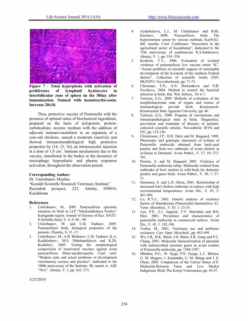
Figure 7 - Total hyperplasia with activation of proliferates of lymphoid hystiocytes in interfollicular zone of spleen on the 30day after immunization. Stained with hematoxylin-eosin. Increase 20х10.
Thus, protective vaccine of Pasteurella with the presence of optimal ratios of biochemical ingredients, prepared on the basis of polypotent, protein-carbohydrate, enzyme medium with the addition of adjuvant immuno-modulator in an organism of a year-old chickens, caused a moderate reactivity and showed immunomorphological high protective properties by [14, 15, 16], an intramuscular injection at a dose of 1,0 cm3. Immune mechanisms due to the vaccine, manifested in the bodies in the dynamics of macrophage hyperplastic and plasma responses activation, throughout the observation period. Corresponding Author: Dr. Umitzhanov Mynbay “Kazakh Scientific Research Veterinary Institute” Raiymbek prospect, 223, Almaty, 050016, Kazakhstan References 1. Umitzhanov, M., 2005. Pasteurellosis epizootic
situation on birds in LLP “Maykudukskaya Poultry” Karaganda region. Journal of Science of Kaz. HATU. S.Seifullin them, V. 4, 9: 96 - 99.
2. Umitzhanov, M. and U.B. Taubaev, 2005. Pasteurellosis birds, biological properties of the parasite. Zharshy, 8: 15 -17.
3. Umitzhanov, M., A.B. Bizhanov, U.B. Taubaev, K.A. Kashkinbaev., M.S. Dzhubandykova and K.Zh. Kushaliev, 2005. Testing the morphological composition of inactivated vaccines against avian pasteurellosis. Mater.mezhd.nauchn. Conf. conf. “Modern state and actual problems of development veterinarnoy science and practice”, dedicated to the 100th anniversary of the Institute. Sb. nauch. tr. ASE “Nivi”. Almaty, V. 1, pp: 262 -271.
4. Aydarbekova, L.J., M. Umitzhanov and B.Sh. Karataev, 2008. Pasteurellosis birds The hyperimmune serum by various methods. KazNAU, Intl. nauchn. Conf. Conference “Innovation in the agricultural sector of Kazakhstan”, dedicated to the 75th anniversary of academician K.S.Sabdenova. Almaty, V. 1, pp: 554 -556.
5. Kashirin, V.V., 2006. Evaluation of residual virulence of pasteurellosis live vaccine strain “K”. “Actual problems of scientific support of sustainable development of the livestock of the southern Federal district”. Collection of scientific works GNU SKZNIVI. Novocherkassk, pp: 71-73.
6. Christmas, T.N., A.N. Borisenkova and O.B. Novikova, 2004. Method to control the bacterial infection in birds. Rat. Wet. Inform., 10: 6-7.
7. Turitsyn, E.G., 2009. Methods of evaluation of the morphofunctional state of organs and tissues of immunological provide birds. Krasnoyarsk: Krasnoyarsk State Agrarian University, pp: 46.
8. Turitsyn, E.G., 2008. Program of vaccinations and immunopathological state in birds. Diagnostics, prevention and treatment of diseases of animals: collected scientific articles. Novosibirsk: IEVS and DV, pp: 133-136.
9. Christensen, J.P., H.H. Dietz and M. Bisgaard, 1998. Phenotypic and genotypic characters of isolates of Pasteurella multocida obtained from back-yard poultry and from two outbreaks of avian cholern in avifauna in Denmark. Avian Pathol., V. 27, 10: 373-381.
10. Permin, A. and M. Bisgaard, 2001. Virulence of Pasteurella multocida subsp. Multocida isolated from outbreaks of fowl cholera in wild birds for domestic poultry and game birds. Avian Pathol., V. 30, 1: 27-31.
11. Simensen, E. and L.D. Olson, 2001. Relationship of increased fowl cholera outbreaks in turkeys with high environmental temperatures. Avian Dis., V. 45, 2: 461-466.
12. Lo, R.Y.C., 2001. Genetic analysis of virulence factors of Mannheimia (Pasteurella) haemolytica A1. Veter. Microbiol., V. 83. 1: 23-35.
13. Aye, P.P., E.J. Angrick, T.Y. Morishita and B.S. Harr, 2001. Prevalence and characteristics of pasteurella multocida in commercial turkeys. Avian Dis., V. 45, 1: 182-190.
14. Teuber, M., 2001. Veterinary use and antibiotic resistance. Curr. Opin. Microbiol., pp: 493-499.
15. Wu, J.R., H.K. Shieh, J.H. Shien, S.R. Gong and P.C. Chang, 2003. Molecular characterization of plasmids with antimicrobial resistant genes in avian isolates of Pasteurella multocida, pp: 1384-1387.
16. Mbuthia, P.G., W. Njagi, P.N. Nyaga, L.C. Bebora, G. M. Mugera, J. Kamundia, U. M. Minga and J. E. Olsen, 2005. Comparison of the Carrier Status of P. Multocida Between Farm and Live Market Indigenous Birds The Kenya Veterinarian, pp: 45-47.
3/23/2014