Embed Size (px)
Citation preview

Life Science Journal, 2012;9( 3) http://www.lifesciencesite.com
2301
Nerve Conduction Velocity of Sciatic Nerve in High Fat Diet Induced Obesity in Rats: Effect of
Corn Oil and Omega 3 Fatty Acids Supplement
Laila Ahmed El sayed, Samah Elattar, and Nashwa Eltablawy
Department of Physiology, Faculty of Medicine, Cairo University, Cairo, Egypt
Abstract: Background: Obesity is a major susceptibility factor leading to the development of various conditions of
the metabolic syndrome. In obese rats, slowing of motor nerve conduction velocity was observed. Fatty acids
metabolism disturbance is very important in the occurrence of peripheral neuropathy. The aim of this work is to
consider the role that balanced diets high in omega 6&9 PUFA (corn oil) or supplying rats with omega 3, play in modulating the impaired nerve function in obese rats. Methods: Thirty two adult male albino rats were randomly
assigned to receive normal chow (NC) (n=8) or high fat diet HFD (n=24), for 12 weeks. After 12 weeks, body
weight and body mass index(BMI) were measured and the NC group(n=8) continue their normal chow diet, Group 1
(NC) and served as a control group and the obese rats were randomly divided into 3 groups, 8 rats each: Group 2:
Ob + HFD group, they continue their high animal fat diet, Group 3: Ob+HFD + corn oil group, they are obese rats
received high fat diet containing corn oil and Group 4: Ob + HFD + Omega 3 group, they are obese rats, fed high
animal fat diet supplemented with omega 3 (0.4 g/kg) daily. After five weeks, the final body weight was measured
and BMI was calculated and blood samples were collected for measuring fasting plasma glucose level and insulin
level and homeostasis model assessment of insulin resistance (HOMA-IR) test were evaluated. Plasma cholesterol,
triglycerides and free fatty acids (FFAs) were measured. The rats were then killed and sciatic nerves were carefully
dissected for measuring the nerve conduction velocity (NCV). Superoxide dismutase activity (SOD), malondialdehyde (MDA) and tumor necrosis factor alpha (TNFα) were estimated in the nerve tissue of the 4 groups.
Results: The results of this study showed a significant increase of body weight (gm) and BMI (kg/m2) in high fat
diet group (p< 0.05) after 12weeks of the start of the diet when compared to the control group (NC). There were
significant elevations in the final weight (gm) and BMI (kg/m2), a significant elevation in insulin level (µIU/l) and HOMA-IR test, a significant increase in nerve malondialdehyde (MDA), and tumor necrosis factor alpha (TNFα) and
a significant decrease in superoxide dismutase activity (SOD) and nerve conduction velocity (NCV) (m/s) after
5weeks of high fat diet in (Ob+HFD) group, when compared to NC group. Changing diet composition for 5weeks in Ob+ HFD+corn oil and Ob+HFD+omega 3 groups, did not induce any significant variation in body weight, BMI,
or fasting blood glucose level as compared to Ob+HFD group. Insulin level (µIU/l) and HOMA-IR test were
significantly decreased in Ob+ HFD+corn oil and Ob+HFD+omega 3 groups compared to Ob+HFD group. Plasma
cholesterol levels (mg/dl), triglycerides (mg/dl), and free fatty acids (FFA) (mmol/l) were significantly decreased
after 5weeks diet in Ob+ HFD+corn oil or Ob+HFD+ Omega 3 groups when compared to mean values of Ob+HFD
group. Tissue malondialdehyde (MDA) and tumor necrosis factor alpha (TNFα) were significantly decreased but
superoxide dismutase (SOD) activity was significantly increased in Ob+HFD+corn oil and Ob+HFD+omega3 groups compared to Ob+HFD. NCV(m/s) in Ob+HFD+ corn oil group was significantly increased compared to Ob+
HFD and their values in Ob+HFD+ corn oil group showed no significant variation as compared to NC group. While
there was a significant increase in NCV in Ob+ HFD+Omega 3 group as compared to Ob+ HFD group, there was
still a significant decrease compared to NC group. Conclusion: The results of this study may have important clinical
and speculative implications. Corn oil or omega 3 supplementation may be effective in obesity induced neuropathy.
The mechanism of their effects is multifactorial including improving insulin sensitivity, correction of dyslipdemia,
reducing oxidative stress and an anti-inflammatory effect. This possibility should be carefully considered and
examined in future trials of essential fatty acid supplementation.
[Laila Ahmed El sayed, Samah Elattar, and Nashwa Eltablawy Nerve Conduction Velocity of Sciatic Nerve in
High Fat Diet Induced Obesity in Rats: Effect of Corn Oil and Omega 3 Fatty Acids Supplement] Life Sci J
2012;9(3):2301-2314] (ISSN:1097-8135). http://www.lifesciencesite.com. 402
Key words: nerve conduction velocity, obesity, oxidative stress, inflammation, corn oil, omega3, insulin resistance.
1. Introduction
Obesity is a pathological condition in which
excess body fat has accumulated to the extent that it may have an adverse effect on health, leading to
reduced life expectancy and/or increased health
problems [1]. Obesity is a major susceptibility factor
leading to the development of various conditions of the metabolic syndrome. A recent study from the

Life Science Journal, 2012;9( 3) http://www.lifesciencesite.com
2302
World Health Organization approximates that,
globally, 1.6 billion adults are overweight with at
least 400 million adults classified as obese [2].
In obese rats, slowing of motor nerve
conduction velocity was observed. Obese Zucker rats
develop neural deficits independently of
hyperglycemia [3]. Evidence for the development of
neuropathic changes at the prediabetic stage, prior to
development of overt hyperglycemia and diabetes
mellitus, is emerging from both clinical [4] and
experimental [5] studies. The pathophysiologic basis of this relationship is not well understood. A number
of pharmacological agents that showed promise in
animal studies have been withdrawn from clinical
trials because of a lack of efficacy or adverse side-
effects [6].
The induction of obesity may be performed in
animals by neuroendocrine, dietary or genetic
changes. [7]. High-fat diet (HFD)-fed rats with
alimentary obesity and hyperinsulinemia, develop
nerve conduction velocity deficit, therefore, represent
an ideal model for evaluating effects of changing dietary composition on manifestations of
neuropathy[8]. The contributions of insulin
resistance, hypertriglyceridemia and/or increased
nonesterified fatty acids (NEFA), and
hypercholesterolemia to this condition remain
unknown [9].
Fatty acids metabolism disturbance is very
important in the occurrence of peripheral neuropathy.
Low plasma omega-6 and omega-3 fatty acids levels
were associated with accelerated decline of peripheral
nerve function with aging [10]. Evidence has
emerged suggesting that both omega-6 and omega-3 fatty acids are also important for peripheral nerve
health and function [11].
The administration of unsaturated fatty acids
especially omega-3 has gained considerable attention
recently. The effect of omega-3 fatty acid on the
treatment of coronary arteries atherosclerosis has
been shown [12]. Consumption of Omega-3 fatty
acids in animal models could be effective in restoring
nerve conduction velocity [13]. Also, omega 6&9
poly unsaturated fatty acids (PUFA) supplied from
corn oil has also been reported to be beneficial in systemic diseases, such as hypertension [14],
cardiovascular disease [15] and cancer [16],
however, little is known about the possible benefits
that dietary omega-3 or omega 6&9 may have for
the nerve conduction disorders in obesity. In this
study, we propose that the lipid changes associated
with obesity might partially explain the reported
neural dysfunction. We consider the role that
balanced diets high in omega 6&9 (corn oil) or
supplying rats with omega 3, can have in modulating
the impaired nerve function in obese rats.
2. Material and methods
Experimental animals and groups
Thirty two adult male albino rats of body weight
80-100 gm, 3 to 4 weeks old, were included in this
study. The rats were supplied by the Animal House
Unit of Kasr Al-Ainy, Faculty of medicine, Cairo
University, housed in cages at room temperature with
normal light & dark cycle. The rats were randomly
assigned to receive normal chow; control group (NC)
[16] (n=8), and HFD [17] (n=24), for 12 weeks. The
composition of the different experimental diets used is shown in table 1.
Table 1: The composition of experimental diets Ingredients
Contents (g/kg diet)
HFD
(animal fat)
HFD (animal fat) +corn oil
Control diet
(NC)
Carbohydrates
-Corn starch 100 100 480
-Sucrose 100 100 100
Fats
- Soybean oil 50 50 50
- Animal fat 500 300 120
- Corn oil 200
Protein
- Casein 190 190 190
This study was carried out in the Physiology and
Biochemistry Departments, Faculty of Medicine,
Cairo University. After 12 weeks, the rats were
classified into:
Group 1: they are the normal rats, received normal
chow and served as normal control. They
continued free access to laboratory rat chow and
tap water and received 4 mL of normal saline
through gavage daily for another 5 weeks (NC).
The HFD-fed rats were divided into 3 groups, 8
rats each:
Group 2: Ob + HFF group, they are obese rats those
continue their high animal fat diet and received
4 mL of normal saline through gavage for 5
weeks.
Group 3: Ob +HFD+ corn oil group, they are obese
rats, received high fat diet. In this group of rats,
corn oil was administered as 20% of the diet,
replacing same percent of animal fat and
received 4 mL of normal saline through gavage
daily. Corn oil contains high omega 6&9
polyunsaturated fat [18]. FA analysis of corn oil
showed that corn oil contained mono unsaturated fatty acids (27.576%), poly
unsaturated fatty acids (PUFA) (57.36%),

Life Science Journal, 2012;9( 3) http://www.lifesciencesite.com
2303
omega 6 (58%) and omega 9 (28%), fatty acids
in the percent shown in table 2 [19].
Table 2: The fatty acid analysis of corn oil
Saturated fatty acids
MUFA PUFA Omega
3 Omega
6 Omega
9
Corn oil
12.948 27.576 57.36 1 58 28
Values are expressed as weight percent (%) of total
fat.
Group 4: Ob + HFD + Omega 3 group, they are
obese rats, fed high fat diet rich in saturated
animal fat for 5 weeks and received omega 3
(0.4 g/kg BW) daily through gavage [20].
Each Omega-3 capsule contains Fish Oil
(Omega-3 Docosahexaenoic acid DHA 12%,
eicosapentaenoic acid EPA 18%) 999 mg,
Vitamin E 1 mg, Gelatin (food grade) 371 mg.
Alpha tocopherol is included in the capsule to
avoid auto-oxidation.
Weight measurements: All rats were weighed in grams and naso-anal
lengths were measured in cm at the end of the 12-
weeks study and at the end of the 5weeks of different
diet trial. The body mass index (BMI) was calculated
(by dividing the body weight in kilograms by the
length in meters squared) [21] at the end of the study
period (after 12weeks of NC or HFD and after
5weeks of corn oil or omega 3 supplementation).
Rats were fasted overnight for at least 6 hours
and blood samples were obtained by introducing a
fine heparinized capillary tube at the inner canthus of the eye into the venous plexus. The blood samples
were delivered into centrifuge tubes to which
anticoagulant was added then centrifuged at 10,000
rpm for 20 minutes and plasma was separated and
stored at -70˚C for measuring fasting blood glucose
level and insulin level and HOMA-IR test were
evaluated. Plasma cholesterol, triglycerides and FFAs
were measured. The rats were then killed and sciatic
nerves were carefully dissected for measuring of
nerve conduction velocity, tissue superoxide
dismutase activity (SOD), malondialdehyde (MDA) and tumor necrosis factor alpha (TNFα).
Biochemical measurements
Measurement of fasting plasma glucose level
Plasma glucose in blood samples was measured
using oxidase- peroxidase method [22].
Measurement of plasma insulin
Plasma insulin levels were analyzed using
enzyme-linked immunosorbent assay ELISA (Dako,
Carpinteria, CA) according to the manufacturer’s
instructions [23].
HOMA-IR test
To estimate insulin resistance, the homeostasis
model assessment for insulin resistance (HOMA-IR:
insulin resistance index) [24] was used, calculated as
the product of fasting insulin (in μIU) and fasting
glucose (in mmol/l) divided by 22.5. A lower index
indicates greater insulin sensitivity.
FFA detection
FFA was measured in plasma samples using
Free Fatty Acid Quantification Kit supplied by
Abcam USA according to manufacturer guide [25].
Measurement of lipid
Plasma total cholesterol was assayed as
described by Siedel et al. [26], while the protocols of
Jacobs and Van Denmark [27] was adopted for the
determination of triglycerides (TAG).
Measurement of MDA
To measure the MDA concentration, 100 mg of
sciatic nerve tissue in 1 mL PBS, pH 7.0 was
homogenized with micropestle in microtube. 20 %
TCA was added to nerve homogenate to precipitate
the protein, and centrifuged. Supernatants were collected and thiobarbituric acid (TBA) solution was
added to the supernatants. After boiling for 10
minutes in water bath, the absorbance was measured.
Concentration of MDA in supernatants of nerve
homogenate was calculated using the standard curve
[28].
Measurement of SOD activity
Superoxide dismutase (SOD) activity in nerve
homogenate was measured through the inhibition of
nitroblue tetrazolium (NBT) reduction by O2-
generated by the xanthine/xanthine oxidase system.
One SOD activity unit was defined as the enzyme amount causing 50% inhibition in 1 mL reaction
solution per milligram tissue protein and the result
was expressed as U/mg protein[29].
Measurement of TNF- α
TNF-α was measured by in nerve tissues using
ELISA (quantikine R&D system USA) according to
the manufacturer`s instructions [30].
Nerve conduction velocity measurements:
Electrophysiological Recording:
The Sciatic nerve was mounted in a nerve
chamber designed for recording of action potential from isolated nerve. It contains 15 stainless wire
electrodes. The nerve was dissected free without any
muscles remnants. About 2 cm of the nerve was
positioned over the electrodes and embedded in
paraffin oil to maximize signal amplitude and prevent
drying. The proximal part of the nerve was stimulated
by 2 platinum stimulating hook electrodes and the
recording electrode was placed 1 cm apart from the
stimulating one.
Electrophysiological measurements were
performed using an AD instruments Power Lab 4/25

Life Science Journal, 2012;9( 3) http://www.lifesciencesite.com
2304
stimulator and Bio AMP amplifier followed by a
computer assisted data analysis. Sciatic nerves were
stimulated with square wave pulses of 200 µsec duration at 1-10 volts for conduction velocities.
Conduction velocity is measured by dividing the
distance between the stimulating and recording
electrodes by latent period, which is the time elapsed
between the application of stimulus until the peak of
the maximum compound action potential(CAP) [31].
Statistical analysis:
Data were analyzed using the statistical package
SPSS version 15. Values were expressed as mean +
standard deviation (SD). Comparisons between groups were done using analysis of variance
(ANOVA) with multiple comparisons post hoc test in
normally distributed quantitative variables while non
para metrical Mann- Whitney test was used for non
normally distributed quantitative variables. P-values
less than 0.05 were considered as statistically
significant [32].
3. Results
Effect of 12 weeks of HFD on body weight and
BMI in rats:
The results of this study showed a significant increase of body weight (gm) and BMI (kg/m2) in
high fat diet group ( p< 0.05) when compared to
control group( NC) after 12weeks of the start of the
diet, indicating presence of obesity in HFD group
(Table 3, Figures1,2).
Effect of 5 weeks of HFD (without
supplementation) on body weight and BMI:
The results reported a significant elevation in
the mean values of final weight measurements (gm)
and BMI(kg/m2) in rats after 5weeks of high animal
fat diet (Ob+HFD), when compared to corresponding values of normal chow group(NC).(Table 4, Figures
3,4)
Effect of 5 weeks of HFD on plasma glucose,
insulin, HOMA-IR in obese rats:
Table 5 and figures 5-7 show that HFD yielded
an insignificant elevation in the mean value of
fasting blood glucose level (mmol/l), while there was
a significant elevation in insulin level (µIU/l) and HOMA-IR test after 5weeks of HFD compared to NC
group. This reflects the effect of HFD on induction of
insulin resistance.
Effect of 5 weeks of HFD on cholesterol levels,
triglycerides and free fatty acids. When observing levels of plasma cholesterol
levels (mg/dl), triglycerides (mg/dl), and free fatty
acids (mmol/l) after 5weeks of high animal fat diet
(Ob+HFD) in table 6 and figures 8-10, the present
results recorded a significant elevation in their plasma
levels compared to NC group, denoting the effect of
obesity and HFD on dyslipidemia and elevation of
plasma lipid levels.
Effect of 5 weeks of HFD on oxidative stress and
inflammation:
Our results recorded a significant increase in
nerve tissue malondialdehyde (MDA), and tumor
necrosis factor alpha (TNFα) and a significant
decrease in superoxide dismutase activity (SOD) after
5weeks of high animal fat diet in obese rats compared
to NC group (Table 7, Figures 11-13). This reflects
impairment of antioxidant activity and the effect of HFD in obese rats on elevation oxidative stress and
increased the inflammatory marker.
Effect of 5 weeks of HFD on nerve conduction
velocity in obese rats:
Interestingly, the current results recorded a
significant decrease in nerve conduction velocity
(NCV) (m/s) after 5weeks of high animal fat diet
(Ob+HFD), compared to values recorded from
normal chow group of rats (NC) (p< 0.05) (Table 8,
Figure 14).
Effect of changing diet composition in obese rats
on different parameters: Ob+ HFD+corn oil
versus Ob+HFD+ Omega 3:
When observing the values of final body weight
(gm) and BMI measurements (kg/m2) in rats after
changing diet composition for 5W, we can observe
that there was no significant variation in these values
in Ob+HFD+ corn oil group or in Ob+HFD+omega 3
groups compared to values recorded in Ob+HFD
group (Table 4 and Figures 3&4).
Furthermore, as shown in table (5) and figures
(5-7) there was no significant variation in fasting
blood glucose level in Ob+HFD+ corn oil and Ob+HFD+ Omega 3 group compared to Ob+HFD
group. As regarding the mean values of fasting
insulin level (µIU/l) and HOMA-IR test after 5 weeks, there was no significant variation between
Ob+HFD+corn oil group when compared to
Ob+HFD+Omega 3, however, the plasma levels of
these parameters in the Ob+HFD+ corn oil and
Ob+HFD+ Omega 3 groups were significantly
decreased when compared to Ob+HFD. Thus, these
results reflect that changing the formula of diet from
HFD only to addition of corn oil or of omega 3 to
HFD improved insulin sensitivity and decreased the
exposure to insulin resistance condition. When observing levels of plasma cholesterol
levels (mg/dl), triglycerides (mg/dl), and free fatty
acids (mmol/l) after 5weeks diet (Ob+HFD+ corn oil
or Ob+HFD+ Omega 3), table 6 and figures 8, 9 &10
show no significant change in the mean values
recorded between these 2 groups, but the mean values
of these parameters in the 2groups were significantly
decreased when compared to mean values recorded in

Life Science Journal, 2012;9( 3) http://www.lifesciencesite.com
2305
Ob+HFD group. This indicates that the corn oil or
omega3 protected against dyslipidemia.
When nerve tissue malondialdehyde level
(MDA), superoxide dismutase activity (SOD) and
tumor necrosis factor alpha level(TNFα) were
estimated after 5weeks of high corn oil diet
(Ob+HFD+ corn oil), or high fat diet supplemented
with omega 3 (Ob+HFD+Omega 3) in male rats, it
can be observed that there was no significant
variation in mean values of MDA, SOD activity or
TNFα in Ob+HFD+ corn oil group compared to Ob+HFD+ Omega 3 group, but the 2 groups showed
significantly decreased tissue malondialdehyde and
(MDA) and tumor necrosis factor alpha (TNFα)
levels and significantly increased superoxide
dismutase activity (SOD) when compared to
Ob+HFD (Table 7, Figures 11-13).
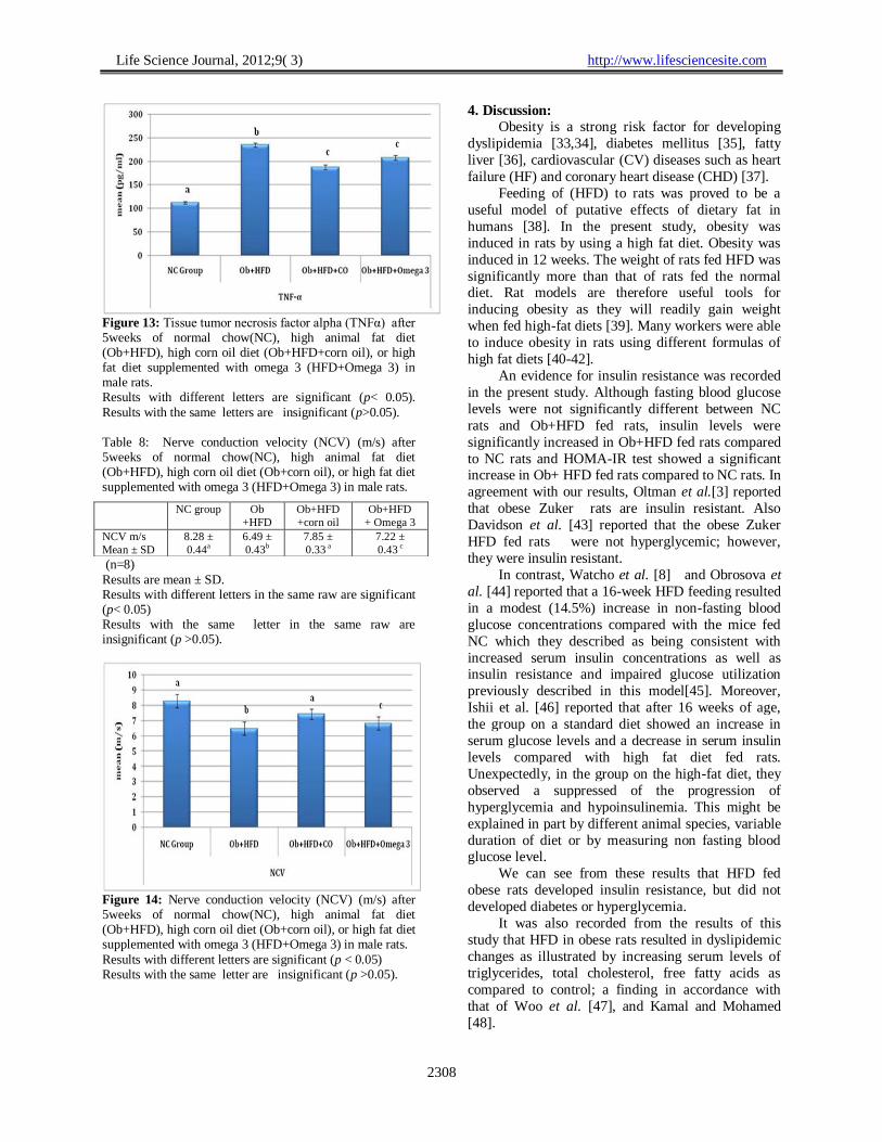
Regarding NCV(m/s) values, both in
Ob+HFD+ corn oil and Ob+ HFD+Omega 3 groups,
there was a significant increase compared to
Ob+HFD and their values in Ob+HFD+ corn oil
group showed no significant variation as compared to
control group. In contrast in Ob+ HFD+Omega 3
group, there was a significant decrease in values of
this group as compared to the control group and
Ob+HFD+ corn oil (Table 8, Figure 14).
Table 3: Body weight measurements (gm) and body
mass index (Kg/m2) in rats after 12weeks of normal
chow (NC) (n=8), high animal fat diet (HFD)(n=24),
in male rats. NC Group HFD group
Body weight (gm)
Mean ± SD
163.88 ± 11.52a 324.25 ± 28.07
b
BMI (Kg/m2)
Mean ± SD
5.93 ± 0.40a 11.69 ± 1.09
b
Results with different letters in the same raw are
significant (p< 0.05)
Results with the same letter in the same raw are
insignificant (p>0.05).
Figure 1: Body weight measurements (gm) and figure 2: body mass index in rats after 12weeks of normal
chow(NC) (n=8), high animal fat diet (HFD)(n=24), in male rats.
Results with different letters are significant (p< 0.05) Results with the same letter are insignificant (p>0.05).
Table 4:Final body weight measurements (gm) and
body mass index(Kg/m2) in rats after 5weeks of
normal chow(NC), high animal fat diet (Ob+HFD),
high corn oil diet (Ob+HFD+corn oil), or high fat diet
supplemented with omega 3 (Ob+HFD+Omega 3) in
male rats.
NC
group
Ob
+HFD
Ob+
corn oil
Ob+HFD
+Omega 3
Final
weight
(gm)
189.50 ± 8.50
a 359.38 ± 38.53
b 347.25 ± 25.76
b 358.13 ± 26.16
b
BMI (kg/ m
2)
5.93 ± 0.40
a
11.96 ± 1.62
b 11.70 ± 0.55
b
11.90 ± 0.43
b
(n=8)
Results are mean ± SD.
Results with different letters in the same raw are
significant (p< 0.05)
Results with the same letter in the same raw are
insignificant (p>0.05).
Figure 3: Final body weight measurements (gm) in
rats after 5weeks of normal chow(NC), high animal
fat diet (Ob+HFD), high corn oil diet (Ob+HFD+corn
oil), or high fat diet supplemented with omega 3 (HFD+Omega 3) in male rats.

Life Science Journal, 2012;9( 3) http://www.lifesciencesite.com
2306
(n=8) -Results with different letters are significant
(p< 0.05).-Results with the same letter are
insignificant (p>0.05).
Figure 4: Body mass index (kg/m2) in rats after 5weeks of normal chow(NC), high animal fat diet (Ob+HFD), high corn oil diet (Ob+HFD+corn oil), or high fat diet
supplemented with omega 3 (HFD+Omega 3) in male rats. n=8 Results with different letters are significant (p< 0.05) Results with the same letter are insignificant (p>0.05).
Table 5: Fasting plasma glucose level (mmol/l), insulin
level(µIU/l) and HOMA-IR test after 5weeks of normal chow(NC), high animal fat diet (Ob+HFD), high corn oil diet rich in polyunsaturated fatty acids( Ob+HFD+corn oil), or high fat diet supplemented with omega 3 (HFD+Omega 3) in male rats.
NC group
Ob+HFD Ob+HFD +corn oil
Ob+HFD
+ Omega 3
Glucose mmol/l
4.69 ± 0.37
5.41 ± 0.30
5.05 ± 0.18
5.45 ± 0.39
Insulin µIU/l
11.64± 0.78 a
20.59 ± 1.20b
11.68 ± 0.74 a
11.55 ± 0.76 a
HOMA-IR
2.43 ± 0.26 a
4.95 ± 0.40 b
2.63 ± 0.23a
2.80 ± 0.31 a
(n=8)
Results are mean ± SD. Results with different letters in the same raw are significant (p< 0.05) Results with the same letter in the same raw are insignificant (p>0.05).
Figure 5: Fasting plasma glucose level (mmol/l) after
5weeks of normal chow(NC), high animal fat diet (Ob+HFD), high corn oil diet rich in polyunsaturated fatty acids ( Ob+HFD+corn oil), or high fat diet supplemented
with omega 3 (HFD+Omega 3) in male rats. Results in the different groups are insignificant to each other (p>0.05).
Figure 6: Insulin level(µIU/l) after 5weeks of normal chow(NC), high animal fat diet (Ob+HFD), high corn oil diet rich in polyunsaturated fatty acids( Ob+HFD+corn oil), or high fat diet supplemented with omega 3 (HFD+Omega 3) in male rats.
Results with different letters are significant (p< 0.05) Results with the same letter are insignificant (p>0.05).
Figure 7: HOMA-IR test after 5weeks of normal chow(NC), high animal fat diet (Ob+HFD), high corn oil diet rich in polyunsaturated fatty acids( Ob+HFD+corn oil), or high fat diet supplemented with omega 3 (HFD+Omega 3) in male rats. Results with different letters are significant (p< 0.05)
Results with the same letter are insignificant (p>0.05). Table 6: Plasma cholesterol levels (mg/dl), triglycerides (mg/dl), and free fatty acids (mmol/l) after 5weeks of normal chow(NC), high animal fat diet (Ob+HFD), high corn oil diet (Ob+HFD+corn oil), or high fat diet supplemented with omega 3 (Ob+HFD+Omega 3) in male rats. NC
group
Ob+HFD Ob+HFD
+corn oil
Ob+HFD
+ Omega 3
Cholesterol
mg/dl
128.79±
2.86 a
188.44 ±
6.3439 b
140.86 ±
5.36 c
153.11 ±
2.00 c
Triglycerides
mg/dl
72.43 ±
6.92a
105.75 ±
2.80b
84.53 ±
1.84 a
81.25 ±
1.64 a
FFA mmol/l 0.17 ±
0.04 a
0.52 ±
0.04 b
0.24 ±
0.02 a
0.27 ±
0.02a
(n=8) Results are mean ± SD. Results with different letters in the same are significant (p<0.05) Results with the same letter in the same raw are insignificant (p>0.05).

Life Science Journal, 2012;9( 3) http://www.lifesciencesite.com
2307
Figure 8: Plasma cholesterol levels (mg/dl) after 5weeks of normal chow(NC), high animal fat diet (Ob+HFD), high corn oil diet (Ob+HFD+corn oil), or high fat diet
supplemented with omega 3 (Ob+HFD+Omega 3) in male rats. Results with different letters are significant (p< 0.05) Results with the same letter are insignificant (p>0.05).
Figure 9: Plasma triglycerides (mg/dl) after 5weeks of normal chow (NC), high animal fat diet (Ob+HFD), high corn oil diet (Ob+HFD+corn oil), or high fat diet supplemented with omega 3 (Ob+HFD+Omega 3) in male rats.
Results with different letters are significant (p< 0.05). Results with the same letter are insignificant (p>0.05).
Figure 10: Plasma free fatty acids (mmol/l) after 5weeks of normal chow(NC), high animal fat diet (Ob+HFD), high corn oil diet (Ob+HFD+corn oil), or high fat diet supplemented with omega 3 (Ob+HFD+Omega 3) in male rats. Results with different letters are significant (p< 0.05). Results with the same letter are insignificant (p>0.05).
Table 7: Tissue malondialdehyde level (MDA), superoxide
dismutase activity (SOD) and tumor necrosis factor alpha level (TNFα) after 5weeks of normal chow(NC), high animal fat diet (Ob+HFD), high corn oil diet (Ob+HFD+corn oil), or high fat diet supplemented with omega 3 (HFD+Omega 3) in male rats. NC
group
Ob+HFD
Ob+HFD
+corn oil
Ob+HFD
+ Omega
3
MDA
nmol/mg ptn
111.36 ±
5.51a
173.10 ±
7.07b
148.51 ±
3.93c
156.30 ±
4.84c
SOD
activity
U/mg ptn
2.03 ±
0.17a
0.45±
0.02b
1.26 ±
0.15c
1.60 ±
0.26c
TNFα
pg/ml
112.15 ±
2.07a
234.89 ±
4.78b
187.79 ±
4.62c
207.23 ±
5.03 c
(n=8) Results are mean ± SD. Results with different letters in the same raw are significant (p< 0.05) Results with the same letter in the same raw are insignificant (p>0.05).
Figure11: Tissue malondialdehyde (MDA) after 5weeks of normal chow(NC), high animal fat diet (Ob+HFD), high corn oil diet (Ob+HFD+corn oil), or high fat diet
supplemented with omega 3 (HFD+Omega 3) in male rats. Results with different letters are significant (p< 0.05). Results with the same letter are insignificant (p>0.05).
Figure 12: Tissue superoxide dismutase activity (SOD) after 5weeks of normal chow(NC), high animal fat diet (Ob+HFD), high corn oil diet (Ob+HFD+corn oil), or high fat diet supplemented with omega 3 (HFD+Omega 3) in male rats. Results with different letters are significant (p< 0.05).
Results with the same letter are insignificant (p>0.05).
Life Science Journal, 2012;9( 3) http://www.lifesciencesite.com
2308
Figure 13: Tissue tumor necrosis factor alpha (TNFα) after 5weeks of normal chow(NC), high animal fat diet (Ob+HFD), high corn oil diet (Ob+HFD+corn oil), or high fat diet supplemented with omega 3 (HFD+Omega 3) in male rats. Results with different letters are significant (p< 0.05).
Results with the same letters are insignificant (p>0.05). Table 8: Nerve conduction velocity (NCV) (m/s) after 5weeks of normal chow(NC), high animal fat diet (Ob+HFD), high corn oil diet (Ob+corn oil), or high fat diet supplemented with omega 3 (HFD+Omega 3) in male rats.
(n=8)
Results are mean ± SD. Results with different letters in the same raw are significant (p< 0.05) Results with the same letter in the same raw are insignificant (p >0.05).
Figure 14: Nerve conduction velocity (NCV) (m/s) after 5weeks of normal chow(NC), high animal fat diet (Ob+HFD), high corn oil diet (Ob+corn oil), or high fat diet supplemented with omega 3 (HFD+Omega 3) in male rats.
Results with different letters are significant (p < 0.05) Results with the same letter are insignificant (p >0.05).
4. Discussion:
Obesity is a strong risk factor for developing
dyslipidemia [33,34], diabetes mellitus [35], fatty
liver [36], cardiovascular (CV) diseases such as heart
failure (HF) and coronary heart disease (CHD) [37].
Feeding of (HFD) to rats was proved to be a
useful model of putative effects of dietary fat in
humans [38]. In the present study, obesity was
induced in rats by using a high fat diet. Obesity was
induced in 12 weeks. The weight of rats fed HFD was
significantly more than that of rats fed the normal diet. Rat models are therefore useful tools for
inducing obesity as they will readily gain weight
when fed high-fat diets [39]. Many workers were able
to induce obesity in rats using different formulas of
high fat diets [40-42].
An evidence for insulin resistance was recorded
in the present study. Although fasting blood glucose
levels were not significantly different between NC
rats and Ob+HFD fed rats, insulin levels were
significantly increased in Ob+HFD fed rats compared
to NC rats and HOMA-IR test showed a significant increase in Ob+ HFD fed rats compared to NC rats. In
agreement with our results, Oltman et al.[3] reported
that obese Zuker rats are insulin resistant. Also
Davidson et al. [43] reported that the obese Zuker
HFD fed rats were not hyperglycemic; however,
they were insulin resistant.
In contrast, Watcho et al. [8] and Obrosova et
al. [44] reported that a 16-week HFD feeding resulted
in a modest (14.5%) increase in non-fasting blood
glucose concentrations compared with the mice fed
NC which they described as being consistent with
increased serum insulin concentrations as well as insulin resistance and impaired glucose utilization
previously described in this model[45]. Moreover,
Ishii et al. [46] reported that after 16 weeks of age,
the group on a standard diet showed an increase in
serum glucose levels and a decrease in serum insulin
levels compared with high fat diet fed rats.
Unexpectedly, in the group on the high-fat diet, they
observed a suppressed of the progression of
hyperglycemia and hypoinsulinemia. This might be
explained in part by different animal species, variable
duration of diet or by measuring non fasting blood glucose level.
We can see from these results that HFD fed
obese rats developed insulin resistance, but did not
developed diabetes or hyperglycemia.
It was also recorded from the results of this
study that HFD in obese rats resulted in dyslipidemic
changes as illustrated by increasing serum levels of
triglycerides, total cholesterol, free fatty acids as
compared to control; a finding in accordance with
that of Woo et al. [47], and Kamal and Mohamed
[48].
NC group Ob
+HFD
Ob+HFD
+corn oil
Ob+HFD
+ Omega 3
NCV m/s
Mean ± SD
8.28 ±
0.44a
6.49 ±
0.43b
7.85 ±
0.33 a
7.22 ±
0.43 c

Life Science Journal, 2012;9( 3) http://www.lifesciencesite.com
2309
Dyslipidemic changes occurs in obesity may be
due to the increased triglycerides content of the liver
due to increased influx of excess non esterified fatty
acids (NEFAs) into the liver[49]. Lipid alterations
affect the structure and function of the nerve
membrane and have been considered as contributory
factors to oxidative stress in obesity [50].
In the present study, it was found that nerve
MDA level was significantly elevated with a
significant decrease in the enzyme superoxide
dismutase activity in HFD fed obese rats compared to NC rats and this was an indication for increased
oxidative stress in these obese rats. Increased
production of reactive oxygen species as well as
reduced antioxidant defense mechanisms have been
suggested to play a role in both humans and animal
obesity induced pathology [51,52].
Interestingly, oxidative stress, the key metabolic
abnormalities previously thought to be caused
primarily by high glucose and shown to contribute to
diabetic neuropathy, clearly manifest in this Ob+HFD
model of pre-diabetic neuropathy characterized by insulin resistance in the absence of overt diabetes or
hyperglycemia.
In this study, our obese rats also developed
nerve disorder, demonstrated as NCV slowing. The
finding that rats fed a high fat diet develop indices of
neuropathy is consistent with studies of obese Zucker
rats [3, 53], and also with clinical studies in which
pre-diabetes and impaired glucose tolerance have
been associated with an early-onset neuropathy [54,
55]. It was previously suggested that impaired
glucose tolerance can directly cause nerve injury [56],
however, it appears that NCV slowing is simply a covariant with other factors related to obesity.
This study shows that velocity of sciatic nerve
conduction in obese rats could depend on dietary fat
modification. The addition of omega 3 to high animal
fat diet or consumption of corn oil rich in omega 6&9
PUFA in addition to animal fat was associated with
increased sciatic nerve conduction velocity in obese
rats, whereas high animal fat diet in obese rats caused
a significant slowing of sciatic nerve conduction
velocity. In addition, we found that omega 6&9
PUFA supplemented food (corn oil) induced a significant improvement of nerve conduction velocity
compared to the enriching food with omega 3. These
data show that omega 3 enrichment or corn oil could
be associated with improved nerve conduction
velocity in obese rats.
The present study provides evidence of the
therapeutic efficacy of omega 6&9 PUFA and
omega 3 on NCV deficits, in the model of
neuropathy associated with obesity. It should be
noticed that the improvement in NCV was not
associated with weight reduction. As seen from the
results of the present study, there is no significant
change in final body weight or BMI between the
obese groups fed HFD with animal fat, corn oil or
supplemented with omega 3 fatty acids.
Some previous studies suggested an association
between insulin resistance, compensatory
hyperinsulinemia, and peripheral neuropathy in
human. Also, higher insulin resistance was
independently associated with the presence of cardiac
autonomic neuropathy (CAN) in Korean type 2
diabetes mellitus (T2DM) patients [57]. In the present study it was shown that either
omega 3 or corn oil supplementation was associated
with improved insulin sensitivity and decreased blood
insulin level and this may play a role in improving
nerve conduction, however the exact mechanism for
this relation is not clear and needs further
investigations.
One study provides evidence that insulin
receptor substrate (IRS) proteins are expressed in the
dorsal root ganglia (DRG) and could play an
important role in the ability of insulin to support peripheral neurons. Elevated serine phosphorylation
of IRS proteins reported in their study to be a major
contributing mechanism underlying the effect of
insulin resistance on neurons [58].
Insulin resistance is an important risk factor for
endothelial dysfunction, and impairment of vascular
function of epineurial arterioles precedes nerve
dysfunction in obese normoglycemia Zucker rats
[59]. It has been shown that improving insulin
sensitivity improves vascular resistance in obese
Zucker rats [60].
We can see from the results of the present study that dyslipidemia may be a contributing factor to
reductions in peripheral nerve conduction velocity.
This dyslipidemia was shown to be mostly corrected
by dietary supplements and this correction appears to
play a role in improvement of nerve conduction
velocity, may be in part by a normalization of fatty
acid composition of nerve membrane and eicosanoid
synthesis, which is depressed in neuropathy and/or by
a direct effect on incorporation of these fatty acids
into the plasma membranes [61]. By changing
membrane properties, omega 6&9 or omega 3 PUFA can modify the activity of transmembrane enzymes,
such as Na,K- Atpase, which is implicated in the
propagation of nerve impulses[62].
Our findings are consistent with studies showing
that high dietary intake of fatty acids prevents the
development and clinical progression of nerve
conduction deficits in diabetic animals as well as in
the general human population [63,11]. In diabetic
rats, the administration of linoleic acid, an n-6 fatty
acid, improved sciatic NCV [11]. In patients with
generalized peroxisomal disorders, congenital

Life Science Journal, 2012;9( 3) http://www.lifesciencesite.com
2310
diseases with impaired myelinogenesis, the
administration of the n-3 fatty acids, DHA,
significantly improved myelin formation alleviating
the symptoms in these patients [64].
PUFA are the major structural components of
the neuronal membrane phospholipids [65] and
therefore, their structural and chemical characteristics
influence membrane functions, such as the activity of
membrane bound proteins, signal transduction and
also neurotransmission [66-68]. It was also reported
that supplementation with sunflower oil, which contains high quantity of linoleic acid, restored NCV
in diabetic rats, and this effect was accompanied by a
modification of phospholipid fatty acid composition
in nerve membranes [10].
In particular, the electrophysiologic effect of the
omega-3 fatty acids seems to be the result of specific
modulation of ion currents, particularly of the
voltage-dependent sodium current and of the L-type
calcium currents across sarcolemmal phospholipids
membranes [68].
Mammals synthesize the long chain PUFA from linoleic acid [18:2(n-6)] and a-linolenic acid [18:3(n-
3)], which are the 2 precursors of (n-6) and (n-3) fatty
acids families provided by the diet. Specific enzymes,
desaturases and elongases, are involved in this
pathway, but the conversion of precursors to long
chain PUFA is generally low in humans.
Consequently, the decrease in bioavailability of
PUFA, affects the fatty acid composition of
membrane phospholipids (PL) with repercussions on
membrane protein functionality [69], eicosanoid
production [70, 71], and peroxisome proliferator-
activated receptor (PPAR) regulation [72, 73]. It was suggested that the rate-limiting nature of -
6-desaturation contributes to the development of
neuropathy. Bypassing the rate-limiting step by using
gamma-linolenic acid (GLA) may have desirable
effects and anti-inflammatory effects [74]. Because
essential fatty acids (EFAs) and their metabolites are
exceptionally important in both the structure and
function of nerves [75], it seemed possible that
neuropathy might be particularly responsive to PUFA
supplementation.
An important observation in the results of this study is that omega-6 fatty acids, supplied by corn oil,
appear to have a beneficial effect on peripheral nerve
function than omega-3 fatty acids, requires
consideration. In fact, omega-6 PUFAs are generally
more highly represented in the nerve membrane than
omega-3 fatty acids and have major effects of excess
than the n-3 fatty acids [76].
The beneficial effects of omega 6&9 or omega
3PUFA may at least partially be related to inhibition
of oxidative stress in peripheral nerve as evidenced in
the present results by improving the antioxidant
enzyme superoxide dismutase activity and decreasing
oxidative stress marker MDA.
Oxidative stress is closely linked to upregulation
of 12/15-lipooxygenase (12/15LO), an enzyme
converting arachidonic acid to 12-
Hydroxyeicosatetraenoic acid (12(S)-HETE), 15(S)-
HETE, and a number of derivatives of these acids.
These lipid-like compounds undergo spontaneous
lipid peroxidation, which leads to induction of
oxidative nitrosative stress, activation of mitogen-
activated protein kinases (MAPKs), and proinflammatory response [77, 78]. MAPK activation
has been demonstrated to play an important role in
peripheral diabetic neuropathy [79, 80].
It was demonstrated that reducing oxidative
stress in epineural vessels improved vascular
relaxation to acetylcholine as well as NCV [52, 81-
83]. The increase in superoxide in the aorta of high
fat fed rats is likely due to increased NAD(P)H
oxidase activity and/or expression, which has been
linked to increased activity of angiotensin in obesity
[84]. Finally, the results of the present study show
that diet supplemented with omega 3 or PUFA rich in
omega 6&9 fatty acids induce an anti-inflammatory
effect as indicated by decreased TNF alpha content in
the sciatic nerve of the obese rats.
Evidence for the importance of low grade
inflammation in diabetic neuropathy is also emerging
from both experimental and clinical studies [85,86].
Our results are in agreement with Ferrucci et al.
[87] and Kapoor and Huang [88] who reported from
their studies that n-3 PUFAs and the gamma linolenic
acid (GLA), an n-6 fatty acid, have been shown to have significant anti-inflammatory properties. PUFAs
inhibit the production of proinflammatory cytokines,
i.e., Il-1β, IL6 and tumor necrosis factor-alpha by
activating transcription factors, such as the
peroxisome proliferator-activated receptors and
nuclear factor kB [89]. As inflammation is one of the
main pathophysiologic processes involved in
peripheral polyneuropathy, it could be extremely
relevant in progression of axonal damage [90].
Studies in normal volunteers indicate that
omega-3 fatty acid supplementation reduced the ability of monocytes to produce IL-1β upon
stimulation with endotoxin. The effect was most
pronounced 10 weeks after stopping the
supplementation and suggests prolonged
incorporation of omega-3 fatty acids into a pool of
circulating monocytes [91]. The capacity of the
monocytes from these donors to synthesize IL-1β
returned to the pre-supplement level 20 weeks after
ending supplementation. Similar results were
observed for IL-1α and TNF [92].

Life Science Journal, 2012;9( 3) http://www.lifesciencesite.com
2311
Previous studies suggested that in patients
affected by peripheral neuropathy, a supplementation
with PUFA may positively influence the axonal
degeneration of the nerve [10].
Conclusion
The results of this study have important clinical
and speculative implications. Based on our findings,
we suggest that corn oil or omega 3 supplementation
may be effective in treatment of obesity induced
neuropathy. The mechanism of their effects is multifactorial including improving insulin sensitivity,
correction of dyslipdemia which could reflect on fatty
acid composition of the nerve membrane structure
and function, reducing oxidative stress and an anti-
inflammatory effect. This possibility should be
carefully considered and examined in future trials of
essential fatty acid supplementation.
Acknowledgment
The authors thank Prof Dr Laila Rashed (MD
biochemistry) for great assistance in biochemical measurements
Corresponding author
Samah Elattar Department of Physiology, Faculty of Medicine,
Cairo University, Cairo, Egypt
References: 1. Haslam DW, James WP. Obesity. Lancet. 2005;366
(9492):1197-209.
2. Díaz M E, Jiménez S, García RG, Bonet M, Wong
I. Overweight, obesity, central adiposity and associated chronic diseases in cuban adults.
MEDICC Review. l 2009; 11 (4):23-8.
3. Oltman CL, Coppey LJ, Gellett JS, Davidson EP, Lund DD, Yorek MA. Progression of vascular and
neural dysfunction in sciatic nerves of Zucker diabetic fatty and Zucker rats. Am J Physiol
Endocrinol Metab. 2005;289(1):E113-22.
4. Singleton JR, Smith AG. Neuropathy associated with prediabetes: what is new in 2007?. Current
Diabetes Reports. 2007; 7: 420–4.
5. Vincent AM, Hayes JM, McLean LL, Vivekanandan-Giri A, Pennathur S, Feldman EL.
Dyslipidemia-induced neuropathy in mice: the role of oxLDL/LOX-1. Diabetes. 2009; 58(10):376–85.
6. Veves A, Backonja M, Malik RA. Painful diabetic neuropathy: epidemiology, natural history, early
diagnosis, and treatment options. Pain Med. 2008 ;9(6):660-74.
7. Von Diemen V, Trindade EN, Trindade MR.
.Experimental model to induce obesity in rats. Acta Cir Bras. 2006;21(6):425-9.
8. Watcho P, Stavniichuk R, Ribnicky DM, Raskin I,.
Obrosova IG. High-Fat Diet-Induced Neuropathy of Prediabetes and Obesity: Effect of PMI-5011, an
Ethanolic Extract of Artemisia dracunculus L. Mediators of Inflammation. 2010;2010 (268547):1-
10.
9. Lupachyk S, Watcho P, Hasanova N, Julius
U, Obrosova IG. Triglyceride, non esterified fatty acids, and prediabetic neuropathy: role for
oxidative-nitrosative stress. Free Radic Biol Med. 2012;52(8):1255-63.
10. Lauretani F, Bandinelli S, Benedetta B, Cherubini
A, Iorio AD, Blè,A, Giacomini V, Corsi AM, Guralnik JM, Ferrucci L. Omega-6 and
omega-3 fatty acids predict accelerated decline of peripheral nerve function in older persons. Eur J
Neurol. 2007 ;4(7): 801–8.
11. Head RJ, McLennan PL, Raederstorff D, Muggli R, Burnard SL, McMurchie EJ. Prevention of nerve
conduction deficit in diabetic rats by polyunsaturated fatty acids. The American journal
of Clinical Nutrition. 2000;71:386S–392S.
12. Cameron, N.E. Metabolic and vascular factors in the pathogenesis of diabetic neuropathy. Diabetes.
1997;46 (12): 531-7.
13. Jarrahi, M., Effect of diet Containing fish oil on nerve Conduction velocity of diabetic albino rats.
Fall, 1999;1.1: 1.1.
14. Weisinger HS, Armitage JA, Sinclair AJ, Vingrys AJ, Burns PL, Weisinger RS. Perinatal omega-3
fatty acid deficiency affects blood pressure later in life. Nat Med. 2001;7:258-9.
15. Marchioli R, Levantesi G, Macchia A,.
Antiarrhythmic mechanisms of n-3 PUFA and the results of the GISSI-Prevenzione Trial. J Membr
Biol. 2005;206:117.
16. Murray NR, Weems C, Chen L,. Protein kinase C
beta II and TGF beta RII in omega-3 fatty acid-mediated inhibition of colon carcinogenesis. J Cell
Biol. 2002;157:915–920.
17. Lee KY, Ahn HC, Kim C, Kim SH, Kim DK, Park HS. Pancreatic Exocrine Response to Long-
Term High-Fat Diets in Rats. JOP. J Pancreas (Online) 2006; 7(4):397-404.
18. Kuda O, Jelenik T, Jilkova Z, Flachs P, Rossmeisl
M. n-3 Fatty acids and rosiglitazone improve insulin sensitivity through additive stimulatory effects on
muscle glycogen synthesis in mice fed a high-fat diet. Diabetologia. 2009;52:951.
19. Giacometti J, Tomljanovic AB, Milin C, Cuk M,
Stasic BR. Olive and corn oil enriched diets changed the phospholipid fatty acid composition in
mice liver after one-thirds hepatectomy. Food and Nutrition Sciences, 2012, 3, 240-248
20. Bas O, Songur A, Sahin O, Mollaoglu H. The
protective effect of fish n-3 fatty acids on cerebral

Life Science Journal, 2012;9( 3) http://www.lifesciencesite.com
2312
ischemia in rat hippocampus. Neurochem Int.
2007;50:548-54.
21. Jeyakumar SM, Vajreswari A, Giridharan NV: Chronic dietary vitamin A supplementation
regulates obesity in an obese mutant WNIN/Ob rat
model. Obesity 2006, 14:52-9.
22. Trinder L. Determination of blood glucose using an oxidase peroxidase system with a non-carcinogenic
chromagen. Ann. Clin. Biochem.1969; 1: 24–9.
23. Delams H. G. Biochemical analysis of human and animal serum for monoclonal antibodies using
ELISA. Biochem. 1986; 14, 214–31.
24. Mathews DR., Hosker JP, Rudenski AS, Naylor BA, Treacher DF, Turner RC. Homeostasis model
assessment: insulin resistance and β-cell function from fasting plasma glucose and insulin
concentrations in man. Diabetologia. 1985:28: 412–9.
25. Erridge C & Samani NJ. Saturated fatty acids do not
directly stimulate Toll-like receptor signaling. Arterioscler Thromb Vasc Biol. 2009;29: 1944-9.
26. Siedel J, Hagele EO, Ziegenhorn J, Wahlefeld AW.
Reagent for the enzymatic determination of serum total cholesterol with improved lipolytic efficiency.
Clinical Chemistry. 1983;29(6):1075–1080.
27. Jacobs NJ, VanDenmark PJ. Enzymatic determination of serum triglyceride.ch.
Biochemistry and Biophysics. 1960;88:250–255.
28. Wills ED. Evaluation of lipid peroxidation in lipids and biological membranes. In: Snell K, Mullock B,
eds.Biochemical toxicology: A practical approach. London: Oxford;1987.
29. Misra HP,Fridovich I:the role of superoxide in the
autooxidation of epinephrine and a simple assay for superoxide dismutase. J Biol Chem.1972;247:3170-
5.
30. Maskos K, Fernandez-Catalan C, Huber R,
Bourenkov GP, Bartunik H, Ellestad GA, et al. Crystal structure of the catalytic domain of human
tumor necrosis factor-α-converting enzyme. Proc. Natl. Acad.Sci. U.S.A. 1998;95: 3408-12.
31. Leal-Cardoso JH, Matos-Brito BG, Lopes-Junior
JEG, Viana-Cardoso KV, Sampaio-Freitas AB, Brasil RO, Coelho-de-Souza AN, Albuquerque
AAC. Effects of estragole on the compound action potential of the rat sciatic nerve. Brazilian Journal of
Medical and Biological Research.2004; 37(8), 1193-8.
32. Chan YH. Biostatistics102: Quantitative Data –
Parametric & Non-parametric Tests. Singapore Med J. 2003;44(8): 391-6.
33. Montani JP, Carroll JF, Dwyer TM, Antic V, Yang
Z, Dulloo AG. Ectopic fat storage in heart, blood vessels and kidneys in the pathogenesis of
cardiovascular diseases. Int J Obes. 2004;28:S58-65.
34. Fried M, Hainer V, Basdevant A, Buchwald H,
Dietel M, Finer N, Greve JW, Horber F, Mathus-Vliegen E, Scopinaro N, Steffen R, Weiner R,
Widhalm K. Interdisciplinary European guidelines on surgery for severe obesity. Rozhl Chir.
2008;87:468.
35. Pagotto U, Vanuzzo D, Vicennati V, Pasquali RG.
Pharmacological therapy of obesity. G Ital Cardiol (Rome) 2008;9(4 Suppl1):835-935.
36. Marović D. Elevated body mass index fatty liver.
Srp Arh Celok Lek. 2008;136:122-125.
37. Lavie CJ, Artham SM, Milani RV, Ventura HO. The obesity paradox: impact of obesity on the prevalence
prognosis of cardiovascular diseases. Postgrad Med. 2008;120:34.
38. Lopez IP, Marti A, Milagro FI, Zulet Md Mde L,
Moreno-Aliaga MJ, Martinez A, De Miguel C. DNA microarray analysis of genes differentially expressed
in diet-induced (cafeteria) obese rats. Obes Res. 2003;11:188.
39. Diemen V, Trindade N, Trindade R. Experimental
model to induce obesity in rats. Acta Cir Bras. 2006;21:425.
40. Kim JH, Hahm DH, Yang DC, Kim JH, Lee HJ,
Shim I. Effect of crude saponin of Korean red ginseng on high-fat diet-induced obesity in the rat. J
Pharmacol Sci. 2005;97:124.
41. Diniz YS, Faine LA, Galhardi CM, Rodrigues HG, Ebaid GX, Burneiko RC, Cicogna AC, Novelli EL.
Monosodium glutamate in standard and high-fiber diets. Metabolic syndrome and oxidative stress in
rats. Nutrition. 2005;21:749.
42. Mercer JG, Archer ZA. Diet-induced obesity in the Sprague-Dawley rat: dietary manipulations and their
effect on hypothalamic neuropeptide energy balance
systems. Biochem Soc Transact. 2005;33:1086.
43. Davidson EP, Coppey LJ, Calcutt NA, Oltman CL, Yorek MA. Diet Induced Obesity in Sprague Dawley
Rats Causes Microvascular and Neural Dysfunction Diabetes Metab Res Rev. 2010 May; 26(4): 306–18.
44. Obrosova IG, Ilnytska O, Lyzogubov VV, Pavlov
IA, Mashtalir N, Nadler JL,. Drel VR. Effects of “Healthy” Diet and Aldose Reductase Inhibition. .
Diabetes. 2007;56:2598–608.
45. Gao Z, Wang ZQ, Zhang X, Butler AA, Zuberi A, Gawronska-Kozak B, Lefevre M, York DA,
Ravussin E, Berthoud HR, McGuinness OP, Cefalu WT, Ye J Inactivation of PKC{theta} leads to
increased susceptibility to obesity and dietary insulin resistance in mice. Am J Physiol Endocrinol Metab.
2007;292:E91 –4.
46. Ishii Y, Ohta T, Sasase T, Morinaga H, Hata T, Miyajima K, Katusda Y, Masuyama T, Shinohara
M, Kakutani M, Matsushita M A high-fat diet inhibits the progression of diabetes mellitus in type 2
diabetic rats. Nutr Res. 2010 ;30(7):483-91.

Life Science Journal, 2012;9( 3) http://www.lifesciencesite.com
2313
47. Woo MN, Bok SH, Lee MK, Kim HJ, Jeon SM, Do
GM, Shin SK, Ha TY, Choi MS. Anti-obesity hypolipidemic effects of a proprietary herb fiber
combination (S&S PWH) in rats fed high-fat diets. J Med Food. 2008;11:169-78.
48. Kamal AA, Mohamed AN. Effect of Carnitine and herbal mixture extract on obesity induced by high fat
diet in rats. Diabet & Metab Synd. 2009;1:1-17.
49. Grundy SM. Metabolic complications of obesity. Obesity, Metabolic Syndrome, and Cardiovascular
Disease. J Clin Endo & Metab. 2004;89(6):2595–2600.
50. Leopold JA, Loscalzo J. Oxidative mechanisms and
athero-thrombotic cardiovascular disease. Drug Discov. 2008;5:5–5.
51. Sonta T, Inoguchi T, Tsubouchi H, Sekiguchi N,
Kobayashi K, Matsumoto S, Utsumi H, Nawata H. Evidence for contribution of vascular NAD(P)H
oxidase to increased oxidative stress in animal models of diabetes and obesity. Free Radic Biol
Med. 2004;37:115–115.
52. Keaney JF, Larson MG, Vasan RS, Wilson PWF, Lipinska I, Corey D.. Obesity and systemic oxidative
stress: clinical correlates of oxidative stress in the Framingham study. Arterioscler Thromb Vasc Biol.
2003;23:434–434.
53. Oltman CL, Davidson EP, Coppey LJ, Kleinschmidt TL, Lund DD, Yorek MA. Attenuation of
vascular/neural dysfunction in Zucker rats treated with Enalapril or Rosuvastatin. Obesity.2008;16:82–
89.
54. Singleton JR, Smith AG, Russell J, Feldman EL. Polyneuropathy with impaired glucose tolerance:
implications for diagnosis and therapy. Curr Treatment Options Neurol. 2005;7:33–42.
55. Summer CJ, Sheth S, Griffin JW, Cornblath DR, Polydefkis M. The spectrum of neuropathy in
diabetes and impaired glucose tolerance. Neurology. 2003; 60:108–111.
56. Smith AG, Singleton JR. Impaired glucose tolerance
and neuropathy. Neurologist. 2008; 14:23–9.
57. Lee RH, Dellon AL. Insulin Resistance. Does it play a role in peripheral neuropathy? Diabetes Care,
1999; 22(11): 1914-5.
58. Grote C W, Morris J K, Ryals J M, Geiger P C, Wright DE. Insulin Receptor Substrate 2 Expression
and Involvement in Neuronal Insulin Resistance in Diabetic Neuropathy. Exp Diabetes Res. 2011; 2011:
212571.
59. Hsueh WA, Quiñones MJ. Role of endothelial dysfunction in insulin resistance. The American
Journal of Cardiology. 2003;92(4) supplement 1:10J-17J.
60. Davidson EP, Coppey LJ, Kleinschmidt TL,
Oltman CL, . Yorek MA. Vascular and Neural
Dysfunctions in Obese Zucker Rats: Effect of
AVE7688 Exp Diabetes Res. 2009; 2009: 912327.
61. Sima AAF, Sugimoto K: Experimental diabetic neuropathy: an update. Diabetologia. 1999;42:773–
88.
62. Chattopadhyay J, Thompson EW, Schmid HH:
Nonesterified fatty acids in normal and diabetic rat sciatic nerve. Lipids. 1992;27:513–7.
63. de Lau LM, Bornebroek M, Witteman JC, Hofman
A, Koudstaal PJ, Breteler MM. Dietary fatty acids and the risk of Parkinson disease: the Rotterdam
study. Neurology. 2005 ;28;64(12):2040-5.
64. Martinez M, Vazquez E. MRI evidence that docosahexaenoic acid ethyl ester improves
myelination in generalized peroxisomal disorders. Neurology. 1998;51(1):26-32.
65. Gibbons A. American Association of Physical
Anthropologists Meeting. Humans' head start: new views of brain evolution. Science. 2002;296:835–7.
66. Lauritzen I, Blondeau N, Heurteaux C, Widmann C,
Romey G, Lazdunski M. Polyunsaturated fatty acids are potent neuroprotectors. The EMBO
Journal. 2000;19:1784–1793.
67. Youdim KA, Martin A, Joseph JA. Essential fatty acids and the brain: possible health
implications.International Journal of Developmental Neuroscience. 2000;18:383–399.
68. Leaf A, Kang JX, Xiao YF, Billman GE, Voskuyl
RA. Experimental studies on antiarrhythmic and antiseizure effects of polyunsaturated fatty acids in
excitable tissues. Journal of Nutritional Biochemistry. 1999;10:440–448.
69. Faas FH, Carter WJ. Altered fatty acid desaturation
and microsomal fatty acid composition in the streptozotocin diabetic rats. Lipids. 1980;15:953–61.
70. Hishinuma T, Yu GS, Takabatake M, Nakagawa Y, Ita K, Nishikawa M, Ishibashi M, Suzuki K,
Matsumoto M, et al. Analysis of the thromboxane/prostacyclin balance in human urine by
gas chromatography/selected ion monitoring: abnormalities in diabetics.Prostaglandins Leukot
Essent Fatty Acids. 1996;54:445–9.
71. Silberbauer K, Schernthaner G, Sinzinger H, Piza-Katzer H, Winter M. Decreased vascular
prostacyclin in juvenile-onset diabetes. N Engl J Med.1979;300:366–7.
72. Desvergne B, Wahli W. Peroxisome proliferator-
activated receptors: nuclear control of metabolism. Endocr Rev. 1999;20:649–88.
73. Kitajka K, Puskas LG, Zvara A, Hackler L Jr,
Barcelo-Coblijn G, Yeo YK, Farkas T. The role of n-3 polyunsaturated fatty acids in brain: modulation of
rat brain gene expression by dietary n-3 fatty acids. Proc Natl Acad Sci USA.2002;99:2619–24.

Life Science Journal, 2012;9( 3) http://www.lifesciencesite.com
2314
74. Horrobin DF. Fatty acid metabolism in health and
disease: the role of L\-6-desaturase. Am J Clin Nutr. 1993;57:732S-7S.
75. Horrobin DF. Essential fatty acids. psychiatric
disorders and neuropathies. In: Horrobin DF, ed.
Omega-6 essential fatty acids: Pathophysiology and roles in clinical medicine. New York: Alan Liss,
1990:305-19.
76. Simopoulos AP, Leaf A, Salem N Jr Workshop on the Essentiality of and Recommended Dietary
Intakes for Omega-6 and Omega-3 Fatty Acids. J Am Coll Nutr. 1999;18(5):487-9.
77. Natarajan R, Nadler JL. Lipid inflammatory
mediators in diabetic vascular disease,”Arteriosclerosis, Thrombosis, and Vascular
Biology. 2004; 24(9): 1542–8.
78. Purves T, Middlemas A, Agthong S, A role for mitogen-activated protein kinases in the etiology of
diabetic neuropathy. FASEB Journal. 2001; 15(13): 2508–14.
79. Price SA, Agthong S, Middlemas AB, Tomlinson
DR. Mitogen-activated protein kinase p38 mediates reduced nerve conduction in experimental diabetic
neuropathy: interactions with aldose reductase. Diabetes. 2004; 53(7): 1851–6.
80. LeRoith D, Fonseca V, Vinik A. Metabolic memory
in diabetes—focus on insulin. Diabetes/Metabolism Research and Reviews. 2005; 21(2):85–90.
81. Dyck P1. Thomas P Coppey LJ, Gellett JS,
Davidson EP, Dunlap JA, Lund DD, Yorek MA. Effect of antioxidant treatment of streptozotocin-
induced diabetic rats on endoneurial blood flow, motor nerve conduction velocity, and vascular
reactivity of epineurial arterioles of the sciatic nerve. Diabetes. 2001;50:1927-37.
82. Coppey LJ, Davidson EP, Rinehart TW, Gellett JS, Oltman CL, Lund DD, Yorek MA. ACE inhibitor or
angiotensin II receptor antagonist attenuates diabetic neuropathy in streptozotocin-induced diabetic rats.
Diabetes. 2006;55:341-8.
83. Davidson EP, Kleinschmidt TL, Oltman CL, Lund DD, Yorek MA. Treatment of streptozotocin-
induced diabetic rats with AVE7688, a
vasopeptidase inhibitor, on vascular and neural disease. Diabetes. 2007;56:355-62.
84. Cai H, Griendling KK, Harrison DG. The vascular
NAD(P)H oxidases as therapeutic targets in
cardiovascular diseases. Trends Pharmacol Sci. 2003; 24:471-8.
85. Doupis J, Lyons TE, Wu S, Gnardellis C, Dinh T,
Veves A. Microvascular reactivity and inflammatory cytokines in painful and painless peripheral diabetic
neuropathy. Journal of Clinical Endocrinology and Metabolism. 2009; 94(6): 2157–63.
86. Nunemaker CS, Chen M, Pei H. 12-Lipoxygenase-
knockout mice are resistant to inflammatory effects of obesity induced by western diet. American
Journal of Physiology. 2008;. 295(5): E1065–75.
87. Ferrucci L, Cherubini A, Bandinelli S, Bartali B, Corsi A, Lauretani F, Martin A, Andres-Lacueva C,
Senin U, Guralnik JM. Relationship of plasma polyunsaturated fatty acids to circulating
inflammatory markers. J Clin Endocrinol Metab. 2006 Feb;91(2):439-46.
88. Kapoor R, Huang YS. Gamma linolenic acid: an
antiinflammatory omega-6 fatty acid. Curr Pharm Biotechnol. 2006 Dec;7(6):531-4.
89. Calder VL, Bondeson J, Brennan FM, Foxwell BM,
Feldmann M. Antigen-specific T-cell downregulation by human dendritic cells following
blockade of NF-kappaB. Scand J Immunol. 2003;57(3):261-70.
90. Di Iorio A, Cherubini A, Volpato S, Sparvieri E,
Lauretani F, Franceschi C, Senin U, Abate G, Paganelli R, Martin A, Andres-Lacueva C, Ferrucci
L. Markers of inflammation, vitamin E and peripheral nervous system function: the InCHIANTI
study. Neurobiol Aging. 2006;27(9):1280-8..
91. Simopoulos AP, Omega-3 fatty acids in
inflammation and autoimmune diseases. Am Coll Nutr. 2002; 21 (6): 495-505
92. Natarajan R, Nadler JL, Lipoxygenases and lipid
signaling in vascular cells in diabetes, Frontiers in Bioscience, 2003;. 8:s783–s95.
8/11/2012


![Life Science Journal 2012;9(4) €¦ · difference systems of nonlinear reaction-diffusion-convection equations, Appl. Num. Math., 59 (10) (2009) 2677-2693. [11] Y. Ming Wang, A modified](https://img.dokumen.tips/doc/110x75/605f2e2ca881ec309a58fc85/life-science-journal-201294-difference-systems-of-nonlinear-reaction-diffusion-convection.jpg)