Embed Size (px)
Citation preview

Life of Photosynthetic Complexes in the Cyanobacterium Synechocystis sp. PCC 6803
by
Cheng I Daniel Yao
A Dissertation Presented in Partial Fulfillment of the Requirements for the Degree
Doctor of Philosophy
Approved April 2011 by the Graduate Supervisory Committee:
Wim Vermaas, Chair
Petra Fromme Robert Roberson Andrew Webber
ARIZONA STATE UNIVERSITY
May 2011

i
ABSTRACT
The cyanobacterium Synechocystis sp. PCC 6803 performs oxygenic
photosynthesis. Light energy conversion in photosynthesis takes place in photosystem I
(PSI) and photosystem II (PSII) that contain chlorophyll, which absorbs light energy that
is utilized as a driving force for photosynthesis. However, excess light energy may lead to
formation of reactive oxygen species that cause damage to photosynthetic complexes,
which subsequently need repair or replacement. To gain insight in the
degradation/biogenesis dynamics of the photosystems, the lifetimes of photosynthetic
proteins and chlorophyll were determined by a combined stable-isotope (15N) and mass
spectrometry method. The lifetimes of PSII and PSI proteins ranged from 1-33 and 30-75
hours, respectively. Interestingly, chlorophyll had longer lifetimes than the chlorophyll-
binding proteins in these photosystems. Therefore, photosynthetic proteins turn over and
are replaced independently from each other, and chlorophyll is recycled from the
damaged chlorophyll-binding proteins.
In Synechocystis, there are five small Cab-like proteins (SCPs: ScpA-E) that
share chlorophyll a/b-binding motifs with LHC proteins in plants. SCPs appear to
transiently bind chlorophyll and to regulate chlorophyll biosynthesis. In this study, the
association of ScpB, ScpC, and ScpD with damaged and repaired PSII was demonstrated.
Moreover, in a mutant lacking SCPs, most PSII protein lifetimes were unaffected but the
lifetime of chlorophyll was decreased, and one of the nascent PSII complexes was
missing. SCPs appear to bind PSII chlorophyll while PSII is repaired, and SCPs stabilize
nascent PSII complexes. Furthermore, aminolevulinic acid biosynthesis, an early step of
chlorophyll biosynthesis, was impaired in the absence of SCPs, so that the amount of
chlorophyll in the cells was reduced.

ii
Finally, a deletion mutation was introduced into the sll1906 gene, encoding a
member of the putative bacteriochlorophyll delivery (BCD) protein family. The Sll1906
sequence contains possible chlorophyll-binding sites, and its homolog in purple bacteria
functions in proper assembly of light-harvesting complexes. However, the sll1906
deletion did not affect chlorophyll degradation/biosynthesis and photosystem assembly.
Other (parallel) pathways may exist that may fully compensate for the lack of Sll1906.
This study has highlighted the dynamics of photosynthetic complexes in their biogenesis
and turnover and the coordination between synthesis of chlorophyll and photosynthetic
proteins.

iii
DEDICATION
This dissertation is dedicated to my parents, An-Ni Yao and Sheng –Long Yao, and my
brother, Cheng-Yu Yao,
for their support throughout all the years during my Doctoral work.

iv
ACKNOWLEDGMENTS
Gratitude goes to my advisor Wim Vermaas
and my Committee Members Petra Fromme, Robert Roberson, and Andrew Webber.
I also thank my friends and colleagues for their supports in many aspects,
Dmitri, Sawsan, Bing, Hatem, Miguel, Christoph, Hongliang, Shuqin,
Dan Brune, Dan Jenk, Cathy, Yifei, Vicki, Ipsita, Raul, Wei, and Srini.
Special thanks to my mentor and host in Sweden, Christiane Funk, for her scientific
advice.

v
Page
LIST OF ABBREVIATIONS..…………………………………………………………… xiii
PSII biogenesis and repair……...……………………………….…..…… 4
Chlorophyll biosynthesis and its regulation……….………………..…… 6
Chlorophyll………………………………..……………….………..…… 10
Small CAB-like proteins…………………..……………….………..…… 12
PAGE…………………………………………………….……………… 21
TABLE OF CONTENTS
LIST OF TABLES…………………………………………………………………………. x
LIST OF FIGURES………………………………………………………...……………… xi
CHAPTER
I. INTRODUCTION………………………………………………………………………. 1
About the cyanobacterium Synechocystis………...……………….……... 1
Photosystems in cyanobacteria………….…………………………...…... 2
Aims of this study……………………………………………….…..…… 12
II. LOCALIZATION OF THE SMALL CAB-LIKE PROTEINS IN PHOTOSYSTEM II 14
Abstract…..…………………………………………………...………….……..... 14
Introduction…………….…………….………………………………….…...…... 15
Materials and Methods…………………………………….………….………….. 18
Growth conditions…………………………………………...……….….. 18
Mutant construction.……………...…………………...………….……... 19
Biochemical preparations…..……………………………….....………… 20
Isolation of His-tagged complexes…..………………...……...……..…... 20
vi
Pigment and protein analysis………………………………….………… 47
CHAPTER Page
Immunoblotting……………………………………………….…….…… 21
Pigment analysis……………………………..………..……………….… 21
Protein analysis by matrix-assisted laser desorption ionization time-of-
flight (MALDI-TOF) mass spectrometry……....………………...….
22
Results……………………………………………………………………...…….. 23
Proteins co-purifying with ScpD-His….……………………..……..…… 23
ScpD-His associates with PSII subunits…...……….………….…...…… 26
Nearest neighbors of ScpD………………………………………....……. 29
ScpC co-migrates with PSII…………………………………………...… 31
ScpE is not associated with PSII……………..………………………….. 35
Discussion…………………………………………………………………..……. 37
III. PHOTOSYSTEM II COMPONENT LIFETIMES IN THE CYANOBACTERIUM
SYNECHOCYSTIS SP. PCC 6803: SMALL CAB-LIKE PROTEINS STABILIZE
BIOSYNTHESIS INTERMEDIATES AND AFFECT EARLY STEPS IN
CHLOROPHYLL SYNTHESIS………………………………………..……………..
43
Abstract…….…………………………………………………………………….. 43
Introduction…………………………………………………………………….… 44
Materials and Methods.…………………………………………………….…….. 46
Growth conditions………….……………………………………….…… 46
Mutant construction…………………………………………….……….. 46
Isotope labeling and isolation of His-tagged complexes...………..…….. 47
vii
PSII dynamics…………..……………………………………….………. 53
A pool of nascent PSII components……………………..………………. 59
The role of SCPs………………………………………..……….………. 60
Discussion…………...…………..……………………………………….………. 66
PSII protein and chlorophyll turnover……….………………….………. 66
SCPs and chlorophyll reutilization..…………………………….………. 68
SCPs stabilize nascent PSII protein complexes………………....………. 69
SCPs and ALA biosynthesis…………………………………….………. 70
IV. LIFETIMES OF PHOTOSYSTEIM I AND PHOTOSYSTEM II PROTEINS IN
THE CYANOBACTERIUM SYNECHOCYSTIS SP. PCC 6803………………..……
72
Abstract.......................…………..……………………………………….………. 72
Introduction………….…………..……………………………………….………. 73
Materials and Methods…………………………………………………................ 74
Strains and growth conditions…......…………………………….………. 74
Isotope labeling and membrane preparation…………………….………. 75
PAGE……………………………………..…………………….………. 75
CHAPTER PAGE
Chlorophyll synthesis upon illumination..………………………………. 50
Oxygen evolution………………………………………………………... 50
Fluorescence spectroscopy…………………..………….……………….. 50
Aminolevulinic acid (ALA) supplementation…...………..……..………. 51
Results…….………………………………………………………………...……. 51
Identification of PSII components………………………………...…..… 51

viii
CHAPTER PAGE
Protein analysis………………………………………………….………. 76
Results……………….…………..……………………………………….………. 76
Identification of photosynthetic protein complexes and photosynthetic
proteins……………….…….……………………………….……….
76
Dynamics of photosystem I and photosystem II..……………….………. 77
Role of SCPs in the photosystem…………………..………….………. 82
Discussion…………….………………………………………………….………. 82
Turnover of PSII and PSI proteins……...……………………….………. 82
Chlorophyll in the photosystems…..…………………………….………. 84
SCPs, chlorophyll, photosynthetic proteins…….……………….………. 84
V. FUNCTION OF SLL1906, A MEMBER OF THE BACTERIOCHLOROPHYLL
DELIVERY FAMILY, IN THE CYANOBACTERIUM SYNECHOCYSTIS SP.
PCC 6803…………………………….………………………………………………..
87
Abstract………………………….……………………………………….………. 87
Introduction…..……………………………….………………………….………. 88
Materials and Methods.....………………...…..………………………….………. 89
Growth conditions……………………………………………….………. 89
Construction of mutants and transformation of Synechocystis sp. PCC
6803…………………………..……….…………………….……….
89
Pigments analysis…………….………………………………….………. 90
Oxygen evolution……………………………….……………….………. 90
Fluorescence spectroscopy……...……………………………….………. 90

ix
CHAPTER PAGE
Results…………………………………...……………………………….………. 90
Construction and characteristics of sll1906 deletion mutants..….………. 90
Pigment composition of the mutants…………………………….………. 92
Chlorophyll degradation of synthesis…………...……………….………. 93
Photosystem biogenesis………….…………………..………….………. 95
Discussion…………….………………………………………………….………. 97
VI. PERSPECTIVE AND OUTLOOK…………………………………………………… 99
References………..……………………….…………………...………………….………. 102

x
LIST OF TABLES
II-2 Mass spectrometry identification of proteins that co-purified with ScpD-His upon
separation by two-dimensional BN/SDS-PAGE after nickel chromatography and
solubilization with 0.04 % β-dodecyl maltoside…....…..………………..….……..…
28
II-3 Mass spectrometry identification of proteins that co-purified with ScpD-His upon
separation by two-dimensional BN/SDS-PAGE after nickel chromatography and
solubilization with 0.8 % β-dodecyl maltoside…………...…..…………..………..…
32
III-1 Average Mascot scores of mass spectrometric identification of tryptic peptides of
PSII proteins……………………………….………….....………………..………..…
53
III-2 Comparison of half-lives and lag time of PSII components in PSI-less and PSI-
less/SCP-less strains…………….……………..………..………………..….……..…
58
IV-1 Comparison of half-lives of PSI and PSII components in the wild-type and SCP-
less strains…………………………………………..………..…….……..………..…
80
IV-2 Comparison of percentage of unlabeled PSI proteins in trimeric and monomeric
forms and PSII proteins in dimeric and monomeric forms at 3, 9, 24, and 48 h of
labeling……………………….………………………………………….…………….
81
V-1 Effects of the sll1906 deletion mutation on doubling time, chlorophyll content, and
oxygen evolution rates of wild type and ΔchlL cells.………..……………..….…...
92
V-2 Protein sequence alignments and possible chlorophyll-binding amino acid residues
in Sll1906 relative to PucC from Rhodobacter capsulatus and Synechocystis psbB
98
Table Page
II-1 Mass spectrometry identification of proteins apparently forming a complex with
ScpD-His (data shown in Figure II-1)………………….…………….……….………
26

xi
LIST OF FIGURES
Figure Page
I-1 Proposed scheme for assembly of the PSII complex in Synechocystis sp. PCC 6803 4
I-2 Chlorophyll biosynthetic pathway………………………...………………....……... 7
I-3 Chlorophyll a structure and its absorption (left) and fluorescence spectra (right) in
diethyl ether…………………………………….…………..…………..…...….…..
11
II-1 Proteins co-purifying with ScpD-His….…………………………………….….….. 24
II-2 ScpD-His is associated specifically with PSII……………………………………... 27
II-3 Closest neighbors of ScpD……….………………………...……………….……… 30
II-4 ScpC co-migrates with PSII……………………..………………………….……… 33
II-5 ScpE is located in the thylakoid membrane...……………………………..……….. 36
II-6 ScpE is not associated with PSII...………………………………………….……… 38
II-7 Model of ScpD binding to PSII………..……………………………………...…… 40
III-1 CBB-stained SDS-PAGE gel of components co-isolating with CP47-His purified
via nickel affinity chromatography…….……………………………………...……
52
III-2 LC-MS/MS spectra of peptides from near the N-terminus (A) and C-terminus (B)
of PsbA (D1) that was 15N-labeled for 1 h (1) and of PsbC (CP43) that was 15N-
labeled for 9 h (2) in PSI-less cells……………………………….………….……..
54
III-3 Turnover of PSII components from PSI-less (A) and PSI-less/SCP-less (B) cells
that were harvested in post-exponential growth phase (OD730~0.9)…….……....….
56
III-4 Turnover of PSII components from PSI-less cells that were harvested in the
exponential growth phase (OD730~0.65)…………………………….…………..….
59

xii
V-1 Segregation of the Δsll1906 strain of Synechocystis sp. PCC 6803…………..…... 91
V-2 HPLC spectra of cyanobacterial pigments………………………………………... 93
V-3 Chlorophyll degradation and light-dependent chlorophyll synthesis………...…… 94
V-4 77 K fluorescence emission spectra of Synechocystis sp. PCC 6803 cells……….. 96
Figure Page
III-5 BN-PAGE followed by SDS-PAGE gel for PSII complexes co-isolating with
CP47-His from the PSI-less strain (A) and the PSI-less/SCP-less strain (B)....……
61
III-6 Unlabeled (14N) and labeled (15N) chlorophyll from the PSI-less/ΔchlL and PSI-
less/SCP-less/ΔchlL strains upon illumination…..………...………...……...………
63
III-7 77 K fluorescence emission spectra of Synechocystis sp. PCC 6803 cells lacking
PSI and ChlL…………………………..…………………………………..………..
64
III-8 MALDI-TOF mass spectra of chlorophyll isolated from 15N-grown PSI-less cells
with (left) or without (right) SCPs that were supplemented with 14N-ALA for 0
(top) or 24 (bottom) hours……………………………………………….………….
65
IV-1 BN-PAGE followed by SDS-PAGE gel using membrane proteins from the wild-
type (A) and SCP-less (B) strain.………………...…….……….…………………..
77
IV-2 Turnover of photosynthetic proteins from the wild-type (A) and SCP-less (B)
cells.…………………………………………………………………………..……..
78

xiii
LIST OF ABBREVIATIONS
ALA………………………… Aminolevulic acid
BCD…………………...……. Bacteriochlorophyll delivery
BN…………………………... Blue native
Cab………………………….. Chlorophyll a/b binding protein
Cyt…………………………. Cytochrome
DM………………………… β-dodecyl maltoside
DPOR……………………… Light-independent protochlorophyllide reductase
ETC………………………... Electron transfer chain
GluTR……………………… Glutamyl-tRNA reductase
HPLC……………………… High performance liquid chromatography
LAHG……………………….. Light-activated heterotrophic growth
LHC…………………………. Light-harvesting complex
LPOR……………………….. Light-dependent protochlorophyllide reductase
MALDI-TOF………………... Matrix-assisted laser desorption ionization time-of-flight
MES……………………….. 2-[N-morpholino]ethanesulfonic acid
MS…………………………. Mass spectrometry
OD730………………………. Optical density at 730 nm
PAGE……………………… Polyacrylamide gel electrophoresis
PBG………………………... Porphobilinogen
POR………………………... Protochlorophyllide oxidoreductase
PSI…………………………. Photosystem I
PSII………………………… Photosystem II

xiv
RC…………………………. PSII reaction center-like complex
RC47………………………. PSII core complex lacking CP43
RCC1………………………. Monomeric PSII core complex
RCC2………………………. Dimeric PSII core complex
SDS………………………... Sodium dodecyl sulfate
SCPs……………….………. Small Cab-like proteins

1
CHAPTER I. INTRODUCTION
About the cyanobacterium Synechocystis — Cyanobacteria, also called “blue-green
algae”, are photoautotrophic organisms capable of oxygenic photosynthesis similar to
that in eukaryotic algae and plants. They are clearly separated from other bacteria, such
as purple and green bacteria, because they utilize water as an electron donor for
photosynthesis. Due to the ability to conduct oxygen-producing photosynthesis, it has
generally been accepted that the ancestors of cyanobacteria in early stages of evolution
gave rise to plastids in eukaryotes by endosymbiotic events. There are many properties,
both in the structure and mechanism of photosynthesis, that are common to
cyanobacteria, algae, and plants. Genetic engineering techniques facilitate studies of gene
function and regulation and are applicable to cyanobacteria. Because of these factors,
cyanobacteria are used as model organisms for studying photosynthesis in higher plants
that have a more complex genetic system.
Synechocystis sp. PCC 6803, a member of coccus-shaped Chroococcales, is a
unicellular cyanobacterium isolated from fresh water. It has been used as a model
cyanobacterium in the study of photosynthesis because of two main advantages: (1)
naturally transformable characteristics that permit foreign DNA to integrate into the
Synechocystis genome by homologous recombination, and (2) the ability to perform
heterotrophic growth that allows characterization of mutants that lack photosynthetic
function. In 1996, Synechocystis sp. PCC 6803 was the first photosynthetic organism for
which its entire genome sequence was determined (Kaneko et al., 1996). With the DNA
sequence information along with the advantages, Synechocystis has been studied
extensively in the field of photosynthesis.

2
Photosystems in cyanobacteria — The signature of oxygenic photosynthesis is to extract
electrons from water, producing protons and oxygen in the process. Light energy that is
absorbed by chlorophylls in two pigment-binding protein complexes, photosystem I (PSI)
and photosystem II (PSII), embedded in the thylakoid membrane, and by phycobilins in
phycobilisomes at the periphery of the thylakoid membrane places the pigment molecules
in their excited state. The excited state is transferred to an oxidizable chlorophyll in the
reaction center. An electron is transferred to a nearby pigment, thereby putting into
motion the light-driven electron transport chain. Then the oxidized pigment molecule is
subsequently re-reduced. The electron transfer chain (ETC) in PSII consists of P680 (a
special chlorophyll a molecule), pheophytin, plastoquinones and other components. The
electron transfer is initiated with primary charge separation at P680, where the electron is
extracted from, and the released electron travels along the ETC across the membrane.
Oxidized P680 is re-reduced by an electron extracted from the water molecule via Tyrz, a
residue in the D1 protein (Barber, 2002). In PSI, the primary charge separation is initiated
by the chlorophyll dimer P700, and electron acceptors include A0 (Chl a), A1
(phylloquinone) and Fx, FA, and FB, the Fe4S4 clusters. The electron is finally used in
reduction of NADP+ by ferredoxin-NADP reductase via ferredoxin. Oxidized P700 is
recovered by receiving an electron from plastocyanin that carries the electron from
cytochrome b6f (cyt b6f) complexes. Overall, the electrons go through these two
photosystem complexes via the ETC to form NADPH and to create a proton gradient that
is utilized by ATP synthase to generate ATP.
The structures of both PSI and PSII have been studied in detail by several groups
(Jordan et al., 2001; Ferreira et al., 2004; Loll et al., 2005; Amunts et al., 2007; Guskov
et al., 2009). PSII in cyanobacteria consists of 20 protein subunits with 35 chlorophyll a
molecules, 12 carotenoid molecules, 2 pheophytin molecules, 25 lipids, a heme molecule,

3
a chloride ion, and metal ions (Gustov et al., 2007). The membrane-intrinsic part of PSII
comprises the antenna proteins CP47 and CP43, the reaction center subunits D1 and D2,
and 13 small subunits including cytochrome (cyt) b-559 (PsbE and PsbF). In addition,
there are three extrinsic proteins (PsbO, PsbU, and PsbV) located at the lumenal side. The
antenna proteins and reaction center proteins bind all 35 chlorophyll a molecules in PSII
(Muh et al., 2008). PSI complexes consist of 12 protein subunits and 127 cofactors
including 96 chlorophylls, 2 phylloquinones, 3 Fe4S4 clusters, 22 carotenoids, and 4 lipids
(Jordan et al., 2001). The multi-protein complex is composed of nine intrinsic proteins
including the two largest subunits among the photosynthetic proteins, PsaA and PsaB,
and three cytosolic proteins (PsaC, PsaD, and PsaE).
Even though the PSI and PSII reaction center complexes are conserved in higher
plants and cyanobacteria, their light-harvesting antennae are very different. Unlike higher
plants that have integral membrane light-harvesting complexes (LHC) primarily
consisting of LHC proteins containing three transmembrane helices and binding
chlorophyll a and b, cyanobacteria possess water-soluble peripheral phycobilisomes as
their primary light-harvesting antenna. These supramolecular complexes are primarily
composed of phycobiliproteins that are covalently attached to phycobilins, open-chain
tetrapyrroles derived from the heme biosynthesis pathway (Bryant, 1994). The absorption
wavelengths of the phycobilins range from 565 nm (phycoerythrins), 575 nm
(phycoerythrocyanins), 615 nm (phycocyanins) to 650 nm (allophycocyanins);
Synechocystis possesses phycocyanins and allophycocyanins. The absorbed energy is
transferred to the chlorophylls (absorption wavelength about 665 nm) in reaction center
complexes by excitation transfer.
4
PSII biogenesis and repair — In oxygenic photosynthesis, PSII is easily damaged
irreversibly by overexcitation; this leads to photoinhibition of PSII activity. In order to
retain PSII homeostasis, PSII biogenesis and repair operate to maintain a level of
functional PSII in the thylakoid membrane. The cyanobacterium Synechocystis sp. PCC
6803 has been used extensively to study PSII biogenesis and repair.
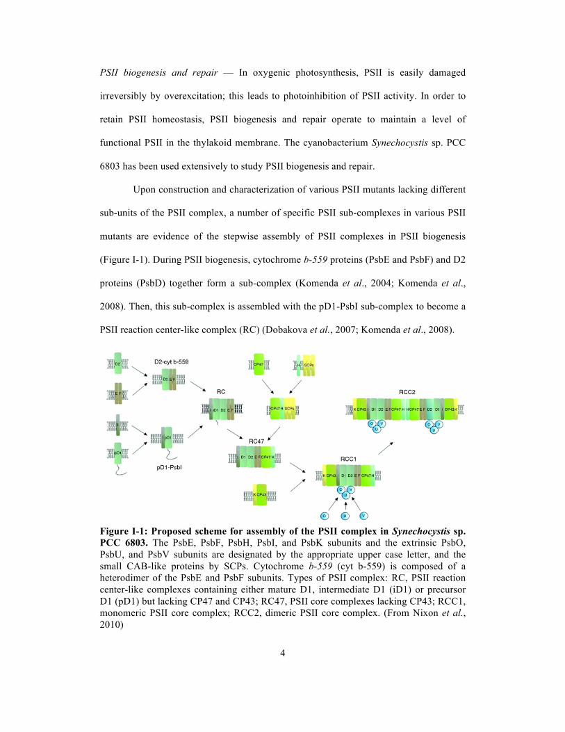
Upon construction and characterization of various PSII mutants lacking different
sub-units of the PSII complex, a number of specific PSII sub-complexes in various PSII
mutants are evidence of the stepwise assembly of PSII complexes in PSII biogenesis
(Figure I-1). During PSII biogenesis, cytochrome b-559 proteins (PsbE and PsbF) and D2
proteins (PsbD) together form a sub-complex (Komenda et al., 2004; Komenda et al.,
2008). Then, this sub-complex is assembled with the pD1-PsbI sub-complex to become a
PSII reaction center-like complex (RC) (Dobakova et al., 2007; Komenda et al., 2008).
Figure I-1: Proposed scheme for assembly of the PSII complex in Synechocystis sp. PCC 6803. The PsbE, PsbF, PsbH, PsbI, and PsbK subunits and the extrinsic PsbO, PsbU, and PsbV subunits are designated by the appropriate upper case letter, and the small CAB-like proteins by SCPs. Cytochrome b-559 (cyt b-559) is composed of a heterodimer of the PsbE and PsbF subunits. Types of PSII complex: RC, PSII reaction center-like complexes containing either mature D1, intermediate D1 (iD1) or precursor D1 (pD1) but lacking CP47 and CP43; RC47, PSII core complexes lacking CP43; RCC1, monomeric PSII core complex; RCC2, dimeric PSII core complex. (From Nixon et al., 2010)

5
The D1 protein (PsbA) in the pD1-PsbI sub-complex has not yet been processed to its
mature form; this precursor (pD1) carries a 16-residue extension at the C-terminus that is
cleaved by CtpA to leave an intermediate eight-amino-acid extension as intermediate D1
(iD1) or to lead to mature D1 (total 344 amino acid residues) in this RC (Komenda et al.
2007; Inagaki et al., 2001). The cleavage of the C-terminal extension of pD1 is required
for assembly of a functional CaMn4 cluster (Nixon et al., 1992; Anbudurai et al., 1994).
Then, the RC-like sub-complex associates with the CP47 (PsbB)-PsbH sub-complex to
form RC47. Subsequently, CP43 (PsbC) and PsbK are associated with RC47. PSII
biogenesis is completed with assembly of the CaMn4 cluster and attachment of the
extrinsic subunits (PsbO, PsbU, and PsbV) of the oxygen-evolving complex to the PSII
intrinsic protein complex. Using Blue Native/PAGE (BN/PAGE) of Synechocystis
complexes, mature PSII is found as two forms: the PSII monomer (RCC1) and the PSII
dimer (RCC2) (Herranen et al., 2004).
The D1 protein is the main PSII subunit damaged during PSII photoinhibition.
Rapid turnover of D1 has been observed in vivo in radioactive pulse-chase labeling
experiments (Ohad et al., 1984). In order to degrade the damaged D1 protein, partial
disassembly of PSII may be required by detachment of the oxygen-evolving complex and
CP43 may become loose from the PSII complex. The damaged D1 protein is likely to be
degraded by members of the FtsH protease families. However, some of the FtsH
proteases have shown to play a more crucial role in D1 degradation: impaired rates of D1
degradation were observed in mutants lacking FtsH2 in Synechocystis sp. PCC 6803
(Silva et al., 2003; Komenda et al., 2006) and FtsH2 and FtsH5 in A. thaliana (Bailey et
al., 2002; Kato et al., 2009). After degradation of the damaged D1 protein, the
replacement of the newly synthesized D1 protein occurs co-translationally into the RC47
6
complex (Zhang et al., 1999). CP43 reattaches to form a PSII core complex, which then
reassembles the CaMn4 cluster and extrinsic proteins into a functional PSII.
During PSII assembly in the PSII biogenesis and repair cycle, there are PSII
assembly factors that aid and/or regulate PSII assembly. These PSII assembly factors
make sure that PSII assembles properly. For example, Ycf48 (Hcf136 in plants) binds to
and stabilizes unassembled pD1 and aids formation of the PSII RC (Plucken et al., 2002;
Komenda et al., 2008). Psb27 is an assembly factor at the lumenal side that is mainly
associated with CP47 and CP43 of monomeric PSII and non-oxygen-evolving PSII
complexes and prevents binding of the oxygen-evolving complex (Kashino et al., 2002;
Nowaczyk et al., 2006; Cormann et al., 2009). Psb28 and Psb29 are important assembly
factors for the CP47 protein (Dobakova et al., 2009; Keren et al., 2005).
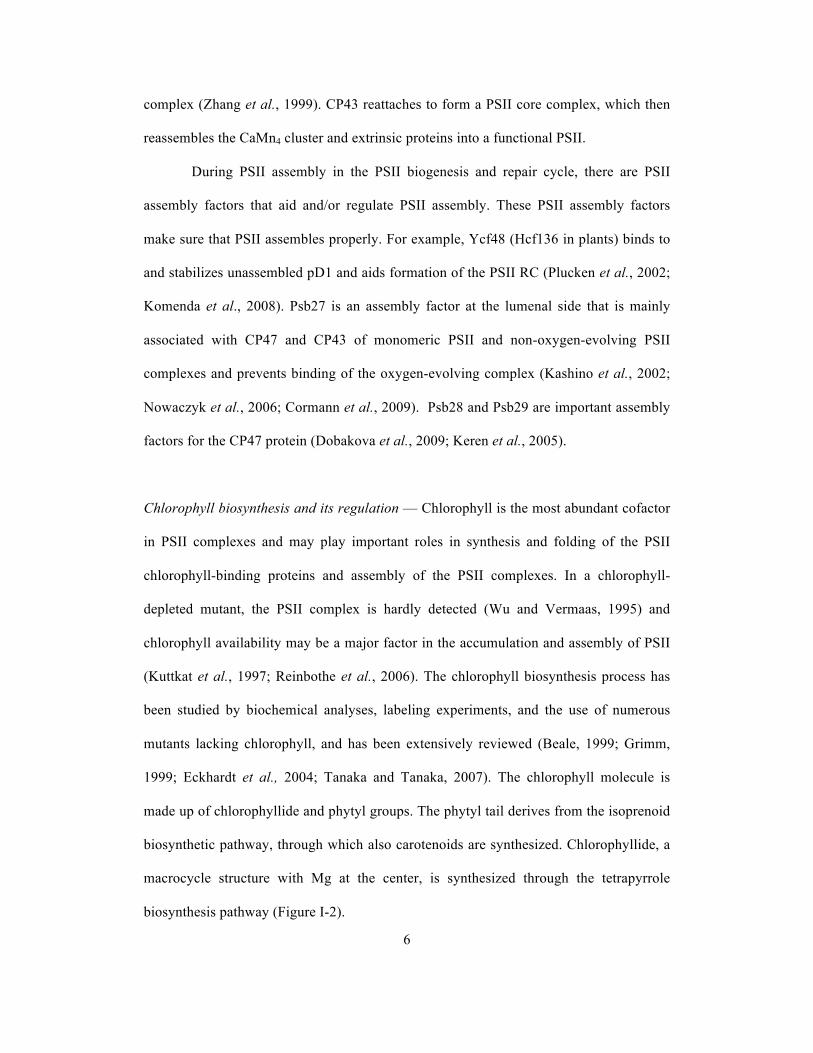
Chlorophyll biosynthesis and its regulation — Chlorophyll is the most abundant cofactor
in PSII complexes and may play important roles in synthesis and folding of the PSII
chlorophyll-binding proteins and assembly of the PSII complexes. In a chlorophyll-
depleted mutant, the PSII complex is hardly detected (Wu and Vermaas, 1995) and
chlorophyll availability may be a major factor in the accumulation and assembly of PSII
(Kuttkat et al., 1997; Reinbothe et al., 2006). The chlorophyll biosynthesis process has
been studied by biochemical analyses, labeling experiments, and the use of numerous
mutants lacking chlorophyll, and has been extensively reviewed (Beale, 1999; Grimm,
1999; Eckhardt et al., 2004; Tanaka and Tanaka, 2007). The chlorophyll molecule is
made up of chlorophyllide and phytyl groups. The phytyl tail derives from the isoprenoid
biosynthetic pathway, through which also carotenoids are synthesized. Chlorophyllide, a
macrocycle structure with Mg at the center, is synthesized through the tetrapyrrole
biosynthesis pathway (Figure I-2).

7
Figure I-2: Chlorophyll biosynthetic pathway. (From Blankenship, 2002)

8
The initial steps in tetrapyrrole biosynthesis, from the biosynthesis of 5-
aminolevulinic acid (ALA) to protoporphyrinogen IX, take place in the cytosol or stroma
of the plastid in plants, whereas the subsequent steps are membrane-bound (Joyard et al.,
2009; Mochizuki et al., 2010). ALA is the first major intermediate in the tetrapyrrole
biosynthetic pathway and its synthesis occurs in three enzymatic steps from glutamate
(the C5-pathway) (Beale, 1999). Glutamyl-tRNA synthetase ligates glutamate with
tRNAGlu. The reaction is followed by the one catalyzed by the glutamyl-tRNA reductase
(GluTR) that reduces the carboxyl group of glutamyl-tRNA to produce glutamate-1-
semialdehyde. Subsequently, glutamate-1-semialdehyde is transaminated by glutamate-1-
semialdehyde aminotransferase to form ALA. ALA is a universal precursor of
tetrapyrrole biosynthesis in all organisms, but this ALA biosynthetic pathway is only
present in plants, algae, most bacteria including cyanobacteria, and archaea. The other
eukaryotic organisms and some bacteria including non-sulfur purple bacteria have an
alternative pathway (Shemin pathway) to synthesize ALA by condensation of succinyl-
CoA with glycine (Mayer and Beale, 1992).
The enzymatic reactions converting ALA to protoporphyrin IX start with
condensation of two ALA molecules to form a pyrrole molecule, porphobilinogen (PBG).
Subsequently, four PBGs are polymerized and ligated to form the first closed tetrapyrrole
ring, uroporphyrinogen III. Protoporphyrin IX is synthesized from oxidation of
protoporphyrinogen IX derived from a series of decarboxylations of uroporphyrinogen
III. Uroporphyrinogen III also can be utilized to produce vitamin B and siroheme via the
siroheme biosynthetic branch.
Protoporphyrin IX can incorporate either Mg2+ or Fe2+, leading molecules into
the chlorophyll branch and heme branch of the tetrapyrrole biosynthesis pathway,
respectively (Vavilin and Vermaas, 2002; Tanaka and Tanaka, 2007). The first step of the

9
chlorophyll branch is catalyzed by Mg-chelatase, a heterotrimer of three subunits, ChlH,
ChlI, and ChlD, that inserts Mg2+ into protoporphyrin. This reaction requires ATP
hydrolysis. The next couple of reactions are catalyzed by methyltransferase and cyclase
to produce protochlorophyllide. Reduction of protochlorophyllide to divinyl-
chlorophyllide is performed by protochlorophyllide oxidoreductase (POR). There are two
types of structurally unrelated PORs, the light-dependent protochlorophyllide reductase
(LPOR) and light-independent protochlorophyllide reductase (DPOR). LPOR contains
only one protein subunit without a cofactor, and NADPH is required for the reaction.
DPOR consists of three subunits, ChlL, ChlN, and ChlB, whose amino acid sequences
show significant similarities to those of NifH, NifD, and NifK in nitrogenase,
respectively (Burke et al., 1993). The enzyme carries two FeS clusters, and the reduction
reaction is ATP-dependent (Sarma et al., 2008; Muraki et al., 2010). Due to lack of
DPOR in angiosperms, protochlorophyllide is accumulated when angiosperm seedlings
are grown in the dark (Griffiths, 1975). The 8-vinyl group of the B ring of divinyl-
chlorophyllide is reduced by divinylchlorophyllide a reductase to form chlorophyllide.
The final step of chlorophyll a synthesis is to esterify chlorophyllide with phytyl
pyrophosphate by chlorophyll synthase.
In order to produce a sufficient amount of chlorophyll to meet the demands of the
chlorophyll-binding proteins but at the same time to prevent accumulation of excess
chlorophyll and its precursors in the cell, the chlorophyll biosynthetic pathway is highly
regulated. Glutamyl-tRNA reduction by GluTR in ALA synthesis is a most important
regulation point in the tetrapyrrole pathway (Beale, 1990). This regulation point is
reasonable because of most of the tetrapyrrole intermediates may cause damage to the
cells in the light and the presence of oxygen. Controlling the GluTR activity is mainly
achieved through feedback regulation by the end products such as heme, and Mg-

10
protoporphyrin IX and its later intermediates (Rieble and Beale, 1991). FLU has been
identified as a regulatory protein to repress GluTR activity as protochlorophyllide is
accumulated in higher plants (Meskauskiene et al., 2001). The other regulation step is at
the major branchpoint where protoporphyrin IX is directed toward either the chlorophyll
or heme branches. The concentration of substrates for the formation of Mg-
protoporphyrin IX such as ATP and Mg2+ affects Mg-chelatase activity. Also, GUN4, a
regulatory protein in plants, functions in enhancing substrate binding and/or product
release of Mg-chelatase (Adhikari et al., 2009; Peter and Grimm, 2009). For the heme
branch, an increase in ferrochelatase activity resulting in increased production of heme
could inhibit synthesis of ALA (Srivastava and Beale, 2005; Beck and Grimm, 2006).
However, the deletion of the C-terminal extension of ferrochelatase that contains one
transmembrane helix with a CAB domain (called ScpA in Synechocystis) reduces its
activity, which results in upregulated ALA synthesis (Sobotka et al., 2008).
Chlorophyll — The chlorophyll a molecule contains five rings (A through E), and is
classified as a chlorin rather than a porphyrin because of the reduction of ring D by
protochlorophyllide reductase (Figure I-3). Chlorophyll a has maximal absorption at 430
(Soret), 578 (Qx), and 662 (Qy) nm in diethyl ether and fluoresces at 670 nm
(Blankenship, 2002). All eukaryotic photosynthetic organisms and cyanobacteria have
chlorophyll a. There are the other types of chlorophylls in photosynthetic organisms. For
example, higher plants have chlorophyll b in their light-harvesting complexes, and purple
bacteria possess bacteriochlorophylls that contain bacteriochlorins, where the B ring is
reduced, instead of chlorins.

11
Figure I-3: Chlorophyll a structure and its absorption (left) and fluorescence spectra (right) in diethyl ether. (From Blankenship, 2002)
Chlorophyll is very efficient in absorbing light energy and has long-lived excited
states (up to a few nanoseconds) to allow the conversion of the excitation energy into an
electrochemical potential via charge separation. However, if the excitation energy is not
used, the excited chlorophyll may drop its energy state to a lower-energy excited state,
the chlorophyll triplet state, that has an even longer lifetime (a few µs). 3O2 can react with
triplet-state chlorophyll to produce 1O2 that is a reactive oxygen species and can damage
cells if there are no efficient quenchers nearby (Triantaphylides and Havaux, 2009).
Therefore, in order to avoid the formation of 1O2 generated from chlorophyll, cooperation
between chlorophyll biosynthesis and synthesis of chlorophyll-binding proteins during
PSII biogenesis and accommodation of chlorophyll during the PSII repair cycle are
critically important as free chlorophyll in the thylakoid membrane and chlorophyll in a
non-functional reaction center, where excitation energy can not be used in charge
separation and where close association with a triplet quencher may not exist, are
considered to be very dangerous.

12
Small CAB-like proteins — The cyanobacterium Synechocystis sp. PCC 6803 possesses
five small CAB-like proteins (ScpA, ScpB, ScpC, ScpD, and ScpE; SCPs refers to all
five proteins), which are predicted to have a single membrane-spanning helix. The
sequence of the helices is similar to the first and third membrane-spanning region of the
Cab protein family in higher plants (Dolganov et al., 1995). Pigment-binding regions of
Cab proteins are conserved in the SCPs, and SCPs appear to bind chlorophyll in vitro
(Funk and Vermaas, 1999; Storm et al., 2008). However, unlike CAB proteins in higher
plants that function in light harvesting or photoprotection, SCPs appear to transiently
bind chlorophyll in the supply of chlorophyll to photosynthetic proteins and to regulate
the tetrapyrrole biosynthesis as a function of chlorophyll availability (Xu et al., 2002; Xu
et al., 2004). Therefore, SCPs may play an important role as a bridge in communication
between chlorophyll and photosynthetic proteins.
Aims of this study — Chapter II discusses the association of a member of the family of
Small Cab-like Proteins (SCPs) with PSII. Because of this association, chlorophylls can
be temporarily stored while PSII components are being replaced. In Chapter III, the
lifetimes of PSII components are studied. The lifetime data give insight regarding the
requirement and correlation between PSII protein synthesis and chlorophyll biosynthesis,
and show that SCPs play important roles in reutilization of chlorophyll and in the stability
of nascent PSII proteins and complexes. The SCP-less mutants also show a significant
decrease in ALA biosynthesis. The fourth chapter addresses the lifetimes of PSII and PSI
proteins in the wild-type strain. Based on a previous study that suggested that SCPs also
stabilize the trimeric PSI complex (Wang et al., 2008), the lifetime of photosynthetic
proteins in the SCP-less mutant is also examined. As of results, there are no changes in

13
the lifetimes of most photosynthetic proteins, except the extrinsic proteins, upon removal
of the SCPs. The last chapter is to characterize the sll1906 gene that is a member of the
putative bacteriochlorophyll delivery (BCD) protein. In this work, the Δsll1906 mutant
was created. However, the chlorophyll biosynthesis/degradation and photosystem
assembly are not affected in the mutant. There may be other pathways fully compensating
for the lack of Sll1906.

14
CHAPTER II. LOCALIZATION OF THE SMALL CAB-LIKE PROTEINS IN
PHOTOSYSTEM II
Abstract
The cyanobacterial SCPs consist of one-helix proteins that resemble transmembrane
regions of the light-harvesting proteins of plants. To determine whether these proteins are
associated with protein complexes in the thylakoid membrane, an abundant member of
the SCP family, ScpD, was marked with a His tag, and proteins co-isolating with His-
tagged ScpD were identified. These proteins included the major PSII components as well
as FtsH, which is involved in degradation of the PSII complex. To ascertain specific
interaction between ScpD and the PSII complex, the His-tagged protein fraction was
subjected to two-dimensional blue native/SDS-PAGE. Again, PSII components were co-
isolated with ScpD-His, and ScpD-His was found to interact most strongly with CP47.
ScpD association was most prominent with the monomeric form of PSII, suggesting
ScpD association with PSII that is being repaired. Using antibodies that recognize both
ScpC and ScpD, we found the ScpC protein, which is very similar in primary structure to
ScpD, to also co-isolate with the PSII complex. In contrast, ScpE did not co-isolate with a
major protein complex in thylakoids. A fourth member of the SCP family, ScpB, could
not be immunodetected, but was found by mass spectrometry in samples co-isolating
with ScpD-His. Therefore, ScpB may be associated with ScpD as well. No association
between SCPs and PSI could be demonstrated. On the basis of these and other data
presented, we suggest that members of the SCP family can associate with damaged PSII
and can serve as a temporary pigment reservoir while PSII components are being
replaced.
(Published in J. Biol. Chem. 282, 267-276, 2007)

15
Introduction In organisms performing oxygenic photosynthesis, sunlight is absorbed by chlorophylls
and other pigments, and absorbed excitation energy is transferred to the reaction centers,
where the photochemical process of converting excitation energy to chemical (redox)
energy takes place. These pigments are bound to proteins to keep them in their proper
location and orientation so that the energy transfer is efficient and rapid and so that toxic
triplet states can be quenched effectively. In plants, the vast majority of pigments,
including chlorophylls a and b and various carotenoids, are bound to a family of integral
membrane proteins called the light-harvesting complex (LHC). Most abundant is LHCII,
the main light-harvesting complex of PS II, which has been crystallized and is known to
consist of three transmembrane helices (B, C, and A), each of which is composed of 20–
34 amino acids (Kuhlbrandt et al., 1994; Liu et al., 2004). The sequences of helices A
and B are very similar and comprise the CAB (chlorophyll a/b-binding) motif, which is
composed of about 25 amino acid residues and includes the domain involved in
chlorophyll binding (Jansson, 1999). Each individual LHCII apoprotein molecule binds
an array of about eight chlorophylls a, six chlorophylls b, three to four carotenoids, and
two lipids (Standfuss et al., 2005). Several other closely related chlorophyll a/b-binding
polypeptides function as light-harvesting antenna for PSII and PSI in plants. Together,
these proteins are known as CAB proteins (Jansson, 1999). The CAB proteins in plants
display a high degree of sequence similarity and are believed to share a common
evolutionary origin (Durnford et al., 1999; Heddad and Adamska, 2002). Their
corresponding nuclear encoded genes belong to an extended cab family that includes also
the genes coding for early light-inducible proteins, which are stress-induced (Adamska,
2001) and probably bind chlorophyll a and lutein (Adamska et al., 1999). The CAB

16
family also includes the PsbS protein (Funk, 2001), which has an important function in
non-photochemical quenching (Li et al., 2000). PsbS is predicted to have four thylakoid
membrane-spanning regions, and it binds chlorophylls a and b as well as carotenoids
(Funk et al., 1994). Moreover, related genes coding for polypeptides with one or two
transmembrane α-helices have been detected in the genomes of Arabidopsis thaliana
(Heddad and Adamska, 2000; Jansson et al., 2000), rice and poplar (Klimmek et al.
2006), Chlamydomonas reinhardtii (Teramoto et al., 2004), and the red alga
Cyanidioschyzon merolae (Ohta et al., 2003).
In contrast to plants, cyanobacteria lack the multihelix CAB proteins. The major
peripheral LHC in some cyanobacteria is the phycobilisome, which is in the cytoplasm, is
bound to the thylakoid membrane, and contributes to the deep blue-green color of
cyanobacteria. However, small CAB-like proteins of <8 kDa have recently been
identified in the genomes of marine and freshwater cyanobacteria (reviewed in Ref.
Bhaya et al., 2002). These proteins are predicted to have a single membrane-spanning α-
helix, which shows significant sequence similarity to the first and third membrane-
spanning regions of the green plant CAB proteins, giving them the name small CAB-like
proteins (SCPs) (Funk and Vermaas, 1999). They were also designated high light-
inducible proteins because their RNA level was found to increase after transfer of cells to
high light and many other stress conditions (He et al., 2001; Mikami et al., 2002). In the
small genome of the cyanobacterium Prochlorococcus marinus MED4, as many as 23
scp or hli genes were identified (Bhaya et al., 2002), and these genes have recently been
detected in the genomes of Prochlorococcus cyanophages (Lindell et al., 2004; Sullivan
et al., 2005), where they are believed to maintain the photosynthetic activity of the host

17
during an infection (Lindell et al., 2004). Although the function of the SCPs is not fully
understood, these findings indicate their importance.
In the cyanobacterium Synechocystis sp. PCC 6803, five SCPs were identified
(Funk and Vermaas, 1999); four of them (ScpB-E) encode proteins of ~ 6 kDa, whereas
the fifth (ScpA) is the C-terminal extension of the ferrochelatase. The genes coding for
ScpB–E are induced under various different stress conditions, including very high light
intensity (>500 µmol m–2 s–1), low temperature, and nitrogen and sulfur starvation (He et
al., 2001; Mikami et al., 2002). A mutant with these four genes inactivated is sensitive to
high intensity illumination and shows alteration in pigmentation and in the ability to
perform non-photochemical dissipation of absorbed light energy (Havaux et al., 2003).
The enhanced expression of the scp genes in response to high intensity illumination is
consistent with the putative function of SCPs in protection against light stress (He et al.,
2001). It was suggested that SCPs play a role in energy dissipation that is analogous to
the process of non-photochemical quenching of higher plants (Havaux et al., 2003), but
the absence of scp genes does not affect fluorescence characteristics (Xu et al., 2004). On
the other hand, a carotenoid closely associated with phycobilin energy transfer is now
recognized to be involved with energy transfer regulation (Mullineaux and Emlyn-Jones,
2005; Rakhimberdieva et al., 2004; Wilson et al., 2006). It also has been hypothesized
that SCPs prevent the formation of reactive oxygen species by serving as transient
carriers of chlorophyll (Xu et al., 2002; Xu et al., 2004).
The presence of the CAB motif in SCPs suggests that SCPs bind chlorophyll
molecules in a similar way as the LHCII of plants. Furthermore, the SCPs seem to
participate in tetrapyrrole biosynthesis and regulate pigment availability. The chlorophyll
synthesis rates in the PSI-less/ΔchlL/ΔscpB, PSI-less/ΔchlL/ΔscpE, and PSI-
less/ΔchlL/ΔscpC/ΔscpD strains decrease when these three mutants are transferred from

18
darkness to light (Xu et al., 2002; Xu et al., 2004). Interestingly, ScpC and ScpD seem to
be functionally complementary (Xu et al., 2004). These two protein sequences are most
similar (87.1% identity) (He et al., 2001), indicating a rather recent gene duplication
(Bhaya et al., 2002) or a reasonably strict primary structure requirement.
ScpD was immunologically detected in thylakoid membranes of Synechocystis
sp. PCC 6803 (Hao et al., 2001). To understand the function of this and other SCPs, it is
important to know which complexes in the membrane they interact with. Here, we used
His-tagged ScpD proteins to identify the main complexes with which ScpD is associated.
After two-dimensional PAGE (blue native (BN) PAGE followed by SDS-PAGE), ScpD
was found to be associated with monomeric PSII, its closest neighbor being CP47. CP43
and Psb28 were also found to interact with ScpD. Although ScpC could be identified in
the PSII fraction and ScpB was found to co-fractionate with ScpD to some degree, ScpE
was found in thylakoids, but did not seem to be associated with PSII.
Materials and Methods
Growth conditions — Synechocystis sp. PCC 6803 strains (wild-type, the PSI-less
strain (ΔpsaAB) (Shen et al., 1993), the PSII-less strain (ΔpsbDIC/ΔpsbDII) (Carpenter
et al., 1990), the PSI-less/PSII-less strain (ΔpsaAB/ΔpsbDIC/ΔpsbDII) (Ermakova-
Gerdes et al., 1996), the CP47-His-tagged HT-3 strain (Bricker et al., 1998), and the
ScpD-His strain (see below)) were cultivated at 30 °C in BG-11 medium (Rippka et al.,
1979). The PSI-less and PSII-less mutants were provided with 15 mM glucose. All
strains except the PSI-less strain were grown at normal (50 µmol photons m–2 s–1) or high
(500 µmol photons m–2 s–1) light intensity as indicated. Because of its light sensitivity, the
PSI-less strain was cultured at 4 µmol photons m–2 s–1. To induce the SCPs, the wild-type

19
and PSII-less strains were grown at high light intensity for 7 h. In the PSI-less/PSII-less
and PSI-less strains, SCPs are induced also at light intensities of 50 and 10 µmol photons
m–2 s–1, respectively (Funk and Vermaas, 1999).
Mutant construction — To generate the ScpD-His strain, a plasmid construct was made
to tag the ScpD protein in Synechocystis with an His6 epitope on its N terminus and to
express the corresponding gene construct under the control of the psbAII promoter. To
construct this plasmid, the scpD gene was amplified by PCR using a mixture of Taq and
Pfu DNA polymerases and gene-specific primers (forward, 5′-
TTATACATATGCATCATCATCATCATCATGGAACTAGCCGCGGATTTCGCCT-
3′; and reverse, 5′-TCGGATCCTTAGAGAGGAGAGCAACCAACCC-3′) with
artificially generated restriction sites for NdeI and BamHI and containing six histidine
codons (CAT) in the forward primer. After restriction, the PCR fragment was cloned into
the NdeI and BamHI sites of the pPSBA plasmid; the resulting plasmid contains the
scpD-His gene construct right behind the psbAII start codon (Lagarde et al., 2000) and
retains the upstream and downstream regions of the Synechocystis psbAII gene. The
ligation mixture was amplified by PCR using pPSBA primers amplifying the entire
psbAII/scpD-His region, and DNA of the desired size was selected. Amplification by
PCR was chosen because transformation of Escherichia coli with the ligation mixture
yielded no colonies, presumably reflecting toxicity of the plasmid to E. coli. The PCR
product containing the scpD-His gene was transformed into the Synechocystis psbAII-
KS strain, in which the psbAII gene was replaced with a kanamycin resistance/sacB
cartridge (Lagarde et al., 2000). The sacB gene codes for a levan sucrase, leading to
sucrose sensitivity of this strain (Ried and Collmer, 1987). After transformation,
Synechocystis cells were grown on BG-11 plates for 4 days. Transformants were then

20
transferred to plates with 5% sucrose, and sucrose-resistant colonies were checked for
kanamycin sensitivity. The resulting strain expressing both wild-type and His-tagged
forms of the ScpD protein was subsequently transformed with chromosomal DNA from a
scpD- strain carrying a spectinomycin resistance cassette insertion (Prentki and Krisch,
1984), and spectinomycin-resistant transformants were selected (Xu et al., 2002).
Insertion of the scpD-His gene at the desired location was confirmed by DNA
sequencing, and deletion of the wild-type scpD gene was confirmed by PCR.
Biochemical preparations — Total membranes from the different Synechocystis strains
were isolated as described (Funk and Vermaas, 1999). Radioactive labeling of cells using
a mixture of L-[35S]methionine and L-[35S]cysteine (>1000 Ci/mmol, final activity of 400
µCi/mL; Tran35S-label, ICN Biomedicals) and isolation of membranes were performed as
described (Komenda et al., 2004). Isolated membranes were solubilized with n-dodecyl
β-maltoside (n-dodecyl β-maltoside/chlorophyll ratios were 20 and 100 (w/w) in the PSI-
containing and PSI-less strains, respectively), and extracted complexes were separated by
BN gel electrophoresis (Schagger and von Jagow, 1991).
Isolation of His-tagged complexes — Cells from Synechocystis sp. PCC 6803 strains
carrying a His tag were pelleted after 4 h of exposure to high light intensity (500 µmol
photons m–2 s–1), resuspended in Buffer A (50 mM MES-NaOH (pH 6.0), 10 mM MgCl2,
5 mM CaCl2, and 25% glycerol), and broken. Thylakoids were prepared as described
(Bricker et al., 1998). The cell homogenate (at 1 mg/mL chlorophyll) was brought to
1.28% β-dodecyl maltoside and incubated for 25 min at 4 °C. The sample was then
loaded onto a Ni2+ metal affinity column. The column was washed with 9 column
volumes (45 ml) of Buffer A containing 0.04% β-dodecyl maltoside and 10 mM

21
imidazole. Subsequently, the column was washed with 10 mL of Buffer A with 0.04% β-
dodecyl maltoside and 30 mM imidazole. Bound ScpD-His was eluted with 0.04% β-
dodecyl maltoside and 100 mM imidazole in Buffer A. The eluate was precipitated by the
addition of an equal volume of 25% polyethylene glycol 8000 in 50 mM MES-NaOH
(pH 6.0) and then resuspended in Buffer A containing 0.04% β-dodecyl maltoside.
PAGE — To the resuspended Ni2+ column eluate was added 0.1 volume of loading
solution containing 750 mM aminocaproic acid and 5% Coomassie Brilliant Blue G-250.
Protein complexes in the eluate were separated by BN-PAGE at 4 °C as described
(Schagger and von Jagow, 1991) using a 5–14% polyacrylamide gradient gel. For the
second dimension, the BN gel lane of interest was incubated for 20 min in a solution
containing 25 mM Tris-HCl (pH 7.5) and 1% SDS and then placed on top of an SDS-12–
20% polyacrylamide gel containing 7 M urea (Komenda et al., 2002). After
electrophoresis, gels were either stained with silver nitrate (Bjellqvist et al., 1993) or
transferred onto polyvinyl difluoride membrane for further analysis by Western blotting.
Immunoblotting — For immunoblotting, the proteins were transferred onto polyvinyl
difluoride membrane (Towbin et al., 1979). Anti-ScpC antibody raised in rabbits against
residues 1–17 of the ScpC protein (MTTRGFRLDQDNRLNNF) was a gift from Dr. A.
Sokolenko (University of Munich). A peptide-directed antibody against a region near the
N-terminus of ScpE (ELQPNQTPVQEDPKFG) was made commercially by Innovagen
AB (Lund, Sweden).
Pigment analysis — Chlorophyll content was determined in 80% acetone and was
calculated as described (Porra et al., 1989).

22
Protein analysis by matrix-assisted laser desorption ionization time-of-flight
(MALDI-TOF) mass spectrometry — Protein identification by peptide mass
fingerprinting and post-source decay tandem mass spectrometry (MS/MS) analysis was
carried out using a Voyager-DE STR mass spectrometer (Applied Biosystems,
Stockholm). In-gel digestion to produce peptides for analysis by mass spectrometry was
carried out essentially as described (Shevchenko et al., 1996) using sequencing-grade
modified trypsin (Promega/SDS Biosciences, Falkenberg, Sweden) or sequencing-grade
chymotrypsin (Roche Diagnostics, Bromma, Sweden). Silver-stained protein bands were
destained prior to in-gel digestion using the method previously described (Gharahdaghi et
al., 1999). To analyze the in gel-digested proteins by MALDI-TOF mass spectrometry,
dried droplet preparations were applied as described (Kussmann et al., 1997). The
matrices used were readymade solutions of α-cyano-4-hydroxycinnamic acid (G2037A)
and 2,5-dihydroxybenzoic acid (G2039A) from Agilent Technologies (Stockholm).
Samples were concentrated and desalted as needed using homemade Stop-and-Go
extraction columns as described (Rappsilber et al., 2003). Data base searches were
carried out on an in-house Mascot server that was licensed to Umeå University by Matrix
Science (www.matrixscience.com) using the current version of the NCBInr Database and
the Synechocystis Protein Database of the European Bioinformatics Institute. The data
bases were searched using peptide mass fingerprint spectra and post-source decay
MS/MS spectra. If appropriate, proteins were identified by sequence queries that included
both types of data. The search parameters restricted the error for peptide masses to 50
ppm and for MS/MS fragments to 0.5 Da. The instrument type specified for MS/MS ion
searches was MALDI-TOF/TOF. By default, the search parameters permitted one missed

23
cleavage site and variable oxidation states of methionine. If appropriate, two or more
missed cleavage sites were allowed.
Results
Proteins co-purifying with ScpD-His — Based on two-phase separation experiments,
ScpD is a prevalent SCP member in the thylakoid membrane (Hao et al., 2001), but its
association with protein complexes in this membrane system remains unknown. To learn
about the function of the SCPs, we decided to tag ScpD with His, to determine its
interaction partners, and to analyze the SCP composition of thylakoid complexes. As
described under “Materials and Methods” we created a Synechocystis mutant in which
scpD had been deleted and replaced with a His-tagged scpD copy, the expression of
which was under the control of the psbAII promoter. After harvesting and rupturing the
cells, the total membranes were solubilized using β-dodecyl maltoside, and ScpD-His-
containing complexes were isolated via nickel column chromatography (Bricker et al.,
1998). Subsequently, the proteins were separated by SDS-PAGE and analyzed by
MALDI-TOF mass spectrometry.
To test the validity of this protocol, we also isolated PSII complexes via CP47-
His using the HT-3 mutant (Bricker et al., 1998) and analyzed them by SDS-PAGE (data
not shown). They were composed of essentially the same subunits as shown in originally
(Bricker et al., 1998; Kashino et al., 2002).
The SDS gel in Figure II-1 shows the washed off fractions and eventual eluate
resulting from the affinity purification of the ScpD-His complex (lanes C–E) and similar
fractions of a chromatography control using wild-type ScpD (lanes A and B). Although
the wild-type fraction collected upon washing the column (lane A) showed no
recognizable pattern, the corresponding fraction from the ScpD-His strain showed a

24
Figure II-1: Proteins co-purifying with ScpD-His. This Coomassie Blue-stained SDS-polyacrylamide gel displays total membrane fractions purified via nickel chromatography. Fractions from wild-type cells (control) are shown in lanes A and B, and fractions from the ScpD-His mutant strain are shown in lanes C–E. Lanes A and C, fractions obtained during the second washing step (0.04% β-dodecyl maltoside and 30 mM imidazole in Buffer A); lanes B, D, and E, fractions obtained in the elution step (0.04% β-dodecyl maltoside and 100 mM imidazole in Buffer A). Lanes D and E show the results of two separate experiments, and the protein bands shown in lane E were analyzed by MALDI-TOF mass spectrometry, resulting in identification as indicated to the right (also see Table II-1). Cytb559, cytochrome b559. pattern of components resembling that of PSII (lane C). Indeed, upon elution with 100
mM imidazole, such components co-eluted with ScpD-His (lanes D and E; representing
results from two independent preparations). The presence of two distinct bands with
apparent masses of 47.3 and 6 kDa was clearly visible in these lanes, and fainter bands
migrating with apparent masses between 4 and 6, 30 and 45, and 70 and 80 kDa could be
observed. The only visible differences between the PSII complexes washed off the
column at 30 mM imidazole (lane C) and at 100 mM imidazole (lane D) were the

25
presence of ScpD-His and an enrichment of CP47 in the latter fraction. It therefore seems
that most of the PSII complexes were washed off the column and that only a minor
fraction was bound to ScpD. The corresponding eluent fraction from the wild-type
control in lane B did not display protein bands, demonstrating the specificity of retention
of the proteins in lanes D and E by ScpD-His.
To identify the proteins that co-purified with ScpD-His, the individual bands in
Figure II-1 (lanes D and E) were digested with trypsin and analyzed by MALDI-TOF
mass spectrometry. If the peptide mass fingerprint spectra of the individual bands were
not sufficient for unequivocal protein identification, post-source decay MS/MS spectra of
individual peptides were acquired for protein identification by sequence queries (Mann
and Wilm, 1994; Perkins et al., 1999). Table II-1 summarizes the results of this analysis.
As expected, the mass spectra showed the presence of ScpD (Ssr2595) in the major band
at an apparent mass of 6 kDa; ScpD was identified with high confidence by its peptide
mass fingerprint spectrum in combination with an MS/MS analysis of the peptide
GFRLDQDNR. The major band at an apparent mass of 47.3 kDa was found to contain
CP47 (Slr0906) and the hypothetical protein Slr0909. Mass spectrometry analysis also
identified CP43 (Sll0851) with an apparent mass of 39.8 kDa, and D2 (Sll0849/Slr0927)
and D1 (Sll1867; two bands) with apparent masses of 36.7 kDa, 34.3 kDa, and 33.5 kDa,
respectively. The band of the D2 protein also contained Slr1128, annotated as a
hypothetical integral membrane protein. In the high mass range, the FtsH proteases
Sll1463 and Slr0228 were present at an apparent mass of 77 kDa, and the FtsH protease
slr1604 was found at an apparent mass of 71 kDa. Furthermore, Sll1021 and Slr0798
(both annotated as hypothetical proteins) were found at apparent masses of 88.1 and 95.1

26
Table II-1: Mass spectrometry identification of proteins apparently forming a complex with ScpD-His (data shown in Figure II-1)
Experimental/theoretical mass
ORFa
Gene product
Mascot search and scoreb
Sequence coverage
r.m.s. error
% ppm 95.1/76.8 kDa Slr0798 Hypothetical
protein SQ 193 35 14
88.1/74.3 kDa Sll1021 Hypothetical protein
PMF 249 44 7
76.7/68.1 kDa Sll1463 FtsH SQ 190 32 14 76.7/68.4 kDa Slr0228 FtsH PMF 150 39 17 70.8/67.1 kDa Slr1604 FtsH PMF 83 24 23 47.3/55.8 kDa Slr0906 CP47 SQ 228 39 15 47.3/52.4 kDa Slr0909 Hypothetical
protein SQ 142 40 11
39.8/51.8 kDa Sll0851 CP43 SQ 143 17 25 36.7/39.4 kDa Sll0849/Slr0927 D2 SQ 157 22 15 36.7/35.6 kDa Slr1128 Hypothetical
protein PMF 173 51 13
34.3/39.6 kDa Sll1867 D1 SQ 78 13 11 33.5/39.6 kDa Sll1867 D1 SQ 132 20 10
6.9/7.7 kDa Ssr2595 ScpD SQ 112 50 13 6.0/7.7 kDa Ssr2595 ScpD SQ 131 50 32 4.8/7.7 kDa Ssl1633 ScpB MIS 105 18 4.8/4.8 kDa Smr0006 Cytochrome
b559 MIS 109
38
a ORF, open reading frame; r.m.s., root mean square; SQ, sequence query including peptide mass fingerprint (PMF) and MS/MS ion search (MIS) data. b Identified by a search in the NCBInr Database. kDa, respectively. In the low mass range, ScpB (Ssl1633) and the small subunit of
cytochrome b559 (Smr0006) were identified at an apparent mass of 4.8 kDa.
ScpD-His associates with PSII subunits — The composition of the affinity-purified
ScpD-His complex indicated that ScpD associated with PSII components. However,
fractions that are isolated by a one-step affinity purification may contain contamination
by nonspecifically bound proteins. For this reason, we subjected the purified ScpD-His

27
Figure II-2: ScpD-His is associated specifically with PSII. ScpD-His and copurified proteins were separated by two-dimensional BN/SDS-PAGE after nickel chromatography and solubilization with 0.04% β-dodecyl maltoside. Proteins were identified by mass spectrometry (see Table II-2). complex to two-dimensional BN/SDS gel electrophoresis. This technique is an accepted
approach to separate protein complexes and their subunits, and it has been successfully
used to study protein complexes from the thylakoid membranes of higher plants
(Thidholm et al., 2002; Aro et al., 2005). Figure II-2 shows an example for the analysis
of the ScpD-His complex at ~200 kDa by two-dimensional BN/SDS gel electrophoresis.

28
Table II-2: Mass spectrometry identification of proteins that co-purified with ScpD-His upon separation by two-dimensional BN/SDS-PAGE after nickel chromatography and solubilization with 0.04% β-dodecyl maltoside
Experimental/ theoretical mass
ORFa
Gene product
Mascot search and scoreb
Sequence coverage
r.m.s. error
% ppm 52.2/55.8 kDa Slr0906 CP47 PMF 213 38 12 38.9/51.8 kDa Sll0851 CP43 SQ 107 15 9 33.5/39.4 kDa Sll0849/Slr0927 D2 PMF 73c 20 17 27.5/39.6 kDa Sll1867 D1 PMF 77c 20 20 7.8/12.2 kDa Slr1645 PsbZ PMF 74c 29 11 7.8/7.0 kDa Ssl2598 PsbH MIS 50 21 7.8/7.7 kDa Ssr2595/Ssl2542 ScpD/ScpC MIS 24c 12 5.8/7.7 kDa Ssr2595/Ssl2542 ScpD/ScpC MIS 25c 12
a ORF, open reading frame; r.m.s., root mean square; SQ, sequence query including peptide mass fingerprint (PMF) and MS/MS ion search (MIS) data. b Identified by a search in the NCBInr Database. c Identified by a search in the Synechocystis sp. PCC 6803 Protein Database of the European Bioinformatics Institute. The pattern of BN gel separation in the first dimension showed two main bands (Figure
II-2). Upon SDS-PAGE in the second dimension and silver staining of the gel followed
by MALDI-TOF mass spectrometry of bands, the corresponding proteins could be
identified. The results are summarized in Table II-2. In the high mass region, we found
the PSII core subunits CP47, CP43, D1, and D2 at apparent masses of 52, 39, 33, and 32
kDa, respectively. In the low mass region, we detected PsbH, PsbZ, and ScpC/ScpD as a
broader band spanning the 6–8 kDa range. It is interesting to note that only the band with
faster migration in the first dimension provided clear evidence for small subunits (Figure
II-2). The slower migrating band may represent PSII dimers at 500 kDa (Thidholm et al.,
2002). The relatively strong affinity of the putative PSII dimer on BN gel for the dye
Coomassie Blue (as seen by the intense coloration of this band relative to the more
rapidly migrating PSII fraction that represents PSII monomers) was unexpected. Equally

29
unexpected was the depletion of ScpD and other low mass polypeptides in this fraction,
as this fraction was isolated by retention on a nickel-nitrilotriacetic acid column,
indicating the association with ScpD-His at the time of isolation. We hypothesize that
ScpD (and possibly ScpC) is associated with monomeric PSII in a way that affects
Coomassie Blue affinity (which would place the SCPs around the PSII monomer) and
that the PSII monomers and dimers/multimers are in dynamic exchange. Another
interesting feature of Figure II-2 is the heavy staining of low mass proteins in the more
rapidly migrating band. Although quantitation of proteins based on silver staining is
tenuous, the high staining intensity of these proteins suggests the association of multiple
polypeptide copies with PSII.
The limited amount of the ScpD-His complex material on the BN gel shown in
Figure II-2 made the analysis of the low mass proteins difficult. Although the
identification of PsbH and PsbZ was unambiguous, MS/MS analysis identified the
peptide GFRLDQDNR, which matches the sequence of ScpD and of its close homolog
ScpC. For this reason, our mass spectrometry data do not allow us to distinguish between
these two SCPs. The purification of the protein complex using His-tagged ScpD implies
that ScpD is present. However, on the basis of the mass spectrometry data, we can neither
confirm nor exclude the presence of ScpC.
Nearest neighbors of ScpD — The purification of ScpD-His complexes by nickel
affinity chromatography and BN gel electrophoresis showed a clear association of ScpD
with PSII, but did not provide evidence regarding the localization of ScpD within the
PSII complex. To obtain information regarding the neighbors of ScpD-His in the PSII
complex, we used a higher β-dodecyl maltoside concentration (0.8% rather than 0.04%)

30
Figure II-3: Closest neighbors of ScpD. ScpD-His and co-purified proteins were separated by BN/SDS-PAGE after nickel chromatography and solubilization with 0.8% β-dodecyl maltoside. The lower panel shows immunostaining of ScpD-His after two-dimensional PAGE using an antibody directed against the His tag. Proteins were identified by mass spectrometry (see Table II-3). Note that Psb28 does not correspond to the sharp dot on the gel, but rather is an underlying band. for solubilization of the ScpD-His complex before separation by two-dimensional
BN/SDS gel electrophoresis. The BN/SDS gel in Figure II-3 shows that the stronger
solubilization of the purified ScpD-His complex resulted in the formation of smaller
subcomplexes. To monitor the separation of different ScpD-His complexes, the SDS gel
was probed by immunoblotting using antibodies directed against the His tag. To make
sure no ScpD-His was overlooked, the antibody concentration used was high, and
therefore, the signal was not linear with the amount of His tag. The composition of the

31
ScpD-containing complexes was analyzed by MALDI-TOF mass spectrometry. Table II-
3 shows the results from the analysis of two different gels and corresponds to protein
bands as indicated in Figure II-3.
ScpD-His was found to be prominently associated with CP47, which was
consistently detected to co-purify with ScpD-His. The weak band below that of CP47 in
Figure II-3 was assigned to CP43 (Table II-3). The diffuse band in the 7–8-kDa region
contained ScpD-His (see immunoblot in the lower panel of Figure II-3), and as this band
was more diffuse than that of the immunoblot, it also might contain ScpC, as our mass
spectra do not allow us to exclude the presence of this protein. A smaller complex toward
the right in Figure II-3 clearly contained ScpD in the diffuse band in the 7–8-kDa range.
In addition, we found Psb28 in a band at an apparent mass of 12.4 kDa. The distinct spot
close to Psb28 is probably an artifact and does not seem to display this protein.
ScpC co-migrates with PSII — As indicated, the detected mass fragment of ScpD is
exactly identical to that of ScpC. Indeed, the primary structures of ScpD and ScpC are
87% identical, and the compelling similarity between these two SCPs indicates that they
may have not only a similar function, but also similar binding partners. Therefore, it was
important to determine the location of ScpC in the thylakoid membrane.
Toward this goal, Synechocystis wild-type cells were pulse-labeled with L-
[35S]Met/Cys for 30 min while growing at 500 µmol photons m–2 s–1, and subsequently,
thylakoid membranes were isolated and analyzed by two-dimensional BN gel
electrophoresis in combination with autoradiography. The autoradiogram of the wild-type
strain in Figure II-4A (first panel) displayed a strong band in the ScpC/ScpD region at 6
kDa that was present in two complexes. The first complex, RCC1, was identified

32
Table II-3: Mass spectrometry identification of proteins that co-purified with ScpD-His upon separation by two-dimensional BN/SDS-PAGE after nickel chromatography and solubilization with 0.8% β-dodecyl maltoside. The results from the analysis of two different gels shown in Figure II-3 are presented. ORF, open reading frame; r.m.s., root mean square; SQ, sequence query including peptide mass fingerprint (PMF) and MS/MS ion search (MIS) data.
Mascot searcha
Sequence coverage
r.m.s. error
Experimental/ theoretical mass
ORF
Gene product
Exp 1 Exp 2 Exp 1
Exp 2
Exp 1
Exp 2
ppm
Experiment 1
Middle lane
47.3/55.8 kDa Slr0906 CP47 PMF167 PMF142 40 33 7 4
39.8/51.8 kDa Sll0851 CP43 SQ50b SQ39b 9 7 21 14
6.9/7.7 kDa Ssr2595 ScpD SQ64b SQ85 50 42 20 22
Right lane
12.4/12.5 kDa Sll1398 Psb28 N.D. SQ136 53 26
8.0/7.7 kDa Ssr2595 ScpD SQ84 MIS65 42 12 12
7.1/7.7 kDa Ssr2595 ScpD SQ97 MIS72 42 12 20
a Identified by a search in the NCBInr Database. b Identified by a search in the Synechocystis sp. PCC 6803 Protein Database of the European Bioinformatics Institute. previously as monomeric PSII consisting of the CP47, CP43, D2, and D1 proteins
(Komenda et al., 2004). The second complex, termed RC47, was smaller and was
depleted in CP43. A comparison with SCP deletion mutants showed that the band with a
molecular mass of 6 kDa was reduced in the ΔscpC/ΔscpD strain (second panel), but
present in the ΔscpB and ΔscpE strains (third and fourth panels, respectively). These
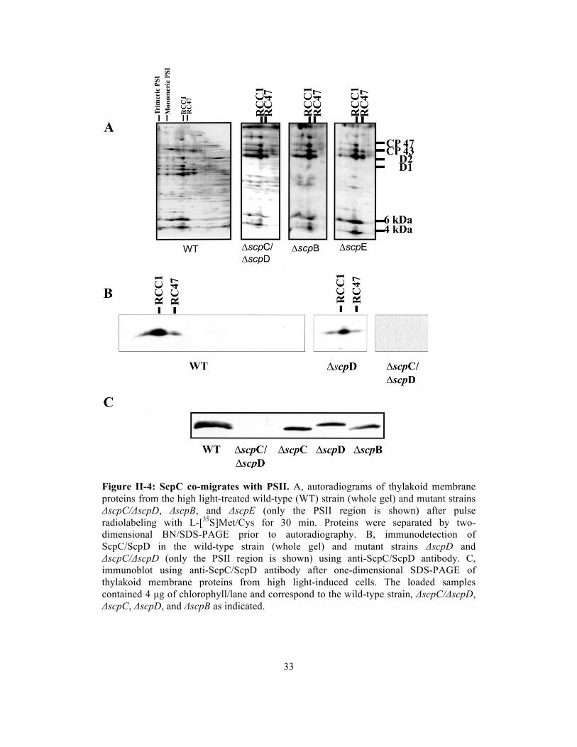
33
Figure II-4: ScpC co-migrates with PSII. A, autoradiograms of thylakoid membrane proteins from the high light-treated wild-type (WT) strain (whole gel) and mutant strains ΔscpC/ΔscpD, ΔscpB, and ΔscpE (only the PSII region is shown) after pulse radiolabeling with L-[35S]Met/Cys for 30 min. Proteins were separated by two-dimensional BN/SDS-PAGE prior to autoradiography. B, immunodetection of ScpC/ScpD in the wild-type strain (whole gel) and mutant strains ΔscpD and ΔscpC/ΔscpD (only the PSII region is shown) using anti-ScpC/ScpD antibody. C, immunoblot using anti-ScpC/ScpD antibody after one-dimensional SDS-PAGE of thylakoid membrane proteins from high light-induced cells. The loaded samples contained 4 µg of chlorophyll/lane and correspond to the wild-type strain, ΔscpC/ΔscpD, ΔscpC, ΔscpD, and ΔscpB as indicated.

34
observations indicate that the high light-induced 6-kDa band of the RCC1 and RC47
complexes contained ScpD and/or ScpC, but most likely not ScpB or ScpE.
To distinguish between ScpD and ScpC in the 6-kDa band of the RCC1 and
RC47 complexes, an antibody was raised against the N terminus of ScpC
(MTTRGFRLDQDNRLNNF), which is identical to that of ScpD except for the third
residue (S in ScpD). Indeed, immunostaining of high light-induced wild-type cells
identified the bands in the 6-kDa region as ScpC and ScpD (Figure II-4B, left panel).
The bands were absent in thylakoids of the high light-induced ΔscpC/ΔscpD strain
(right panel). Therefore, the minor band with a molecular mass of 6 kDa seen in the
autoradiogram of the ΔscpC/ΔscpD strain (Figure II-4A, second panel) belongs to
other co-migrating proteins. In the ΔscpD deletion mutant, the antibody unambiguously
identified ScpC co-migrating with RCC1 and RC47 (Figure II-4B, middle panel).
Interestingly, after applying the same procedure to a PSII-less mutant, ScpC and ScpD
were found to co-migrate with small complexes or as free proteins (data not shown),
suggesting that these SCPs do not readily associate with large complexes such as PSI.
Separation of thylakoid preparations from high light-induced ΔscpC and ΔscpD strains
by one-dimensional SDS electrophoresis made it possible to identify the lower migrating
band as ScpD (Figure II-4C, third lane) and the upper band as ScpC (fourth lane). In
the wild-type strain, the ScpD protein appeared to be dominant (Figure II-4C), but the
ratio between ScpC and ScpD amounts was highly variable among various strains and
conditions, suggesting that ScpC and ScpD are indeed functionally equivalent.
Interestingly, in the ΔscpB strain, the level of ScpD was decreased, and ScpC appeared
to be absent altogether (fifth lane). ScpC/ScpD immunodetection by two-dimensional

35
BN/SDS-PAGE of extracts from the ΔscpB strain showed that ScpD remained associated
with PSII complexes (RCC1 and RC47) as in the wild-type control (data not shown).
ScpE is not associated with PSII — Now that ScpD and ScpC have been positively
correlated with PSII complexes, we set out to determine the location of the other two
small SCPs as well (ScpB and ScpE; ScpA is a C-terminal extension of ferrochelatase).
We were unable to generate antibodies against ScpB. However, we could elicit antibodies
against the peptide ELQPNQTPVQEDPKFG, which is a sequence that is part of the N-
terminal region of ScpE. These antibodies were used for detection of ScpE on SDS-
polyacrylamide gels of membrane preparations from the wild-type and PSII-less strains
(Vermaas et al., 1990) that were grown at 500 µmol photons m–2 s–1 (high light) for 7 h to
induce the expression of ScpE from the PSI-less/PSII-less strain (Ermakova-Gerdes et
al., 1995), in which SCPs are induced also at light intensities of 50 µmol photons m–2 s–1
(Funk and Vermaas, 1999), and from the PSI-less strain that, because of its light
sensitivity, was grown at 10 µmol photons m–2 s–1. As negative controls, membranes from
the wild-type strain grown at normal light intensity (50 µmol photons m–2 s–1) and from
the ΔscpE strain grown at 500 µmol photons m–2 s–1 were included (Figure II-5A and D).
Although no ScpE was detected in membranes from the ΔscpE strain or the wild-type
strain grown at 50 µmol m–2 s–1, the antibody immunostained ScpE in the other strains
and in the wild-type strain grown at high light intensity.
To further localize ScpE, the total membrane fraction of the wild-type strain
grown at high light intensity was separated into fractions enriched in thylakoid
membranes, plasma membranes, and outer membranes using two-phase partitioning
(Norling et al., 1998). In these fractions, ScpE was immunodetected exclusively in the

36
Figure II-5: ScpE is located in the thylakoid membrane. The subcellular location of ScpE was detected by immunoblotting. After breaking the cells, the total membranes were separated by one-dimensional SDS-PAGE and analyzed using anti-ScpE antibody. A, wild-type (WT), PSI-less (PSI–), PSII-less (PSII–), and PSI-less/PSII-less mutant cells were grown at 50 (Control), 500 (HL), and 10 (PSI-less mutant) µmol m–2 s–1. B, thylakoid (TM), cytoplasmic (PM), and outer (OM) membranes were purified by two-phase partitioning (Aro et al., 2005); separated by SDS-PAGE; and analyzed by immunoblotting using anti-ScpE antibody. C, total membranes of high light-treated wild-type and nickel chromatography-purified PSII complexes (CP47-His) (Bricker et al., 1998) and ScpD-His complexes were analyzed by immunoblotting using anti-ScpE antibody. D, shown is the accumulation of ScpE in the high light-induced wild-type, ΔscpB, ΔscpC/ΔscpD, and ΔscpE strains. Equal amounts of cells were loaded in each lane. The proteins were separated by denaturing SDS-PAGE, transferred onto polyvinyl difluoride membrane, and probed with anti-ScpE antibody.

37
thylakoid membrane fraction (Figure II-5B). To investigate whether ScpE is associated
with PSII, oxygen-evolving PSII was isolated from the HT-3 mutant (Bricker et al.,
1998), which contains a hexahistidine tag at the C terminus of the CP47 protein. Also the
ScpD-His fraction eluted after nickel column purification (Figure II-1) was analyzed.
Although an immunoreaction was obtained from the positive control (total membranes
isolated from high light-stressed wild-type strain), ScpE could not be detected in either
PSII or the ScpD-His fraction (Figure II-5C). Deletion of ScpB or ScpC and ScpD did not
alter the presence of ScpE, even though the ScpE abundance was decreased in the
ΔscpC/ΔscpD strain (Figure II-5D).
To determine the potential association of ScpE with membrane complexes, a
two-dimensional BN/SDS-polyacrylamide gel was challenged with anti-ScpE
antibodies.As indicated in Figure II-6, ScpE was not stably associated with one of the
photosystems, but instead was present in small complexes and in free form in the wild-
type strain as well as in the PSI-less mutant. This is in agreement with the fact that no
ScpE protein was detected in isolated PSII (Figure II-5).
Discussion
In various plant genomes, one-helix proteins with high similarity to the first and third
helices of the plant chlorophyll a/b-binding antenna proteins have been detected (Heddad
and Adamska, 1999; Jansson et al., 2000; Klimmek et al., 2006; Teramoto et al., 2004;
Ohta et al., 2003). However, their function still remains enigmatic. In this work, we have
shown that ScpC–E of Synechocystis sp. PCC 6803 are found in thylakoid membranes.
ScpC and ScpD are associated with PSII, whereas ScpE is not associated with larger
membrane complexes.

38
Figure II-6: ScpE is not associated with PSII. Shown is the localization of ScpE after two-dimensional BN/SDS-PAGE analysis of thylakoid membrane proteins from the PSI-less and high light-induced wild-type (WT) strains. Thylakoid membrane proteins were separated in the first dimension by BN-PAGE and in the second dimension by denaturing SDS-PAGE using a 12–20% linear gradient polyacrylamide gel, blotted onto polyvinyl difluoride membrane, and immunostained using anti-ScpE antibodies.
Plant one-helix proteins (Jansson et al., 2000) are apparently involved in
pigment-related processes other than light harvesting, and a light-harvesting function can
also be excluded for the SCPs of Synechocystis sp. PCC 6803 (Xu et al., 2002; Xu et
al., 2004). The whole cab gene family has been suggested to have originally evolved to
serve a function in photoprotection, and the role in light harvesting may be a derived
function (Jansson, 2005).
Instead, the SCPs affect steps in the chlorophyll biosynthesis pathway (Xu et al.,
2002) and chlorophyll stability in the cell, even in darkness (Xu et al., 2004).
Interestingly, the one-helix CAB-like proteins in different organisms exhibit regulatory
responses opposite to those of their relatives, the light-harvesting proteins: at high light
intensity, when the expression of the LHC proteins is repressed, the one-helix

39
proteins/SCPs are up-regulated. Also, the SCPs are up-regulated under many stress
conditions (He et al., 2001). This indicates a function in protection in a broad sense; they
might provide either direct protection (for example, as a pigment carrier) or indirect
protection by regulating pigment metabolism. From a sequence perspective, it is likely
that they bind pigments (chlorophylls and carotenoids), as chlorophyll-binding residues
in the CAB family are conserved in SCPs, and deletion of SCPs leads to a decrease in
chlorophyll and carotenoid content of cells (Xu et al., 2004).
According to the results of this study, ScpD is clearly associated with PSII. PSII
core proteins co-purified with ScpD-His and formed a complex that was retained during
BN-PAGE, at least when PSII was monomeric. Association of SCPs with PSII has not
been observed thus far in crystallography studies of PSII (Zouni et al., 2001; Kern et al.,
2005; Ferreira et al., 2004) or in proteomic PSII association studies (Kashino et al.,
2002), as SCPs are apparent only under conditions of high light exposure. In PSII, ScpD
seems to be associated most closely with CP47; CP47 was the most prominent band after
purification of ScpD-His. However, after solubilization in the presence of increased
detergent concentration, also Psb28 was detected in association with ScpD. Psb28
(Sll1398) has been found to be a substoichiometric subunit of PSII (Kashino et al., 2002)
and is thought to have a regulatory function. As it is considered to reside on the stromal
side of PSII, it might stabilize the complex between ScpD and CP47 (Figure II-7).
Another interesting association of the light-stressed ScpD-His/PSII complex is
that with several FtsH proteases. Deletion analysis of single FtsH protease in
Synechocystis has shown that Slr1604 is crucial for the survival of cells, a phenotype
that also has been observed in the deletion of Slr0228 (Nixon et al., 2005). However,
deletion of Sll1463 does not show a phenotype. Our mass spectrometry analysis showed

40
Figure II-7: Model of ScpD binding to PSII. Stress conditions cause PSII monomerization. ScpD and probably also ScpC bind to the monomers and are located close to CP47. Substoichiometric Psb28 might stabilize the binding between CP47 and ScpC/ScpD.
that this gene was expressed and that its product was apparently associated with PSII.
Therefore, it is most likely involved in repair of light-stressed PSII. The co-purification
of PSII and FtsH with ScpD-His suggests that ScpD is associated with the PSII repair
process. This provides an explanation for the fact that ScpD was found to be associated
with monomeric PSII (Figure II-2), which is often correlated with PSII repair processes.
Indeed, the relatively weak Coomassie Blue staining of the monomeric PSII band
suggests that ScpD forms a protective shield around PSII, most prominently interacting
with the chlorophyll-binding protein CP47, which is near the periphery of the complex.
As ScpD has chlorophyll-binding potential but does not contribute to light
harvesting (Xu et al., 2004), an attractive hypothesis is that ScpD polypeptides serve as a
chlorophyll storage device while PSII is repaired and components are replaced. Indeed,
the rate of turnover of chlorophyll is much lower than that of the PSII protein

41
components with which it is associated (Vaviline et al., 2005). Multiple ScpD and/or
ScpC proteins may be associated with PSII, thus providing an explanation for the high
level of SCPs relative to PSII (Figure II-2).
ScpC is part of PSII as well. Upon BN/SDS-PAGE of complexes from a PSII-
less mutant, ScpC and ScpD migrated as low molecular mass complexes or as free
proteins (data not shown). After denaturing PAGE, ScpC migrated at a slightly higher
molecular mass compared with ScpD. ScpC and ScpD substitute for each other's function
(Xu et al., 2004) and have been hypothesized to form a complex (He et al., 2001).
Although ScpD is generally more abundant than ScpC in light-stressed wild-type cells,
the ScpC/ScpD ratio may vary. ScpB was found by mass spectrometry analysis in the
fraction co-isolating with ScpD-His (Figure II-1). Although this suggests that ScpB is
associated with ScpD, the pulse-labeling pattern of PSII in the absence of scpB was
similar to that of the control (Figure II-4). ScpE is present in thylakoids, but is not found
to be associated with ScpD. Interestingly, ScpB and ScpE have the strongest influence on
chlorophyll biosynthesis (Xu et al., 2002). ScpA is the C-terminal part of ferrochelatase.
In this study, we did not include ScpA, suggested to have an important function in
regulating the tetrapyrrole pathway at the branch point between chlorophyll biosynthesis
versus heme/phycobilin biosynthesis. It has been shown that decreased ferrochelatase
activity improves photoautotrophic growth of a PSII mutant because of increased supply
of chlorophyll (Sobotka et al., 2005) and that the ScpA domain is important for the
ferrochelatase function.
Based on the considerations provided here, the most plausible explanation
regarding the function of ScpC and ScpD in light-stressed PSII is pigment storage during
protein turnover. As such pigments may not be able to transfer energy to functional

42
photosystems and as there is no evidence for high chlorophyll fluorescence from
pigments while photosystems are being repaired, excitations of pigments associated with
SCPs should be quenched efficiently. However, these pigments should not be in
excitation transfer contact with PSII pigments so that they do not decrease light-
harvesting efficiency under conditions in which light is not in excess. There may be
parallels between PSII interaction with SCPs versus the interaction between IsiA and
PSI. IsiA has been found to form a ring around PSI, which is functional in light
harvesting, but also empty rings that are effective energy dissipaters have been detected
(Kouril et al., 2005). Similarly, ScpD and ScpC may form a ring around damaged PSII
centers, aid in repair, and dissipate absorbed energy as needed.

43
CHAPTER III. PHOTOSYSTEM II COMPONENT LIFETIMES IN THE
CYANOBACTERIUM SYNECHOCYSTIS SP. PCC 6803: SMALL CAB-LIKE
PROTEINS STABILIZE BIOSYNTHESIS INTERMEDIATES AND AFFECT EARLY
STEPS IN CHLOROPHYLL SYNTHESIS
Abstract
In order to gain insight in the lifetimes of PSII chlorophyll and proteins, a combined
stable-isotope labeling (15N) / mass spectrometry method was used to follow both old and
new pigments and proteins. PSI-less Synechocystis cells were grown to exponential or
post-exponential phase and then diluted in BG-11 medium with 15N-ammonium and 15N-
nitrate. PSII was isolated, and the masses of PSII protein fragments and chlorophyll were
determined. Lifetimes of PSII components ranged from 1.5 h to 40 h, implying that at
least some of the proteins and chlorophyll turned over independently from each other.
Also, a significant amount of nascent PSII components accumulated in thylakoids when
cells were in post-exponential growth phase. In a mutant lacking small Cab-like proteins
(SCPs), most PSII protein lifetimes were unaffected but the lifetime of chlorophyll and
the amount of nascent PSII components that accumulated were decreased. In the absence
of SCPs one of the PSII biosynthesis intermediates, the monomeric PSII complex without
CP43, was missing. Therefore, SCPs may stabilize nascent PSII protein complexes.
Moreover, upon SCP deletion the rate of chlorophyll synthesis and the accumulation of
early tetrapyrrole precursors were drastically reduced. When 14N-aminolevulinic acid
(ALA) was supplemented to 15N-BG-11 cultures, the mutant lacking SCPs incorporated
much more exogenous ALA into chlorophyll than the control demonstrating that ALA
biosynthesis was impaired in the absence of SCPs. This illustrates the major effects that

44
non-stoichiometric PSII components such as SCPs have on intermediates and assembly,
but not on the lifetime of PSII proteins.
Introduction
Cyanobacteria, algae, and plants can use sunlight and water to carry out oxygenic
photosynthesis. In these organisms, linear photosynthetic electron transfer is catalyzed by
the thylakoid-embedded protein complexes PSII, cyt b6f, and PSI. Linear electron transfer
provides electrons to NADP producing NADPH and transfers protons across the
thylakoid membrane leading to a proton gradient that is used for ATP synthesis. NADPH
and ATP can be used for carbon fixation producing organic compounds. These organic
compounds, along with oxygen produced in water splitting in PSII, enable heterotrophic,
aerobic life on Earth.
The photosystems are multi-protein subunits that non-covalently bind different
cofactors, including chlorophyll a, carotenoids, quinones, lipids, and several inorganic
ions. During photosynthesis, components of PSII complexes turn over rapidly, at least in
comparison to PSI complexes (Powles, 1984; Sonoike, 2006). Of the proteins in the PSII
complex the PsbA (D1) protein turns over most rapidly in the light (Mattoo et al., 1984;
Ohad et al., 1984). This rapid turnover presumably is due to redox chemistry at the water-
splitting complex and/or to reactive oxygen species that are generated from oxygen
reacting with the triplet state chlorophyll formed upon charge recombination between the
primary donor P680+ and the primary acceptor pheophytin (Phe)- (Vass et al., 1992;
Macpherson et al., 1993; Krieger-Liszkay, 2005). According to pulse-chase experiments,
the D1 protein has a half time of 30 min to 1 h under intense illumination (Prasil et al.,
1992). However, the other PSII components appear to have a much lower turnover rate.
For example, the halftime of the PsbB (CP47) protein was estimated to be about 12 h

45
(Schuster et al., 1988; Mattoo et al., 1999), and the lifetime of total chlorophyll in
Synechocystis cells is over a week (Vavilin et al., 2005). If this vast disparity in the
lifetime of PSII components indeed is true, then careful orchestration of the synthesis,
assembly, and repair of photosynthetic complexes is required as free chlorophyll in the
cell would be harmful in the light and in the presence of oxygen and as PSII polypeptides
that are not incorporated in a complex may not be stable in the membrane (Mullet et al.,
1990; Krieger-Liszkay, 2005; Triantaphylides and Havaux, 2009).
In the cyanobacterium Synechocystis sp. PCC 6803, there are five small Cab-like
proteins (ScpA-E), which are single-helix membrane proteins that are located in the
thylakoid membrane (Funk and Vermaas, 1999). The presence of the CAB (chlorophyll
a/b-binding) motif in SCPs suggests that SCPs bind chlorophyll molecules at motifs
similar to those of LHCII in plants (Jansson, 1994; Jansson et al., 2000; Vavilin et al.,
2007; Storm et al., 2008). SCPs appear to play an important role in early stages of
tetrapyrrole biosynthesis and may regulate chlorophyll availability (Xu et al., 2002).
However, unlike CAB proteins that are associated with functional PSII in plants and are
involved in light harvesting and non-photochemical quenching (NPQ), at least two of the
SCPs (ScpC and ScpD) have been found to be associated with damaged and/or nascent
PSII complexes (Yao et al., 2007). One SCP (ScpA) is fused with ferrochelatase,
suggesting a regulatory role in tetrapyrrole biosynthesis (Sobotka et al., 2008).
Furthermore, SCPs may prevent the formation of reactive oxygen species by serving as
transient carriers of chlorophyll (Xu et al., 2004), and SCPs appear to be involved in PSII
re-assembly or/and repair processes by temporarily binding chlorophyll while PSII
protein components are being replaced (Vavilin et al., 2007).
Here we expand on the role of SCPs and show that they stabilize nascent PSII
complexes and increase the presence of early chlorophyll biosynthesis precursors in the

46
cell. Stable-isotope labeling and mass spectroscopy allow for a detailed analysis of
lifetimes of components of the PSII complex and illustrate that while different
components degrade at different rates, degradation of only chlorophyll and to some
degree D1 is significantly affected by SCPs.
Materials and Methods
Growth conditions — Synechocystis sp. PCC 6803 strains, which included the PSI-less
strain (ΔpsaAB) (Shen et al., 1993), the PSI-less/SCP-less (ΔscpABCDE) strain (Xu et al.,
2002, Xu et al., 2004), the CP47-His PSI-less strain carrying a His tag at the C-terminus
of the CP47 (PsbB) protein (see mutant construction), and the CP47-His PSI-less/SCP-
less strain, were cultivated at 30°C in BG-11 medium (Rippka et al., 1979) with 5 mM
glucose and buffered with 10 mM N-tris(hydroxymethyl)-2-aminoethanesulfonic acid
(TES)-NaOH (pH 8.0). Because of the light sensitivity of PSI-less strains, cells were
cultured at a light intensity of 4 µmol photons m-2 s-1. Cell growth was monitored by
measuring the optical density at 730 nm in a 1-cm cuvette using a Shimadzu UV-160
spectrophotometer.
Mutant construction — To generate strains with His-tagged PsbB (CP47), a pUC19-
psbB-His6-gentamycin(Gm)R plasmid was constructed. A DNA region upstream of the
His6-tag including part of psbB gene from the HT-3 strain (Bricker et al., 1998) and a
DNA region of wild-type Synechocystis downstream of the psbB gene stop codon were
amplified by PCR with artificially generated restriction sites for EcoRI right after the stop
codon of psbB. These two PCR fragments were digested with EcoRI and ligated. The
ligated DNA fragment containing natural KpnI sites at both ends was cloned into the
KpnI site of the pUC19 plasmid. The GmR gene from the pHP45-GmR plasmid was

47
introduced into this plasmid by using the EcoRI restriction site. The plasmid was
introduced into the PSI-less and PSI-less/SCP-less strains to create the CP47-His PSI-less
and CP47-His PSI-less/SCP-less strains. Insertion of the psbB-His gene at the desired
location, replacing the native psbB gene, was confirmed by DNA sequencing, and
segregation of the mutant genome in Synechocystis was confirmed by PCR.
Isotope labeling and isolation of His-tagged complexes — CP47-His PSI-less cultures
were grown to OD730~0.65 (exponential phase) or 0.9 (post-exponential phase) and were
diluted four-fold in BG-11 medium containing 4.5 mM Na15NO3 and 2 mM 15NH4Cl. Cell
samples were collected at 1, 3, 9, 24 and 48 hours after the dilution. Cell pellets were
resuspended in Buffer A (50 mM 2-(N-morpholino) ethanesulfonic acid hydrate (MES)-
NaOH (pH 6.0), 10 mM MgCl2, and 25% glycerol), and broken by Bead Beater (BioSpec
Products, Bartlesville, OK). Cell homogenates were prepared as described (Bricker et al.,
1998). The cell homogenate (at 0.2 mg/mL chlorophyll) was brought to 1% β-dodecyl
maltoside and incubated for 35 min at 4°C and centrifuged. The supernatant was then
loaded on an affinity column with 3 ml of Ni-NTA agarose (Qiagen). The column was
washed with 10 bed (Ni matrix) volumes of Buffer A containing 0.04% β-dodecyl
maltoside and 10 mM imidazole. CP47-His and its associated proteins were eluted with
0.04% β-dodecyl maltoside and 100 mM imidazole in Buffer A. The eluted samples were
precipitated with an equal volume of 50 mM MES-NaOH and 25% PEG 6000 (pH 6.0)
and centrifuged. Isolated CP47-His complex samples were resuspended in Buffer A with
0.04% β-dodecyl maltoside.
Pigment and protein analysis — Isolated CP47-His complexes corresponding to 2 µg of
chlorophyll were resuspended in 10 volumes of ice-cold 100% acetone with 0.1% NH4Cl,

48
and then incubated at -80 °C for 2 h and centrifuged. Chlorophyll from the supernatant
was purified by HPLC using a Waters Spherisorb S10ODS2 semi-prep column (250 X 10
mm) eluted with a water/methanol-acetone gradient, and the mass distribution was
determined by MALDI-TOF (Vavilin et al., 2005). The pellet, which contained proteins,
was dissolved in SDS sample buffer (86 mM Tris-HCl (pH 8.0), 2.5% SDS, 20 mM
dithiothreitol, and 0.25 M sucrose) and loaded on an SDS/12-20% polyacrylamide
gradient gel containing 7 M urea (Komenda et al., 2002). The gel was stained with 0.15%
Coomassie Brilliant Blue R-250 in a solution of 50% methanol and 10% acetic acid. In-
gel digestion to produce peptides for analysis by mass spectrometry (LC-MS/MS) was
carried out essentially as described (Shevchenko et al., 1996) using sequencing-grade
modified trypsin (Promega/SDS Bioscience). For Blue Native (BN)-2D gels, isolated
CP47-His complex samples corresponding to 2 µg chlorophyll were loaded. BN-PAGE
was performed on a 5-14% polyacrylamide gradient gel as described (Schagger and von
Jagow, 1991). For separation of proteins in the second dimension, the lanes of the BN gel
were excised and incubated with 25 mM Tris/HCl, pH 7.5 containing 1% SDS (v/v) for
30 min at room temperature. The lanes were then layered onto 1.5-mm-thick SDS-PAGE
gels. SDS-PAGE and gel staining were performed as described above.
Peptides in trypsin digests were separated using a Dionex Ultimate 3000 liquid
chromatography system equipped with both a HPG 3400M high pressure gradient pump
and a LPG 3400 MB low pressure gradient pump together with a WPS300TB
autosampler and a FLM 3100B column compartment. Solvents used for peptide
chromatography were X: water with 0.1% formic acid and Y: acetonitrile with 0.1%
formic acid. The LPG 3400 MB pump supplied 5% Y and 95% X at a constant flow rate
of 5 µL/min and was used to load 3 µL trypsin-digested samples onto a Dionex Acclaim
PepMap 100 C18, 5 µm, precolumn cartridge (300 µm ID x 5 mm length). Sample

49
loading proceeded for 6 min, after which a valve in the column compartment placed the
precolumn cartridge in line with a Dionex Acclaim PepMap 100 C18, 3 µm, capillary
column (75 µm ID x 15 cm length) operated at a flow rate of 300 nL/min. Tryptic
peptides were then separated using the following linear gradient: 0 to 4 min, 5% Y; 4 to
11.5 min, 5 to 20% Y; 11.5 to 51.5 min, 20 to 50% Y; 51.5 to 59 min, 50 to 65% Y; 59 to
69 min, 65 to 95% Y; 69 to 72 min, 95% Y; 72 to 74 min, 95 to 5% Y, with the
remainder at each time being solvent X.
Peptides eluting from the column were analyzed using a Bruker MicrOTOF-Q
mass spectrometer equipped with an online nanospray source. Calibration was performed
prior to running the first sample using sodium iodide clusters sprayed from a 200 µM
solution in acetone. The inlet capillary of the mass spectrometer was set at -1500 V
relative to the spray needle, and nitrogen drying gas was supplied at 180 oC and a flow
rate of 3 L/min. Spectra were acquired over an m/z range of 50 to 1800. Automatic
MS/MS analysis with argon as the collision gas occurred at peak intensities greater than
2000 counts, with doubly charged precursors preferred. Collision energy settings for
doubly charged ions were 16 eV at m/z = 350, 28 eV at m/z = 800, and 44 eV at m/z =
1200 and beyond. Collision energy settings for triply charged ions were 14 eV at m/z =
350, 24 eV at m/z = 800, and 45 eV at m/z = 1200 or greater. Data were acquired with a
digitizer rate of 2 GHz with a spectra summation rate of 2 Hz. After summation of two
spectra, acquisition of MS/MS spectra on each precursor was excluded for one minute.
Data analysis, including deconvolution, was performed using Bruker Data
Analysis 3.4 or 4.0 software and compound mass lists exported to Biotools 3.1 as Mascot
Generic (mgf) files. Peak lists were then submitted online to the Matrix Science website
(www.matrixscience.com) to search databases for peptide identification using the Mascot
search engine.

50
The percentage of unlabeled protein remaining as a function of time was
corrected for growth of the culture (OD730) during the time of labeling in order to be able
to compare all data to those at time 0. For example, if cells doubled every 24 h and
unlabeled protein at 24 h was 30% of the total intensity for that protein fragment (the
remaining 70% of the protein being labeled), the % of unlabeled protein (relative to the
amount at time 0) was entered as 2×30%=60% in Figure III-3 and III-4.
Chlorophyll synthesis upon illumination — PSI-less/ΔchlL and PSI-less/SCP-less/ΔchlL
strains were grown in regular BG-11 medium with 5 mM glucose and 10 mM TES/NaOH
(pH 8.0) in darkness for a week (Wu and Vermaas, 1995). Subsequently, cells were
diluted four-fold in BG-11 medium with 5 mM glucose and 10 mM TES/NaOH (pH 8.0)
and also containing 4.5 mM Na15NO3 and 2 mM 15NH4Cl. Cells were then exposed to
continuous illumination at an intensity of 4 µmol photons m-2 s-1. Culture samples were
collected after 1, 3, 6, 9, 12, and 24 hours. Pigments were extracted from the cells with
100% methanol. Chlorophyll was purified by HPLC and analyzed by MALDI-TOF.
Oxygen evolution — Oxygen evolution measurements were performed on intact cells at
30 °C using a Clark-type electrode (Hansatech, Cambridge, UK). Electron acceptors were
2.0 mM K3Fe(CN)6 and 0.4 mM 2,5-dimethyl-p-benzoquinone. The light intensity (after
filtering through a water filter and a filter transmitting >550 nm light) was saturating
(2,500 µmol photons m-2 s-1).
Fluorescence spectroscopy — Fluorescence emission spectra of intact cells were
measured at 77 K using a SPEX Fluorolog 2 instrument (SPEX Industries, Edison, NJ).
Measurements were carried out with excitation and emission slit widths of 1 and 0.25

51
mm, respectively, which correspond to bandwidths of 4 and 1 nm. The excitation
wavelength was 435 nm.
Aminolevulinic acid (ALA) supplementation — PSI-less and PSI-less/SCP-less strains
were propagated for 2 weeks in 15N BG-11 medium lacking unlabeled nitrate but
containing 9 mM Na15NO3, 10 mM TES-NaOH (pH 8.0), and 5 mM glucose. As needed,
cell cultures were diluted from OD730=0.9 to an OD730 of about 0.3 with fresh 15N BG-11
medium. After two weeks, 4 mM ALA (14N) was added to the culture. After cells were
grown for an additional 24 h, pigments were extracted from the cells with 100%
methanol. Chlorophyll was purified by HPLC and analyzed by MALDI-TOF.
Results
Identification of PSII components — In order to determine the lifetime of PSII
components, CP47-His PSI-less Synechocystis cells were grown in the presence of 15NO3-
and 15NH4+ for a specific time period. After harvesting and breaking the cells, the total
membrane fraction was solubilized using β-dodecyl maltoside, and PSII complexes were
isolated via Ni-column chromatography. Subsequently, PSII proteins were separated by
SDS-PAGE and analyzed by LC-MS/MS mass spectrometry. The PsbA (D1), PsbB
(CP47), PsbC (CP43), PsbD (D2), PsbE and PsbF (cytochrome b559), PsbH, PsbO, and
Psb27 proteins were identified in the gel (Figure III-1) and their identity was confirmed
by mass spectrometry analysis. Mascot scores for PSII proteins are indicated in Table III-
1. All proteins were identified reliably, but the score for PsbF was rather low as only a
single peptide was identified for this component. In addition, Slr0909, a protein of
unknown function whose gene is located 3.5 kbp downstream of the psbB gene in the
Synechocystis genome, and the translation elongation factor Tu (Sll1099) were identified

52
in the fraction purified on the Ni column. The pattern of proteins co-isolating with the
His-tagged CP47 was identical in the PSI-less strain and the PSI-less/SCP-less strain
(Figure III-1).
Figure III-1: CBB-stained SDS-PAGE gel of components co-isolating with CP47-His purified via nickel affinity chromatography. A. Fraction from PSI-less cells obtained during the washing step (0.04% β-dodecyl maltoside and 10 mM imidazole in Buffer A). B. Protein ladder. C and D. Fractions from PSI-less cells (C) and PSI-less/SCP-less cells (D) obtained in the elution step (0.04% β-dodecyl maltoside and 100 mM imidazole in Buffer A). Proteins were identified by LC-MS/MS. TEF (translation elongation factor-Tu, Sll1099) and Slr0909 co-purified with the PSII proteins.

53
Table III-1: Average Mascot scores of mass spectrometric identification of tryptic peptides of PSII proteins. The average Mascot score for each protein was calculated from the individual Mascot scores of each sample collected within 9 hours of the start of 15N-labeling for all proteins except the PsbA protein; for PsbA, only samples collected within 3 hours of the start of 15N-labeling were used. Longer labeling times had insufficient unlabeled peptides that are used for determining the Mascot score.
PSII dynamics — When using a stable isotope (15N) rather than traditional pulse-chase
labeling with a radiolabeled tracer, both old (unlabeled) and new (labeled) peptides can
be monitored at the same time using mass spectrometry. In Figure III-2, an example of
the analysis is shown for D1 and CP43 peptides. In Figure III-2A the peptides are from
close to the N-terminus (residues 65-85 of D1, unlabeled base mass of 2,057; and 123-
139 of CP43, unlabeled base mass of 2,052). These masses increase to 2,080 and 2,071,
respectively, if these peptides are fully 15N labeled. Figure III-2A shows LC-MS mass
spectra of the D1 peptide after 1 h of 15N-labeling and of the CP43 peptide after 9 h of
labeling. The group of peaks on the left side of the spectra represents old peptides
(unlabeled); slightly heavier molecules are due to natural abundance of isotopes. The
group of peaks to the right side of the spectra consists of newly synthesized peptides
(fully and partially 15N-labeled). The m/z values are half the theoretical mass due to the
double charge of the peptide: (M+2H)2+. The spectra of these peptides at 0 h labeling
time (Figure III-2, inserts) show a distribution of peaks contributed mainly by the natural
abundance of 13C. The distinction between peaks with 15N-label and isotope peaks due to
natural isotope abundance is clear (Figure III-2). By comparing the summed amplitude of
Protein PsbA PsbB PsbC PsbD PsbE PsbF PsbH PsbO Psb27
Average Mascot Score
245±93 689±222 540±152 382±131 191±45 40±14 72±28 330±114 169±87

54
Figure III-2: LC-MS/MS spectra of peptides from near the N-terminus (A) and C-terminus (B) of PsbA (D1) that was 15N-labeled for 1 h (1) and of PsbC (CP43) that was 15N-labeled for 9 h (2) in PSI-less cells. The inserts show controls that were labeled for 0 h. All peptides were detected as doubly charged (z=2) molecules.

55
the peaks of old vs. new peptides, the ratio between the two can be determined. The N-
terminal peptide of the D1 protein had only 46% unlabeled peptides left after 1 h of 15N
labeling, indicating rapid turnover of this protein even at low light intensity (4 µmol
photons m-2 s-1) (Figure III-2A). In contrast, CP43 peptides still were 59% unlabeled after
9 h of 15N labeling (Figure III-2A).
Ribosome pausing has been postulated to occur upon translation of psbA message
(Zhang and Aro, 2002). If so, then the labeling rate of a C-terminal region of the D1
protein is expected to have a delay relative to that of the N-terminal region. In order to
test whether significant ribosome pausing occurs that would delay the labeling of C-
terminal regions of the D1 protein, the labeling of a D1 peptide near the C-terminus
(Figure III-2B) was compared with that shown in Figure III-2A. After 1 h of labeling the
amount of labeled D1 near the C-terminus was 52%, whereas it was 54% near the N-
terminus. This suggests that if ribosome pausing occurs, it is no more than a couple of
minutes. A similar observation was made for labeling of a CP43 peptide near the C-
terminus: after 9 h the amount of labeling (41%) was identical for a peptide near the C-
terminus vs. near the N-terminus (Figure III-2).
Labeling of the PSII proteins as well as chlorophyll was followed over time
(Figure III-3). The cell number increased during the 15N-labeling period, and therefore
the total amount of PSII proteins increased as well. What is indicated in Figure III-3A is
the amount of remaining unlabeled protein in the cells over the labeling period relative to
the amount of unlabeled protein at time 0. Interestingly, the amount of unlabeled material
initially increased in the first 6-9 hours for many of the PSII proteins (except PsbA and
PsbD) as well as for chlorophyll. This increase in the amount of unlabeled protein largely
was absent in the PSI-less/SCP-less strain (Figure III-3B). The half-lives of PSII
components were determined by monitoring the disappearance of old (unlabeled)

56
Figure III-3: Turnover of PSII components from PSI-less (A) and PSI-less/SCP-less (B) cells that were harvested in post-exponential growth phase (OD730~0.9). The amount of unlabeled proteins and chlorophyll in these strains was followed during a 48-h period after the start of 15N-labeling. 100% indicates the amount present at the start of labeling. PsbA: with solid line; PsbB: with solid line; PsbC: with solid line; PsbD: with solid line; PsbE: with solid line; PsbF: with solid line; PsbH: with dashed line; PsbO: with dashed line; Psb27: with dashed line; and chlorophyll (Chl): with dashed line. Numbers on the y-axis represent the percentage of unlabeled proteins/chlorophyll relative to time 0. Shown are the average results of two independent experiments ± S.D.

57
peptides in the time period between 9 and 48 hours after the start of labeling, whereas for
PsbA and PsbD the time range starting at time 0 h was used (Table III-2). Table III-2
shows that the half-life time of most PSII proteins is independent of the presence of SCPs
and that the various PSII proteins have greatly different lifetimes. The half-life of the D1
protein was 1.5 h in the PSI-less strain whereas the D2 protein was 5-fold more stable
with a half-time of 7.5 h. The CP47 and CP43 proteins and the PsbH protein that
participates in the binding of chlorophyll together with CP47 (Muh et al., 2008) are about
2-fold more stable than D2, with half-times of 13-15 h. The cytochrome b559 proteins
(PsbE and PsbF proteins), which are the anchor proteins for PSII (Stewart and Brudvig,
1998), are the most stable intrinsic PSII proteins with half times of about a day. The two
luminal proteins, PsbO and Psb27, have lifetimes similar to that of some of the slower-
degrading integral membrane proteins. PsbO, the Mn-stabilizing protein in the oxygen-
evolving complex, has a particularly long half-life (24-33 hours), presumably because it
can be dissociated from damaged PSII and reused for repaired/new PSII. The Psb27
protein, which is an assembly factor mainly associated with CP47 and CP43 of
monomeric PSII and non-oxygen-evolving PSII complexes (Kashino et al., 2002;
Cormann et al., 2009), had a 13-15 h halftime. Interestingly, in the PSI-less background
strain chlorophyll had a half-life time of about 40 h, longer than any of the PSII proteins.
This illustrates that chlorophyll is largely reutilized when chlorophyll-binding proteins
turn over.
Absence of SCP has been shown to reduce the lifetime of chlorophylls (Vavilin
et al., 2007). Removal of SCPs does not greatly alter the half-life time of most PSII
proteins. In the PSI-less/SCP-less mutant, the lifetimes of the CP43, PsbE, PsbF, and
Psb27 proteins were slightly shorter compared to those in the PSI-less strain (Table III-1),

58
Table III-2: Comparison of half-lives and lag time of PSII components in PSI-less and PSI-less/SCP-less strains. The half-life times were calculated from the decrease in the percentage of unlabeled protein correcting for the increase in unlabeled protein that occurred for the longer-lived polypeptides and the chlorophyll, particularly in PSI-less cells in post-exponential phase (OD730~0.9). Listed are the average results of two to five independent experiments ± S.D.
Half-life time (h) / lag time (h)
Strains PSI-less PSI-less/SCP-less
PsbA (D1) 1.5±0.5a < 1 2.5±0.5 < 1
PsbB (CP47) 15±1 6 15±2 1
PsbC (CP43) 13±1 3 11±2 1
PsbD (D2) 7.5±1 < 1 7±0.5 < 1
PsbE 28±2 7 25±2 2
PsbF 23±3 6 20±2 2
PsbH 14±1 3 15±1 1
PsbO 33±4 6 24±2 1
Psb27 15±1 3 13±1 1
Chlorophyll 40±2 9 20±1 3
a In exponential growth phase (OD730~0.65) the half-life time of the D1 protein is 1 h. This difference is due to a small contribution of unlabeled D1 that is incorporated into PSII complexes in cells at higher density (OD730~0.9). and the PsbO lifetime seemed to have been affected a little more. However, the biggest
effect of the SCP deletion was on the lifetime of chlorophyll, which was about 50%
shorter relative to that in the PSI-less strain. The fact that absence of SCPs affects the
lifetime of PSII-associated chlorophyll more than that of PSII proteins confirms the
notion that SCPs aid in chlorophyll stabilization and recycling upon PSII degradation and
reassembly (Vavilin et al., 2007). However, as the chlorophyll lifetime in the PSI-
less/SCP-less strain exceeds that of the main chlorophyll-binding PSII proteins, CP47 and
CP43, some chlorophyll recycling occurs even in the absence of SCPs.
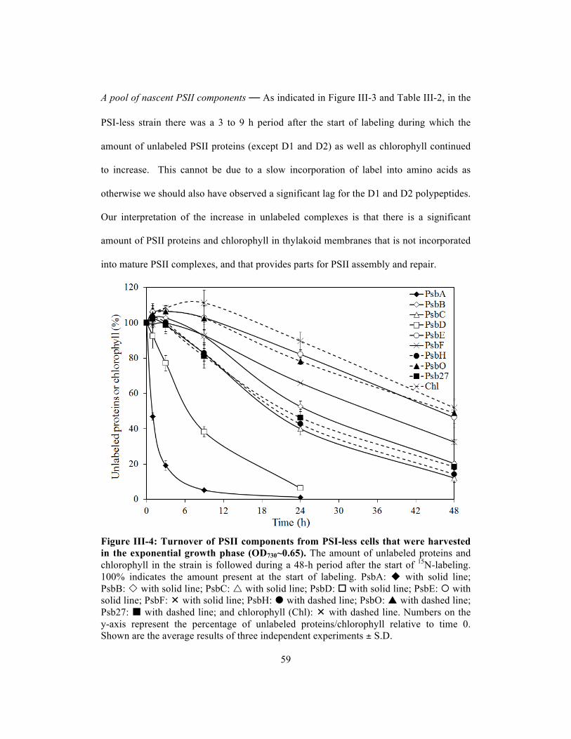
59
A pool of nascent PSII components — As indicated in Figure III-3 and Table III-2, in the
PSI-less strain there was a 3 to 9 h period after the start of labeling during which the
amount of unlabeled PSII proteins (except D1 and D2) as well as chlorophyll continued
to increase. This cannot be due to a slow incorporation of label into amino acids as
otherwise we should also have observed a significant lag for the D1 and D2 polypeptides.
Our interpretation of the increase in unlabeled complexes is that there is a significant
amount of PSII proteins and chlorophyll in thylakoid membranes that is not incorporated
into mature PSII complexes, and that provides parts for PSII assembly and repair.
Figure III-4: Turnover of PSII components from PSI-less cells that were harvested in the exponential growth phase (OD730~0.65). The amount of unlabeled proteins and chlorophyll in the strain is followed during a 48-h period after the start of 15N-labeling. 100% indicates the amount present at the start of labeling. PsbA: with solid line; PsbB: with solid line; PsbC: with solid line; PsbD: with solid line; PsbE: with solid line; PsbF: with solid line; PsbH: with dashed line; PsbO: with dashed line; Psb27: with dashed line; and chlorophyll (Chl): with dashed line. Numbers on the y-axis represent the percentage of unlabeled proteins/chlorophyll relative to time 0. Shown are the average results of three independent experiments ± S.D.

60
To test our interpretation of the increase in unlabeled polypeptide being due to a
reservoir of unfinished PSII complexes in the cell, the labeling experiment with PSI-less
cells was repeated with cells from exponential phase (OD730~0.65) instead of post-
exponential phase (OD730~0.9) as in exponential phase the reservoir may be expected to
be smaller due to a faster de novo PSII biosynthesis (Figure III-4). The lifetimes of the
PSII components were similar to those measured in cells in post-exponential phase.
However, the increase in the amount of unlabeled protein is much less and the time over
which an increase is observed is much shorter. These results support our interpretation
and demonstrate that cells in the post-exponential growth phase tend to accumulate PSII
proteins and chlorophyll to be ready for later use.
The role of SCPs — As indicated in Figure III-3B, the increase in the amount of
unlabeled PSII protein in cells in the post-exponential phase is much smaller in the PSI-
less/SCP-less strain relative to in the PSI-less control, whereas the half-life time of the
PSII proteins is not much affected. This is very much comparable to what was shown
above for cells in exponential phase. These data suggest that pools of nascent PSII
proteins and protein-bound chlorophyll may be stabilized by SCPs.
In order to test the interpretation that SCPs are involved with stabilization of
nascent PSII complexes and/or intermediates, isolated PSII samples from the PSI-less
strain and the PSI-less/SCP-less strain were run on BN-SDS PAGE. As indicated in
Figure III-5, the main difference between PSII complexes from the PSI-less and PSI-
less/SCP-less strains was that the RC47(1) complex, corresponding to the PSII monomer
form without the CP43 protein, was missing in the PSII preparation from the PSI-
less/SCP-less strain. However, the RC47(2) band, corresponding to the RC47 dimer, was

61
Figure III-5: BN-PAGE followed by SDS-PAGE gel for PSII complexes co-isolating with CP47-His from the PSI-less strain (A) and the PSI-less/SCP-less strain (B). The bands on BN-PAGE (top) are unstained and are colored by the native chlorophyll and Coomassie Brilliant Blue. The SDS-PAGE gel was stained with Coomassie Brilliant Blue. RCC(2): mature PSII dimer; RCC-RC47: PSII dimer lacking CP43 in one of the monomers; RC47(2): PSII dimer lacking CP43 in both monomers; RCC(1): PSII monomer; and RC47(1): PSII monomer lacking CP43. present in preparations from both strains. In PSII biogenesis, RC47 complexes are
assembled from RC complexes (D1, D2, cytb559 and PsbI proteins) and sub-CP47
complexes (CP47, PsbH and SCP proteins) (Promnares et al., 2006, Nixon et al., 2010).
SCP association could aid the stability of the RC47 complex before dimerization occurs.

62
These observations suggest that our interpretation of SCPs stabilizing intermediates in
formation of the mature PSII complex is reasonable.
Another role of the SCPs involves chlorophyll binding and stabilization, and
earlier work has shown that deletion of SCPs in the PSI-less background strain also
affects the chlorophyll content per cell and the accumulation of chlorophyll precursors
(Xu et al., 2002; Xu et al., 2004). However, the location of this SCP effect has not yet
been pinpointed. The stability of PSII is not affected by SCPs as shown in Table III-2. In
line with earlier observations (Vavilin et al., 2007), we do not see evidence of SCPs
stably binding a large amount of chlorophyll as the oxygen evolution rate in the PSI-less
strain was 2480±80 µmol O2 (mg Chl)-1 h-1 whereas in the PSI-less/SCP-less strain this
rate was 2730±180 µmol O2 (mg Chl)-1 h-1. Thus, the number of PSII reaction centers on
a per-chlorophyll basis is similar regardless of the presence of SCPs.
Earlier work had indicated the lack of accumulation of chlorophyll precursors in
SCP-less strains (Xu et al., 2004) suggesting an early block in chlorophyll biosynthesis in
the absence of SCPs. The 15N-labeling approach provides an opportunity to monitor the
effects of the absence of SCPs in more detail. After growth of the PSI-less/ΔchlL and
PSI-less/SCP-less/ΔchlL strains in darkness for 5 days, the strains had little chlorophyll
left as ChlL is required for light-independent protochlorophyllide reduction and
chlorophyll synthesis (Figure III-6). After incubation in darkness, the culture was diluted
four-fold with medium containing 4.5 mM Na15NO3 and 2 mM 15NH4Cl and the culture
was transferred to continuous illumination at 4 µmol photons m-2 s-1. As indicated in
Figure III-6, during the first 6 hours of illumination, the PSI-less/ΔchlL strain synthesized
primarily labeled (15N) chlorophyll along with some unlabeled (14N) chlorophyll.
Unlabeled chlorophyll may have been synthesized in part from accumulated

63
Figure III-6: Unlabeled (14N) and labeled (15N) chlorophyll from the PSI-less/ΔchlL and PSI-less/SCP-less/ΔchlL strains upon illumination. Cells were grown in the dark for 5 days. The cultures were illuminated after they had been diluted with three volumes of BG-11 medium containing Na15NO3 and 15NH4Cl. Chlorophyll was extracted from the cells for analysis at the times indicated. Open and closed symbols represent unlabeled (14N) and labeled (15N) chlorophyll, respectively, in the PSI-less/ΔchlL (squares) and PSI-less/SCP-less/ΔchlL (triangles) strains. Shown are the average results of two independent experiments ± S.D. protochlorophyllide or other available precursors. However, in the PSI-less/SCP-
less/ΔchlL strain the amount of unlabeled chlorophyll decreased, and labeled (15N)
chlorophyll was synthesized very slowly (about 20-fold slower than in the PSI-less/ΔchlL
strain). The decrease in unlabeled chlorophyll and the slow increase in labeled
chlorophyll suggest that in the SCP-less mutant the amount of chlorophyll precursors is
greatly diminished, and tetrapyrrole biosynthesis is impaired.
A very slow synthesis of PSII in the PSI-less/SCP-less/ΔchlL strain was
confirmed by 77 K fluorescence emission spectra after 24 hours of illumination (Figure
III-7). A peak at 695 nm corresponds to CP47-associated chlorophyll and reflects intact

64
Figure III-7: 77 K fluorescence emission spectra of Synechocystis sp. PCC 6803 cells lacking PSI and ChlL. Cells had been grown in darkness for one week and were then transferred to continuous light (4 µmol photons m-2 s-1) for 0 h (PSI-less/ΔchlL: dotted line; PSI-less/SCP-less/ΔchlL: dashed line) or 24 h (PSI-less/ΔchlL : solid line; PSI-less/SCP-less/ΔchlL: dashed/dotted line). The spectra were normalized to 100 at 683 nm, where phycobilisomes and some chlorophylls emit maximally. The excitation wavelength was 435 nm. a.u., arbitrary units. PSII complexes. While in the PSI-less/ΔchlL strain a significant amount of PSII
complexes was present after 24 h of illumination, in the PSI-less/SCP-less/ΔchlL strain
only very little 695 nm emission was observed.
The very slow chlorophyll biosynthesis and the lack of a large amount of
unlabeled chlorophyll synthesis upon illumination of the PSI-less/SCP-less/ΔchlL strain
suggest that early intermediates in chlorophyll biosynthesis may have been depleted.
Therefore, we wished to determine whether aminolevulinic acid (ALA), an early
intermediate in tetrapyrrole biosynthesis, was depleted in strains lacking SCPs. As
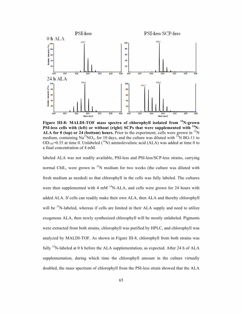
65
Figure III-8: MALDI-TOF mass spectra of chlorophyll isolated from 15N-grown PSI-less cells with (left) or without (right) SCPs that were supplemented with 14N-ALA for 0 (top) or 24 (bottom) hours. Prior to the experiment, cells were grown in 15N medium, containing Na15NO3, for 10 days, and the culture was diluted with 15N BG-11 to OD730=0.35 at time 0. Unlabeled (14N) aminolevulinic acid (ALA) was added at time 0 to a final concentration of 4 mM. labeled ALA was not readily available, PSI-less and PSI-less/SCP-less strains, carrying
normal ChlL, were grown in 15N medium for two weeks (the culture was diluted with
fresh medium as needed) so that chlorophyll in the cells was fully labeled. The cultures
were then supplemented with 4 mM 14N-ALA, and cells were grown for 24 hours with
added ALA. If cells can readily make their own ALA, then ALA and thereby chlorophyll
will be 15N-labeled, whereas if cells are limited in their ALA supply and need to utilize
exogenous ALA, then newly synthesized chlorophyll will be mostly unlabeled. Pigments
were extracted from both strains, chlorophyll was purified by HPLC, and chlorophyll was
analyzed by MALDI-TOF. As shown in Figure III-8, chlorophyll from both strains was
fully 15N-labeled at 0 h before the ALA supplementation, as expected. After 24 h of ALA
supplementation, during which time the chlorophyll amount in the culture virtually
doubled, the mass spectrum of chlorophyll from the PSI-less strain showed that the ALA

66
used for chlorophyll synthesis primarily was 15N-ALA, which cells had synthesized
themselves. In contrast, newly synthesized chlorophyll from the PSI-less/SCP-less strain
was primarily unlabeled as the amounts of labeled (original) and unlabeled (newly
synthesized) chlorophyll were about the same. These results suggest that the ALA
amount is limiting as a consequence of the SCP deletion and that therefore SCPs appear
to play an important role in the very early steps of tetrapyrrole biosynthesis (ALA
formation).
Discussion
PSII protein and chlorophyll turnover — Previous work with radioisotopes had indicated
that different PSII polypeptides differed in their lifetimes (Schuster et al., 1988; Mattoo
et al., 1999), and work from our group had indicated that the chlorophyll lifetime in the
cell was very long (Vavilin et al., 2005; Vavilin et al., 2007). It is clear that careful
orchestration of the synthesis, assembly, and repair of photosynthetic complexes is
required, but until now very few data were available on the lifetimes of longer-lived
protein components, and there is little known about how this orchestration may occur and
how it is regulated. Using stable-isotope labeling combined with mass spectrometry,
both labeled (new) and unlabeled (old) peptides can be detected. The rate of
disappearance of unlabeled peptides over time determines the turnover rate of the
proteins, and this approach allowed an accurate and comprehensive determination of
lifetimes of PSII components.
PSII complexes were isolated from PSI-less Synechocystis cultures grown at a
light intensity of 4 µmol photons m-2 s-1, making use of a His-tag attached to the PsbB
(CP47) protein. Therefore, only polypeptides and complexes associated with PsbB with
an exposed C-terminal His tag will be detected. The fraction obtained after His-tag

67
affinity purification contained at least nine PSII proteins (seven intrinsic proteins
including the reaction center proteins PsbA and PsbD, the chlorophyll-binding proteins
PsbB and PsbC, the cytochrome b559 proteins PsbE and PsbF, and PsbH, and two lumenal
proteins (PsbO and Psb27)). Other PSII proteins may not have stained sufficiently well to
be detected. Moreover, a translation elongation factor, Tu (Sll1099), and Slr0909 co-
isolated with the PSII complex. In a previous study, Sll1099 was also seen from co-
isolating with PSII-associated His-ScpB (Kufryk et al., 2008). Slr0909 is a protein of
unknown function, but it is encoded about 3 kbp downstream from psbB and slr0909
could possibly be co-transcribed with psbB and an intervening gene of unknown function.
As indicated in Table III-2, PSII proteins have very different half-life times
ranging from 1.5 hours up to 33 hours. Therefore, PSII proteins (or groups of such
polypeptides) turn over and are replaced independently from each other, and remaining
PSII proteins appear to be reused for assembly into a functional PSII complex. Our
results confirm the radiolabeling-based interpretations of other groups (Mattoo et al.,
1984; Ohad et al., 1984) that rapidly synthesized D1, the PSII protein with the shortest
lifetime, is actually incorporated into PSII complexes. The fast turnover of D1 in the
PSII complex (5 to 20 times faster than turnover rates of the other PSII proteins) poses a
challenge for the PSII complex as D1 is a central rather than a peripheral part of the
complex. However, the lipids around the reaction center may facilitate the replacement of
damaged D1 proteins (Loll et al., 2007). The other PSII proteins from the damaged
complex may stay together and be re-assembled around a new D1 polypeptide, or
subcomplexes may form a pool from which new complexes are formed (Smith and
Howe, 1993; Zak et al., 2001; Nixon et al., 2005). In the latter case, PSII complexes may
be assembled from “old” polypeptides originating from different PSII complexes.

68
The lifetime of chlorophyll in various mutant strains has been studied earlier by
means of stable-isotope labeling, and because chlorophyll is much more stable than
chlorophyll-binding proteins according to the lifetimes of chlorophyll-binding proteins
estimated from the pulse-chase method, we have suggested that chlorophyll is recycled
upon degradation of chlorophyll-binding PSII components (Vavilin et al., 2007). In this
study, we experimentally support the earlier interpretations by comparing the half-life
times of the main chlorophyll-binding proteins in PSII (D1, D2, CP47, and CP43)
(t1/2=1.5-15 h) with the half-life time of chlorophyll extracted from isolated PSII (t1/2=40
h) (Table III-2). Therefore, chlorophyll in damaged chlorophyll-binding proteins can be
re-utilized.
SCPs and chlorophyll reutilization — In cyanobacteria the SCP proteins bind
chlorophyll, may associate damaged PSII centers, and serve as a temporary pigment
reservoir while PSII components are being replaced (Storm et al., 2008, Promnares et al.,
2006, Yao et al., 2007, Xu et al., 2004). We have now analyzed how SCPs affect the
lifetimes of PSII polypeptides and chlorophyll. In line with earlier observations (Vavilin
et al., 2007), the lifetime of PSII chlorophyll was reduced by half in the PSI-less/SCP-
less strain compared to the PSI-less strain. However, the lifetimes of the chlorophyll-
binding proteins in PSII (D1, D2, CP47, and CP43) and of PsbH that participates in the
binding of chlorophyll remained essentially unchanged (Table III-2). This indicates that
SCPs function primarily in reutilization of chlorophyll and do not affect the stability of
chlorophyll-binding proteins.
Interestingly, whereas the lifetimes of PSII proteins were not changed or were
decreased slightly in the SCP-less strain, the lifetime of D1 was increased (Table III-2
and Figure III-3). The longer lifetime of D1 may have been caused by slower assembly of

69
PSII due to the lack of chlorophyll availability, which affects D1 translation and
processing in Synechocystis (He and Vermaas, 1998). Chlorophyll availability is thought
to be lower in the absence of SCPs as the rate of chlorophyll synthesis was decreased 4-
fold in the PSI-less/SCP-less strain, and the chlorophyll content per cell was almost 4-
fold less than in the PSI-less strain (Figure III-6) (Vavilin et al., 2007, Xu et al., 2004).
Therefore, during the replacement of the D1 protein in the PSII repair cycle, the proteins
may be waiting for available chlorophylls to assemble PSII, which appears to take longer
in the absence of SCPs.
SCPs stabilize nascent PSII protein complexes — As shown in Figure III-3, unlabeled
PSII proteins (except D1 and D2) increased for the first 3 to 9 hours of 15N-labeling if
late-log cultures are used and SCPs are present. The D1 and D2 proteins did not show
this increase in unlabeled protein, excluding the possibility that there is an extensive pool
of unlabeled amino acids that remains available for many hours after the start of labeling.
Instead, the most plausible explanation is that the increase in unlabeled protein originates
from PSII proteins that were present at time 0 but that were not associated with PsbB
with an exposed C-terminal His-tag at that time. Figure III-5 shows that the His-tag-
isolated samples consist of mature PSII complexes in monomer and dimer forms as well
as RC47 (PSII without CP43) dimers and RC47/mature PSII dimers. These complexes
are in line with what was observed previously (Herranen et al., 2004; Komenda et al.,
2006). However, RC47 monomers were found to occur only in the presence of SCPs,
suggesting that SCPs can stabilize biosynthetic intermediates of PSII complexes. Indeed,
even when cells are in late-log stage, in the PSI-less/SCP-less mutant very little synthesis
of unlabeled PSII components is observed after the start of labeling (Figure III-3B),
70
suggesting that no significant accumulation of such PSII biosynthesis intermediates
occurs if SCPs are absent.
SCPs have been found to be associated with PSII (Yao et al., 2007; Kufryk et al.,
2008). Analysis of isolated His-tagged ScpD complexes suggests that ScpD binds to
CP47 proteins in the vicinity of PsbH, and SCPs have been found to be associated with
PSII throughout its biogenesis, from just CP47 proteins to monomeric PSII complexes
(Promnares et al., 2006; Yao et al., 2007). Therefore, even though SCPs do not stabilize
PSII components in mature PSII complexes (Table III-2), there is significant SCP-
induced stabilization of nascent PSII complexes.
SCPs and ALA biosynthesis — In addition, SCPs also appear to be involved, directly or
indirectly, in ALA biosynthesis. This effect results in slow chlorophyll biosynthesis in the
absence of SCPs (Figure III-6) and in slow generation of PSII complexes (Figure III-7).
In contrast to the PSI-less strain that does not use much exogenous ALA and that
therefore can generate and utilize sufficient internal ALA, the PSI-less/SCP-less mutant
readily uses exogenous ALA for chlorophyll biosynthesis (Figure III-8). The most
straightforward interpretation of these results is that deletion of SCPs in the PSI-less
background strain severely impairs the ALA biosynthesis pathway. However, ALA
synthesis is thought to occur in the cytoplasm (Joyard et al., 2009), and SCPs are
transmembrane proteins located in thylakoid membranes. This suggests that the SCP-
mediated regulation of ALA synthesis is not a direct effect but rather may be influenced
by intermediate events.
ALA synthesis is a tightly regulated step that is negatively regulated by, for
example, protochlorophyllide that accumulates in darkness in plants (Richter et al.,
2010). However, based on results of earlier studies, there is no accumulation of

71
protochlorophyllide or earlier intermediates such as Mg-protoporphyrin IX in PSI-less
and PSI-less/SCP-less strains if chlorophyll biosynthesis has not been impaired (Xu et al.,
2004). However, chlorophyllide, which is chlorophyll without the phytyl tail,
accumulates as the chlorophyll biosynthesis rate and chlorophyll content per cell decrease
in the PSI-less/SCP-less strain (Xu et al., 2004). It is possible that chlorophyllide, which
is more hydrophilic than chlorophyll, may serve as a signal for ALA biosynthesis
enzymes to negatively regulate the output of ALA, thus reducing overall chlorophyll
biosynthesis in the PSI-less/SCP-less strain (Figure III-8). Indeed, in plants reduced
expression and activity of chlorophyll synthase also has been shown to cause a feedback-
controlled inactivation of ALA synthesis (Shalygo et al., 2009).
In conclusion, stable-isotope labeling (15N), mass spectrometry and well-defined
mutants are a powerful combination to provide insights in assembly and synthesis of PSII
polypeptides and associated cofactors; chlorophyll already is associated with
subcomplexes that are intermediates in PSII synthesis. SCPs aid the stability of nascent
PSII complexes in PSII biogenesis and also affect the flux from ALA through the
tetrapyrrole biosynthesis pathway to chlorophyll. This work illustrates the multiple levels
of control and regulation that pigments and SCPs have in PSII synthesis and assembly.
72
CHAPTER IV. LIFETIMES OF PHOTOSYSTEM I AND PHOTOSYSTEM II
PROTEINS IN THE CYANOBACTERIUM SYNECHOCYSTIS SP. PCC 6803
Abstract
In order to study the dynamics of photosystem II (PSII) and photosystem I (PSI), the
lifetimes of photosynthetic proteins were determined by a combined stable-isotope
labeling (15N) / mass spectrometry method. Upon labeling, newly synthesized proteins
and chlorophyll were heavier due to isotope incorporation, and old and new pew proteins
in the two photosystems could be distinguished. The lifetimes of PSI and PSII proteins
ranged from 30 to 75 h and from less than 1 h to 11 h, respectively, and nascent PSI
proteins accumulate in the thylakoid membrane. PSI complexes indeed were much more
stable than PSII complexes, and the lifetimes of PSII proteins at higher light intensity
were decreased from those determined at lower light intensity in a previous study.
However, the lifetime of chlorophyll was longer than that of chlorophyll-binding
proteins, implying that chlorophyll is recycled. Moreover, the dynamics of PSI and PSII
complexes showed that the interchange between monomeric and multimeric forms of PSI
and PSII, if multimers indeed are not artifacts, occurs on a timescale of about an hour or
less. In the SCP-less mutant, the lifetimes of most PSI proteins and the amount of nascent
PSI proteins in the membrane were not affected. Also, from an earlier study, the rate of
chlorophyll biosynthesis and the lifetime of chlorophyll did not change. These results
indicate that the function of SCPs might not involve the PSI complex and its proteins.

73
Introduction
Oxygenic photosynthesis in cyanobacteria, algae, and plants is catalyzed mainly by two
multisubunit complexes, photosystem I (PSI) and photosystem II (PSII). PSI and PSII are
embedded in the thylakoid membrane and are composed of multiple protein subunits that
bind chlorophyll, carotenoids, and other cofactors. Electrons provided by PSII originate
from water, and PSI provides additional energy from light to produce NADPH. A proton
gradient established across the thylakoid membrane upon electron transfer is used for
ATP synthesis, and NAPDH is utilized for cellular processes including carbon fixation
leading to the synthesis of organic compounds.
In cyanobacteria, the PSII complex consists of 20 protein subunits, together
binding 35 chlorophylls; moreover, extrinsic proteins are located on the lumenal side
(Guskov et al., 2009). According to BN gel results and PSII crystal structures,
cyanobacterial PSII complexes are present in dimeric and monomeric forms (Guskov et
al., 2009; Herranen et al., 2004). However, the composition and structure of PSI
complexes is very different than that of PSII complexes. Compared with PSII, PSI has
less protein subunits (12 of them) but a lot more chlorophyll (96 of them), and its
extrinsic proteins are located on the cytosolic side (Jordan et al., 2001). Cyanobacterial
PSI complexes were found in trimeric and monomeric forms in vitro (Herranen et al.,
2004; Jordan et al., 2001), but trimeric PSI was never observed in the crystal structure
and BN protein gels so far in plants (Jensen et al., 2003; Amunts et al., 2007). However,
the most interesting difference between the two photosystems is their stability. The major
challenge that PSII complexes face is the photodamage that is caused by oxidizing
species. Some components of PSII complexes show a high turnover rate; in particular the
D1 protein that binds many cofactors including part of P680 turns over on the timescale

74
of about an hour (Ohad et al., 1984). In contrast to PSII, PSI is more stable. Therefore,
the dynamics of PSII and PSI complexes may be different.
From previous studies, in the cyanobacterium Synechocystis sp. PCC 6803 the
lifetimes of PSII components at low light intensity (4 µmol photons m-2 s-1) had been
determined to range from 1.5 h for the D1 protein up to 40 h for PSII-associated
chlorophyll (Yao et al., submitted). However, the lifetime of chlorophyll extracted from
whole cells in wild type is over 200 h (Vavilin et al., 2007). As most chlorophyll is
associated with PSI in cyanobacteria (Guskov et al., 2009; Jordan et al., 2001), this
would suggest that PSI-associated chlorophyll is more stable than PSII chlorophyll. In
order to gain comprehensive knowledge in the lifetimes of PSI and PSII proteins and
their associated chlorophyll, stable-isotope labeling (15N), BN/SDS-PAGE, and mass
spectrometry were applied to monitor the fate of old and newly synthesized proteins over
time. In addition, Small Cab-like proteins (SCPs), single transmembrane helix proteins,
have shown to be involved in PSII chlorophyll recycling. There is also evidence that
SCPs stabilize PSI complexes (Wang et al., 2008). In this study, the lifetimes of PSII and
PSI proteins and chlorophyll will be determined with and without SCPs, and the
dynamics of PSII and PSI complexes between their different forms will be discussed.
Materials and Methods
Strains and growth conditions The wild-type (WT) and ΔscpABCDE (SCP-less)
strains (Xu et al., 2002, Xu et al., 2004) of Synechocystis sp. PCC 6803 were grown
photoautotropically in liquid BG-11 medium (Rippka et al., 1979) at 30 °C at a light
intensity of 75 µmol photons m-2 s-1. Cell growth was monitored by measuring the optical
density at 730 nm in a 1-cm cuvette using a Shimadzu UV-160 spectrophotometer.

75
Isotope labeling and membrane preparation Cell cultures were grown to an
OD730~0.65 and were diluted four-fold in BG11 medium containing 4.5 mM Na15NO3
and 2 mM 15NH4Cl. Cell samples were collected at 3, 9, 24 and 48 hours after the
dilution. Cell pellets were resuspended in a mixture of 50 mM 2-(N-morpholino)
ethanesulfonic acid hydrate (MES)-NaOH (pH 6.0), 10 mM MgCl2, and 25% glycerol,
and broken by Bead Beater (BioSpec Products, Bartlesville, OK). Cell membranes were
prepared exactly as described (Bricker et al., 1998) and were stored at -80°C.
PAGE Wild type and SCP-less membrane samples corresponding to 10 µg of
chlorophyll were solubilized in 1% β-dodecyl maltoside for 45 min on ice in darkness;
subsequently 0.1 volume of loading solution containing 750 mM aminocaproic acid and
5% Coomassie Brilliant Blue G-250 were added. Protein complexes in the membrane
were separated in the first dimension by blue-native (BN) electrophoresis at 4 °C in a 5-
14% polyacrylamide gel according to Schagger and von Jagow (1991). For the second
dimension, the BN gel lanes were incubated for 45 min at room temperature in a solution
containing 25 mM Tris-HCl (pH 7.5), 2% SDS, and 10% mercaptoethanol. The lanes
were then layered onto a 1.5-mm-thick SDS/12-20% polyacrylamide gradient gel
containing 7 M urea (Komenda et al., 2002). The gel was stained with 0.15% CBB R-250
in a solution of 50% methanol and 10% acetic acid. In-gel digestion to produce peptides
for analysis by mass spectrometry (LC-MS/MS) was carried out essentially as described
(Shevchenko et al., 1996) using sequencing-grade modified trypsin (Promega/SDS
Bioscience).
Protein analysis Peptides in trypsin digests were separated using a Dionex Ultimate
3000 liquid chromatography system equipped with both a HPG 3400M high pressure
76
gradient pump and a LPG 3400 MB low pressure gradient pump together with a
WPS300TB autosampler and a FLM 3100B column compartment. A Bruker MicrOTOF-
Q mass spectrometer equipped with an online nanospray source was used for protein
identification. Instrumental setups for HPLC and mass spectrometer and data analysis
were described earlier (Yao et al., submitted)
Results
Identification of photosynthetic protein complexes and photosynthetic proteins
Membrane protein complexes from wild-type Synechocystis sp. PCC 6803 cells were
separated by blue native (BN)-PAGE and then proteins from each individual protein
complex were separated by SDS-PAGE (Figure IV-1). Various PSI complexes (PSI
supercomplex, and trimeric and monomeric PSI complexes) and PSII complexes (dimeric
and monomeric complete PSII complexes, and the CP43-less PSII monomer (RC47))
were identified in membranes from both the wild-type and SCP-less strains. Protein
complexes other than photosynthetic complexes, such as NDH complexes, were seen as
well. The profile of membrane proteins and complexes were very similar to that in a
study from Herranen et al. (2001). In order to determine the lifetimes of PSII and PSI
proteins, 2D BN/SDS-PAGE as performed with membrane protein samples from wild-
type and SCP-less cells grown in the presence of 15NO3- and 15NH4
+ for a specific time
period (0 h, 3 h, 9 h, 24 h, and 48 h). PsaA, PsaB, PsaD, PsaF, PsaL, and PsaE from
protein spot 1-6 (respectively) in trimeric PSI and PsbB, PsbC, PsbD, and PsbA from
protein spot 11-14 (respectively) in monomeric PSII were identified and used to
determine their labeling.
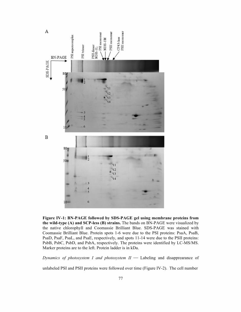
77
Figure IV-1: BN-PAGE followed by SDS-PAGE gel using membrane proteins from the wild-type (A) and SCP-less (B) strains. The bands on BN-PAGE were visualized by the native chlorophyll and Coomassie Brilliant Blue. SDS-PAGE was stained with Coomassie Brilliant Blue. Protein spots 1-6 were due to the PSI proteins: PsaA, PsaB, PsaD, PsaF, PsaL, and PsaE, respectively, and spots 11-14 were due to the PSII proteins: PsbB, PsbC, PsbD, and PsbA, respectively. The proteins were identified by LC-MS/MS. Marker proteins are to the left. Protein ladder is in kDa. Dynamics of photosystem I and photosystem II Labeling and disapprearance of
unlabeled PSI and PSII proteins were followed over time (Figure IV-2). The cell number

78
Figure IV-2: Turnover of photosynthetic proteins from the wild-type (A) and SCP-less (B) cells. The amount of unlabeled proteins in the wild-type strain is followed during a 48-h period after the start of 15N-labeling. 100% indicates the amount present at the start of labeling. PSI proteins are in dashed line, and PSII proteins are in solid line. PsaA: with dashed line; PsaB: with dashed line; PsaD: with dashed line; PsaE: with dashed line; PsaF: with dashed line; PsaL: with dashed line; PsbA: with solid line; PsbB: with solid line; PsbC: with solid line; and PsbD: with solid line. Numbers on the y-axis represent the percentage of unlabeled proteins/chlorophyll relative to time 0. Shown are the average results of two independent experiments ± error.

79
increased during the 15N-labeling period, and therefore the total amount of proteins
increased as well. What is indicated in Figure IV-2A is the amount of unlabeled protein
in the culture volume relative to the amount in the same volume at time 0. Interestingly,
the amount of unlabeled material increases initially in the first 3 hours for all of the PSI
proteins (Figure IV-2A dashed lines) in the wild-type strain. In contrast to PSI proteins,
PSII reaction center proteins (D1 and D2) do not show such an increase, and PSII antenna
proteins (CP47 and CP43) have a minor lag in the exponential decrease of unlabeled
protein (Figure IV-2A solid lines). The half-lives of PSI and PSII components (Table IV-
1) were determined by monitoring the disappearance of old (unlabeled) peptides in the
time period between 9 and 48 hours after the start of labeling, whereas for PSII proteins
PsbA and PsbD half-lives were calculated from time 0 and for PsbB and PsbC starting at
3 hours after labeling. In wild type, the half-lives of PSI reaction center proteins (PsaA
and PsaB) are 40 h whereas the other intrinsic PSI proteins, PsaF, which is involved in
docking of plastocyanin, and PsaL, which plays a role in formation of the PSI trimer in
cyanobacteria (Chitnis and Chitnis, 1993; Fischer et al., 1998; Karapetyan et al., 1999),
have half-life times of 50 and 30 h, respectively. The extrinsic proteins (PsaD and PsaE)
have longer half-lives (70-75 h) presumably because they can be dissociated from
damaged PSI and be reused for repaired or new PSI. The fact that extrinsic proteins have
longer half-lives than the intrinsic proteins also has been seen for PSII (Yao et al.,
submitted). PSII proteins have much shorter half-lives than PSI proteins. Compared with
the lifetimes of PSII proteins in the earlier study (Yao et al., submitted), the lifetimes of
PSII proteins are much shorter in this experiment, presumably due to the higher light
intensity during culture conditions. The unlabeled D1 protein had largely disappeared at
the 3-h timepoint. The D2 protein was more stable but had a halftime of a little over 3 h.

80
Table IV-1: Comparison of half-lives of PSI and PSII components in the wild-type and SCP-less strains. The half-life times were calculated from the decrease in the percentage of unlabeled protein correcting for the increase in unlabeled protein that occurred for the longer-lived polypeptides. Listed are the average results of two independent experiments ± error.
The CP43 and CP47 proteins, the chlorophyll-binding proteins, have halftimes of 6.5 and
11 h, respectively. These results are in line with the concept that PSII complexes turn
over much faster than PSI complexes.
As indicated in Figure IV-2A, in the wild-type strain there was a 3 h period after
the start of labeling during which the amount of unlabeled PSI proteins continued to
increase. This cannot be due to a slow incorporation of label into amino acids as D1
polypeptides were almost fully labeled after 3-h period. Therefore, our interpretation of
the increase in unlabeled complexes is that there are nascent PSI proteins in thylakoid
membranes that are not incorporated into mature PSI complexes. For the PSII proteins,
the PsbB and PsbC proteins show only very small lags in the exponential decrease of
unlabeled proteins 3 hours after the start of labeling.
Half-life time (h)
Strains Wild-type SCP-less
PsaA 40±7 50±4
PsaB 40±7 50±4
PsaD 75±7 50±7
PsaE 70±7 53±4
PsaF 50±7 40±2
PsaL 30±1 30±6
PsbA (D1) < 1 < 1
PsbB (CP47) 11±2.5 10.5±1
PsbC (CP43) 6.5±1.5 6±0.5
PsbD (D2) 3.3±1 3±0.3

81
Table IV-2: Comparison of percentage of unlabeled PSI proteins in trimeric and monomeric forms and PSII proteins in dimeric and monomeric forms at 3, 9, 24, and 48 h of labeling. Listed are the average results of one to ten peptides.
3 h 9 h 24 h 48 h
Trimer 96±1 78±2 46±3 14±1.5 PsaA
Monomer 95±1 78±1 46±2 13±0.5
Trimer 91±1 75±2 43±2 13±1.5 PsaB
Monomer 91±2 74±2 43±1
Trimer 94±1 80±1 55±1 18.5±0.1 PsaE
Monomer 92±1 78±1 57±1
Trimer 94±0 79±3 51±2 16.5±1.5 PsaF
Monomer 93±2 78±3 54±2 11.5
Trimer 94±1 76±2 45±1 10±0.5 PsaL
Monomer 93±1 78±1
Dimer 2.5 PsbA
Monomer 4.0±1.5
Dimer 80±2 50±3 16±2 3.0±1.5 PsbB
Monomer 77±1 47±1 15±2 3.5±1.0
Dimer 72±2 37±1 5.5±0.5 1.1±0.2 PsbC
Monomer 69±2 34±1 5.5±0.5 1.1±0.1
Dimer 59 16±2 0.9±0.1 PsbD
Monomer 58±2 16±1 0.8±0.1
PSI is thought to exist in both trimeric and monomeric forms, and PSII is thought
to exist in dimeric and monomeric forms, based on results of crystal structures and
separation of isolated complexes under non-denaturing conditions. In order to get insights
in the conversion dynamics of PSI between trimeric and monomeric forms and of PSII
between dimeric and monomeric forms, the percentage of labeled and unlabeled PSI and
PSII proteins from different forms was monitored upon different times of labeling. Table
IV-2 shows the percentage of unlabeled proteins at different times of labeling. Each PSI
or PSII protein had a similar lifetime when the complex was in monomeric vs. multimeric

82
form. These results demonstrate that the different forms of PSI or PSII, if relevant under
in vivo conditions, are in equilibrium, and the dynamics of PSI and PSII switching
between the different forms occurs on a timescale of about an hour or less.
Role of SCPs in the photosystems In a previous study (Yao et al., submitted), deletion
of SCPs in a PSI-less background strain did not change the lifetimes of PSII proteins
except of the PsbO protein. This is consistent with current results showing that deletion
of SCPs in the wild-type background strain had little effect on the lifetimes of the PSII
proteins (Table IV-1, Figure IV-2B solid lines). The half-life time of the intrinsic PSI
proteins was not much affected by removal of the SCPs either while the half-life time of
the extrinsic PSI proteins (PsaD and PsaE) decreased (Table IV-1). Also, the amount of
nascent PSI proteins accumulating in the membrane was not significantly changed upon
the removal of SCPs (Figure IV-2). These results indicate that SCPs might not influence
the PSI proteins.
Discussion
Pulse-chase methods using radioisotopes have been widely used to estimate the lifetimes
of polypeptides. However, this method is difficult for relatively stable proteins as other
proteins may be labeled much faster and more intensely. PSI proteins were known to be
long-lived proteins, but their lifetimes have not been specifically determined. Using
stable-isotope labeling combined with mass spectrometry, we can determine the lifetimes
of PSI (long-lived) and PSII (short-lived) proteins in parallel.
Turnover of PSII and PSI proteins Photosystem complexes and proteins were
separated by the BN/SDS-PAGE and identified by LC-MS/MS (Figure IV-1). The

83
lifetimes of PSII proteins including the reaction center proteins D1 and D2 and the
chlorophyll-binding proteins CP47 and CP43 were determined (Table IV-1). However,
compared with the lifetime of the PSII proteins in the PSI-less background strain grown
at 4 µmol photons m-2 s-1 in a previous study (Yao et al., submitted), the lifetimes of the
PSII proteins in the wild-type strain were decreased by 50% except for the CP47 protein
whose lifetime was only decreased by 30%. In the current experiments, the wild-type
strain was cultured at a light intensity that was almost 20 times stronger than that used for
the PSI-less strain in the previous study. Light intensity is a main factor to determine the
length of the lifetimes of PSII core proteins although PSII in the PSI-less strain may
easily occur in the acceptor side of photoinhibition under the same light condition
compared with the wild-type strain because of over-reduction of plastoquinone pool due
to the lack in PSI complexes. The lifetimes of four PSII core proteins showed a logical
progression: the D1 protein, where the processes of water oxidation and charge
separation take place, appears to experience the most photodamage (Aro et al., 1993); the
further the other PSII proteins are from the D1 protein, the longer is their lifetime.
In contrast to PSII proteins, PSI proteins are stable. PsaA and PsaB, the reaction
center proteins, had similar lifetimes to the other intrinsic proteins, PsaF and PsaL (Table
IV-1). This result demonstrates that the processes of charge separation and electron
transport in PSI are very stable and do not rapidly induce protein damage. The PSI
extrinsic proteins, PsaD and PsaE, exhibit longer lifetimes than the PSI intrinsic proteins.
This phenomenon has also been seen in PSII in a previous study (Yao et al., submitted)
as PsbO had a longer lifetime than any of the PSII intrinsic proteins. Extrinsic proteins
presumably can be easily dissociated from damaged photosystems and reused for
repaired/new photosystems.

84
As indicated in Table IV-2, the PSI and PSII proteins have the same percentage
of unlabeled peptides in the monomeric vs. multimeric form of the photosystems
throughout the period of the labeling. These results support the notion that the PSI and
PSII complexes dynamically interchange between monomers and multimers. Although
the PSII and PSI complexes in X-ray crystals always show up as multimers in
cyanobacteria (Jordan et al., 2001; Ferreira et al., 2004; Loll et al., 2005; Guskov et al.,
2009), BN gels show both multimers and monomers of PSI and PSII complexes
(Herranen et al., 2004), and freeze-fractured micrograph of plant thyakoids reveals PSII
dimers and monomers in stacked and non-stacked regions of grana, respectively
(Staehelin, 1976; Staehelin, 2003). A possible explanation is that the exchange between
monomers and multimers in the thylakoid membrane may be highly dynamic in vivo.
Chlorophyll in the photosystems In the cyanobacterium Synechocystis sp. PCC 6803,
PSI is an abundant membrane protein complex in the thylakoid membrane binding over
90% of the chlorophyll in cells. Previous studies with 15N-labeling had demonstrated that
the lifetime of chlorophyll in the wild-type cells was very long (>200 h), whereas it was
much shorter (80 h) in PSI-less cells (Vavilin et al., 2007). This suggests that PSI-
associated chlorophyll has a long lifetime. Indeed, PSI proteins have a relatively long
lifetime as well (Table IV-1). However, the lifetime of chlorophyll is more than 5 times
longer than that of the PSI chlorophyll-binding proteins (PsaA and PsaB). This suggests
that chlorophyll is recycled upon degradation of PSI chlorophyll-binding proteins and can
be reincorporated into new or existing complexes. The recycling of chlorophyll is also
seen in PSII (Vavilin et al., 2007; Yao et al., submitted).

85
SCPs, chlorophyll, and photosynthetic proteins Based on previous studies (Vavilin et
al., 2007; Yao et al., submitted), the lifetime of PSII chlorophyll is reduced in the PSI-
less/SCP-less strain. SCPs function in reutilization of PSII chlorophyll while PSII
components are being replaced, and do not affect the lifetimes of PSII chlorophyll-
binding proteins. As shown in Table IV-1, the lifetimes of PSI chlorophyll-binding
proteins were not affected by removal of SCPs either. In addition, the lifetime of
chlorophyll in PSI-containing strains did not change or decreased slightly upon deletion
of SCPs (Vavilin et al., 2007). As PSI complexes contain most of chlorophyll in
Synechocystis cells, SCPs do not appear to play an important role in recycling of
chlorophyll from PSI.
The absence of SCPs led to a significant decrease in the amount of nascent PSII
proteins/complexes in the PSI-less background strain (Yao et al., submitted). The loss of
nascent PSII proteins in the PSI-less/SCP-less strain was interpreted to be due to decrease
in the supply of chlorophyll and the reduction of the amount of the PSII complexes.
However, as shown in Figure IV-2, there is a small increase in the amount of unlabeled
PsbB proteins in the SCP-less strain relative to in the wild type. As SCPs stabilize the
nascent PSII complexes such as RC47 complexes (Yao et al., submitted), the increase in
unlabeled PsbB proteins in the thylakoid membrane may come from the destabilization of
nascent PSII complexes in the SCP-less strain. However, the amount of nascent PSI
proteins did not change much upon removal of SCPs as PSI proteins are predominant
photosynthetic proteins in wild-type Synechocystis cells. Additionally, the lifetimes of
PSI proteins and chlorophyll were not affected. SCPs may not function to stabilize
nascent PSI proteins and complexes.
In conclusion, the lifetimes of PSI proteins are much longer than the lifetimes of
PSII proteins. However, recycling of chlorophyll occurs in both photosystems. PSII and
86
PSI complexes dynamically exchange between monomeric and multimeric complexes, if
multimeric complexes indeed exist in vivo, on the order of an hour or less. Upon the
removal of SCPs, SCPs have no effects on the lifetimes of most of the photosynthetic
proteins, and the lifetime of chlorophyll is mostly unaffected as SCPs only recycle PSII
chlorophyll but not PSI chlorophyll. Overall, SCPs do not seem to play an important role
in PSI complexes and their components.

87
CHAPTER V. FUNCTION OF SLL1906, A MEMBER OF THE
BACTERIOCHLOROPHYLL DELIVERY FAMILY, IN THE CYANOBACTERIUM
SYNECHOCYSTIS SP. PCC 6803
Abstract
A deletion mutation was introduced into the sll1906 gene in the cyanobacterium
Synechocystis sp. PCC 6803 to examine the function of Sll1906, the corresponding
member of the bacteriochlorophyll delivery (BCD) family. The Sll1906 sequence
contains possible chlorophyll-binding sites. The pigment profile indicated that the
chlorophyll and carotenoids contents were not altered in the mutant, and no chlorophyll
precursors accumulated. According to the oxygen evolution and 77 K fluorescence
emission spectra, the PSII activity and PSII/PSI ratio remained the same upon deletion of
the gene. The sll1906 deletion was also introduced into the ΔchlL background mutant
strain, in which chlorophyll is synthesized in the light only. When grown in light-
activated heterotrophic growth (LAHG) conditions, the rate of chlorophyll degradation in
the ΔchlL/Δsll1906 mutant was similar to that in the ΔchlL background strain. When cells
were returned to continuous illumination after a week of growth under LAHG conditions,
both the rate of chlorophyll synthesis and chlorophyll-dependent photosystem biogenesis
were monitored. The deletion of the sll1906 gene affected neither. Although sll1906
deletion did not affect chlorophyll degradation/biosynthesis and photosystem assembly,
Sll1906 could still be involved in these processes as other pathways may compensate in
the absence of Sll1906.
(In press, Proc. Photosynth. Congress in Beijing 2010)
88
Introduction
Chlorophyll is a key pigment in the process of photosynthesis and chlorophyll a is
present in all oxygenic phototrophs. However, in the light chlorophyll may give rise to
harmful reactive oxygen species if chlorophyll excitation would not be quenched
efficiently. Therefore, the concentration of free chlorophyll (not bound to proteins and
not close to carotenoids) is minimized in the cell. This is achieved by highly regulating
chlorophyll synthesis in conjunction with synthesis of photosynthetic proteins. However,
even though chlorophyll biosynthesis has been well studied, it is unknown how, for
example, chlorophyll delivery from chlorophyll synthase to chlorophyll-binding proteins
occurs. The existence of chlorophyll transfer proteins in oxygenic phototrophs may be
expected but has not been demonstrated. However, members of a putative BCD family
have been identified in purple bacteria (Saier et al., 1999). The BCD proteins have 12
putative transmembrane segments and exhibit similar topological features. The topology
of the PucC protein with 12 membrane-spanning segments has been examined in
Rhodobacter capsulatus; both the N and C termini of the protein are located in the
cytoplasm (LeBlanc and Beatty, 1996). The function of PucC is thought to be a
shepherding activity that allows the light-harvesting complex (LH) 1 and 2 to assemble
properly; the N terminus of the protein is important for its function (Jaschke et al, 2008;
LeBlanc and Beatty, 1996). In the cyanobacterium Synechocystis sp. PCC 6803, a PucC
homolog is found. This homolog, Sll1906, is also a member of the BCD family. In this
work, the Δsll1906 mutant was created and analyzed in terms of chlorophyll transfer
potential.

89
Materials and Methods
Growth conditions Synechocystis sp. PCC 6803 wild type and mutant strains were
grown photoautotrophically with air bubbling at 30 °C in BG-11 medium at a light
intensity of 40 µmol photons m-2 s-1. When the strains were grown in liquid culture under
light-activated heterotrophic growth (LAHG) conditions, cells were kept in complete
darkness except for one 15-min light period (white light at 20 µmol photons m-2 s-1) every
24 h, and the cultures were supplemented with 5 mM glucose. Cell growth was monitored
by measuring the optical density at 730 nm in a 1-cm cuvette using a Shimadzu UV-160
spectrophotometer.
Construction of mutants and transformation of Synechocystis sp. PCC 6803
Synechocystis sp. PCC 6803 Δsll1906 mutants were generated by transformation of
Synechocystis cells with a plasmid containing the sll1906 gene with the section from 25
bp (BamHI) downstream of the start codon to 306 bp (BclI) upstream of the stop codon
replaced by a kanamycin (Km) resistance cassette from pUC4K. Transformants were
selected by screening for kanamycin resistance and subcultured at increasing
concentrations of antibiotics to allow segregation of wild-type and mutant genome copies
to occur, thus leading to homozygous strains. Segregation was confirmed by PCR using
Synechocystis sp. PCC 6803 DNA from transformants as a template and one forward
primer (CTTACAACAGGCCCTACAAG) and two reverse primers: one is
CATCGGATACGTCCACCAAG that hybridizes to the sll1906 gene, and the other is
CATGAGTGACGACTGAATCC that is used to check insertion of the Kmr gene.
Construction of the ΔchlL mutant was described earlier (Wu and Vermaas, 1995).

90
Pigments analysis Pigments were extracted from Synechocystis cells with 100%
methanol with 0.1% NH4OH. Chlorophyll content of the cells was measured by a
Shimadzu UV-160 spectrophotometer. Total pigment content was analyzed by HPLC
using a Waters Spherisorb S10ODS2 (250 mm × 10 mm) Semi-Prep column. The column
was eluted with H2O, methanol, and acetone at a flow rate of 2.0 mL/min using the
following gradient program: 0 to 1 min, 90% of methanol in water; 1 to 6 min, 90 to
100% of methanol in water; 6 to 10 min, 0 to 25 % of acetone in methanol; 10-12 min, 25
to 60% of acetone in methanol; 12 to 21 min, 60 to 100% of acetone in methanol; and 21
to 25 min, 100% acetone.
Oxygen evolution Oxygen evolution measurements were performed at 30 °C using a
Clark-type electrode (Hansatech, Cambridge, UK). Intact cells were used, and 2.0 mM
K3Fe(CN)6 and 0.4 mM 2,5-dimethyl-p-benzoquinone were used as electron acceptors.
The light intensity (after filtering through a water filter and a filter transmitting >550 nm
light) was saturating (2500 µmol photons m-2 s-1).
Fluorescence spectroscopy Fluorescence emission spectra of intact cells were
measured at 77 K using a SPEX Fluorolog 2 instrument (SPEX Industries, Edison, NJ).
Measurements were carried out with excitation and emission slit widths of 1 and 0.25
mm, respectively, which correspond to bandwidths of 4 and 1 nm. The excitation
wavelength was 435 nm.
Results
Construction and characteristics of sll1906 deletion mutants In order to examine the
function of Sll1906, a Δsll1906 mutant was generated with an insertional deletion in the
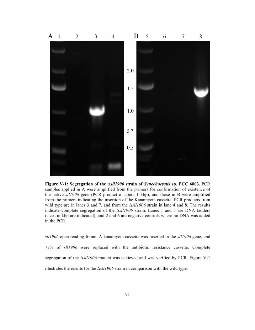
91
Figure V-1: Segregation of the Δsll1906 strain of Synechocystis sp. PCC 6803. PCR samples applied in A were amplified from the primers for confirmation of existence of the native sll1906 gene (PCR product of about 1 kbp), and those in B were amplified from the primers indicating the insertion of the Kanamycin cassette. PCR products from wild type are in lanes 3 and 7, and from the Δsll1906 strain in lane 4 and 8. The results indicate complete segregation of the Δsll1906 strain. Lanes 1 and 5 are DNA ladders (sizes in kbp are indicated), and 2 and 6 are negative controls where no DNA was added in the PCR. sll1906 open reading frame. A kanamycin cassette was inserted in the sll1906 gene, and
77% of sll1906 were replaced with the antibiotic resistance cassette. Complete
segregation of the Δsll1906 mutant was achieved and was verified by PCR. Figure V-1
illustrates the results for the Δsll1906 strain in comparison with the wild type.

92
Table V-1: Effects of the sll1906 deletion mutation on doubling time, chlorophyll content, and oxygen evolution rates of wild type and ΔchlL cells. Listed are the average results of two or three independent experiments ± S.D. ND: Not determined.
Deletion of Sll1906 was found to have no significant impact on photoautotrophic
growth, the amount of chlorophyll per cell, and PSII-driven oxygen evolution (Table V-
1). This lack of a significant difference between strains with or without the sll1906 gene
was found also at a higher light intensity (110 µmol photons m-2 s-1) (data not shown) and
in ΔchlL background (ΔchlL) strains.
Pigment composition of the mutants To determine the effect of deletion of Sll1906 in
wild-type strains in terms of their content of pigments such as chlorophyll, chlorophyll
precursors, and carotenoids, cells were extracted with 100% methanol, and the extracts
were subjected to HPLC analysis. Chlorophyll and chlorophyll precursors such as Mg-
protoporphyrin IX, Mg-protoporphyrin 13-monomethyl ester, and protochlorophyllide
would have been detected by HPLC by means of 410 nm absorbance if such precursors
accumulated (Figure V-2A). However, no chlorophyll precursors accumulated in either
strain. The chlorophyll (C) content was similar in the wild type and Δsll1906 mutant,
consistent with the results reported in Table V-1. Pheophytin a (P) was found in both
strains at equally low levels. As shown in Figure V-2B, there is no change in the
Strain Cell doubling time
(h) Chlorophyll content (µg chl/(ml⋅OD730))
Oxygen evolution (µmol O2/(mg chl⋅h)
Wild type 12±2 3.55±0.21 424±36
Δsll1906 13±2 3.63±0.25 442±31
ΔchlL 12±1 2.60±0.14 ND
ΔchlL/Δsll1906 13±1 2.65±0.21 ND
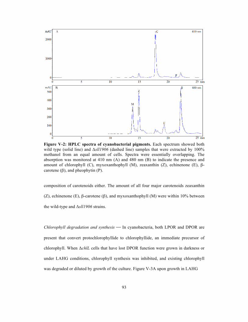
93
Figure V-2: HPLC spectra of cyanobacterial pigments. Each spectrum showed both wild type (solid line) and Δsll1906 (dashed line) samples that were extracted by 100% methanol from an equal amount of cells. Spectra were essentially overlapping. The absorption was monitored at 410 nm (A) and 480 nm (B) to indicate the presence and amount of chlorophyll (C), myxoxanthophyll (M), zeaxanthin (Z), echinenone (E), β-carotene (β), and pheophytin (P). composition of carotenoids either. The amount of all four major carotenoids zeaxanthin
(Z), echinenone (E), β-carotene (β), and myxoxanthophyll (M) were within 10% between
the wild-type and Δsll1906 strains.
Chlorophyll degradation and synthesis In cyanobacteria, both LPOR and DPOR are
present that convert protochlorophyllide to chlorophyllide, an immediate precursor of
chlorophyll. When ΔchlL cells that have lost DPOR function were grown in darkness or
under LAHG conditions, chlorophyll synthesis was inhibited, and existing chlorophyll
was degraded or diluted by growth of the culture. Figure V-3A upon growth in LAHG

94
Figure V-3: Chlorophyll degradation and light-dependent chlorophyll synthesis. Chlorophyll levels (µg/ml-OD730) were monitored in the ΔchlL (triangle with dashed line) and ΔchlL/Δsll1906 (open square with solid line) strains upon transfer to LAHG conditions at time 0 (A) or upon transfer to continuous illumination (40 µmol photons m-2
s-1) at time 0 after cells had been grown under LAHG conditions for 2 weeks (B). shows the chlorophyll content in the ΔchlL strains with and without the sll1906 gene

95
conditions for 6 days. No significant difference in chlorophyll degradation was observed.
To examine whether the chlorophyll biosynthesis rate was affected by Sll1906, the cells
were grown in LAHG conditions for at least about a week until the amount of chlorophyll
was minimal, and subsequently the rate of synthesis of chlorophyll was determined upon
continuous illumination at 40 µmol photons m-2 s-1. As shown in Figure V-3B, the
Δsll1906 mutant exhibited the same rate of chlorophyll synthesis as the wild type.
Therefore, deletion of Sll1906 did not affect the process of chlorophyll degradation and
synthesis.
Photosystem biogenesis In order to study the effects of deletion of Sll1906 on PSI and
PSII, 77 K fluorescence emission spectra of whole cells were measured upon excitation at
435 nm. A major peak at 725 nm is characteristic for PSI-associated chlorophyll, and two
smaller peaks at 685 and 695 nm correspond to phycobilisomes and chlorophylls, and
CP47-associated chlorophyll, respectively (Figure V-4). Deletion of Sll1906 did not
change the PSII/PSI ratio regardless of wild-type or chlL- backgrounds (Figure V-4A).
However, the PSII/PSI ratio increased in the chlL- mutants. According to Table V-1, the
chlorophyll content was reduced about 25% upon deletion of chlL. Therefore, it most
likely is a decrease in amount of PSI that caused the increase of the PSII/PSI ratio.
To see whether the absence of Sll1906 affected the biogenesis of PSII and PSI
upon chlorophyll synthesis, the ΔchlL and ΔchlL/Δsll1906 strains were monitored by 77
K fluorescence emission spectra at different stages of greening after a week of culturing
under LAHG conditions. At time 0, there were no peaks at 695 and 725 nm, which means
that very little or no PSII and PSI was present (Figure V-4B). During 24 hours of
illumination, deletion of Sll1906 did not have an impact on the rate of PSII and PSI
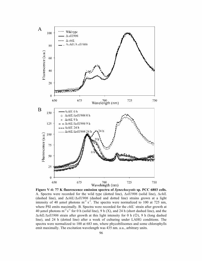
96
Figure V-4: 77 K fluorescence emission spectra of Synechocystis sp. PCC 6803 cells. A. Spectra were recorded for the wild type (dotted line), Δsll1906 (solid line), ΔchlL (dashed line), and ΔchlL/Δsll1906 (dashed and dotted line) strains grown at a light intensity of 40 µmol photons m-2 s-1. The spectra were normalized to 100 at 725 nm, where PSI emits maximally. B. Spectra were recorded for the chlL- strain after growth at 40 µmol photons m-2 s-1 for 0 h (solid line), 9 h (X), and 24 h (short dashed line), and the ΔchlL/Δsll1906 strain after growth at this light intensity for 0 h (), 9 h (long dashed line), and 24 h (dotted line) after a week of culturing under LAHG conditions. The spectra were normalized to 100 at 683 nm, where phycobilisomes and some chlorophylls emit maximally. The excitation wavelength was 435 nm. a.u., arbitrary units.

97
biogenesis upon chlorophyll synthesis. The results show that deletion of Sll1906 did not
appear to alter the delivery of chlorophyll to chlorophyll-binding proteins or aiding
photosystem assembly.
Discussion
The putative BCD family was introduced in an earlier study (Saier et al., 1999), and the
BCD proteins in different organisms have been named as PucC or bacteriochlorophyll
synthase. The function of the BCD family has been examined in Rhodobacter capsulatus,
which contains three members of the BCD family, PucC, LhaA, and ORF428. The LhaA
and PucC proteins were reported to enhance correct LH complex assembly (Young et al.,
1998; Jaschke et al, 2008). The Sll1906 protein in Synechocystis has a 24-27% amino
acid sequence identity with Rhodobacter BCD members and has a hydropathy profile
similar to that of the LhaA/PucC proteins. The BCD family proteins sequences of purple
bacteria (Rhodobacter capsulatus and Rhodopseudomonas palustris) and cyanobacteria
(Synechocystis sp. PCC 6803, Prochlorococcus marinus 9211, and Synechococcus sp.
CC9902) were analyzed. Transmembrane segments (TMSs) 1 and 2, and TMSs 7 and 8
as well as their connecting loop regions are well conserved, and the loop region between
TMSs 4 and 5 as well as TMS5 is also well conserved. These results are consistent with
an earlier study (Saier et al., 1999). Interestingly, the rest of the protein sequence (over
50%) has low or no similarity between BCD family of purple bacteria and cyanobacteria
but is highly conserved within purple bacteria and moderately conserved within
cyanobacteria.
Sll1906 was suggested to be involved in tetrapyrrole delivery for assembly of
chlorophyll-binding complexes (Young and Beatty, 1998). However, Synechocystis cells

98
Table V-2. Protein sequence alignments and possible chlorophyll-binding amino acid residues in Sll1906 relative to PucC from Rhodobacter capsulatus and Synechocystis psbB. Underlined residues represent possible chlorophyll binding sites. Sll1906 protein sequences 1 and 2 represent residues 22-49 and 76-87. CP47 protein sequence 2 represents residues 13-25.
lacking sll1906 have normal chlorophyll content and chlorophyll synthesis (Table V-1
and Figure V-3), whereas also the tetrapyrrole biosynthesis pathway was not disrupted in
the mutant (no accumulation in chlorophyll precursors) (Figure V-2A). Also, judging
from the 77 K fluorescence emission spectra, lack of Sll1906 did not impair PSII and PSI
assembly (Figure V-4).
In order to examine if Sll1906 possibly binds chlorophyll, Table V-2 shows
examples of the Sll1906 protein sequences that contain possible chlorophyll-binding
amino acid residues (underlined). Protein sequence 1 is highly conserved in all organisms
possessing BCD proteins and contain a few amino acid residues that could bind
chlorophyll. Protein sequence 2 is conserved in cyanobacteria only but aligns well with
part of PsbB containing a histidine that binds chlorophyll (Muh et al., 2008). Therefore,
Sll1906 may have chlorophyll-binding ability.
In conclusion, the Δsll1906 mutant did not show significant effects on pigment
content and photosystem assembly. However, this does not necessarily mean that Sll1906
is not involved in these processes as other (parallel) pathways may exist that may fully
compensate for the lack of Sll1906.
Organisms Protein sequence 1 Protein sequence 2
Rb capsulatus PucC RLSLFQITVGMTLTLLAGTLNRVMIVEL KSDTHKSALGLRR
Syn 6803 Sll1906 RLGLFQMGLGIMSLLTLGVLNRVLIDEL LSDSQRLWGYH-R
PsbB (CP47) LNDPGRLISVHLM

99
CHAPTER VI. PERSPECTIVE AND OUTLOOK
A combination of stable-isotope labeling (15N) and mass spectrometry is a powerful
method to provide insights in the dynamics of photosystems in the membrane. In this
study, the lifetimes of PSI and PSII proteins have been determined to range from 30-75 h
and 1-33 h, respectively. The wide range of lifetimes indicates that the damaged
photosynthetic proteins can be replaced independently, and the other proteins can be re-
used and assembled into functional photosynthetic complexes. Indeed, Synechocystis
cells have nascent photosynthetic proteins in the membrane for the replacement of
damaged proteins or for photosystem biogenesis when photosynthetic complexes turn
over. If there are changes in environment or growth conditions, cells may be able to
response rapidly by utilizing these nascent proteins and retain a homeostasis of the
photosystems. It would be interesting to examine the lifetimes of photosynthetic proteins
and change in the pool of nascent photosynthetic proteins upon transition between
different growth conditions. Also, the IsiA (CP43’) protein, an iron-stress-induced
protein, can form circular aggregates (rings) of up to 18 subunits around trimeric PSI in a
first ring and possibly up to 25 subunits in a second ring under iron-deficient or some
other stress conditions (Bibby et al., 2001; Boekema et al., 2001; Yeremenko et al.,
2004). IsiA proteins may serve as a light-harvesting antenna and as an energy dissipator
for PSI (Bibby et al., 2001; Berera et al., 2009). By applying a combination of stable-
isotope labeling and mass spectrometry on strains with and without IsiA proteins under
iron-sufficient/deficient conditions, the effects of IsiA on lifetimes of PSI proteins and
the dynamics of PSI complexes switching between monomeric and trimeric forms can be
studied.

100
Chlorophyll is the most abundant cofactor in the photosystems. A total of 131
chlorophyll molecules are in PSII and PSI combined, and synthesis of one chlorophyll
molecule from glutamate requires 9 ATP and 13 NADPH. It is a significant investment
just on cofactors for cyanobacteria to practice photosynthesis. Synechocystis cells have
evolved an efficient way of reutilizing chlorophyll from the damaged chlorophyll-binding
proteins in the PSI and PSII complexes. As known from this study, SCPs play an
important role in PSII chlorophyll recycling. However, what factors function in the
recycling of PSI chlorophyll still needs to be investigated. Although chlorophyll can be
utilized, highly active chlorophyll synthesis is required as there is always a significant
amount of chlorophyll-binding proteins synthesized for both photosystem repair and
biogenesis to retain a homeostasis of photosystems and cell growth.
As chlorophyll is a strong photo-oxidizer, free chlorophyll is very dangerous for
cells by rapidly producing reactive oxygen species in the presence of light and oxygen,
and cellular damage may happen as quick as in a few seconds. However, chlorophyll in
chlorophyll-binding proteins is relatively safe as efficient quenchers, such as carotenoids,
are nearby, as demonstrated by the long half-lives of chlorophyll-binding proteins that do
not turn over on a timescale of up to 40 h. Therefore, cooperation between chlorophyll
biosynthesis and synthesis of chlorophyll-binding proteins is important. This study shows
that SCPs are possible candidates as bridges in communication between chlorophyll and
PSII proteins as upon removal of SCPs the rate of chlorophyll synthesis and the amount
of chlorophyll molecules are reduced while the amount of PSII proteins decrease and the
accumulation of nascent PSII proteins in membranes disappears. Interestingly, the
phenotype is exhibited only in the absence of PSI. However, PSI possesses about 90% of
chlorophyll in the Synechocystis cells. The deletion of SCPs in the PSI-containing strain
can not inhibit ALA synthesis significantly because chlorophyll synthesis needs to meet

101
the demand of chlorophyll from PSI. It is as yet unknown which protein(s), if any, that
function like SCPs serve PSI, and whether such protein(s) can function like SCPs for
PSII. The Sll1906 protein, a member of putative bacteriochlorophyll delivery family, may
possibly function as a bridge between chlorophyll and chlorophyll-binding proteins.
However, deletion of Sll1906 does not show any phenotype in either chlorophyll
biosynthesis/degradation or photosystem assembly. In order to understand how the
synthesis of chlorophyll and chlorophyll-binding proteins is coordinated and chlorophyll
is delivered to the chlorophyll-binding proteins, a continuous search for factor(s) that
function like the SCPs in PSII but, for example, work in PSI is critical.

102
References
Adamska, I., Roobol-Boza, M., Lindahl, M., and Andersson, B. (1999) Isolation of pigment-binding early light-inducible proteins from pea. Eur. J. Biochem. 260, 453–460 Adamska, I. (2001) The Elip family of stress proteins in the thylakoid membranes of pro- and eukaryota. Advances in Photosynthesis and Respiration: Regulation of Photosynthesis (Aro, E. M., and Andersson, B., eds) Vol. 11, pp. 487–505, Kluwer Academic Publishers, Dordrecht, The Netherlands Adhikari, N. D., Orler, R., Chory, J., Froehlich, J. E., and Larkin, R. M. (2009) Porphyrins promote the association of GENOMES UNCOUPLED 4 and a Mg-chelatase subunit with chloroplast membranes. J. Biol. Chem. 284, 24783-24796 Amunts, A., Drory, O., and Nelson, N. (2007) The structure of a plant photosystem I supercomplex at 3.4 Å resolution. Nature 447, 58-63 Anbudurai, P. R., Mor, T. S., Ohad, I., Shestakov, S. V., and Pakrasi, H. B. (1994) The ctpA gene encodes the C-terminal processing protease for the D1 protein of the photosystem-II reaction-center complex. Proc. Natl. Acad. Sci. USA 91, 8082-8086 Aro, E. M., Virgin, I., and Andersson, B. (1993) Photoinhibition of photosystem II – inactivation, protein damage and turnover. Biochim. Biophys. Acta 1143, 113-134 Aro, E. M., Suorsa, M., Rokka, A., Allahverdiyeva, Y., Paakkarinen, V., Saleem, A., Battchikova, N., and Rintamaki, E. (2005) Dynamics of photosystem II: a proteomic approach to thylakoid protein complexes. J. Exp. Bot. 56, 347–356 Bailey, S., Thompson, E., and Nixon, P. J. (2002) A critical role for the Var2 FtsH homologue of Arabidopsis thaliana in the photosystem II repair cycle in vivo. J. Biol. Chem. 277, 2006-2011 Barber, J. (2002) P680: what is it and where is it? Bioelectrochemistry 55, 135-138 Beale, S. I. (1990) Biosynthesis of the tetrapyrrole pigment precursor, aminolevulinic acid, from glutamate. Plant Physiol. 93, 1273-1279 Beale, S. I. (1999) Enzymes of chlorophyll biosynthesis. Photosyn. Res. 60, 43-73 Beck, C. F. and Grimm, B. (2006) Involvement of Tetrapyrroles in Cellular Regulation in Advances in Photosynthesis and Respiration: Chlorophylls and bacteriochlorophylls, Vol. 25 (Grimm, B., Porra, R. J., Rudiger, W., and Scheer, H. eds.) pp. 223-235, Springer, Dordrecht Berera, R., van Stokkum, I. H. M., D’Haene, S., Kennis, J. T. M., van Grondelle, R., and Dekker, J. P. (2009) A mechanism of energy dissipation in cyanobacteria. Biophys. J. 96, 2261-2267

103
Bhaya, D., Dufresne, A., Vaulot, D., and Grossman, A. (2002) Analysis of the hli gene family in marine and freshwater cyanobacteria. FEMS Microbiol. Lett. 215, 209–219 Bibby, T. S., Nield, J., and Barber, J. (2001) A photosystem II-like protein, induced under iron-stress, forms an antenna ring around the photosystem I trimer in cyanobacteria. Nature 412, 743-745 Bjellqvist, B., Pasquali, C., Ravier, F., Sanchez, J. C., and Hochstrasser, D. (1993) A nonlinear wide-range immobilized pH gradient for 2-dimensional electrophoresis and its definition in a relevant pH scale. Electrophoresis 14, 1357–1365 Blankenship, R. E. (2002) Molecular Mechanisms of Photosynthesis. Blackwell Science, Malden Boekema, E. J., Hifney, A., Yakushevska, A. E., Piotrowski, M., Keegstra, W., Berry, S., Michel, K. P., Pistorius, E. K., and Kruip, J. (2001) A giant chlorophyll-protein complex induced by iron deficiency in cyanobacteria. Nature 412, 745-748 Bricker, T. M., Morvant, J., Masri, N., Sutton, H. M., and Frankel, L. K. (1998) Isolation of a highly active photosystem II preparation from Synechocystis 6803 using a histidine-tagged mutant of CP47. Biochim. Biophys. Acta 1409, 50–57 Bryant, D. A. (1994) The Molecular Biology of Cyanobacteria. Kluwer Academic Press, Dordrecht Burke, D. H., Hearst, J. E., and Sidow, A. (1993) Early evolution of photosynthesis: clues from nitrogenase and chlorophyll iron proteins. Proc. Natl. Acad. Sci. USA. 90, 7134-7138 Carpenter, S. D., Charité, J., Eggers, B., and Vermaas, W. F. J. (1990) The psbC start codon in Synechocystis sp. PCC 6803. FEBS Lett. 260, 135-137 Chitnis, V. P. and Chitnis, P. R. (1993) PsaL subunit is required for the formation of photosystem I trimers in the cyanobacterium Synechocystis sp. PCC 6803. FEBS Lett. 336, 330 –334 Cormann, K. U., Bangert, J. A., Ikeuchi, M., Rögner, M., Stoll, R., and Nowaczyk, M. M. (2009) Structure of Psb27 in solution: Implications for transient binding to photosystem II during biogenesis and repair. Biochemistry 48, 8768-8770 Dobakova, M., Tichy, M., and Komenda, J. (2007) Role of the PsbI protein in photosystem II assembly and repair in the cyanobacterium Synechocystis sp. PCC 6803. Plant Physiol. 145, 1681-1691

104
Dobakova, M., Sobotka, R., Tichy, M., and Komenda, J. (2009) Psb28 protein is involved in the biogenesis of the photosystem II inner antenna CP47 (PsbB) in the cyanobacterium Synechocystis sp PCC 6803. Plant Physiol. 149, 1076-1086 Dolganov, N. A. M., Bhaya, D., and Grossman, A. R. (1995) Cyanobacterial protein with similarity to the chlorophyll a/b binding proteins of higher plants: Evolution and regulation. Proc. Natl. Acad. Sci. USA 92, 636-640 Durnford, D. G., Deane, J. A., Tan, S., McFadden, G. I., Gantt, E., and Green, B. R. (1999) A phylogenetic assessment of the eukaryotic light-harvesting antenna proteins, with implications for plastid evolution. J. Mol. Evol. 48, 59–68 Eckhardt, U., Grimm, B., and Hortensteiner, S. (2004) Recent advances in chlorophyll biosynthesis and breakdown in higher plants. Plant Mol. Biol. 56, 1-14 Ermakova-Gerdes, S., Shestakov, S., and Vermaas, W. (1996) Targeted random mutagenesis in the D2 protein of photosystem II: Identification of functionally important residues at the lumenal side of the thylakoid. Plant Mol. Biol. 30, 243-254 Ferreira, K. N., Iverson, T. M., Maghlaoui, K., Barber, J., and Iwata, S. (2004) Architecture of the photosynthetic oxygen-evolving center. Science 303, 1831–1838 Fischer, N., Hippler, M., Setif, P., Jacquot, J. P., and Rochaix, J. D. (1998) The PsaC subunit of photosystem I provides an essential lysine residue for fast electron transfer to ferredoxin. EMBO J. 17, 849-858 Funk, C., Schroder, W. P., Green, B. R., Renger, G., and Andersson, B. (1994) The intrinsic 22 kDa protein is a chlorophyll-binding subunit of photosystem II. FEBS Lett. 342, 261–266 Funk, C. and Vermaas, W. (1999) A cyanobacterial gene family coding for single-helix proteins resembling part of the light-harvesting proteins from higher plants. Biochemistry 38, 9397–9404 Funk, C. (2001) The PsbS protein: A Cab-protein with a Function of its Own in Advances in Photosynthesis and Respiration: Regulation of Photosynthesis (Aro, E. M., and Andersson, B., eds) Vol. 11, pp. 453–467, Kluwer Academic Publishers, Dordrecht, The Netherlands Gharahdaghi, F., Weinberg, C. R., Meagher, D. A., Imai, B. S., and Mische, S. M. (1999) Mass spectrometric identification of proteins from silver-stained polyacrylamide gel: A method for the removal of silver ions to enhance sensitivity. Electrophoresis 20, 601–605 Griffiths, W. T. (1975) Characterization of of the terminal stages of chlorophyll(ide) synthesis in etioplast membrane preparations. Biochem. J. 152, 623-635 Grimm, B. (1999) The Metabolic Pathway of Tetrapyrrole Biosynthesis in Peroxidizing Herbicides (Böger, P. and Wakabayashi, K. eds), pp 213-244, Springer-Verlag, Berlin
105
Guskov, A., Kern, J., Gabdulkhakov, A., Broser, M., Zouni, A., and Saenger, W. (2009) Cyanobacterial photosystem II at 2.9 Å resolution and the role of quinones, lipids, channels and chloride. Nat. Struct. Biol. 16, 334-342 Hao, L. M., Schmidt, K., Paulsen, H., and Funk, C. (2001) Isolation and reconstitution of hypothetical pigment carrier proteins. Proceedings of the 12th International Congress on Photosynthesis (Larkum, T., and Critchley, C., eds) pp. S31–005, Commonwealth Scientific and Industrial Research Organisation Publishing, Melbourne, Australia Havaux, M., Guedeney, G., He, Q., and Grossman, A. R. (2003) Elimination of high-light-inducible polypeptides related to eukaryotic chlorophyll a/b-binding proteins results in aberrant photoacclimation in Synechocystis PCC6803. Biochim. Biophys. Acta 1557, 21–33 He, Q. and Vermaas, W. (1998) Chlorophyll a availability affects psbA translation and D1 precursor processing in vivo in Synechocystis sp. PCC 6803. Proc. Natl. Acad. Sci. USA 95, 5830-5835 He, Q., Dolganov, N., Bjorkman, O., and Grossman, A. R. (2001) The high light-inducible polypeptides in Synechocystis PCC6803 – expression and function in high light. J. Biol. Chem. 276, 306–314 Heddad, M. and Adamska, I. (2000) Light stress-regulated two-helix proteins in Arabidopsis thaliana related to the chlorophyll a/b-binding gene family. Proc. Natl. Acad. Sci. U. S. A. 97, 3741–3746 Heddad, M. and Adamska, I. (2002) The evolution of light stress proteins in photosynthetic organisms. Comp. Funct. Genom. 3, 504–510 Herranen, M., Battchikova, N., Zhang, P., Graf, A., Sirpio, S., Paakkarinen, V., and Aro, E. M. (2004) Towards functional proteomics of membrane protein complexes in Synechocystis sp. PCC 6803. Plant Physiol. 134, 470-481 Inagaki, N., Yamamoto, Y., and Satoh, K. (2001) A sequential two-step proteolytic process in the carboxyl-terminal truncation of precursor D1 protein in Synechocystis sp PCC 6803. FEBS Lett. 509, 197-201 Jansson, S. (1994) The light-harvesting chlorophyll a/b binding proteins. Biochim. Biophys. Acta 1184, 1-19 Jansson, S. (1999) A guide to the Lhc genes and their relatives in Arabidopsis. Trends Plant Sci. 4, 236–240 Jansson, S., Andersson, J., Kim, S. J., and Jackowski, G. (2000) An Arabidopsis thaliana protein homologous to cyanobacterial high-light-inducible proteins. Plant Mol. Biol. 42, 345–351

106
Jansson, S. (2005) A Protein Family Saga: From Photoprotection to Light-Harvesting in Photoprotection, Photoinhibition, Gene Regulation and Environment (Demmig-Adams, B., ed) pp. 145–153, Springer, Dordrecht, The Netherlands Jaschke, P., LeBlanc, H., Lang, A., and Beatty, J. (2008) The PucC protein of Rhodobacter capsulatus mitigates an inhibitory effect of light-harvesting 2 α and β proteins on light-harvesting complex 1. Photosynth. Res. 95, 279-284 Jensen, P. E., Haldrup, A., Rosgaard, L., and Scheller, H. V. (2003) Molecular dissection of photosystem I in higher plants: topology, structure and function. Physiol. Plant. 119, 313-321 Jordan, P., Fromme, P., Witt, H. P., Klukas, O., Saenger, W., and Krauss, N. (2001) Three-dimensional structure of cyanobacterial photosystem I at 2.5 Å resolution. Nature 411, 909-917 Joyard, J., Ferro, M., Masselon, C., Seigneurin-Berny, D., Salvi, D., Garin, J., and Rolland, N. (2009) Chloroplast proteomics and the compartmentation of plastidial isoprenoid biosynthetic pathway. Mol. Plant 2, 1154-1180 Kaneko, T., Sato, S., Kotani, H., Tanaka, A., Asamizu, E., Nakamura, Y., Miyajima, N., Hirosama, M., Sugiura, M., Sasamoto, S., Kimura, T., Hosouchi, T., Matsuno, A., Muraki, A., Nakazaki, N., Naruo, K., Okumura, S., Shimpo, S., Takeuchi, C., Wada, T., Watanabe, A., Yamada, M., Yasuda, M., and Tabata, S. (1996) Sequence analysis of the genome of the unicellular cyanobacterium Synechocystis sp. PCC 6803. II. Sequence determintation of the entire genome and assignment of potential protein-coding regions. DNA Res. 3, 109-136 Karapetyan, N. V., Holzwarth, A. R., and Rogner, M. (1999) The photosystem I trimer of cyanobacteria: molecular organization, excitation dynamics and physiological significance. FEBS Lett. 460, 395–400 Kashino, Y., Lauber, W. M., Carroll, J. A., Wang, Q., Whitmarsh, J., Satoh, K., and Pakrasi, H. B. (2002) Proteomic analysis of a highly active photosystem II preparation from the cyanobacterium Synechocystis sp PCC 6803 reveals the presence of novel polypeptides. Biochemistry 41, 8004–8012 Kato, Y., Miura, E., Ido, K., Ifuku, K., and Sakamoto, W. (2009) The variegated mutants lacking chloroplastic FtsHs are defective in D1 degradation and accumulate reactive oxygen species. Plant Physiol. 151, 1790-1801 Keren, N., Ohkawa, H., Welsh, E. A., Liberton, M., and Pakrasi, H. B. (2005) Psb29, a conserved 22-kD protein, functions in the biogenesis of photosystem II complexes in Synechocystis and Arabidopsis. Plant Cell 17, 2768-2781 Kern, J., Loll, B., Zouni, A., Saenger, W., Irrgang, K. D., and Biesiadka, J. (2005) Cyanobacterial photosystem II at 3.2 angstrom resolution – the plastoquinone binding pockets. Photosynth. Res. 84, 153–159

107
Klimmek, F., Sjödin, A., Noutsos, C., Leister, D., and Jansson, S. (2006) Abundantly and rarely expressed Lhc protein genes exhit distinct regulation patterns in plants. Plant Physiol. 140, 793–804 Komenda, J., Lupinkova, L., and Kopecky, J. (2002) Absence of the psbH gene product destabilizes photosystem II complex and bicarbonate binding on its acceptor side in Synechocystis PCC 6803. Eur. J. Biochem. 269, 610–619 Komenda, J., Reisinger, V., Muller, B. C., Dobakova, M., Granvogl, B., and Eichacker, L. A. (2004) Accumulation of the D2 protein is a key regulatory step for assembly of the photosystem II reaction center complex in Synechocystis PCC 6803. J. Biol. Chem. 279, 48620–48629 Komenda, J., Barker, M., and Kuvikova, S. (2006) The FtsH protease Slr0228 is important for quality control of photosystem II in the thylakoid membrane of Synechocystis sp PCC 6803. Plant Cell Physiol. 46, 1477-1483 Komenda, J., Kuvikova, S., Granvogl, B., Eichacker, L. A., Diner, B. A., and Nixon, P. J. (2007) Cleavage after residue Ala352 in the C-terminal extension is an early step in the maturation of the D1 subunit of photosystem II in Synechocystis PCC 6803. Plant Cell 19, 2839-2854 Komenda, J., Nickelsen, J., Tichy, M., Prasil, O., Eichacker L. A., and Nixon, P. J. (2008) The cyanobacterial homologue of HCF136/YCF48 is a component of an early photosystem II assembly complex and is important for both the efficient assembly and repair of photosystem II in Synechocystis sp. PCC 6803. J. Biol. Chem. 283, 22390-22399 Kouril, R., Arteni, A. A., Lax, J., Yeremenko, N., D’Haene, S., Rogner, M., Matthijs, H. C., Dekker, J. P., and Boekema, E. J. (2005) Structure and functional role of supercomplexes of IsiA and photosystem I in cyanobacterial photosynthesis. FEBS Lett. 579, 3253–3257 Krieger-Liszkay, A. (2005) Singlet oxygen production in photosynthesis. J. Exp. Bot. 56, 337-346 Krieger-Liszkay, A., Fufezan, C., and Trebst, A. (2008) Singlet oxygen production in photosystem II and related protection mechanism. Photosynth. Res. 98, 551-564 Kufryk, G., Hernandez-Prieto, M. A., Kieselbach, T., Miranda, H., Vermaas, W., and Funk, C. (2008) Association of small CAB-like proteins (SCPs) of Synechocystis sp. PCC 6803 with photosystem II. Photosynth. Res. 95, 135-145 Kühlbrandt, W., Wang, D. N., and Fujiyoshi, Y. (1994) Atomic model of plant light-harvesting complex by electron crystallography. Nature 367, 614–621 Kussmann, M., Nordhoff, E., Rahbek-Nielsen, H., Haebel, S., Rossel- Larsen, M., Jakobsen, L., Gobom, J., Mirgorodskaya, E., Kroll-Kristensen, A., Palm, L., and Roepstorff, P. (1997) Matrix-assisted laser desorption/ionization mass spectrometry

108
sample preparation techniques designed for various peptide and protein analysis. J. Mass Spectrom. 32, 593–601 Kuttkat, A., Edhofer, I., Eichacker, L. A., and Paulsen, H. (1997) Light-harvesting chlorophyll a/b-binding protein stably inserts into etioplast membranes supplemented with Zn-pheophytin a/b. J. Biol. Chem. 272, 20451-20455 Lagarde, D., Beuf, L., and Vermaas, W. (2000) Increased production of zeaxanthin and other pigments by application of genetic engineering techniques to Synechocystis sp strain PCC 6803. Appl. Environ. Microbiol. 66, 64–72 LeBlanc, H. N. and Beatty, J. T. (1996) Topological analysis of the Rhodobacter capsulatus PucC protein and effects of C-terminal deletions on light-harvesting complex II. J. Bacteriol. 178, 4801-4806 Li, X. P., Bjorkman, O., Shih, C., Grossman, A. R., Rosenquist, M., Jansson, S., and Niyogi, K. K. (2000) A pigment-binding protein essential for regulation of photosynthetic light harvesting. Nature 403, 391–395 Lindell, D., Sullivan, M. B., Johnson, Z. I., Tolonen, A. C., Rohwer, F., and Chisholm, S. W. (2004) Transfer of photosynthesis genes to and from Prochlorococcus viruses. Proc. Natl. Acad. Sci. U. S. A. 101, 11013–11018 Liu, Z., Yan, H., Wang, K., Kuang, T., Zhang, J., Gui, L., An, X., and Chang, W. (2004) Crystal structure of spinach major light-harvesting complex at 2.72 angstrom resolution. Nature 428, 287–292 Loll, B., Kern, J., Saenger, W., Zouni, A., and Biesiadka, J. (2005) Towards complete cofactor arrangement in the 3.0 Å resolution structure of photosystem II. Nature 438, 1040-1044 Loll, B., Kern, J., Saenger, W., Zouni, A., and Biesiadka, J. (2007) Lipids in photosystem II: Interaction with protein and cofactors. Biochim. Biophys. Acta 1767, 509-519 MacPherson, A. N., Telfer, A., Barber, J., and Truscott, T. G. (1993) Direct detection of singlet oxygen from isolated photosystem II reaction centres. Biochim. Biophys. Acta 1143, 301-309 Mann, M. and Wilm, M. (1994) Error tolerant identification of peptides in sequence databases by peptide sequence tags. Anal. Chem. 66, 4390–4399 Mattoo, A. K., Hoffman-Falk, H., Marder, J. B., and Edelman, M. (1984) Regulation of protein metabolism: coupling of photosynthetic electron transport to in vivo degradation of the rapidly metabolized 32-kilodalton protein of the chloroplast membranes. Proc. Natl. Acad. Sci. USA 81, 1380-1384 Mattoo, A. K., Giardi, M. T., Raskind, A., and Edelman, M. (1999) Dynamic metabolism of photosystem II reaction center proteins and pigments. Physiol. Plant. 107, 454-461

109
Mayer, S. and Beale, S. (1992) Succinyl-coenzyme A synthetase and its role in aminolevulinic acid biosynthesis in Euglena gracilis. Plant Physiol. 99, 482-487 Meskauskiene, R., Nater, M., Goslings, D., Kessler, F., Op den Camp, R., and Apel, K. (2001) FLU: A negative regulator of chlorophyll biosynthesis in Arabidopsis thaliana. Proc. Natl. Acad. Sci. USA 98, 12826-12831 Mikami, K., Kanesaki, Y., Suzuki, I., and Murata, N. (2002) The histidine kinase Hik33 perceives osmotic stress and cold stress in Synechocystis sp PCC 6803. Mol. Microbiol. 46, 905–915 Mochizuki, N., Tanaka, R., Grimm, B., Masuda, T., Moulin, M., Smith, A. G., Tanaka, A., and Terry, M. J. (2010) The cell biology of tetrapyrroles: a life and death struggle. Trends Plant Sci. 15, 488-498 Muh, F., Renger, T., and Zouni, A. (2008) Crystal structure of cyanobacterial photosystem II at 3.0 Å resolution: A closer look at the antenna system and the small membrane-intrinsic subunits. Plant Physiol. Biochem. 46, 238-264 Mullet, J., Gamble-Klein, P., and Klein, R. (1990) Chlorophyll regulates accumulation of the plastid-encoded chlorophyll apoproteins CP43 and D1 by increasing apoprotein stability. Proc. Natl. Acad. Sci. USA 87, 4038-4042 Mullineaux, C. W., and Emlyn-Jones, D. (2005) State transitions: an example of acclimation to low-light stress. J. Exp. Bot. 56, 389–393 Muraki, N., Nomata, J., Ebata, K., Mizogucho, T., Shiba, T., Tamiaki, H., Kurisu, G., and Fujita, Y. (2010) X-ray crystal structure of the light-independent protochlorophyllide reductase. Nature 465, 110-114 Nixon, P. J., Trost, J. T., and Diner, B. A. (1992) Role of the carboxyl terminus of polypeptide D1 in the assembly of a function water-oxidizing manganese cluster in photosystem II of the cyanobacterium Synechocystis sp PCC 6803 – assembly requires a free carboxyl group at C-terminal position 344. Biochemistry 31, 10859-10871 Nixon, P. J., Barker, M., Boehm, M., de Vries, R., and Komenda, J. (2005) FtsH-mediated repair of the photosystem II complex in response to light stress. J. Exp. Bot. 56, 357–363 Nixon, P., Michoux, F., Yu, J., Boehm, M., and Komenda, J. (2010) Recent advances in understanding the assembly and repair of photosystem II. Ann. Bot. 106, 1-16 Norling, B., Zak, E., Andersson, B., and Pakrasi, H. (1998) 2D-isolation of pure plasma and thylakoid mebranes from the cyanobacterium Synechocystis sp. PCC 6803. FEBS Lett. 436, 189–192 Nowaczyk, M. M., Hebeler, R., Schlodder, E., Meyer, H. E., Warscheid, B., and Rögner, M. (2006) Psb27, a cyanobacterial lipoprotein, is involved in the repair cycle of photosystem II. Plant Cell 18, 3121-3131

110
Ohad, I., Kyle, D. J., and Arntzen, C. J. (1984) Membrane-protein damage and repair – removal and replacement of activated 32-kilodalton polypeptide in chloroplast membranes. J. Cell Biol. 99, 481-485 Ohta, N., Matsuzaki, M., Misumi, O., Miyagishima, S. Y., Nozaki, H., Tanaka, K., Shin, I. T., Kohara, Y., and Kuroiwa, T. (2003) Complete sequence and analysis of the plastid genome of the unicellular red alga cyanidioschyzon merolae. DNA Res. 10, 67–77 Perkins, D. N., Pappin, D. J., Creasy, D. M., and Cottrell, J. S. (1999) Probability-based protein identification by searching sequence databases using mass spectrometry data. Electrophoresis 20, 3551–3567 Peter, E. and Grimm, B. (2009) GUN4 is required for posttranslational comtrol plant tetrapyrrole biosynthesis. Mol. Plant 2, 1198-1210 Plucken, H., Muller, B., Grohmann, D., Westhoff, P., and Eichacker, L. A. (2002) The HCF136 protein is essential for assembly of the photosystem II reaction center in Arabidopsis thaliana. FEBS Lett. 532, 85-90 Porra, R. J., Thompson, W. A., and Kriedemann, P. E. (1989) Determination of accurate extinction coefficients and simultaneous-equations for assaying chlorophyll-a and chlorophyll-b extracted with 4 different solvents – verification of the concentration of chlorophyll standards by atomic-absorption spectroscopy. Biochim. Biophys. Acta 975, 385–394 Powles, S. B. (1984) Photoinhibition of photosynthesis induced by visible light. Ann. Rev. Plant Physiol. 35, 15-44 Prasil, O., Adir, N., and Ohad, I. (1992) Mechanisms of photoinhibition and recovery processes in Topics in Photosynthesis: Dynamics of photosystem II. Vol. II (Barber, J. ed) pp. 293-348, Elsevier, Amsterdam Prentki, P. and Krisch, H. M. (1984) In vitro insertional mutagenesis with a selectable DNA fragment. Gene 29, 303–313 Promnares, K., Komenda, J., Bumba, L., Nebesarova, J., Vacha, F., and Tichy, M. (2006) Cyanobacterial small chlorophyll binding protein ScpD (HliB) is located on the periphery of photosystem II in the vicinity of PsbH and CP47 subunits. J. Biol. Chem. 281, 32705-32713 Rakhimberdieva, M. G., Stadnichuk, I. N., Elanskaya, I. V., and Karapetyan, N. V. (2004) Carotenoid-induced quenching of the phycobilisome fluorescence in photosystem II-deficient mutant of Synechocystis sp. FEBS Lett. 574, 85–88 Rappsilber, J., Ishihama, Y., and Mann, M. (2003) Stop and go extraction tips for matrix-assisted laser desorption/ionization, nanoelectrospray, and LC/MS sample pretreatment in proteomics. Anal. Chem. 75, 663–670

111
Reinbothe, C., Bartsch, S., Eggink, L. L., Hoober, J. K., Brusslan, J., Andrade-Paz, R., Monnet, J., and Reinbothe, S. (2006) A role for chlorophyllide a oxygenase in the regulated import and stabilization of light-harvesting chlorophyll a/b proteins. Proc. Natl. Acad. Sci. USA 103, 4777-4782 Richter, A., Peter, E., Pors, Y., Lorenzen, S., Grimm, B., and Czarnecki, O. (2010) Rapid dark repression of 5-aminolevulinic acid synthesis in green Barley leaves. Plant Cell Physiol. 51, 670-681 Rieble, S. and Beale, S. I. (1991) Purification of glutamyl-tRNA reductase from Synechocystis sp. PCC 6803. J. Biol. Chem. 266, 9740-9744 Ried, J. L. and Collmer, A. (1987) An NPTI-SACB-SACR cartridge for constructing directed, unmarked mutations in gram-negative bacteria by marker exchange-eviction mutagenesis. Gene 57, 239–246 Rippka, R., Deruelles, J., Waterbury, J. B., Herdman, M., and Stanier, R. T. (1979) Generic assignments, strain histories and properties of pure cultures of cyanobacteria. J. Gen. Microbiol. 111, 1–61 Saier, M., Beatty, J., Goffeau. A., Harley, K., Heijne, W., Huang, S., Jack, D., Jahn, P., Lew, K., Liu, J., Pao, S., Paulsen, I., Tseng, T., and Virk, P. (1999). The major facilitator superfamily. J. Mol. Microbiol. Biotechnol. 1, 257-279 Sarma, R., Barney, B. M., Hamilton, T. L., Jones, A., Seefeldt, L. C., and Peters, J. W. (2008) Crystal structure of the L protein of Rhodobacter sphaeroides light-independent protochlorophyllide reductase with MgADP bound: a homologue of the nitrogenase Fe protein. Biochemistry 47, 13004-13015 Schagger, H., and von Jagow, G. (1991) Blue native electrophoresis for isolation of membrane-protein complexes in enzymatically active form. Anal. Biochem. 199, 223–231 Schuster, G., Timberg, R., and Ohad, I. (1988) Turnover of thylakoid photosystem II proteins during photoinhibition of Chlamydomonas reinhardtii. Eur. J. Biochem. 177, 403-410 Shalygo N., Czarnecki, O., Peter, E., and Grimm, B. (2009) Expression of chlorophyll synthase is also involved in feedback-control of chlorophyll biosynthesis. Plant Mol. Biol. 71, 425-436 Shen, G., Boussiba, S., and Vermaas, W. F. J. (1993) Synechocystis sp PCC 6803 strains lacking photosystem I and phycobilisomes function. Plant Cell 5, 1853–1863 Shevchenko, A., Wilm, M., Vorm, O., and Mann, M. (1996) Mass spectrometric sequencing of proteins from silver stained polyacrylamide gels. Anal. Chem. 68, 850–858 Silva, P., Thompson, E., and Bailey, S. (2003) FtsH is involved in the early stages of repair of photosystem II in Synechocystis sp. PCC 6803. Plant Cell 15, 2152-2164

112
Smith, D. and Howe, C. J. (1993) The distribution of photosystem-I and photosystem-II polypeptides between the cytoplasmic and thylakoid membranes of cyanobacteria. FEMS Microbiol. Lett. 110, 341-347 Sobotka, R., Komenda, J., Bumba, L., and Tichy, M. (2005) Photosystem II assembly in CP47 mutant of Synechocystis sp PCC 6803 is dependent on the level of chlorophyll precursors regulated by ferrochelatase. J. Biol. Chem. 280, 31595–31602 Sobotka, R., McLean, S., Zuberova, M., Hunter, C. N., and Tichy, M. (2008) The C-terminal extension of ferrochelatase is critical for enzyme activity and for functioning of the tetrapyrrole pathway in Synechocystis strain PCC 6803. J. Bacteriol. 190, 2086-2095 Sonoike, K. (2006) Photoinhibition and protection of photosystem I in Advances in Photosynthesis and Respiration: The Light-driven Plastocyanin - Ferredoxin Oxidoreductase, Vol. 24 (Golbeck, J. ed.) pp. 657-668, Springer, Dordrecht Srivastava, A. and Beale, S. I. (2005) Glutamyl-tRNA reductase of Chlorobium vibrioforme is a dissociable homodimer that contains one tightly bound heme per subunit. J. Bacteriol. 187, 4444-4450 Staehelin, L. A. (1976) Reversible particle movements associated with unstacking and restacking of chloroplast membranes in vitro. J. Cell Biol. 71, 136-158 Staehelin, L. A. (2003) Chloroplast structure: from chlorophyll granules to supra-molecular architecture of thylakoid membranes. Photosynth. Res. 76, 185-196 Standfuss, J., Terwisscha van Scheltinga, A. C., Lamborghini, M., and Kuhlbrandt, W. (2005) Mechanisms of photoprotection and nonphotochemical quenching in pea light-harvesting complex at 2.5A resolution. EMBO J. 24, 919–928 Stewart, D. H. and Brudvig, G. W. (1998) Cytochrome b559 of photosystem II. Biochim. Biophys. Acta 1367, 63-87 Storm, P., Hernandez-Prieto, M., Eggink, L. L., Hoober, J. K., and Funk, C. (2008) The small CAB-like proteins of Synechocystis sp. PCC 6803 bind chlorophyll. Photosynth. Res. 98, 479-488 Sullivan, M. B., Coleman, M. L., Weigele, P., Rohwer, F., and Chisholm, S. W. (2005) Three Prochlorococcus cyanophage genomes: Signature features and ecological interpretations. PLoS Biol. 3, e144 Tanaka, R. and Tanaka, T. (2007) Tetrapyrrole biosynthesis in higher plants. Annu. Rev. Plant Biol. 58, 321-346 Teramoto, H., Itoh, T., and Ono, T. A. (2004) High-light inducible Lhc-like genes newly identified in Chamydomonas reinhartii. Plant Cell Physiol. 45, 1221–1232

113
Thidholm, E., Lindstrom, V., Tissier, C., Robinson, C., Schroder, W. P., and Funk, C. (2002) Novel approach reveals localisation and assembly pathway of the PsbS and PsbW proteins into the photosystem II dimer. FEBS Lett. 513, 217–222 Towbin, H., Staehelin, T., and Gordon, J. (1979) Electrophoretic transfer of proteins from polyacrylamide gels to nitrocellulose sheets – procedure and some applications. Proc. Natl. Acad. Sci. U. S. A. 76, 4350–4354 Triantaphylides, C. and Havaux, M. (2009) Singlet oxygen in plants: production, detoxification and signaling. Trends Plant Sci. 14, 219-228 Vass, I., Styring, S., Hundal, T., Koivuniemi, A., Aro, E. M., and Andersson, B. (1992) Reversible and irreversible intermediates during photoinhibition of photosystem II: Stable reduced QA species promote chlorophyll triplet formation. Proc. Natl. Acad. Sci. USA 89, 1408-1412 Vavilin, D. and Vermaas, W. (2002) Regulation of the tetrapyrrole biosynthetic pathway leading to heme and chlorophyll in plants and cyanobacteria. Physiol. Plant. 115, 9-24 Vavilin, D., Brune, D. C., and Vermaas, W. (2005) 15N labeling to determine chlorophyll synthesis and degradation in Synechocystis sp PCC 6803 strains lacking one or both photosystems. Biochim. Biophys. Acta 1708, 91–101 Vavilin, D., Yao, D., and Vermaas, W. (2007) Small Cab-like proteins retard degradtion of photosystem II-associated chlorophyll in Synechocystis sp. PCC 6803 – kinetic analysis of pigment labeling with N-15 and C-13. J. Biol. Chem. 282, 37660-37668 Wang, Q., Jantaro, S., Lu, B., Majeed, W., Bailey, M., and He, Q. (2008) The high light-inducible polypeptides stabilize trimeric photosystem I complex under high light conditions in Synechocystis PCC 6803. Plant Physiol. 147, 1239-1250 Wilson, A., Ajlani, G., Verbavatz, J. M., Vass, I., Kerfeld, C. A., and Kirilovsky, D. (2006) A soluble carotenoid protein involved in phycobilisome-related energy dissipation in cyanobacteria. Plant Cell 18, 992–1007 Wu, Q. and Vermaas, W. F. J. (1995) Light-dependent chlorophyll a biosynthesis upon chlL deletion in wild-type and photosystem I-less strains of the cyanobacterium Synechocystis sp PCC 6803. Plant Mol. Biol. 29, 933-945 Xu, H., Vavilin, D., Funk, C., and Vermaas, W. (2002) Small Cab-like proteins regulating tetrapyrrole biosynthesis in the cyanobacterium Synechocystis sp PCC 6803. Plant Mol. Biol. 49, 149–160 Xu, H., Vavilin, D., Funk, C., and Vermaas, W. (2004) Multiple deletions of small cab-like proteins in the cyanobacterium Synechocystis sp PCC 6803 – Consequences for pigment biosynthesis and accumulation. J. Biol. Chem. 279, 27971–27979

114
Yao, D., Kieselbach, T., Komenda, J., Promnares, K., Prieto, M. A., Tichy, M., Vermaas, W., and Funk, C. (2007) Localization of the small CAB-like proteins in photosystem II. J. Biol. Chem. 282, 267-276 Yeremenko, N., Kouril, R., Ihalainen, S., D’Haene, S., van Oosterwijk, N., Andrizhiyevskaya, E. G. Keegstra, W., Dekker, H. L., Hagemann, M., Boekema, E. J., Matthijs, H. C. P., and Dekker, J. P. (2004) Supramolecular organization and dual function of the IsiA chlorophyll-binding protein in cyanobacteria. Biochemistry 43, 10308-10313 Young, C. S. and Beatty, J. T. (1998) Topological model of the Rhodobacter capsulatus light-harvesting complex I assembly protein LhaA. J. Bacteriol. 180, 4742-4745 Young, C. S., Reyes, R. C., and Beatty, J. T. (1998) Genetic complementation and kinetic analyses of Rhodobacter capsulatus ORF1696 mutants indicate that the ORF1696 protein enhances assembly of the light-harvesting I complex. J. Bacteriol. 180, 1759-1765 Zak, E., Norling, B., Maitra, R., Huang, F., Andersson, B., and Pakrasi, H. B. (2001) The initial steps of biogenesis of cyanobacterial photosystems occur in plasma membranes. Proc. Natl. Acad. Sci. USA 98, 13443-13448 Zhang, L. X., Paakkarinen, V., van Wijk, K. J., and Aro, E. M. (1999) Co-translational assembly of the D1 protein into photosystem II. J. Biol. Chem. 274, 16062-16067 Zhang, L. and Aro, E. M. (2002) Synthesis, membrane insertion and assembly of the chloroplast-encoded D1 protein into photosystem II. FEBS Lett. 512, 13-18 Zouni, A., Witt, H. T., Kern, J., Fromme, P., Krauss, N., Saenger, W., and Orth, P. (2001) Crystal structure of photosystem II from Synechococcus elongates at 3.8 angstrom resolution. Nature 409, 739–743