Embed Size (px)
Citation preview
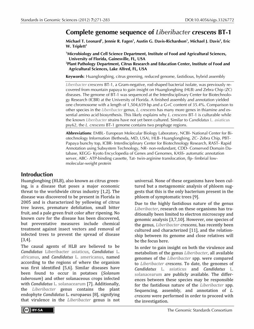
Standards in Genomic Sciences (2012) 7:271-283 DOI:10.4056/sigs.3326772
The Genomic Standards Consortium
Complete genome sequence of Liberibacter crescens BT-1 Michael T. Leonard1, Jennie R. Fagen1, Austin G. Davis-Richardson1, Michael J. Davis2, Eric W. Triplett1 1Microbiology and Cell Science Department, Institute of Food and Agricultural Sciences,
University of Florida, Gainesville, FL, USA 2Plant Pathology Department, Citrus Research and Education Center, Institute of Food and
Agricultural Sciences, Lake Alfred, FL, USA
Keywords: Huanglongbing, citrus greening, reduced genome, fastidious, hybrid assembly
Liberibacter crescens BT-1, a Gram-negative, rod-shaped bacterial isolate, was previously re-covered from mountain papaya to gain insight on Huanglongbing (HLB) and Zebra Chip (ZC) diseases. The genome of BT-1 was sequenced at the Interdisciplinary Center for Biotechnolo-gy Research (ICBR) at the University of Florida. A finished assembly and annotation yielded one chromosome with a length of 1,504,659 bp and a G+C content of 35.4%. Comparison to other species in the Liberibacter genus, L. crescens has many more genes in thiamine and es-sential amino acid biosynthesis. This likely explains why L. crescens BT-1 is culturable while the known Liberibacter strains have not yet been cultured. Similar to Candidatus L. asiaticus psy62, the L. crescens BT-1 genome contains two prophage regions.
Abbreviations: EMBL- European Molecular Biology Laboratory, NCBI- National Center for Bi-otechnology Information (Bethesda, MD, USA), HLB- Huanglongbing, ZC- Zebra Chip, PBT- Papaya bunchy top, ICBR- Interdisciplinary Center for Biotechnology Research, RAST- Rapid Annotation using Subsystem Technology, NR- non-redundant, CDD- Conserved Domain Da-tabase, KEGG- Kyoto Encyclopedia of Genes and Genomes, KASS- automatic annotation server, ABC- ATP-binding cassette, Tat- twin-arginine translocation, flp- fimbrial low-molecular-weight protein
Introduction Huanglongbing (HLB), also known as citrus green-ing, is a disease that poses a major economic threat to the worldwide citrus industry [1,2]. The disease was discovered to be present in Florida in 2005 and is characterized by yellowing of citrus tree leaves, premature defoliation, small bitter fruit, and a pale green fruit color after ripening. No known cure for the disease has been discovered, but preventative measures include chemical treatment against insect vectors and removal of infected trees to prevent the spread of disease [3,4]. The causal agents of HLB are believed to be Candidatus Liberibacter asiaticus, Candidatus L. africanus, and Candidatus L. americanus, named according to the regions of where the organism was first identified [5,6]. Similar diseases have been found to occur in potatoes (Solanum tuberosum) and other solanaceous crops infected with Candidatus L. solanacearum [7]. Additionally, the Liberibacter genus contains the plant endophyte Candidatus L. europaeus [8], signifying that virulence in the Liberibacter genus is not
universal. None of these organisms have been cul-tured but a metagenomic analysis of phloem sug-gests that this is the only bacterium present in the phloem of symptomatic trees [9]. Due to the highly fastidious nature of the genus Liberibacter, research on these organisms has tra-ditionally been limited to electron microscopy and genomic analysis [3,7,10]. However, one species of the genus, Liberibacter crescens, has recently been cultured and characterized [11], and the relation-ship between its genome and close relatives will be the focus here. In order to gain insight on both the virulence and metabolism of the genus Liberibacter, all available genomes of the Liberibacter spp. were compared to Liberibacter crescens. To date, the genomes of Candidatus L. asiaticus and Candidatus L. solanacearum are publicly available. The differ-ences between these species may be responsible for the fastidious nature of the Liberibacter spp. Sequencing, assembly, and annotation of L. crescens were performed in order to proceed with the investigation.
Liberibacter crescens BT-1
272 Standards in Genomic Sciences
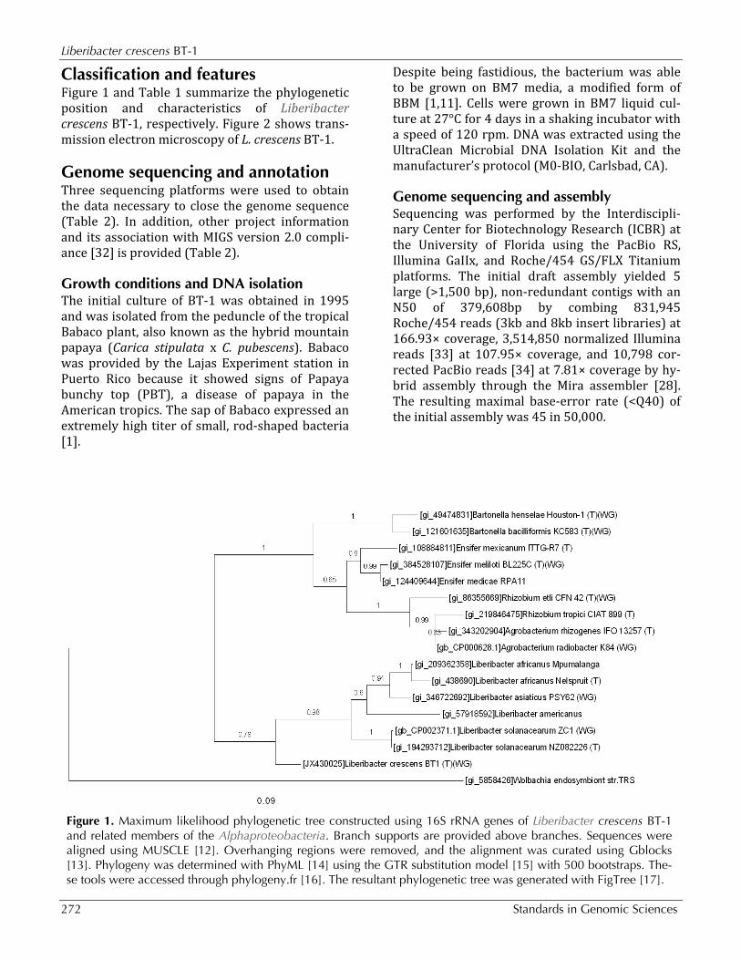
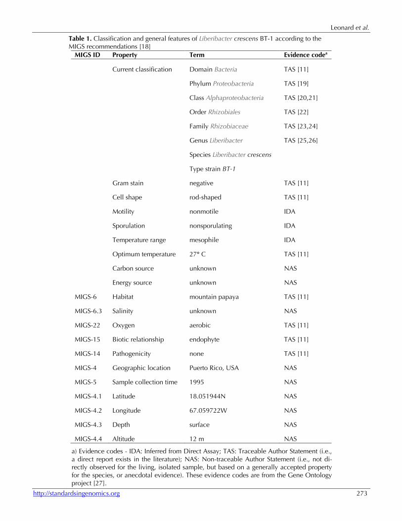
Classification and features Figure 1 and Table 1 summarize the phylogenetic position and characteristics of Liberibacter crescens BT-1, respectively. Figure 2 shows trans-mission electron microscopy of L. crescens BT-1.
Genome sequencing and annotation Three sequencing platforms were used to obtain the data necessary to close the genome sequence (Table 2). In addition, other project information and its association with MIGS version 2.0 compli-ance [32] is provided (Table 2).
Growth conditions and DNA isolation The initial culture of BT-1 was obtained in 1995 and was isolated from the peduncle of the tropical Babaco plant, also known as the hybrid mountain papaya (Carica stipulata x C. pubescens). Babaco was provided by the Lajas Experiment station in Puerto Rico because it showed signs of Papaya bunchy top (PBT), a disease of papaya in the American tropics. The sap of Babaco expressed an extremely high titer of small, rod-shaped bacteria [1].
Despite being fastidious, the bacterium was able to be grown on BM7 media, a modified form of BBM [1,11]. Cells were grown in BM7 liquid cul-ture at 27°C for 4 days in a shaking incubator with a speed of 120 rpm. DNA was extracted using the UltraClean Microbial DNA Isolation Kit and the manufacturer’s protocol (M0-BIO, Carlsbad, CA).
Genome sequencing and assembly Sequencing was performed by the Interdiscipli-nary Center for Biotechnology Research (ICBR) at the University of Florida using the PacBio RS, Illumina GaIIx, and Roche/454 GS/FLX Titanium platforms. The initial draft assembly yielded 5 large (>1,500 bp), non-redundant contigs with an N50 of 379,608bp by combing 831,945 Roche/454 reads (3kb and 8kb insert libraries) at 166.93× coverage, 3,514,850 normalized Illumina reads [33] at 107.95× coverage, and 10,798 cor-rected PacBio reads [34] at 7.81× coverage by hy-brid assembly through the Mira assembler [28]. The resulting maximal base-error rate (<Q40) of the initial assembly was 45 in 50,000.
Figure 1. Maximum likelihood phylogenetic tree constructed using 16S rRNA genes of Liberibacter crescens BT-1 and related members of the Alphaproteobacteria. Branch supports are provided above branches. Sequences were aligned using MUSCLE [12]. Overhanging regions were removed, and the alignment was curated using Gblocks [13]. Phylogeny was determined with PhyML [14] using the GTR substitution model [15] with 500 bootstraps. The-se tools were accessed through phylogeny.fr [16]. The resultant phylogenetic tree was generated with FigTree [17].
Leonard et al.
http://standardsingenomics.org 273
Table 1. Classification and general features of Liberibacter crescens BT-1 according to the MIGS recommendations [18]
MIGS ID Property Term Evidence codea
Current classification Domain Bacteria TAS [11]
Phylum Proteobacteria TAS [19]
Class Alphaproteobacteria TAS [20,21]
Order Rhizobiales TAS [22]
Family Rhizobiaceae TAS [23,24]
Genus Liberibacter TAS [25,26]
Species Liberibacter crescens
Type strain BT-1
Gram stain negative TAS [11]
Cell shape rod-shaped TAS [11]
Motility nonmotile IDA
Sporulation nonsporulating IDA
Temperature range mesophile IDA
Optimum temperature 27° C TAS [11]
Carbon source unknown NAS
Energy source unknown NAS
MIGS-6 Habitat mountain papaya TAS [11]
MIGS-6.3 Salinity unknown NAS
MIGS-22 Oxygen aerobic TAS [11]
MIGS-15 Biotic relationship endophyte TAS [11]
MIGS-14 Pathogenicity none TAS [11]
MIGS-4 Geographic location Puerto Rico, USA NAS
MIGS-5 Sample collection time 1995 NAS
MIGS-4.1 Latitude 18.051944N NAS
MIGS-4.2 Longitude 67.059722W NAS
MIGS-4.3 Depth surface NAS
MIGS-4.4 Altitude 12 m NAS
a) Evidence codes - IDA: Inferred from Direct Assay; TAS: Traceable Author Statement (i.e., a direct report exists in the literature); NAS: Non-traceable Author Statement (i.e., not di-rectly observed for the living, isolated sample, but based on a generally accepted property for the species, or anecdotal evidence). These evidence codes are from the Gene Ontology project [27].
Liberibacter crescens BT-1
274 Standards in Genomic Sciences
Figure 2. Transmission electron microscopy of L. crescens BT-1. Negative stain. Scale bar represents 500 nm.
Table 2. Project information MIGS ID Property Term
MIGS-31 Finishing quality Finished
MIGS-28 Libraries used Three libraries: one illumina library, two 454 paired-end libraries (3kb and 8kb insert size), one PacBio standard library
MIGS-29 Sequencing platforms Illumina GAIIx, 454 GS FLX Titanium, PacBio RS
MIGS-31.2 Fold coverage 6121.2x illumina, 166.9× 454, 31.7× PacBio
MIGS-30 Assemblers Mira v3.4.0.1 [28]; Amos v3.0.0 [29]; WGS v7.0.1.22 [30]; Velvet v1.2.06 [31]
MIGS-32 Gene calling method Glimmer [31]
Genome Database release
Genbank ID CP003789
Genbank Date of Release On publication, or August 31, 2013
GOLD ID
Project relevance Agriculture
Leonard et al.
http://standardsingenomics.org 275
Contigs were subject to an NcoI restriction digest (in silico) and mapped to an OpGen optical map of BT-1 using the same enzyme [35]. Optical mapping yielded a circular map of approximately 1.5 Mbp. Misjoined contigs and contig redundancy were in-dicated by comparison of the scaffold to the optical map of L. crescens, and were manually corrected with the CLC Genomics Workbench (CLCbio, Katrinebjerg, Denmark). Intrascaffold gaps were closed by further passes of the Mira hybrid assembly combining the current scaffold with varying combinations of read data. Omitting certain read technologies at further hy-brid assembly iterations allowed more successful assemblies at different points of the genome. Pseu-do 454-like paired-end reads were generated from the scaffold to allow very large contigs to be em-ployed in further iterations of Mira hybrid assem-bly. Pseudo 454-like reads conformed to the 19 kb upper limit of Mira read length and consisted of a 34 kb insert size. Additionally, subsets of the origi-nal Illumina paired-end reads and normalized Roche/454 reads were entered into the read pool to avoid problematic reads. Contigs of each hybrid assembly pass were manually corrected for misjoined contigs and combined by Minimus2 [29] to yield a circular genomic sequence.
Genome annotation Genome annotation was performed by the Rapid Annotation using Subsystem Technology (RAST)
pipeline [36]. RAST employs tRNAscan-SE [37] to identify tRNA genes, Niels Larsen’s "search_for_rnas" (available from the author) to identify rRNA encod-ing genes, and GLIMMER [38] to identify candidate protein-encoding genes. RAST compares the set of candidate protein-encoding genes to a collection of protein families, referred to as FIGfams [36], in or-der to correct CDS starting positions and place the genome in a phylogenic context. The candidate pro-tein set was compared to the National Center for Biotechnology Information (NCBI) non-redundant (nr) database, SwissProt database, European Bioin-formatics Institute (EBI) phage database, and COG subset of the NCBI Conserved Domain Database (CDD) through the NCBI BLAST suite. Additionally, predicted proteins were annotated through the Kyoto Encyclopedia of Genes and Ge-nomes (KEGG) automatic annotation server (KAAS). KAAS employs NCBI BLAST to search the KEGG Orthology database [39].
Genome properties The genome consists of one circular chromosome of 1,504,659 bp (35.35% GC content). 1,433 genes were predicted, 1,379 of which are protein-coding genes. 1,039 of protein coding genes were as-signed to a putative function with the remaining being annotated as hypothetical proteins. The properties and the statistics of the genome are summarized in Tables 3 and 4.
Table 3. Nucleotide content and gene count levels of the genome Attribute Value % of totala
Genome size (bp) 1,504,659 100
DNA coding region (bp) 1,264,794 84.05
DNA G+C content (bp) 531,980 35.35
Total genesb 1433 100
RNA genes 54 3.77
Protein-coding genes 1379 96.23
Genes in paralog clusters 870 63.08
Genes assigned to COGs 857 62.14
Genes assigned Pfam domains 1057 76.65
Genes with signal peptides 84 6.09
Genes with transmembrane helices 327 23.71
a) The total is based on either the size of the genome in base pairs or the total number of protein coding genes in the annotated ge-nome.
b) Does not include pseudogenes or other genes.
Liberibacter crescens BT-1
276 Standards in Genomic Sciences
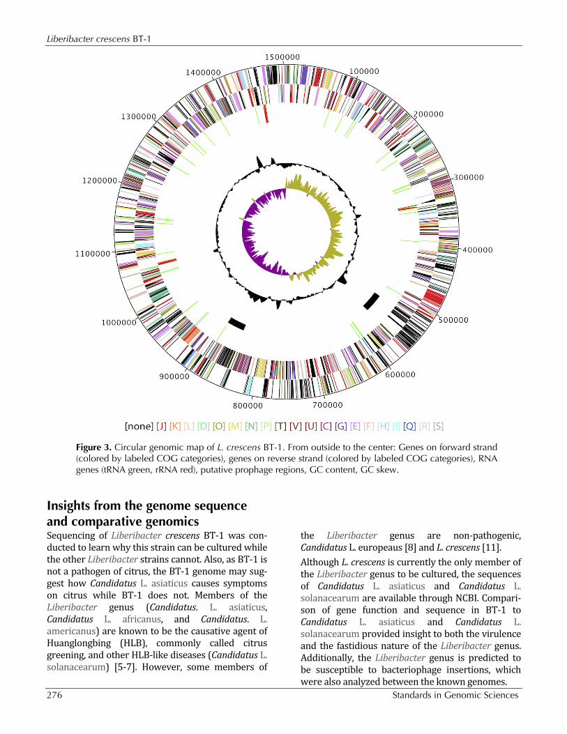
Figure 3. Circular genomic map of L. crescens BT-1. From outside to the center: Genes on forward strand (colored by labeled COG categories), genes on reverse strand (colored by labeled COG categories), RNA genes (tRNA green, rRNA red), putative prophage regions, GC content, GC skew.
Insights from the genome sequence and comparative genomics Sequencing of Liberibacter crescens BT-1 was con-ducted to learn why this strain can be cultured while the other Liberibacter strains cannot. Also, as BT-1 is not a pathogen of citrus, the BT-1 genome may sug-gest how Candidatus L. asiaticus causes symptoms on citrus while BT-1 does not. Members of the Liberibacter genus (Candidatus. L. asiaticus, Candidatus L. africanus, and Candidatus. L. americanus) are known to be the causative agent of Huanglongbing (HLB), commonly called citrus greening, and other HLB-like diseases (Candidatus L. solanacearum) [5-7]. However, some members of
the Liberibacter genus are non-pathogenic, Candidatus L. europeaus [8] and L. crescens [11]. Although L. crescens is currently the only member of the Liberibacter genus to be cultured, the sequences of Candidatus L. asiaticus and Candidatus L. solanacearum are available through NCBI. Compari-son of gene function and sequence in BT-1 to Candidatus L. asiaticus and Candidatus L. solanacearum provided insight to both the virulence and the fastidious nature of the Liberibacter genus. Additionally, the Liberibacter genus is predicted to be susceptible to bacteriophage insertions, which were also analyzed between the known genomes.
Leonard et al.
http://standardsingenomics.org 277
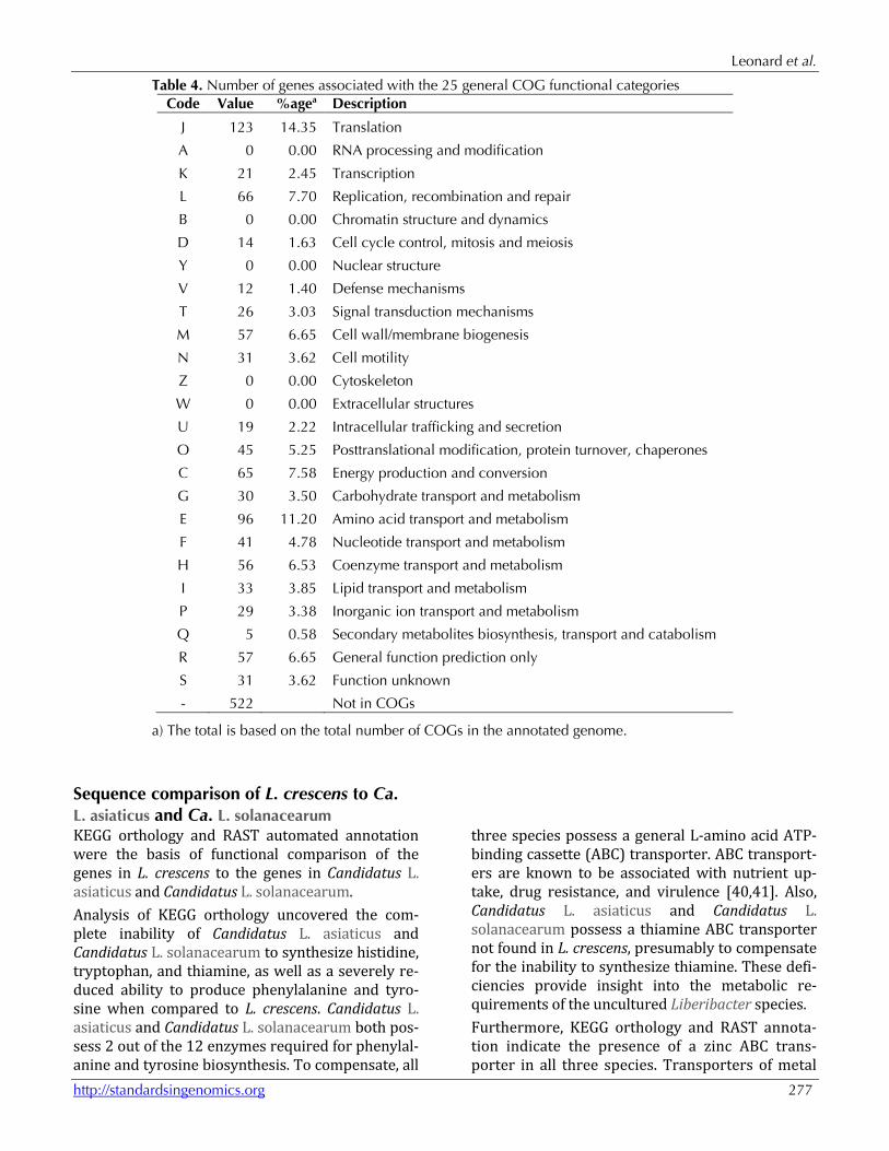
Table 4. Number of genes associated with the 25 general COG functional categories Code Value %agea Description
J 123 14.35 Translation
A 0 0.00 RNA processing and modification
K 21 2.45 Transcription
L 66 7.70 Replication, recombination and repair
B 0 0.00 Chromatin structure and dynamics
D 14 1.63 Cell cycle control, mitosis and meiosis
Y 0 0.00 Nuclear structure
V 12 1.40 Defense mechanisms
T 26 3.03 Signal transduction mechanisms
M 57 6.65 Cell wall/membrane biogenesis
N 31 3.62 Cell motility
Z 0 0.00 Cytoskeleton
W 0 0.00 Extracellular structures
U 19 2.22 Intracellular trafficking and secretion
O 45 5.25 Posttranslational modification, protein turnover, chaperones
C 65 7.58 Energy production and conversion
G 30 3.50 Carbohydrate transport and metabolism
E 96 11.20 Amino acid transport and metabolism
F 41 4.78 Nucleotide transport and metabolism
H 56 6.53 Coenzyme transport and metabolism
I 33 3.85 Lipid transport and metabolism
P 29 3.38 Inorganic ion transport and metabolism
Q 5 0.58 Secondary metabolites biosynthesis, transport and catabolism
R 57 6.65 General function prediction only
S 31 3.62 Function unknown
- 522 Not in COGs
a) The total is based on the total number of COGs in the annotated genome.
Sequence comparison of L. crescens to Ca. L. asiaticus and Ca. L. solanacearum KEGG orthology and RAST automated annotation were the basis of functional comparison of the genes in L. crescens to the genes in Candidatus L. asiaticus and Candidatus L. solanacearum. Analysis of KEGG orthology uncovered the com-plete inability of Candidatus L. asiaticus and Candidatus L. solanacearum to synthesize histidine, tryptophan, and thiamine, as well as a severely re-duced ability to produce phenylalanine and tyro-sine when compared to L. crescens. Candidatus L. asiaticus and Candidatus L. solanacearum both pos-sess 2 out of the 12 enzymes required for phenylal-anine and tyrosine biosynthesis. To compensate, all
three species possess a general L-amino acid ATP-binding cassette (ABC) transporter. ABC transport-ers are known to be associated with nutrient up-take, drug resistance, and virulence [40,41]. Also, Candidatus L. asiaticus and Candidatus L. solanacearum possess a thiamine ABC transporter not found in L. crescens, presumably to compensate for the inability to synthesize thiamine. These defi-ciencies provide insight into the metabolic re-quirements of the uncultured Liberibacter species. Furthermore, KEGG orthology and RAST annota-tion indicate the presence of a zinc ABC trans-porter in all three species. Transporters of metal
Liberibacter crescens BT-1
278 Standards in Genomic Sciences
ions have been shown to play a role in bacterial virulence, including ABC transporters of iron, zinc, and manganese [42,43]. Although the zinc trans-porter was located in L. crescens through RAST annotation, it was not detected by KEGG orthology. This discrepancy is attributed to a low sequence similarity between the protein compo-nents of the zinc ABC transporter (ZnuA, ZnuB, ZnuC) in L. crescens compared to Candidatus L. asiaticus and Ca. L. solanacearum, at 43.6%,
55.3%, and 48.5% average similarity for each component, respectively (Table 5). In contrast, the similarity of each component between Candidatus L. asiaticus and Candidatus L. solanacearum is 78.6%, 93.0%, and 92.2% respectively (Table 5). Sequence similarity was determined through se-quence alignment using the EMBOSS Water tool [44] and the EBLOSUM62 scoring matrix. This var-iation in zinc ABC transport proteins may contrib-ute to the virulence of the Liberibacter genus.
Table 5. Species similarity of zinc ABC transporter components ZnuA ZnuB ZnuC L. crescens to Candidatus L. asiaticus 43.1% 55.2% 46.9% L. crescens to Candidatus L. solanacearum 44.0% 55.4% 50.0%
Average 43.6% 55.3% 48.5% Ca. L. asiaticus to Ca. L. solanacearum 78.6% 93.0% 92.2%
Also present in L. crescens, but not in Candidatus L. asiaticus and Candidatus L. solanacearum, is a twin-arginine translocation (Tat) protein export pathway and an additional iron ABC transporter. The significance of these two transporters is not currently known, but their existence may explain why L. crescens, is less fastidious than Candidatus L. asiaticus and Candidatus L. solanacearum.
Present in Candidatus L. asiaticus and Candidatus L. solanacearum, but not in L. crescens, are several components of a fimbrial low-molecular-weight protein (flp) pilus system. These pili are involved in tight adherence and are encoded by the Tad family proteins [7]. Diversity in the flp pilus oper-on is predicted to contribute to variation in viru-lence among pathogenic species [45-48], and pro-vides further insight to the virulence of the Liberibacter genus.
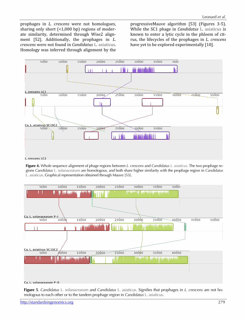
Phages in the genomes of Candidatus L. asiaticus and L. crescens Recently, two prophages, SC1 and SC2, were found to exist in tandem in Candidatus L. asiaticus through DNA isolation from diseased citrus phlo-em and an insect vector of the family Psyllidae [10]. Candidatus L. solanacearum is known to host two prophage regions as well, not in tandem, with one region maintaining a high degree of similarity with the prophage regions in Candidatus L. asiaticus and the other containing a small segment with lower similarity [7]. Two putative prophages
were found in the L. crescens genome through the use of the Prophage Finder tool [49], the Phage_Finder [50] tool, and the methods de-scribed in Casjens et al (2003).
Prophage boundary identification is an inexact process due to the diversity of bacteriophages, and is made even more difficult by the possibility of evolutionary decay of prophages that do not enter a lytic cycle. Additionally, prophage bounda-ries are indicated by a multitude of factors, but not defined by any particular criteria. Position of nearby tRNAs close to the predicted prophage re-gion may be indicative of a boundary, as tRNAs are often sites of phage insertion [50]. A sharp shift in G+C content at the predicted prophage region may also indicate the range of phage insertion, but only if the phage G+C content differs dramatically from the host. Certain genes are unique to phage ge-nomes, and non-phage genes were not typically found to be present between phage genes in an inserted phage. From a genomic standpoint, prophage regions are also indicated by regions not present in closely related species, as well as long strings of unidentified proteins in similar orienta-tion [51]. From the above criteria, the locations and bound-aries of two prophages in L. crescens were pre-dicted to extend from base pair 523,789-564,039 in prophage LC1 and from base pair 848,435-886,798 in prophage LC2. Unlike the two prophages in Candidatus L. asiaticus, the
Leonard et al.
http://standardsingenomics.org 279
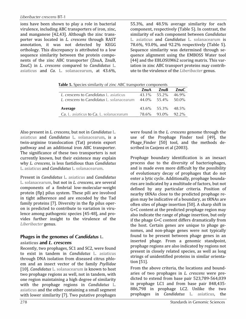
prophages in L. crescens were not homologues, sharing only short (<1,000 bp) regions of moder-ate similarity, determined through Wise2 align-ment [52]. Additionally, the prophages in L. crescens were not found in Candidatus L. asiaticus. Homology was inferred through alignment by the
progressiveMauve algorithm [53] (Figures 3-5). While the SC1 phage in Candidatus L. asiaticus is known to enter a lytic cycle in the phloem of cit-rus, the lifecycles of the prophages in L. crescens have yet to be explored experimentally [10].
Figure 4. Whole sequence alignment of phage regions between L. crescens and Candidatus L. asiaticus. The two prophage re-gions Candidatus L. solanacearum are homologous, and both share higher similarity with the prophage region in Candidatus L. asiaticus. Graphical representation obtained through Mauve [53].
Figure 5. Candidatus L. solanacearum and Candidatus L. asiaticus. Signifies that prophages in L. crescens are not ho-mologous to each other or to the tandem prophage region in Candidatus L. asiaticus.
Liberibacter crescens BT-1
280 Standards in Genomic Sciences
Interestingly, the same zinc ABC transporter men-tioned above is present in the LC2 region. Prophage insertions have been known to add functions to hosts, making the host more competi-tive [54]. In addition to metabolic variation, the differences in extra-chromosomal genomic con-tent between species of the Liberibacter genus may also be indicative of the virulence and fastidi-ous nature of the genus.
Conclusion Liberibacter crescens BT-1 is the first member of the Liberibacter genus to be cultured. The com-plete genome sequences of Candidatus L. asiaticus and Candidatus L. solanacearum have been deter-mined through isolation from the disease vectors [7,9], but any attempt to culture these species typ-ically depends on employing a co-culture with in-sect or plant host cells [5]. Genomic sequencing of L. crescens BT-1 was performed in an attempt to find possible indications for virulence in Candidatus L. asiaticus and Candidatus L.
solanacearum, as well as an explanation for the fastidious nature of these pathogens. Assembly of L. crescens yielded a complete ge-nome containing two predicted prophages. Se-quence comparison of Candidatus L. asiaticus to L. crescens indicated that the species are 75.5% simi-lar [11]. However, the prophage regions are not homologous. Sequencing and analysis of the L. crescens genome provided insight to the metabolic requirements of Candidatus L. asiaticus, which ap-pears to lack the ability to synthesize thiamine and several essential amino acids. Less is known about the virulence of Candidatus L. asiaticus, alt-hough bacteriophages have also been known to add virulence to an otherwise non-pathogenic bacterium [54]. Further genomic analysis indicat-ed that virulence in Candidatus L. asiaticus could also be due to a zinc ABC transporter. While the sequencing of L. crescens gave much insight into the Liberibacter genus, further experiments must be conducted to verify these predictions.
References 1. Davis MJ, Mondal SN, Chen H, Rogers ME,
Brlansky RH. Co-cultivation of ‘ Candidatus Liberibacter asiaticus ’ with Actinobacteria from Citrus with Huanglongbing. Plant Dis 2008; 92:1547-1550. http://dx.doi.org/10.1094/PDIS-92-11-1547
2. Kapur SP, Kapoor SK, Cheema SS, Dhillon RS. Effect of greening disease on tree and fruit charac-ters of Kinnow mandarin. Punjab Horticultural Journal 1978; 50:76-79.
3. Bové J. Huanglongbing: A Destructive, Newly-Emerging, Century-Old Disease of Citrus. J Plant Pathol 2006; 88:7-37.
4. Zhang M, Powell CA, Guo Y, Doud MS, Duan Y. A Graft-Based Chemotherapy Method for Screen-ing Effective Molecules and Rescuing Huanglongbing-Affected Citrus Plants. Phyto-pathology 2012; 102:567-574. PubMed http://dx.doi.org/10.1094/PHYTO-09-11-0265
5. Sechler A, Schuenzel EL, Cooke P, Donnua S, Thaveechai N, Postnikova E. Stone aL, Schneider WL, Damsteegt VD, Schaad NW. Cultivation of 'Candidatus Liberibacter asiaticus', 'Ca. L. africanus', and 'Ca. L. americanus' associated with huanglongbing. Phytopathology 2009; 99:480-486. PubMed http://dx.doi.org/10.1094/PHYTO-99-5-0480
6. Teixeira DC. 'Candidatus Liberibacter americanus', associated with citrus huanglongbing (greening disease) in Sao Paulo State, Brazil. Int J Syst Evol Microbiol 2005; 55:1857-1862. PubMed http://dx.doi.org/10.1099/ijs.0.63677-0
7. Lin H, Lou B, Glynn JM, Doddapaneni H, Civerolo EL, Chen C, Duan Y, Zhou L, Vahling CM. The Complete Genome Sequence of ‘Candidatus Liberibacter solanacearum’, the Bac-terium Associated with Potato Zebra Chip Dis-ease. PLoS ONE 2011; 6:e19135. PubMed http://dx.doi.org/10.1371/journal.pone.0019135
8. Raddadi N, Gonella E, Camerota C, Pizzinat A, Tedeschi R, Crotti E, Mandrioli M, Attilio Bianco P, Daffonchio D, Alma A. ‘Candidatus Liberibacter europaeus’ sp. nov. that is associated with and transmitted by the psyllid Cacopsylla pyri apparently behaves as an endophyte rather than a pathogen. Environ Microbiol 2011; 13:414-426. PubMed http://dx.doi.org/10.1111/j.1462-2920.2010.02347.x
9. Tyler HL, Roesch LF, Gowda S, Dawson WO, Triplett EW. Confirmation of the sequence of 'Candidatus Liberibacter asiaticus' and assess-ment of microbial diversity in Huanglongbing-infected citrus phloem using a metagenomic ap-
Leonard et al.
http://standardsingenomics.org 281
proach. Mol Plant Microbe Interact 2009; 22:1624-1634. PubMed http://dx.doi.org/10.1094/MPMI-22-12-1624
10. Zhang S, Flores-Cruz Z, Zhou L, Kang BH. Fleites La, Gooch MD, Wulff Na, Davis MJ, Duan Y-P, Gabriel DW. 'Ca. Liberibacter asiaticus' carries an excision plasmid prophage and a chromoso-mally integrated prophage that becomes lytic in plant infections. Molecular plant-microbe interac-tions. Mol Plant Microbe Interact 2011; 24:458-468. PubMed http://dx.doi.org/10.1094/MPMI-11-10-0256
11. Fagen J, Leonard MT, McCullough CM, Triplett EW, Davis MJ. Liberibacter crescens gen.nov; sp. nov. first cultured member of the Liberibacter ge-nus. International Journal of Systematic and Evo-lutionary Bacteriology 2012.
12. Edgar RC. MUSCLE: multiple sequence alignment with high accuracy and high throughput. Nucleic Acids Res 2004; 32:1792-1797. PubMed http://dx.doi.org/10.1093/nar/gkh340
13. Castresana J. Selection of conserved blocks from multiple alignments for their use in phylogenetic analysis. Mol Biol Evol 2000; 17:540-552. Pub-Med http://dx.doi.org/10.1093/oxfordjournals.molbev.a026334
14. Guindon S, Gascuel OA. Simple, Fast, and Accu-rate Algorithm to Estimate Large Phylogenies by Maximum Likelihood. Syst Biol 2003; 52:696-704. PubMed http://dx.doi.org/10.1080/10635150390235520
15. Anisimova M, Gascuel O. Approximate likeli-hood-ratio test for branches: A fast, accurate, and powerful alternative. Syst Biol 2006; 55:539-552. PubMed http://dx.doi.org/10.1080/10635150600755453
16. Dereeper A, Guignon V, Blanc G, Audic S, Buffet S, Chevenet F, Dufayard JF, Guindon S, Lefort V, Lescot M, et al. Phylogeny.fr: robust phylogenetic analysis for the non-specialist. Nucleic Acids Res 2008; 36:W465-W469. PubMed http://dx.doi.org/10.1093/nar/gkn180
17. Rambaut A. FigTree. <http://tree.bio.ed.ac.uk/software/figtree/%3E.
18. Field D, Garrity G, Gray T, Morrison N, Selengut J, Sterk P, Tatusova T, Thomson N, Allen MJ, Angiuoli SV, et al. The minimum information about a genome sequence (MIGS) specification. Nat Biotechnol 2008; 26:541-547. PubMed http://dx.doi.org/10.1038/nbt1360
19. Garrity GM, Bell JA, Lilburn T. Phylum XIV. Proteobacteria phyl. nov. In: Garrity GM, Brenner DJ, Krieg NR, Staley JT (eds), Bergey's Manual of Systematic Bacteriology, Second Edition, Volume 2, Part B, Springer, New York, 2005, p. 1.
20. Validation List No. 107. List of new names and new combinations previously effectively, but not validly, published. Int J Syst Evol Microbiol 2006; 56:1-6. PubMed http://dx.doi.org/10.1099/ijs.0.64188-0
21. Garrity GM, Bell JA, Lilburn T. Class I. Alphaproteobacteria class. nov. In: Garrity GM, Brenner DJ, Krieg NR, Staley JT (eds), Bergey's Manual of Systematic Bacteriology, Second Edi-tion, Volume 2, Part C, Springer, New York, 2005, p. 1.
22. Kuykendall LD. Order VI. Rhizobiales ord. nov. In: Garrity GM, Brenner DJ, Krieg NR, Staley JT (eds), Bergey's Manual of Systematic Bacteriolo-gy, Second Edition, Volume 2, Part C, Springer, New York, 2005, p. 324.
23. Skerman VBD, McGowan V, Sneath PHA. Ap-proved Lists of Bacterial Names. Int J Syst Bacteriol 1980; 30:225-420. http://dx.doi.org/10.1099/00207713-30-1-225
24. Conn HJ. Taxonomic relationships of certain non-sporeforming rods in soil. J Bacteriol 1938; 36:320-321.
25. Jagoueix S, Bové JM, Garnier M. The Phloem-Limited Bacterium of Greening Disease of Citrus Is a Member of the alpha Subdivision of the Proteobacteria. Int J Syst Bacteriol 1994; 44:379-386. PubMed http://dx.doi.org/10.1099/00207713-44-3-379
26. Garnier M, Jagoueix-Eveillard S, Cronje PR, Le Roux HF, Bové JM. Genomic characterization of a liberibacter present in an ornamental rutaceous tree, Calodendrum capense, in the Western Cape Province of South Africa. Proposal of 'Candidatus Liberibacter africanus subsp. capensis'. Int J Syst Evol Microbiol 2000; 50:2119-2125. PubMed http://dx.doi.org/10.1099/00207713-50-6-2119
27. Ashburner M, Ball CA, Blake JA, Botstein D, But-ler H, Cherry JM, Davis AP, Dolinski K, Dwight SS, Eppig JT, et al. Gene Ontology: tool for the unification of biology. The Gene Ontology Con-sortium. Nat Genet 2000; 25:25-29. PubMed http://dx.doi.org/10.1038/75556
28. Chevreux B, Wetter T, Suhai S. Genome se-quence assembly using trace signals and addi-tional sequence information. … Science and Bi-ology: Proceedings of the … 1999.
Liberibacter crescens BT-1
282 Standards in Genomic Sciences
29. Sommer DD, Delcher AL, Salzberg SL, Pop M. Minimus: a fast, lightweight genome assembler. BMC Bioinformatics 2007; 8:64. PubMed http://dx.doi.org/10.1186/1471-2105-8-64
30. Myers EW. A Whole-Genome Assembly of Dro-sophila. Science 2000; 287:2196-2204. PubMed http://dx.doi.org/10.1126/science.287.5461.2196
31. Zerbino DR, Birney E. Velvet: algorithms for de novo short read assembly using de Bruijn graphs. Genome Res 2008; 18:821-829. PubMed http://dx.doi.org/10.1101/gr.074492.107
32. Field D, Garrity G, Gray T, Morrison N, Selengut J, Sterk P, Tatusova T, Thomson N, Allen MJ, Angiuoli SV, et al. The minimum information about a genome sequence (MIGS) specification. Nat Biotechnol 2008; 26:541-547. PubMed http://dx.doi.org/10.1038/nbt1360
33. Brown CT, Howe A, Zhang Q, Pyrkosz AB, Brom TH. A Reference-Free Algorithm for Computa-tional Normalization of Shotgun Sequencing Da-ta. eprint arXiv 2012;1203:1-18.
34. Koren S, Schatz MC, Walenz BP, Martin J, How-ard JT, Ganapathy G, Wang Z. Rasko Da, McCombie WR, Jarvis ED and others. Hybrid er-ror correction and de novo assembly of single-molecule sequencing reads. Nat Biotechnol 2012; 30:693-700. PubMed http://dx.doi.org/10.1038/nbt.2280
35. Latreille P, Norton S, Goldman BS, Henkhaus J, Miller N, Barbazuk B, Bode HB, Darby C, Du Z, Forst S, et al. Optical mapping as a routine tool for bacterial genome sequence finishing. BMC Genomics 2007; 8:321. PubMed http://dx.doi.org/10.1186/1471-2164-8-321
36. Aziz RK, Bartels D, Best AA, DeJongh M, Disz T, Edwards RA, Formsma K, Gerdes S, Glass EM, Kubal M, et al. The RAST Server: rapid annota-tions using subsystems technology. BMC Ge-nomics 2008; 9:75. PubMed http://dx.doi.org/10.1186/1471-2164-9-75
37. Lowe TM, Eddy SR. tRNAscan-SE: a program for improved detection of transfer RNA genes in ge-nomic sequence. Nucleic Acids Res 1997; 25:955-964. PubMed
38. Salzberg SL. Delcher aL, Kasif S, White O. Micro-bial gene identification using interpolated Markov models. Nucleic Acids Res 1998; 26:544-548. PubMed http://dx.doi.org/10.1093/nar/26.2.544
39. Moriya Y, Itoh M, Okuda S, Yoshizawa AC, Kanehisa M. KAAS: an automatic genome annota-tion and pathway reconstruction server. Nucleic
Acids Res 2007; 35:W182-W185. PubMed http://dx.doi.org/10.1093/nar/gkm321
40. Garmory HS, Titball RW. MINIREVIEW ATP-Binding Cassette Transporters Are Targets for the Development of Antibacterial Vaccines and Ther-apies. Infect Immun 2004; 72:6757-6763. Pub-Med http://dx.doi.org/10.1128/IAI.72.12.6757-6763.2004
41. Testerman TL, Conn PB, Mobley HLT, Mcgee DJ. Nutritional Requirements and Antibiotic Re-sistance Patterns of Helicobacter Species in Chemically Defined Media Nutritional Require-ments and Antibiotic Resistance Patterns of Heli-cobacter Species in Chemically Defined Media. Infect Immun 2006.
42. Hantke K. Bacterial zinc uptake and regulators. Curr Opin Microbiol 2005; 8:196-202. PubMed http://dx.doi.org/10.1016/j.mib.2005.02.001
43. Panina EM. Comparative genomics of bacterial zinc regulons: Enhanced ion transport, pathogen-esis, and rearrangement of ribosomal proteins. Proc Natl Acad Sci USA 2003; 100:9912-9917. PubMed http://dx.doi.org/10.1073/pnas.1733691100
44. Rice P, Longden I, Bleasby A. EMBOSS: the Euro-pean Molecular Biology Open Software Suite. Trends Genet 2000; 16:276-277. PubMed http://dx.doi.org/10.1016/S0168-9525(00)02024-2
45. Boyd JM, Dacanay A, Knickle LC, Touhami A, Brown LL, Jericho MH, Johnson SC, Reith M. Contribution of Type IV Pili to the Virulence of Aeromonas salmonicida subsp. salmonicida in At-lantic Salmon (Salmo salar L.). Infect Immun 2008; 76:1445-1455. PubMed http://dx.doi.org/10.1128/IAI.01019-07
46. Clock SA, Planet PJ, Perez BA, Figurski DH. Outer Membrane Components of the Tad (Tight Adher-ence) Secreton of Aggregatibacter actinomycetemcomitans. J Bacteriol 2007; 190:980-990. PubMed http://dx.doi.org/10.1128/JB.01347-07
47. Li T, Xu Z, Zhang T, Li L, Chen H, Zhou R. The genetic analysis of the flp locus of Actinobacillus pleuropneumoniae. Arch Microbiol 2011; 194:167-176. PubMed http://dx.doi.org/10.1007/s00203-011-0741-6
48. Wairuri CK, van der Waals JE, van Schalkwyk A, Theron J. Ralstonia solanacearum Needs Flp Pili for Virulence on Potato. Mol Plant Microbe Inter-act 2011; 25:546-556. PubMed http://dx.doi.org/10.1094/MPMI-06-11-0166
Leonard et al.
http://standardsingenomics.org 283
49. Bose M, Barber RD. Prophage Finder: a prophage loci prediction tool for prokaryotic genome se-quences. In Silico Biol 2006; 6:223-227. PubMed
50. Fouts DE. Phage_Finder: automated identification and classification of prophage regions in com-plete bacterial genome sequences. Nucleic Acids Res 2006; 34:5839-5851. PubMed http://dx.doi.org/10.1093/nar/gkl732
51. Casjens S. Prophages and bacterial genomics: what have we learned so far? Mol Microbiol 2003; 49:277-300. PubMed http://dx.doi.org/10.1046/j.1365-2958.2003.03580.x
52. Bateman A, Birney E, Durbin R, Eddy SR, Howe KL, Sonnhammer EL. The Pfam protein families
database. Nucleic Acids Res 2000; 28:263-266. PubMed http://dx.doi.org/10.1093/nar/28.1.263
53. Darling AE, Mau B, Perna NT. progressiveMauve: Multiple Genome Alignment with Gene Gain, Loss and Rearrangement. PLoS ONE 2010; 5:e11147. PubMed http://dx.doi.org/10.1371/journal.pone.0011147
54. Winstanley C, Langille MGI, Fothergill JL, Kukavica-ibrulj I, Paradis-bleau C, Sanschagrin F, Thomson NR, Winsor GL, Quail MA, Lennard N, et al. Newly introduced genomic prophage is-lands are critical determinants of in vivo competi-tiveness in the Liverpool Epidemic Strain of Pseu-domonas aeruginosa. Genome Res 2009; 19:12-23. PubMed http://dx.doi.org/10.1101/gr.086082.108