Embed Size (px)
Citation preview

Leukemia
Tuesday, February 26, 13

Leukemia • Acute – Acute Myelogenous Leukemia (AML)– Acute Lymphoblastic Leukemia (ALL)
• Chronic– Chronic Myelogenous Leukemia (CML)– Chronic Lymphocytic Leukemia (CLL)
• Hairy Cell Leukemia
Tuesday, February 26, 13

ACUTE MYELOGENOUS LEUKEMIA (AML)
Tuesday, February 26, 13

What is AML?Clonal, malignant disease of hematopoietic tissue characterized by:
1. Accumulation of abnormal blast cells, principally in the marrow
2. Impaired production of normal cells
Tuesday, February 26, 13

Tuesday, February 26, 13

Etiology and Pathogenesis
Tuesday, February 26, 13

Predisposing Conditions
• Environmental Factors
• Acquired Diseases
• Inherited or Congenital conditions
Tuesday, February 26, 13

Molecular Pathogenesis• Somatic mutation of hematopoietic
multipotent cell or a more differentiated, lineage-restricted progenitor cell
• Somatic mutation occurs from chromosomal translocation in majority of patients
Tuesday, February 26, 13

Tuesday, February 26, 13

Molecular Pathogenesis• Translocation occurs at region of
proto-oncogene
• Fusion of genes disrupts normal cell pathway and predisposes to a malignant transformation of the cell
• Mutant product often has a
Tuesday, February 26, 13

Examples of Mutated Genes • Core Binding Factor– 2 subunits• CBF- β• RUNX1
• Retinoic Acid receptor – α• HOX family• MLL
Tuesday, February 26, 13

Frequency of AML cases that involve genes listed previously differ according to age group
• Patient age > 50 yrs 20%
• Patient age < 50 yrs 6%
Tuesday, February 26, 13

• Core Binding Gene– Involved in myeloid or lymphoid
differentiation and maturation
• Primary mutation alone not sufficient to cause AML - additional activation mutations required
Tuesday, February 26, 13

Additional Proto-oncogene Mutations
• Hematopoietic Tyrosine Kinases FLT3 and Kit
• N-RAS and K-RAS• FES• FOS• GATA-1• JUN B• MPL
• WT1• WNT• N• P53• PU.1• RB• PM1
Tuesday, February 26, 13

• Interaction of proto-oncogene mutations with loss of function mutations in hematopoietic transcriptions cause acute leukemia phenotype
• Minimum of 2 gene classes proposed:– CLASS I (ex: RUNX1 gene)– CLASS II (ex: core binding factor)
Tuesday, February 26, 13

FLT3 • Encodes tyrosine kinase receptor in
normal myeloid and lymphoid progenitors.
• Internal tandem duplications on chromosome 13 occurs in approx. 25-33% of adult AML cases
Tuesday, February 26, 13
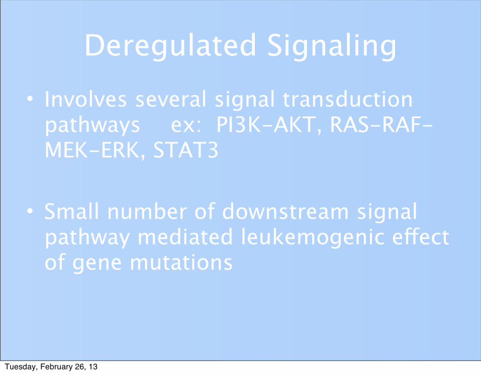
Deregulated Signaling • Involves several signal transduction
pathways ex: PI3K-AKT, RAS-RAF-MEK-ERK, STAT3
• Small number of downstream signal pathway mediated leukemogenic effect of gene mutations
Tuesday, February 26, 13

Mode of Inheritance• Little evidence seen for influence of
inherited factors
• Identical twin of patient with acute leukemia has heightened risk of developing disease
Tuesday, February 26, 13

Epidemiology• AML is predominant form of leukemia
during neonatal period.
• 15,000 new cases of AML annually
• 9,000 patients in US die each year
Tuesday, February 26, 13

AML Incidence rates• 2.3 per 100,000 per year
• Higher among men than women (2.9 v. 1.9)
• MC leukemia in adults (80% of cases)
• Majority of patients > 65 yrs of age
Tuesday, February 26, 13

Incidence Rates
Tuesday, February 26, 13

Signs and Symptoms• Pallor
• Fatigue (50%)
• Weakness
• Palpitations
• Dyspnea on exertion
• Bruising (5%)
• Anorexia (30-40%)
• Weight loss (30-40%)
• Gingival Bleeding
• Conjunctival Hemorrhages
• Pustules and minor pyogenic infections of skin
• Petechiae
• Epistaxis
• Fever
• Splenomegaly and Hepatomegaly (33% of patients)
Tuesday, February 26, 13

Signs and Symptoms• Cough• Diaphoresis• Headache• Bone Pain• Lymphadenopathy
• Sinusitis, pneumonia, pyelonephritis and meningitis uncommon until chemotherapy
Tuesday, February 26, 13

Diagnosis• CBC with peripheral blood smear
• Bone Marrow aspirate and biopsy
• Chest x-ray
• Histochemical Studies
• Cytogenetics and Immunophenotyping
• Clotting Studies (PT, PTT, D-dimer, fibrinogen)
Tuesday, February 26, 13

Hematological Findings
• Anemia
• Thrombocytopenia
• Leukocytosis
Tuesday, February 26, 13

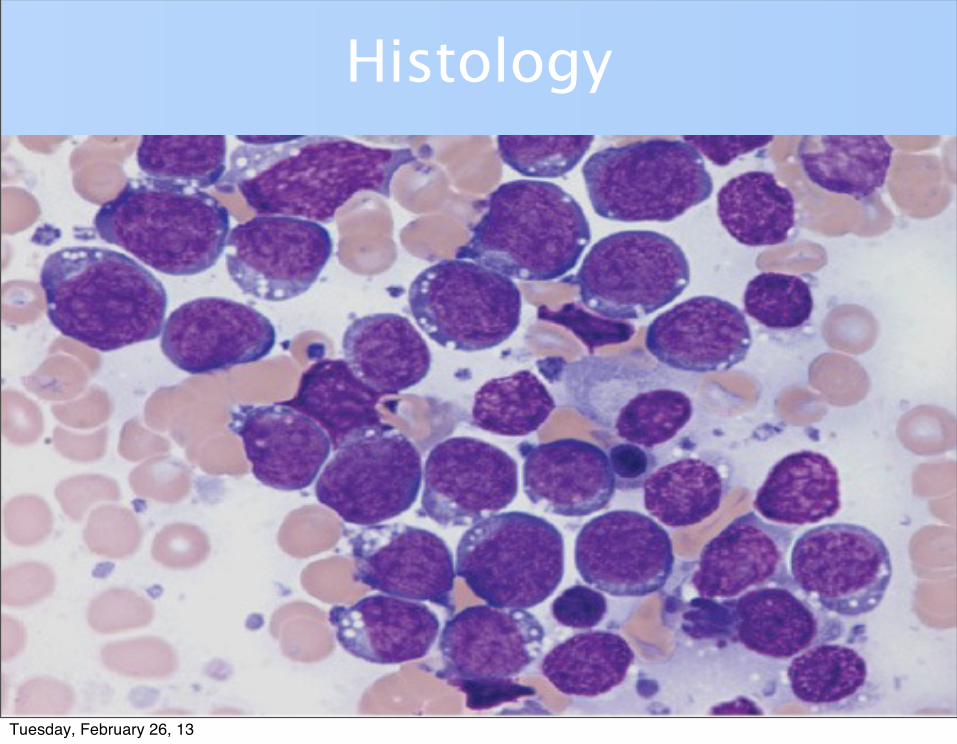
Morphology and Cytology• Blasts in peripheral blood in 90% cases
• More than 20% blasts in bone marrow
• Auer Rods (cytoplasmic granules)
• Positive myeloperoxidase reaction in 3% blasts
Tuesday, February 26, 13

AML Histology
Tuesday, February 26, 13

AML Histology
Tuesday, February 26, 13

AML Histology
Tuesday, February 26, 13

AML Histology
Tuesday, February 26, 13

• Classification/Subtypes– French-American-British Classification• 8 major subtypes• Based on morphology and cytochemistry
• WHO Classificiation– Based on molecular, morphologic, and
clinical features
Tuesday, February 26, 13

Tuesday, February 26, 13
Treatment• Chemotherapy
• Supportive Care
Tuesday, February 26, 13

Chemotherapy• Current standard induction treatment
for AML involves drug regimens with two or more agents that include anthracyclines or an anthraquinone and cytarabine
• Remission rates in the studies cited range from approximately 55 to 90 percent in adult subjects, depending
Tuesday, February 26, 13

Prognosis• Age of diagnosis• Comorbidities• Chromosomal Findings• Symptomatic Interval Preceding
Diagnosis• Presenting Leukocyte count• Circulating Myeloblast count• FAB classification
Tuesday, February 26, 13

AML: Five year relative survival Rates (1996-2004)
AML: Five year relative survival Rates (1996-2004)Age (years) AML (expressed as # of
cases per 100)<45 50.0
45-54 28.5
55-64 17.9
65-74 7.4
>75 1.8
<65 35.8
>65 4.5Tuesday, February 26, 13

Acute Lymphoblastic Leukemia (ALL)
Tuesday, February 26, 13

What is it?• Acute lymphoblastic leukemia (ALL) is
a neoplastic disease that results from multistep somatic mutations in a single lymphoid progenitor cell at one of several discrete stages of development
Tuesday, February 26, 13

Pathogenesis
• Acquired genetic abnormalities are a hallmark of ALL.
• > 75% of all cases have recurring cytogenetic or molecular lesions with prognostic and therapeutic relevance
Tuesday, February 26, 13

Pathogenesis• Chromosomal changes include:
1. abnormalities in the number (ploidy) 2. structure of chromosomes. - Translocation (MC abnormality) - Inversion - Deletion - Point Mutations - Amplifications
Tuesday, February 26, 13

Pathogenesis• Although the frequency of particular
genetic subtypes differs between childhood and adult cases, the general mechanisms underlying the induction are similar
• Mechanisms include:– aberrant expression of oncoproteins – chromosomal translocations that generate
fusion genes encoding transcription factors Tuesday, February 26, 13

Pathogenesis
Primary genetic rearrangement by itself is insufficient to induce overt leukemia.
Cooperative mutations are necessary for leukemic transformation and include genetic and epigenetic changes in key growth regulatory pathways
Tuesday, February 26, 13

Epigenetic Changes
• Hypermethylation of tumor-suppressor genes
• Hypomethylation of oncogenes and abnormalities in post-transcriptional control mechanisms
• These changes are reversible and do not alter the DNA sequence, yet they can alter gene expression in subtle ways that
Tuesday, February 26, 13

Epigenetic Changes• Analysis of changes applied to developing
new biomarkers for risk assignment or disease monitoring, and to the design of alternative treatment in ALL.
• Evidence indicates that the methylation of multiple genes in ALL is associated with a worse outcome.
Tuesday, February 26, 13

Frequencies of Common Genetic Aberrations in Childhood and Adult Acute Lymphoblastic LeukemiaFrequencies of Common Genetic Aberrations in Childhood and Adult Acute Lymphoblastic LeukemiaFrequencies of Common Genetic Aberrations in Childhood and Adult Acute Lymphoblastic Leukemia
Abnormality Children (%) Adult (%)
Hyperdiploidy (>50 chromosomes)
23-29 6-7
Hypodiploidy (<45 chromosomes)
1 2
t(1;19)(q23;p13.3) [TCF3-PBX1]
4 in white, 12 in black 2-3
t(9;22)(q34;q11.2) [BCR-ABL1]
2-3 25-30
t(4;11)(q21;q23) [MLL-AF4]
2 3-7
t(8;14)(q23;q32.3) 2 4
t(12;21)(p13;q22) [ETV6-RUNX1]
20-25 0-3
NOTCH1 mutations* 7 15
HOX11L2 overexpression* 20 13
LYL1 overexpression* 9 15
TAL1 overexpression* 15 3
HOX11 overexpression* 7 30
MLL-ENL fusion 2 3
Abnormal 9p 7-11 6-30
Abnormal 12p 7-9 4-6
del(7p)/del(7q)/monosomy 7
4 6-11
+8 2 10-12
Intrachromosomal amplification of chromosome 21 (iAMP21)
2 ?
Tuesday, February 26, 13

Etiology• Uncertain
• Proposed Risk Factors:1. Genetic Syndromes (ex: Philadelphia chromosome)
2. Environmental Factors
3. Viral infections (ex: EBV and HIV)
4. Smoking
Tuesday, February 26, 13

Incidence Rates
Tuesday, February 26, 13

Incidence Rates• 5,730 new cases in U.S. in 2011
• 2/3 of ALL cases in children (peak incidence ages 2 – 5)
• Comprises less than 20% of leukemia in young adults
• May be B-cell, T-cell, or null-type (non-B, non-T cell)
Tuesday, February 26, 13

Signs and Symptoms• Pallor
• Fatigue
• Shortness of breath
• Easy bruising
• Petechiae
• Weight loss / failure to thrive
• Fever
• Splenomegaly and/or hepatomegaly
• Lymphadenopathy
• Multiple bruises
• Unexplained infections
Tuesday, February 26, 13

Diagnosis• CBC Chemistry studies to check for
organ dysfunction
• Bone marrow aspirate and biopsy
• Genetic/Immunological studies
• Lumbar puncture
Tuesday, February 26, 13

Hematological Findings• Anemia
• WBC < 5,000 (or > 25,000)
• Leukocytosis
• Thrombocytopenia
Tuesday, February 26, 13

Histology
Tuesday, February 26, 13

Histology
Tuesday, February 26, 13
Histology
Tuesday, February 26, 13

L1 - 85% of childhood ALLL2 - Majority of adult ALL
Tuesday, February 26, 13

PrognosisFavorable Factors:
1. Age 3 to 7 yrs2. WBC count < 25,000/µL3. FAB L1 morphology4. Leukemic cell karyotype with > 50 chromosomes and t(12;21)5. No CNS disease at diagnosis
Tuesday, February 26, 13

Adverse Prognostic Factors in adult ALLAdverse Prognostic Factors in adult ALLAdverse Prognostic Factors in adult ALL
Factors B- Cell Precursor T Cell
Age (years) >35 >35
Leukocyte Count (x109/L)
>30 >100
Immunophenotype Pro-B (CD10–) Pre-T
Genetics t(9;22) [BCR-ABL1] HOX11L2 expression ?
t(4;11) [MLL-AF4] ERG expression ?
Hypodiploidy ?
Treatment Response Delayed remission (> 4 weeks)
Delayed remission (> 4 weeks)
Minimal residual disease >10–4 after induction
Minimal residual disease >10–4 after induction
Tuesday, February 26, 13

TREATMENT
Tuesday, February 26, 13

Treatment
• Chemotherapy
• Sometimes stem cell transplantation or radiation therapy
Tuesday, February 26, 13

Treatment4 General phases of chemotherapy
1. Remission Induction
2. CNS prophylaxis
3. Post remission consolidation or intensification
4. MaintenanceTuesday, February 26, 13

Remission Induction• Initial goal is to quickly induce
complete remission
• The induction regimen typically includes a glucocorticoid (prednisone, prednisolone, or dexamethasone), vincristine, and L-asparaginase for children or an anthracycline for adults.
Tuesday, February 26, 13

• After Induction Chemotherapy:
• Bone marrow biopsy is obtained
• If > 5% of blasts with > 20% cellularity, then retreatment necessary.
• Stem cell transplant may be necessary if retreatment fails.
Tuesday, February 26, 13

CNS Prophylaxis
• Important site of leukemic infiltration is meninges
• Prophylaxis and treatment may include high-dose intrathecal methotrexate, cytosine arabinoside, and corticosteroids
Tuesday, February 26, 13

Post remission consolidation or intensification
• Goal is to prevent leukemic regrowth
• Therapy lasts several months and involves drugs with different MOA than drugs used in induction regimen
• Continued low-dose post-remission therapy must be used to ensure prolonged survival. Otherwise recurrence rates can be as high as 90%
Tuesday, February 26, 13

Maintenance
• Most regimens include therapy with methotrexate and mercaptopurine
• Duration typically 2.5-3 years
• Patients in continuous complete remission for 2.5 years, risk of relapse is 20%, usually within 1st year
Tuesday, February 26, 13

Treatment• Continued Supportive Care:
• Transfusions….· Platelets >20,000· Hgb >8
• Empiric antibiotic treatment when fever present
Tuesday, February 26, 13

Relapse• Defined as the reappearance of leukemic cells at
any site in the body.
• Most relapses occur during treatment or within the first 2 years after its completion
• Molecular studies suggest that in some cases, especially those with the ETV6-RUNX1 fusion, subsequent mutations of the residual preleukemic clone that were not eradicated during initial treatment account for the "late relapse.”
• The marrow remains the most common site of Tuesday, February 26, 13

Relapse
• Between 50 - 70% of children and 40 - 50% of adults who achieve complete remission after initial therapy but then suffer a relapse may be able to go into a second complete remission
Tuesday, February 26, 13

Relapse• Treatment for relapse after a first remission may
be standard chemotherapy or experimental drugs, or more aggressive treatments such as stem cell transplants.
Depends on:• Children who relapse 3 or more years after
achieving a first complete remission have an excellent chance for a second remission without aggressive treatments.
• Those who relapse fewer than 6 months following initial treatment, especially while on chemotherapy, have about a 20% chance of long-term freedom from disease. In such cases,
Tuesday, February 26, 13

5 yr survival rates• Children: 80%
• Adults: 40%
• Percentages include children/adults with all levels of risk factors. For children/adults with high-risk disease, survival rates are much lower, while survival rates are higher for some children/adults with low-risk disease.
Tuesday, February 26, 13

• The overall 5-year relative survival for 2001-2007 from 17 SEER geographic areas was 64.4%.
• Five-year relative survival by race and sex was: – 63.9% for white men; – 64.7% for white women; – 60.5% for black men; – 64.1% for black women.
Tuesday, February 26, 13

Sources• Lichtman MA, Kipps TJ, Seligsohn U,
Kaushansky K, Prchal Jt: Williams Hematology, 8th Edition: http:www.accessmedicine.com
• Merck Manual Professional: Acute Leukemia, http://www.merckmanuals.com/professional/
Tuesday, February 26, 13