Embed Size (px)
Citation preview

LETTERdoi:10.1038/nature10099
Single-molecule fluorescence reveals sequence-specific misfolding in multidomain proteinsMadeleine B. Borgia1,2, Alessandro Borgia2, Robert B. Best1, Annette Steward1, Daniel Nettels2, Bengt Wunderlich2,Benjamin Schuler2 & Jane Clarke1
A large range of debilitating medical conditions1 is linked to proteinmisfolding, which may compete with productive folding particularlyin proteins containing multiple domains2. Seventy-five per cent ofthe eukaryotic proteome consists of multidomain proteins, yet it isnot understood how interdomain misfolding is avoided. It has beenproposed that maintaining low sequence identity between covalentlylinked domains is a mechanism to avoid misfolding3. Here we usesingle-molecule Forster resonance energy transfer4,5 to detect andquantify rare misfolding events in tandem immunoglobulin domainsfrom the I band of titin under native conditions. About 5.5 per cent ofmolecules with identical domains misfold during refolding in vitroand form an unexpectedly stable state with an unfolding half-time ofseveral days. Tandem arrays of immunoglobulin-like domains inhumans show significantly lower sequence identity between neigh-bouring domains than between non-adjacent domains3. In particu-lar, the sequence identity of neighbouring domains has been found tobe preferentially below 40 per cent3. We observe no misfolding for atandem of naturally neighbouring domains with low sequence iden-tity (24 per cent), whereas misfolding occurs between domains thatare 42 per cent identical. Coarse-grained molecular simulations pre-dict the formation of domain-swapped structures that are in excellentagreement with the observed transfer efficiency of the misfoldedspecies. We infer that the interactions underlying misfolding are veryspecific and result in a sequence-specific domain-swapping mech-anism. Diversifying the sequence between neighbouring domainsseems to be a successful evolutionary strategy to avoid misfoldingin multidomain proteins.
Multidomain proteins comprise covalently linked, frequently sim-ilar domains, resulting in high effective local protein concentration. Itis therefore probable that these proteins have evolved to avoid inter-domain misfolding in vivo. Co-translational, ‘domain-by-domain’folding is assumed to be important for avoiding misfolding6, but manymultidomain proteins that are long lived or subject to tensile forces willfold and unfold numerous times during their lifetimes, resulting inneighbouring domains being unfolded at the same time, and thereforemay be particularly vulnerable to misfolding. The giant muscle proteintitin, for example, undergoes reversible domain unfolding that mayhave a role in muscle elasticity7.
Single-molecule techniques are ideal for detecting rare events8 suchas misfolding in native conditions. Indeed, the first evidence for themisfolding of adjacent domains in long tandem arrays of the well-characterized twenty-seventh domain from the I band of titin, I27,was obtained using single-molecule atomic force microscopy9 (AFM).An alternative approach is single-molecule Forster resonance energytransfer4,5 (FRET), whose great sensitivity allows the detection of verysmall populations. FRET permits the mapping of intramolecular dis-tances by means of the distance-dependent efficiency of excitationenergy transfer between donor and acceptor fluorophores attached tospecific positions of the protein10 (for details, see Supplementary Fig. 1).
We proposed that denaturation of tandem constructs of titindomains with guanidinium chloride (GdmCl), followed by rapid
refolding into native conditions, might allow formation of misfoldedspecies, which should be detectable using single-molecule FRET11. Welabelled a tandem construct of I27 (I27–I27) in the A strand of domain1 (E3C) and the G strand of domain 2 (N83C) (Fig. 1a) with a donor(Alexa Fluor 488) and an acceptor (Alexa Fluor 594) fluorophore,attached by means of cysteine residues engineered on the proteinsurface. For a misfolded domain formed by strands from domains 1and 2, we predicted that these two strands must be adjacent for thedomain to have the mechanical properties observed in the previousAFM experiments9. The correctly folded tandem domain would thenhave low transfer efficiency, whereas a misfolded state would have hightransfer efficiency. A monomer of I27 was labelled in the correspond-ing positions to provide a model for the misfolded state (Fig. 1b).Labelling was found to have little effect on the stability of I27 (Sup-plementary Fig. 2), and doubly labelled proteins that had not previ-ously been unfolded in GdmCl (‘never unfolded’) showed single,correctly folded populations with transfer efficiencies of E 5 0.37 6 0.01and 0.93 6 0.01 for the tandem I27–I27 and monomer I27, respectively(Fig. 2a, b). (Uncertainties quoted in the text are s.d.)
We conducted refolding experiments by diluting unfolded I27–I27into refolding buffer. The resulting transfer efficiency histograms thenshowed two populations (Fig. 2c): one corresponding to the correctlyfolded native state (E 5 0.37) and one with precisely the same trans-fer efficiency as the analogously labelled monomer (E 5 0.93). This
1University of Cambridge Chemical Laboratory, Lensfield Road, Cambridge CB2 1EW, UK. 2Biochemisches Institut, Universitat Zurich, Winterthurerstrasse 190, 8057 Zurich, Switzerland.
E3C
Domain 1
N83C
Domain 2
E3C
N83C
E3C
N83C
a b
d
Terminal domain Central domain
Terminal domain Central domain
E
D
F
A′
C
B
c
AA′
B
C
A
F
D
E
G
G
N′
C′
Figure 1 | Structures of native and misfolded I27 constructs. a, Nativelyfolded I27–I27 tandem repeat with labelling positions highlighted (goldenspheres). b, Native I27 crystal structure (Protein Data Bank ID, 1TIT). c, One of thedomain-swapped, misfolded state structures formed in Go-model simulations.d, Schematic of the misfolded state topology in c: hydrogen bonds that areperpendicular to the direction of applied force in AFM mechanical unfolding areshown by dashed lines (circled). Four other misfolded state topologies werepopulated in the simulations (Supplementary Fig. 5b). We note that we cannotdistinguish between such topologies from the results presented here.
0 0 M O N T H 2 0 1 1 | V O L 0 0 0 | N A T U R E | 1
Macmillan Publishers Limited. All rights reserved©2011

observation reveals that the A strand of the first domain and the Gstrand of the second domain are arranged as in the monomer. Aquantitative analysis (Supplementary Table 1) showed that5.5 (6 0.2)% of the molecules are found in the misfolded form.
On the basis of the results of the AFM studies9, we had supposed thatthe misfolded species consisted of a single strand-swapped titindomain with the remaining sequence unstructured and, thus, withan unfolding time similar to that of a native domain (t < 34 min; ref.12). We therefore investigated the unfolding kinetics of the misfoldedstate13 (Fig. 3a). At high GdmCl concentrations, the decays in thenumber of high-transfer-efficiency events (E . 0.8), correspondingto the misfolded state, are fitted well by single exponentials (Fig. 3b)with rate constants slightly higher than the unfolding rate constants forwild-type I27 determined in ensemble measurements12 (Fig. 3c). Wecan also estimate the unfolding rate constant of the correctly foldedspecies, which agrees well with the ensemble data (Fig. 3c) (see Sup-plementary Fig. 3 and Supplementary Information). In the absence ofdenaturant, however, the misfolded species was surprisingly long lived,
converting to the correctly folded form only on a timescale of days(Supplementary Fig. 4). The formation of the misfolded structure is thusunder kinetic, rather than thermodynamic, control. Its remarkablekinetic stability clearly distinguishes the misfolded species describedhere from short-lived, partly folded intermediates14–16 sometimes termed‘misfolded’ because they contain some non-native interactions17.
An explanation for the slow unfolding under native conditions issuggested by folding simulations of I27–I27 with a Go-like model18.In these simulations, only native interactions are attractive, and inter-actions between a given pair of residues are considered equal, indepen-dently of whether they are in the same or different domains. Althoughmost trajectories result in two correctly folded domains, misfoldedspecies with two fully folded, strand-swapped domains are occasionallyformed. Five different strand-swapped topologies were observed
0.0 0.2 0.4 0.6 0.8 1.0 1.2
Transfer efficiency, E
Num
ber
of e
vent
s (×
1,00
0)
E = 0.94
E = 0.37
E = 0.15
a
b
c
d
0
1
2
3
4
5
0
1
2
3
4
5
0
1
2
3
4
5
0
2
4
6
8
10
12
Figure 2 | Transfer efficiency histograms of doubly labelled I27 constructs.a, Never-unfolded I27–I27. b, Never-unfolded monomeric I27. c, RefoldedI27–I27. d, Refolded I27–I27–I27; fits of individual populations shown ascoloured lines for clarity. Histograms are fitted with normal or log-normaldistributions. The peak in the grey shaded area consists of events frommolecules without an active acceptor fluorophore28. We note that in theseexperiments a short, four-amino-acid linker (Arg-Ser-Glu-Leu) is includedbetween the domains in the I27–I27 tandem to allow direct comparison withprevious AFM and aggregation experiments3,9. Never-unfolded I27–I27–I27 isshown in Supplementary Fig. 6.
0 500 1,000 1,5000
100
200
300
Time (s)
Num
ber
of e
vent
s (E
> 0
.8)
0.8 0.9 1.0
0 900Time (s)
1,800
1.10
20
40
60
E
Num
ber
of e
vent
s
kobs = (3.0 ± 0.3) × 10–3 s–1
a
b
c
–8
–7
–6
–5
–4
–3
–2
3 3.5 4 4.5 5 5.5 6
ln(k
obs)
[GdmCl] (M)
Figure 3 | Unfolding kinetics. a, Evolution of transfer efficiency histogramsover time (E $ 0.7) from single-molecule double-jump experiments in whichrefolded/misfolded I27–I27 (doubly labelled) was unfolded in 3.5 M GdmCl.Histograms were constructed for a moving window of 120 s that was shifted by30 s for each increment (colour scale). b, The number of events with E . 0.8 foreach histogram in a was summed and the resulting kinetics fitted with a singleexponential decay. The observed rate constants (kobs) are unaffected bydifferent window sizes or the use of non-overlapping windows. c, Unfoldingrate constants for the wild-type I27 monomer (black; ensemble data from ref.12) and for the misfolded and natively folded states of I27–I27 from single-molecule measurements (red and blue, respectively), as functions of GdmClconcentration. Each point was obtained by fitting data sets composed bymerging at least six repeats, with error bars representing the standard error ofthe fit (Methods). For some data points, the error bars are smaller than thesymbols. I27 domains have the same unfolding rate constants in tandem repeatproteins as in isolated domains.
RESEARCH LETTER
2 | N A T U R E | V O L 0 0 0 | 0 0 M O N T H 2 0 1 1
Macmillan Publishers Limited. All rights reserved©2011
(Fig. 1c, d and Supplementary Fig. 5). Such an extensively misfoldedstructure explains its persistence: correct folding cannot occur whileeither misfolded domain remains folded. Because refolding rate con-stants are much higher than unfolding rate constants under nativeconditions, the simultaneous unfolding of both domains is very unlikelyand conversion to the native state is extremely slow19.
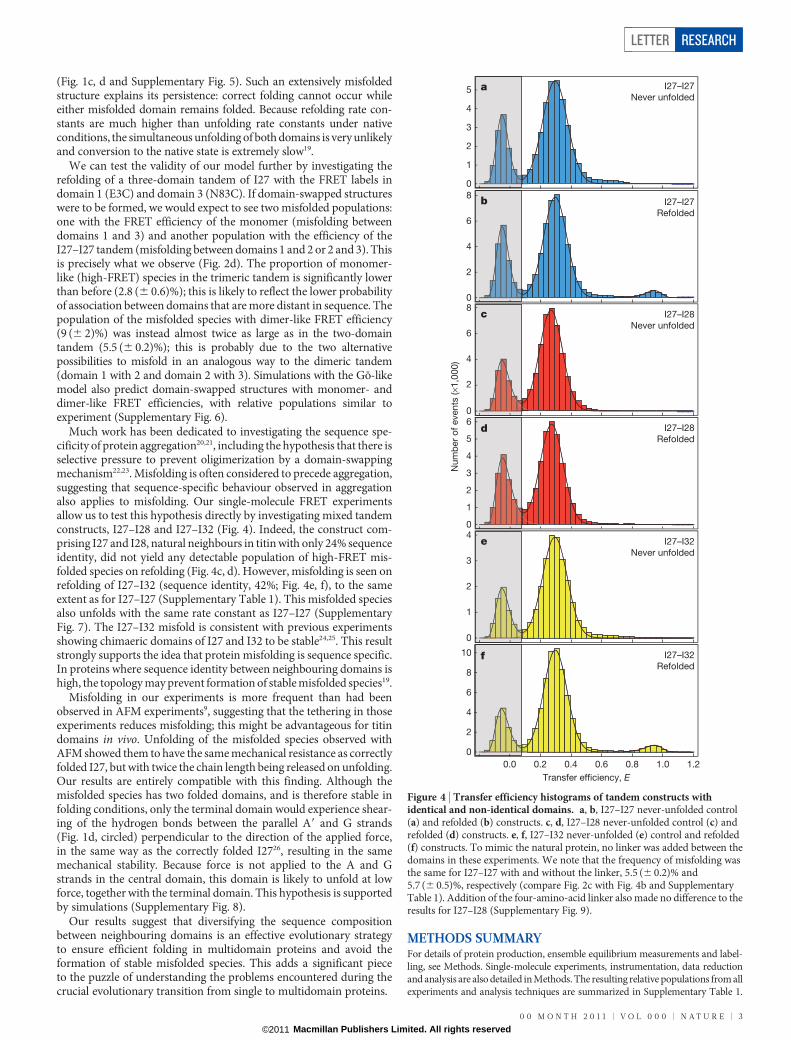
We can test the validity of our model further by investigating therefolding of a three-domain tandem of I27 with the FRET labels indomain 1 (E3C) and domain 3 (N83C). If domain-swapped structureswere to be formed, we would expect to see two misfolded populations:one with the FRET efficiency of the monomer (misfolding betweendomains 1 and 3) and another population with the efficiency of theI27–I27 tandem (misfolding between domains 1 and 2 or 2 and 3). Thisis precisely what we observe (Fig. 2d). The proportion of monomer-like (high-FRET) species in the trimeric tandem is significantly lowerthan before (2.8 (6 0.6)%); this is likely to reflect the lower probabilityof association between domains that are more distant in sequence. Thepopulation of the misfolded species with dimer-like FRET efficiency(9 (6 2)%) was instead almost twice as large as in the two-domaintandem (5.5 (6 0.2)%); this is probably due to the two alternativepossibilities to misfold in an analogous way to the dimeric tandem(domain 1 with 2 and domain 2 with 3). Simulations with the Go-likemodel also predict domain-swapped structures with monomer- anddimer-like FRET efficiencies, with relative populations similar toexperiment (Supplementary Fig. 6).
Much work has been dedicated to investigating the sequence spe-cificity of protein aggregation20,21, including the hypothesis that there isselective pressure to prevent oligimerization by a domain-swappingmechanism22,23. Misfolding is often considered to precede aggregation,suggesting that sequence-specific behaviour observed in aggregationalso applies to misfolding. Our single-molecule FRET experimentsallow us to test this hypothesis directly by investigating mixed tandemconstructs, I27–I28 and I27–I32 (Fig. 4). Indeed, the construct com-prising I27 and I28, natural neighbours in titin with only 24% sequenceidentity, did not yield any detectable population of high-FRET mis-folded species on refolding (Fig. 4c, d). However, misfolding is seen onrefolding of I27–I32 (sequence identity, 42%; Fig. 4e, f), to the sameextent as for I27–I27 (Supplementary Table 1). This misfolded speciesalso unfolds with the same rate constant as I27–I27 (SupplementaryFig. 7). The I27–I32 misfold is consistent with previous experimentsshowing chimaeric domains of I27 and I32 to be stable24,25. This resultstrongly supports the idea that protein misfolding is sequence specific.In proteins where sequence identity between neighbouring domains ishigh, the topology may prevent formation of stable misfolded species19.
Misfolding in our experiments is more frequent than had beenobserved in AFM experiments9, suggesting that the tethering in thoseexperiments reduces misfolding; this might be advantageous for titindomains in vivo. Unfolding of the misfolded species observed withAFM showed them to have the same mechanical resistance as correctlyfolded I27, but with twice the chain length being released on unfolding.Our results are entirely compatible with this finding. Although themisfolded species has two folded domains, and is therefore stable infolding conditions, only the terminal domain would experience shear-ing of the hydrogen bonds between the parallel A9 and G strands(Fig. 1d, circled) perpendicular to the direction of the applied force,in the same way as the correctly folded I2726, resulting in the samemechanical stability. Because force is not applied to the A and Gstrands in the central domain, this domain is likely to unfold at lowforce, together with the terminal domain. This hypothesis is supportedby simulations (Supplementary Fig. 8).
Our results suggest that diversifying the sequence compositionbetween neighbouring domains is an effective evolutionary strategyto ensure efficient folding in multidomain proteins and avoid theformation of stable misfolded species. This adds a significant pieceto the puzzle of understanding the problems encountered during thecrucial evolutionary transition from single to multidomain proteins.
METHODS SUMMARYFor details of protein production, ensemble equilibrium measurements and label-ling, see Methods. Single-molecule experiments, instrumentation, data reductionand analysis are also detailed in Methods. The resulting relative populations from allexperiments and analysis techniques are summarized in Supplementary Table 1.
Num
ber
of e
vent
s (×
1,00
0)
0.0 0.2 0.4 0.6 0.8 1.0 1.2Transfer efficiency, E
I27–I27Never unfolded
I27–I27Refolded
I27–I28Never unfolded
I27–I28Refolded
I27–I32Never unfolded
I27–I32Refolded
a
b
c
d
e
f
0
1
2
3
4
5
0
2
4
6
8
0
2
4
6
8
0
1
2
3
4
5
6
0
1
2
3
4
0
2
4
6
8
10
Figure 4 | Transfer efficiency histograms of tandem constructs withidentical and non-identical domains. a, b, I27–I27 never-unfolded control(a) and refolded (b) constructs. c, d, I27–I28 never-unfolded control (c) andrefolded (d) constructs. e, f, I27–I32 never-unfolded (e) control and refolded(f) constructs. To mimic the natural protein, no linker was added between thedomains in these experiments. We note that the frequency of misfolding wasthe same for I27–I27 with and without the linker, 5.5 (6 0.2)% and5.7 (6 0.5)%, respectively (compare Fig. 2c with Fig. 4b and SupplementaryTable 1). Addition of the four-amino-acid linker also made no difference to theresults for I27–I28 (Supplementary Fig. 9).
LETTER RESEARCH
0 0 M O N T H 2 0 1 1 | V O L 0 0 0 | N A T U R E | 3
Macmillan Publishers Limited. All rights reserved©2011

Folding simulations using a Go-like model were run using the CHARMM code27 asdescribed in Methods. For details of mechanical unfolding simulations, seeMethods.
Full Methods and any associated references are available in the online version ofthe paper at www.nature.com/nature.
Received 6 January; accepted 1 April 2011.
Published online 29 May 2011.
1. Gregersen, N., Bross, P., Vang, S. & Christensen, J. H. Protein misfolding andhuman disease. Annu. Rev. Genomics Hum. Genet. 7, 103–124 (2006).
2. Jaenicke, R. & Seckler, R. Protein misassembly in vitro. Adv. Protein Chem. 50, 1–59(1997).
3. Wright, C. F., Teichmann, S. A., Clarke, J. & Dobson, C. M. The importance ofsequence diversity in the aggregation and evolution of proteins. Nature 438,878–881 (2005).
4. Joo, C., Balci, H., Ishitsuka, Y., Buranachai, C. & Ha, T. Advances in single-moleculefluorescencemethods formolecularbiology.Annu.Rev.Biochem.77,51–76(2008).
5. Schuler, B. & Eaton, W. A. Protein folding studied by single-molecule FRET. Curr.Opin. Struct. Biol. 18, 16–26 (2008).
6. Fedorov, A. N. & Baldwin, T. O. Cotranslational protein folding. J. Biol. Chem. 272,32715–32718 (1997).
7. Li, H. et al. Reverse engineering of the giant muscle protein titin. Nature 418,998–1002 (2002).
8. Borgia, A., Williams, P. M. & Clarke, J. Single-molecule studies of protein folding.Annu. Rev. Biochem. 77, 101–125 (2008).
9. Oberhauser, A. F., Marszalek, P. E., Carrion-Vasquez, M. & Fernandez, J. M. Singleproteinmisfolding events capturedbyatomic forcemicroscopy.NatureStruct. Biol.6, 1025–1028 (1999).
10. Stryer, L. Fluorescence energy transfer as a spectroscopic ruler. Annu. Rev.Biochem. 47, 819–846 (1978).
11. Gambin, Y. et al. Direct single-molecule observation of a protein living in twoopposed native structures. Proc. Natl Acad. Sci. USA 106, 10153–10158 (2009)CrossRef.
12. Fowler, S. B. & Clarke, J. Mapping the folding pathway of an immunoglobulindomain: structural detail from phi value analysis and movement of the transitionstate. Structure 9, 355–366 (2001).
13. Hofmann, H. et al. Single-molecule spectroscopyof protein folding ina chaperonincage. Proc. Natl Acad. Sci. USA 107, 11793–11798 (2010).
14. Ivarsson, Y., Travaglini-Allocatelli, C., Brunori, M. & Gianni, S. Folding andmisfolding in a naturally occurring circularly permuted PDZ domain. J. Biol. Chem.283, 8954–8960 (2008).
15. Gianni, S. et al. Structural characterization of a misfolded intermediate populatedduring the folding process of a PDZ domain. Nature Struct. Mol. Biol. 17,1431–1437 (2010).
16. Korzhnev, D. M., Religa, T. L., Banachewicz, W., Fersht, A. R. & Kay, L. E. A transientand low-populated protein-folding intermediate at atomic resolution. Science329, 1312–1316 (2010).
17. Capaldi, A. P., Kleanthous, C. & Radford, S. E. Im7 folding mechanism: misfoldingon a path to the native state. Nature Struct. Biol. 9, 209–216 (2002).
18. Yang, S. et al. Domain swapping is a consequence of minimal frustration. Proc. NatlAcad. Sci. USA 101, 13786–13791 (2004).
19. Arora, P., Hammes, G. G. & Oas, T. G. Folding mechanism of a multipleindependently-folding domain protein: double B domain of protein A.Biochemistry 45, 12312–12324 (2006).
20. Jaenicke, R. Folding and association of proteins. Prog. Biophys. Mol. Biol. 49,117–237 (1987).
21. Straub, J. E. & Thirumalai, D. Principles governing oligomer formation inamyloidogenic peptides. Curr. Opin. Struct. Biol. 20, 187–195 (2010).
22. Bennett, M. J., Sawaya, M. R. & Eisenberg, D. Deposition diseases and 3D domainswapping. Structure 14, 811–824 (2006).
23. Mitraki, A. Protein aggregation from inclusion bodies to amyloid and biomaterials.Adv. Protein Chem. Struct. Biol. 79, 89–125 (2010).
24. Borgia, A., Steward, A. & Clarke, J. An effective strategy for the design of proteinswith enhanced mechanical stability. Angew. Chem. Int. Ed. 47, 6900–6903 (2008).
25. Balamurali, M. M. et al. Recombination of protein fragments: a promisingapproach toward engineering proteins with novel nanomechanical properties.Protein Sci. 17, 1815–1826 (2008).
26. Lu, H., Isralewitz, B., Krammer, A., Vogel, V. & Schulten, K. Unfolding of titinimmunoglobulin domains by steered molecular dynamics simulation. Biophys. J.75, 662–671 (1998).
27. Brooks, B.R.et al. CHARMM: Aprogram for macromolecular energy, minimization,and dynamics calculations. J. Comput. Chem. 4, 187–217 (1983).
28. Dahan, M. et al. Ratiometric measurement and identification of single diffusingmolecules. Chem. Phys. 247, 85–106 (1999).
Supplementary Information is linked to the online version of the paper atwww.nature.com/nature.
Acknowledgements This work was supported by the Wellcome Trust (grant number,064417), the Swiss National Science Foundation (to B.S.) and the Swiss NationalCenter of Competence in Research in Structural Biology (to B.S.). M.B.B. was supportedby a UK Medical Research Council studentship. A.B. is supported by a Marie CurieIntra-European Fellowship. R.B.B. is supported by a Royal Society University ResearchFellowship. J.C. is a Wellcome Trust Senior Research Fellow. We thank H. Hofmann,A. Soranno and A. Hoffmann for discussions and contributions to data analysis.
Author Contributions M.B.B., A.B., B.S. and J.C. designed the investigation. M.B.B. andA.B. performed the experiments. R.B.B. performed the simulations. D.N. and B.W. builtthe single-molecule instrumentation. D.N. provided data analysis software. A.S. clonedthe gene of the trimeric tandem construct. M.B.B. performed the analysis. M.B.B., J.C.and B.S. wrote the paper.
Author Information Reprints and permissions information is available atwww.nature.com/reprints. The authors declare no competing financial interests.Readers are welcome to comment on the online version of this article atwww.nature.com/nature. Correspondence and requests for materials should beaddressed to J.C. ([email protected]) or B.S. ([email protected]).
RESEARCH LETTER
4 | N A T U R E | V O L 0 0 0 | 0 0 M O N T H 2 0 1 1
Macmillan Publishers Limited. All rights reserved©2011

METHODSProtein expression and labelling. Cysteine residues were introduced by site-directed mutagenesis: for the two-domain constructs, E3C in domain 1 (alwaysI27), N83C in domain 2 if I27 or I32, and K83C in domain 2 if I28 (with thenumbering relative to a single domain); and for the three-domain construct ofI27, E3C in domain 1 and N83C in domain 3. DNA sequencing confirmed themutagenesis.
I27 monomer and the I27–I27, I27–I27–I27, I27–I28 and I27–I32 tandems,with the engineered surface cysteines, were expressed as described previously29,30.Labelling was carried out using Alexa Fluor 488 (donor) and Alexa Fluor 594(acceptor) maleimides (Invitrogen) according to the manufacturer’s procedures.The dyes were mixed simultaneously with reduced protein in equimolar ratios andincubated at 4 uC for ,10 h. Unreacted dye was removed by gel filtration and thedifferently labelled variants were separated by ion-exchange chromatography(MonoQ 5/50 GL; GE Healthcare Biosciences AB). I27 has two intrinsic cysteinesthat were not removed as they are buried in the native state and all labelling wascarried out on folded protein in native conditions.Ensemble measurements. Equilibrium measurements were performed for thedoubly labelled I27 monomer to check the effect of labelling (SupplementaryFig. 2). Experiments were performed in GdmCl on a Cary Eclipse fluorimeter(Varian Inc.) monitoring intrinsic tryptophan fluorescence as described previ-ously29, but with lower protein concentrations (0.05–0.5mM) and the additionof 0.001% Tween 20.Single-molecule instrumentation. Observations of single-molecule fluorescencewere made using a custom-built confocal microscope equipped with a continuous-wave, 488-nm solid-state laser (FCD488-010, JDSU) and an Olympus UplanApo360/1.20W objective. After passage through a dichroic mirror that separatesexcitation and emission light (500DCXR, Chroma Technology), fluorescenceemission passed through a 100-mm pinhole and was split into donor and acceptorfluorescence by a second dichroic mirror (585DCXR, Chroma Technology).Donor fluorescence then passed a filter (ET525/50M, Chroma Technology) beforebeing focused onto a single-photon avalanche diode (MPD 100ct, Micro PhotonDevices). Similarly, acceptor fluorescence passed a filter (QT 650/100) before beingfocused onto a single-photon avalanche diode (SPCM-AQR-13, PerkinElmerOptoelectronics). The arrival time of every photon was recorded with a two-channel,time-correlated, single-photon counting module (PicoHarp300, PicoQuant). Allmeasurements were performed with a laser power of 100mW, measured at the backaperture of the objective (beam waist, 8 mm).Single-molecule equilibrium measurements. All experiments were performed atprotein concentrations between 0.5 and 25 pM, in the same final solution conditions:PBS; 0.001% Tween 20; 140 mM b-mercaptoethanol; 20 mM cysteamine hydro-chloride (single-molecule buffer). Tween 20 (Pierce) was used to prevent surfaceadhesion of the proteins31, whereas the photoprotective agents b-mercaptoethanol(Sigma) and cysteamine hydrochloride (Sigma) were used to minimize chromo-phore damage32. Experiments on never-unfolded proteins were conducted by mix-ing protein in PBS (0.01% Tween, 10 mM b-mercaptoethanol) 1:99 with 0.04 MGdmCl in single-molecule buffer (to mimic the final conditions in the refoldingexperiments). Refolding experiments were performed by mixing protein unfolded in4.4 M GdmCl (PBS, 0.01% Tween, 10 mM b-mercaptoethanol) 1:100 with single-molecule buffer, to a final GdmCl concentration of 0.04 M. Measurements over 8 to10 h were made for all constructs with one or more repeats. The absence of aggre-gates was ensured in all experimental conditions as previously described33.Fluorescence bursts were identified by combining successive photons separated by150ms or less, and events comprising 35 or more photons were kept for analysis.Transfer efficiencies were corrected for quantum yields, cross-talk and direct excita-tion as described previously34,35.
Populations of correctly folded and misfolded molecules in the transfer effi-ciency histograms were quantified using two methods. Where possible, peaks werefitted using a Gaussian distribution (for populations where 0.1 # E # 0.8) or a log-normal distribution (for populations where E $ 0.8), and the resulting fits inte-grated. Transfer efficiencies quoted in the main text denote the average E valueobtained from these fits, with standard deviations calculated from multiple experi-ments. The populations of misfolded species were determined relative to the sumof natively folded and misfolded populations. Alternatively, ranges of transferefficiencies were chosen for each population, the corresponding number of burstswere summed and the relative populations of misfolded species were computed.
Only the latter method could be used in the ‘never-unfolded’ control measure-ments and all experiments involving I27–I28 tandems, as no misfolded populationwas observed. The resulting relative populations from all experiments and analysistechniques are summarized in Supplementary Table 1.Single-molecule kinetic measurements. Unfolding of the misfolded species wasachieved by mixing a previously refolded protein sample (prepared as for refoldingexperiments above, but with higher protein concentration) with GdmCl in single-molecule buffer. To obtain sufficient statistics, at least six repeat measurements weremade for each GdmCl concentration and the resulting photon trajectories weremerged to give one data set. A moving-window analysis13 was applied to the merged,time-resolved photon trajectory with a window size (Dt) of 120 s. Transfer efficiencyhistograms were calculated from the bursts in that time window, and the windowwas shifted byDt/3 to form each successive time point (Fig. 3a). A time t 5 ts 1Dt/2,where ts is the start time of the window, was assigned to each histogram, and thenumber of events with E $ 0.8 in each histogram as a function of time was fitted to asingle exponential (Fig. 3b). The resulting rate constants were robust for differentwindow sizes and non-overlapping windows. Standard errors were taken from thecovariance matrix of the fit (weighted by the average inverse variance of the residualsof the data points with respect to an unweighted fit).
The analysis of the unfolding of the native state from the same type of experi-ment is detailed in Supplementary Information and Supplementary Fig. 5.Simulations. A coarse-grained Go-like model was generated on the basis of thestructure of I27 (Protein Data Bank ID, 1TIT36) using a standard procedure37.Briefly, all bond lengths are fixed by constraints, harmonic terms are used for theangles, a knowledge-based potential is used for the torsion angles and non-bondedinteractions are treated with a Go-like potential in which only interactions formedbetween residues in the folded protein are attractive (with relative strengths givenby the Miyazawa–Jernigan matrix), all others being repulsive. Two or three ident-ical I27 sequences were linked by four-residue repulsive linkers to treat the two andthree domain tandems, respectively. Interactions between residues i and j in dif-ferent domains were treated exactly like the interactions between those residues inthe same domain, and interactions with the linker were repulsive. A simulationtemperature was chosen such that the folding barrier was at least 3kBT, by using asa lower bound the free-energy barrier projected onto the fraction of native con-tacts. Folding simulations were run, starting from fully extended configurations,using Langevin dynamics with a friction of 0.1 ps21 and a time step of 10 fs. Thefinal structures were clustered using a simple leader–follower algorithm with a cut-off of 0.15 on the r.m.s. distance between contact maps.
Mechanical unfolding simulations were performed in which a force of 150 pNwas applied to the ends of the I27–I27 tandem, starting from structures belongingto either the folded cluster or one of the misfolded clusters, and monitoringunfolding by measuring the end–end distance. The CHARMM molecular simu-lation package was used for all calculations27.
29. Scott, K. A., Steward, A., Fowler, S. B. & Clarke, J. Titin; a multidomain protein thatbehaves as the sum of its parts. J. Mol. Biol. 315, 819–829 (2002).
30. Steward,A., Toca-Herrera, J. L.&Clarke, J. Versatile cloning system for constructionof multimeric proteins for use in atomic force microscopy. Protein Sci. 11,2179–2183 (2002).
31. Schuler, B., Lipman, E. A. & Eaton, W. A. Probing the free-energy surface for proteinfolding with single-molecule fluorescence spectroscopy. Nature 419, 743–747(2002).
32. Nettels, D. et al. Single-molecule spectroscopy of the temperature-inducedcollapse of unfolded proteins. Proc. Natl Acad. Sci. USA 106, 20740–20745(2009).
33. Hillger, F., Nettels, D., Dorsch, S. & Schuler, B. Detection and analysis of proteinaggregation with confocal single molecule fluorescence spectroscopy. J. Fluoresc.17, 759–765 (2007).
34. Schuler, B. Application of single molecule Forster resonance energy transfer toprotein folding. Protein Folding Protocols (eds Bai, Y. & Nussinov, R.) 115–138(Humana Press, 2007).
35. Hoffmann, A. et al. Mapping protein collapse with single-molecule fluorescenceand kinetic synchrotron radiation circular dichroism spectroscopy. Proc. NatlAcad. Sci. USA 104, 105–110 (2007).
36. Improta, S., Politou, A. S. & Pastore, A. Immunoglobulin-like modules from titinI-band: extensible components of muscle elasticity. Structure 4, 323–337 (1996).
37. Karanicolas, J. & Brooks, C. L. III. Improved Go-like models demonstrate therobustness of protein folding mechanisms towards non-native interactions. J. Mol.Biol. 334, 309–325 (2003).
LETTER RESEARCH
Macmillan Publishers Limited. All rights reserved©2011

Supplementary Information:
S1: Supplementary Methods
Singular value decomposition (SVD) of native state unfolding.
To analyse the unfolding of the native state from single-molecule double-jump experiments performed
on I27-I27 (as detailed in Methods), singular value decomposition (SVD) was employed due to the
overlap of the native and denatured populations.
A moving-window analysis (see Methods) was applied to the transfer efficiency range -0.2 to 0.6 (20
bins). The resulting E histograms were combined into an m × n matrix, A, for m=20 bins and n time
points. SVD analysis decomposes the matrix A into three matrices38: U, S and V:
€
A = U S VT
U is an m × m matrix whose columns form the “output” basis vectors (eigenvectors of AAT). S is an m
× n diagonal matrix whose elements are the singular values, or weighting factors for every basis vector
(the square roots of eigenvalues of AAT and ATA). VT is a transposed n × n matrix whose columns
form the “output” amplitude vectors (eigenvectors of ATA) that correspond to the kinetics of the
process. The output of the SVD allows a reduced representation of the data matrix to be produced,
which describes the data in terms of basis vectors and their associated time-dependent amplitude
vectors, and the number of non-zero singular values required to reconstruct the data matrix are an
estimate of the number of species involved in the reaction. The amplitude vectors containing
significant kinetic information were fitted with a single exponential process giving the rate constants
for the unfolding of the native state. See Fig. 3c and Supplementary Fig. 3.

S2: Supplementary Table
Supplementary Table 1: Relative populations of the misfolded statea for all tandem constructs refolded and ‘never-unfolded’ calculated as described in Methods.
Percentage of Misfolded Species
Tandem Refolded ‘Never-unfolded’
I27(C3)-I27(C83) 5.7±0.5 0.7±0.6
I27(C3)-I32(C83) 4.6±0.6 0.8±0.3
Link
er-fr
eeb
I27(C3)-I28(C83)c 0.6±0.1 0.5±0.2
I27(C3)-I27(C38) 5.5±0.2 0.7±0.1
I27(C3)-I28(C83)c,d 0.4 0.4
Link
er
I27(C3)-I27-I27(C83):
Dimer-like misfolded statee
Monomer-like misfolded statef
9±2
2.8±0.6
2.1±0.1g
0.6±0.3
a The misfolded state is defined as bursts with E≥0.8 or the integrated fit of the histogram peak for the high FRET population. b In linker-free constructs there are no added residues between domains, while linker constructs have four added amino acids (RSEL) between domains. c Only analysed by summing bursts assigned to each ‘population’ (as described in Methods) d Values quoted for one measurement made for each experiment. e Misfolded state where the dyes have the transfer efficiency of the natively folded two-domain tandem. f Misfolded state where the dyes have the transfer efficiency of the natively folded monomer. g Due to the overlap between the E ranges of the correctly folded and dimer-like misfolded state in the refolded tandem histogram, this value was estimated by quantifying the number of bursts within the E range corresponding to the dimer-like species that fall outside the fit of the correctly folded peak in the never-unfolded tandem histogram.
All errors quoted are the unbiased sample standard deviation calculated from two 8-10 hr measurements.

S3: Supplementary Figures
continuous wave laser(488 nm)0.0 0.2 0.4 0.6 0.8 1.0 1.2
Transfer Efficiency, E
Numb
er o
f eve
nts (
x 1,0
00)
0
1
2
3
4
5
MNb
a
Supplementary Figure 1: Overview of a Single-Molecule FRET Experiment. The diagram on the right shows the main components of the instrument used. For these experiments, the proteins are labelled in specific positions with a suitable donor and acceptor fluorophore. If a protein molecule diffuses through the focused laser beam (circled; upper right), excitation of the donor may result in emission of a fluorescence photon or the transfer of the excitation energy to the acceptor, which can then emit a photon. (a) A typical signal trace of fluorescence photons detected in the donor and acceptor channels; each burst corresponds to a freely diffusing molecule passing thorough the confocal volume. The efficiency of the energy transfer, E, depends on the separation R of donor and acceptor via E(R) = (1+(R/R0)6)-1, where R0 is the Förster radius characteristic of the dye pair4,10. FRET can thus be used to map intramolecular distances in the range of 3 to 8 nm for the R0 of 5.4 nm39 used in this work. Experimentally, E can be calculated for each burst via E = (nA/nA+nD) where nA and nD are the corrected number of acceptor and donor photons detected, respectively (see Methods). (b) A two-dimensional histogram of a refolded sample of I27-I27, which shows subpopulations that can be attributed to the correctly folded (N) and misfolded (M) states, and molecules without an active acceptor28 (shaded area).

Supplementary Figure 2: Comparison of Ensemble Equilibrium Denaturation for I27wt and Doubly-Labelled I27. Fraction of protein folded as a function of denaturant concentration for I27wt (open circles) (data from reference 12) and doubly-labelled I27 () with
€
ΔGD−NH2O of 7.6±0.1 and 8.0±0.1 kcal mol-1, respectively
(calculated using the average m-value, <m>,12). Mutation and labelling do not significantly alter the stability of I27.

Supplementary Figure 3. SVD analysis of the Native State Unfolding in Single-Molecule Measurements. (a) Transfer efficiency histograms as a function of time (progressing from blue to red) of refolded doubly-labelled I27-I27 unfolding in 3.95 M (as described in Methods), for bursts where -0.2 ≤ E ≤ 0.6 (constructed as described in Supplementary Information). (b) Reconstructed transfer efficiency histograms from an SVD of the data in (a) using two components (which describes the process within experimental error). (c) Time evolution of the second basis vector from the SVD of the data in (a) (progressing from blue to red). (d) Kinetics from the second amplitude vector of the SVD of the data in (a) fitted to a single exponential process. It is noted that the first amplitude vector did not contain any kinetic information.

Supplementary Figure 4: Depletion of the Misfolded Species of I27-I27 Over Time in Absence of Denaturant. The points represent the fractional population of the misfolded species. Data are fitted with a single exponential function yielding a rate constant of 2.7 x 10⁻⁶ s-1.

Supplementary Figure 5: I27-I27 Gō-like Model Folding Simulations: Trajectories and Topologies. (a) Two observed folding trajectories resulting in misfolded, strand-swapped topologies (i) and (ii) correspond to the topologies (vi) and (iv) in panel (b), respectively.) Attachment sites of the dyes are indicated by spheres (C3 in domain 1 and C83 in domain 2, blue and red, respectively). In all the misfolded topologies the central misfolded domain forms first. (b) All topologies seen in 125 completed trajectories. The number of trajectories which resulted in each topology annotated in bold: (i) the correctly folded, native tandem, (ii)-(vi) five strand-swapped topologies observed, classified as misfolded topologies. The N- and C- terminal strands, where the dyes are located, are shown in bold in the misfolded topologies.

0.0 0.2 0.4 0.6 0.8 1.0 1.20
2
4
6
8
E
Num
ber o
f Eve
nts (
x1,00
0)
E = 0.15
AA’
B
D
E
F
G
C
nter
A’
B
A
C
F
G
E
D 8D
E
F
G cter
B
A’A
C
1
3
G
AA’
B
C
F
BA
A’
C
D
E
F
A’A
B
C
E
D
F
G
D
E
G
nter
cter
G
A’A
B
C
F
E
D
B
AA’
C
G
D
E
F A
A’
B
C
F
DE
G
nter
cter
c d
e f
g h
a b
Supplementary Figure 6: I27-I27-I27 Gō-like Model Folding Simulations: Structures, Histogram and Topologies. (a) Natively folded I27-I27-I27 and (b) its “never-unfolded” histogram. 64 folding trajectories were generated: the numbers in black denote the number of trajectories which resulted in each topology shown. In total 18 trajectories resulted in “dimer-like” misfolded structures: the structure (c) and topology (d) are shown for one species formed, but all topologies are like those formed for I27-I27 (Supplementary Fig. 4) with a third domain correctly folded. 4 trajectories resulted in “monomer-like” misfolded structures, both of which are shown: structures (e & g) and topologies (f & h). In all structures the dye attachment sites are shown with golden spheres. In the misfolded topology diagrams the N- and C-terminal strands, where the dyes are located, are shown in bold.

Supplementary Figure 7: Unfolding Rate Constants: I27wt from ensemble measurements (black circles12); native state of I27-I27 (with an RSEL linker) measured in single-molecule experiments (blue diamonds); misfolded states (all measured in single-molecule experiments) of I27-I32 (linker-free), I27-I27 (linker-free) and I27-I27 (with an RSEL linker), green, light blue and red diamonds, respectively. Note that error bars are plotted for all data points.

Supplementary Figure 8: Unfolding Trajectories in the Presence of a Pulling Force. Trajectories from simulations, run with a constant pulling force of 150 pN acting between the ends of the chain, for randomly chosen structures for each of the six I27-I27 topologies generated in the Gō-like Model Folding Simulations (Supplementary Figure 5). Three unfolding trajectories, initiated with a different random seed (denoted by different colours), were generated for each topology, monitoring the distance between the chain termini (simulations were terminated once the length exceeded 500 Å). (i)-(vi) numbering is as in Supplementary Figure 5. Only the native topology (i) shows a stable intermediate corresponding to having one domain unfolded with the other remaining folded; all misfolded topologies essentially unfold in one step as observed for the misfolded species in the AFM experiments9.

Supplementary Figure 9: Addition of a Linker does not Result in Misfolding in I27-I28. Transfer efficiency histograms for doubly-labelled I27-I28, with an RSEL linker between the domains, (a) ‘never-unfolded’ and (b) refolded. No misfolded species are formed. Histograms are fitted with Gaussian or log-normal distributions.

S4: Supplementary References:
38 Henry, E. R. & Hofrichter, J. Singular Value Decomposition - Application to
Analysis of Experimental-Data. Methods in Enzymology 210, 129-192, (1992).
39 Schuler, B., Lipman, E. A. & Eaton, W. A. Probing the free-energy surface for
protein folding with single-molecule fluorescence spectroscopy. Nature 419,
743-747, (2002).