Embed Size (px)
Citation preview

BIOCHEMISTRY
MODULE Biological Oxidation, Electron transfer Chain and Oxidative Phosphorylation
Biochemistry
126
Notes
9
BIOLOGICAL OXIDATION,ELECTRON TRANSFERCHAIN AND OXIDATIVE
PHOSPHORYLATION
9.1 INTRODUCTION
Chemically, oxidation is defined as the removal of electrons and reduction asthe gain of electrons. Thus, oxidation is always accompanied by reduction of anelectron acceptor. This principle of oxidation-reduction applies equally tobiochemical systems and is an important concept underlying understanding ofthe nature of biologic oxidation. Many biologic oxidations can take placewithout the participation of molecular oxygen, eg, dehydrogenations. The lifeof higher animals is absolutely dependent upon a supply of oxygen forrespiration, the process by which cells derive energy in the form of ATP fromthe controlled reaction of hydrogen with oxygen to form water. In addition,molecular oxygen is incorporated into a variety of substrates by enzymesdesignated as oxygenases; many drugs, pollutants, and chemical carcinogens(xenobiotics) are metabolized by enzymes of this class, known as the cytochromeP450 system. Administration of oxygen can be lifesaving in the treatment ofpatients with respiratory or circulatory failure.
OBJECTIVES
After reading this lesson, you will be able to
describe biological oxidation
explain Electron transfer chain
describe oxidation phosphorylation

127
Biological Oxidation, Electron transfer Chain and Oxidative Phosphorylation
BIOCHEMISTRY
MODULEBiochemistry
Notes
9.2 BIOLOGICAL OXIDATION-REDUCTION
9.2.1 Redox potential – free energy changes
In reactions involving oxidation and reduction, the free energy change isproportionate to the tendency of reactants to donate or accept electrons. Freeenergy change expressed as oxidation-reduction or redox potential. The redoxpotential of a system is usually compared with the potential of the hydrogenelectrode (0.0 volts at pH 0.0). However, for biologic systems, the redoxpotential is normally expressed at pH 7.0, at which pH the electrode potentialof the hydrogen electrode is -0.42 volts. Enzymes involved in oxidation andreduction are called oxidoreductases and are classified into four groups:oxidases, dehydrogenases, hydroperoxidases, and oxygenases. Oxidases useoxygen as a hydrogen acceptor. Oxidases catalyze the removal of hydrogen froma substrate using oxygen as a hydrogen acceptor and form water or hydrogenperoxide as a reaction product.
9.2.2 Some oxidases contain copper
Cytochrome oxidase is a hemoprotein widely distributed in many tissues, havingthe typical heme prosthetic group present in myoglobin, hemoglobin, and othercytochromes. It is the terminal component of the chain of respiratory carriersfound in mitochondria and transfers electrons resulting from the oxidation ofsubstrate molecules by dehydrogenases to their final acceptor, oxygen. Theenzyme is poisoned by carbon monoxide, cyanide, and hydrogen sulfide. It hasalso been termed cytochrome a3. It is now known that cytochromes a and a3are combined in a single protein, and the complex is known as cytochrome aa3.It contains two molecules of heme, each having one Fe atom that oscillatesbetween Fe3+ and Fe2+ during oxidation and reduction. Furthermore, two atomsof Cu are present, each associated with a heme unit.
9.2.3 Other oxidases are Flavoproteins
Flavoprotein enzymes contain flavin mononucleotide (FMN) or flavin adeninedinucleotide (FAD) as prosthetic groups. FMN and FAD are formed in the bodyfrom the vitamin riboflavin. FMN and FAD are usually tightly – but notcovalently – bound to their respective apoenzyme proteins. Metalloflavoproteinscontain one or more metals as essential cofactors. Examples of flavoproteinenzymes include L-amino acid oxidase, an FMN-linked enzyme found in kidneywith general specificity for the oxidative deamination of the naturally occurringL-amino acids.
9.2.4 Dehydrogenases cannot use oxygen as a hydrogen acceptor
There are a large number of enzymes in this class. They perform two mainfunctions:

BIOCHEMISTRY
MODULE Biological Oxidation, Electron transfer Chain and Oxidative Phosphorylation
Biochemistry
128
Notes
1. Transfer of hydrogen from one substrate to another in a coupled oxidation-reduction reaction. These dehydrogenases are specific for their substratesbut often utilize common coenzymes or hydrogen carriers, eg, NAD+(Figure 9.1). Since the reactions are reversible, these properties enablereducing equivalents to be freely transferred within the cell. This type ofreaction, which enables one substrate to be oxidized at the expense ofanother, is particularly useful in enabling oxidative processes to occur inthe absence of oxygen, such as during the anaerobic phase of glycolysis.
NAD+ + AH2 ←⎯→ NADH + H+ + A
Fig. 9.1: NAD acting as a hydrogen carrier in the dehydrogenase reaction.
2. As components in the respiratory chain of electron transport from substrateto oxygen.
9.2.5 Many dehydrogenases depend on Nicotinamide Coenzymes
These dehydrogenases use nicotinamide adenine dinucleotide (NAD+) ornicotinamide adenine dinucleotide phosphate (NADP+)—or both—and areformed in the body from the vitamin niacin. These coenzymes are reduced bythe specific substrate of the dehydrogenase and reoxidized by a suitable electronacceptor. They may freely and reversibly dissociate from their respectiveapoenzymes. Generally, NAD-linked dehydrogenases catalyze oxidoreductionreactions in the oxidative pathways of metabolism, particularly in glycolysis, inthe citric acid cycle, and in the respiratory chain of mitochondria. NADP-linkeddehydrogenases are found characteristically in reductive syntheses, as in theextramitochondrial pathway of fatty acid synthesis and steroid synthesis— andalso in the pentose phosphate pathway.
9.2.6 Other dehydrogenases depend on Riboflavin
The flavin groups associated with these dehydrogenases are similar to FMN andFAD occurring in oxidases. They are generally more tightly bound to theirapoenzymes than are the nicotinamide coenzymes. Most of the riboflavin-linkeddehydrogenases are concerned with electron transport in (or to) the respiratorychain. NADH dehydrogenase acts as a carrier of electrons between NADH andthe components of higher redox potential. Other dehydrogenases such assuccinate dehydrogenase, acyl-CoA dehydrogenase, and mitochondrial glycerol-3-phosphate dehydrogenase transfer reducing equivalents directly from thesubstrate to the respiratory chain. Another role of the flavin-dependentdehydrogenases is in the dehydrogenation of reduced lipoate, an intermediate inthe oxidative decarboxylation of pyruvate and α-ketoglutarate. The electron-

129
Biological Oxidation, Electron transfer Chain and Oxidative Phosphorylation
BIOCHEMISTRY
MODULEBiochemistry
Notes
transferring flavoprotein is an intermediary carrier of electrons between acyl-CoA dehydrogenase and the respiratory chain.
9.2.7 Cytochromes may also be regarded as dehydrogenases
The cytochromes are iron-containing hemoproteins in which the iron atomoscillates between Fe3+ and Fe2+ during oxidation and reduction. Except forcytochrome oxidase, they are classified as dehydrogenases. In the respiratorychain, they are involved as carriers of electrons from flavoproteins on the onehand to cytochrome oxidase on the other. Several identifiable cytochromes occurin the respiratory chain, ie, cytochromes b, c1, c, a, and a3 (cytochrome oxidase).Cytochromes are also found in other locations, eg, the endoplasmic reticulum(cytochromes P450 and b5), and in plant cells, bacteria, and yeasts.
9.2.8 Hydroperoxidases use hydrogen peroxide or organic peroxide assubstrate
Two type of enzymes found both in animals and plants fall into this category:peroxidases and catalase. Hydroperoxidases protect the body against harmfulperoxides. Accumulation of peroxides can lead to generation of free radicals,which in turn can disrupt membranes and perhaps cause cancer and atherosclerosis.
9.2.9 Peroxidases reduce peroxides using various electron acceptors
Peroxidases are found in milk and in leukocytes, platelets, and other tissuesinvolved in eicosanoid metabolism. The prosthetic group is protoheme. In thereaction catalyzed by peroxidase, hydrogen peroxide is reduced at the expenseof several substances that will act as electron acceptors, such as ascorbate,quinones, and cytochrome c. The reaction catalyzed by peroxidase is complex,but the overall reaction is as follows: In erythrocytes and other tissues, theenzyme glutathione peroxidase, containing selenium as a prosthetic group,catalyzes the destruction of H2O2 and lipid hydroperoxides by reducedglutathione, protecting membrane lipids and hemoglobin against oxidation byperoxides.
9.2.10 Catalase uses hydrogen peroxide as electron donor and electronacceptor
Catalase is a hemoprotein containing four heme groups. In addition to possessingperoxidase activity, it is able to use one molecule of H2O2 as a substrate electrondonor and another molecule of H2O2 as an oxidant or electron acceptor (Figure9.2). Under most conditions in vivo, the peroxidase activity of catalase seemsto be favored. Catalase is found in blood, bone marrow, mucous membranes,

BIOCHEMISTRY
MODULE Biological Oxidation, Electron transfer Chain and Oxidative Phosphorylation
Biochemistry
130
Notes
kidney, and liver. Its function is assumed to be the destruction of hydrogenperoxide formed by the action of oxidases. Peroxisomes are found in manytissues, including liver. They are rich in oxidases and in catalase, Thus, theenzymes that produce H2O2 are grouped with the enzyme that destroys it.However, mitochondrial and microsomal electron transport systems as well asxanthine oxidase must be considered as additional sources of H2O2.
2H2O2 Catalase⎯⎯⎯⎯→ 2H2O + O2
Fig. 9.2: Catalase uses hydrogen peroxide as electron donor and electron acceptor.
9.2.11 Cytochromes P450 are monooxygenases important for thedetoxification of many drugs
Cytochromes P450 are an important superfamily of heme-containingmonooxgenases, and more than 1000 such enzymes are known. Both NADH andNADPH donate reducing equivalents for the reduction of these cytochromes,which in turn are oxidized by substrates in a series of enzymatic reactionscollectively known as the hydroxylase cycle (Figure 9.3). In liver microsomes,cytochromes P450 are found together with cytochrome b5 and have an importantrole in detoxification. Benzpyrene, aminopyrine, aniline, morphine, andbenzphetamine are hydroxylated, increasing their solubility and aiding theirexcretion. Many drugs such as Phenobarbital have the ability to induce theformation of microsomal enzymes and of cytochromes P450. Mitochondrialcytochrome P450 systems are found in steroidogenic tissues such as adrenalcortex, testis, ovary, and placenta and are concerned with the biosynthesis ofsteroid hormones from cholesterol.
Reduced cytochrome P450 ⎯→ Oxidized cytochrome P450
RH + O2 ⎯→ R – OH + H2O
Fig. 9.3: Reduction and oxidation of cytochrome.
9.2.12 Superoxide dismutase protects aerobic organisms against oxygentoxicity
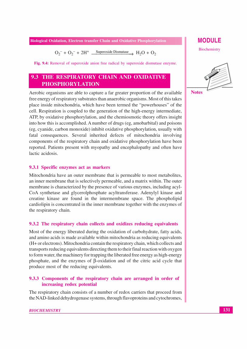
Transfer of a single electron to O2 generates the potentially damaging superoxideanion free radical (O2"-) (Figure 9.4), the destructive effects of which areamplified by its giving rise to free radical chain reactions. The ease with whichsuperoxide can be formed from oxygen in tissues and the occurrence ofsuperoxide dismutase, the enzyme responsible for its removal in all aerobicorganisms (although not in obligate anaerobes) indicate that the potentialtoxicity of oxygen is due to its conversion to superoxide. Superoxide is formedwhen reduced flavins – present, for example, in xanthine oxidase – arereoxidized univalently by molecular oxygen.
131
Biological Oxidation, Electron transfer Chain and Oxidative Phosphorylation
BIOCHEMISTRY
MODULEBiochemistry
Notes
O2– + O2
– + 2H+ Superoxide Dismutase⎯⎯⎯⎯⎯⎯⎯⎯→ H2O + O2
Fig. 9.4: Removal of superoxide anion free radical by superoxide dismutase enzyme.
9.3 THE RESPIRATORY CHAIN AND OXIDATIVEPHOSPHORYLATION
Aerobic organisms are able to capture a far greater proportion of the availablefree energy of respiratory substrates than anaerobic organisms. Most of this takesplace inside mitochondria, which have been termed the “powerhouses” of thecell. Respiration is coupled to the generation of the high-energy intermediate,ATP, by oxidative phosphorylation, and the chemiosmotic theory offers insightinto how this is accomplished. A number of drugs (eg, amobarbital) and poisons(eg, cyanide, carbon monoxide) inhibit oxidative phosphorylation, usually withfatal consequences. Several inherited defects of mitochondria involvingcomponents of the respiratory chain and oxidative phosphorylation have beenreported. Patients present with myopathy and encephalopathy and often havelactic acidosis.
9.3.1 Specific enzymes act as markers
Mitochondria have an outer membrane that is permeable to most metabolites,an inner membrane that is selectively permeable, and a matrix within. The outermembrane is characterized by the presence of various enzymes, including acyl-CoA synthetase and glycerolphosphate acyltransferase. Adenylyl kinase andcreatine kinase are found in the intermembrane space. The phospholipidcardiolipin is concentrated in the inner membrane together with the enzymes ofthe respiratory chain.
9.3.2 The respiratory chain collects and oxidizes reducing equivalents
Most of the energy liberated during the oxidation of carbohydrate, fatty acids,and amino acids is made available within mitochondria as reducing equivalents(H+ or electrons). Mitochondria contain the respiratory chain, which collects andtransports reducing equivalents directing them to their final reaction with oxygento form water, the machinery for trapping the liberated free energy as high-energyphosphate, and the enzymes of β-oxidation and of the citric acid cycle thatproduce most of the reducing equivalents.
9.3.3 Components of the respiratory chain are arranged in order ofincreasing redox potential
The respiratory chain consists of a number of redox carriers that proceed fromthe NAD-linked dehydrogenase systems, through flavoproteins and cytochromes,

BIOCHEMISTRY
MODULE Biological Oxidation, Electron transfer Chain and Oxidative Phosphorylation
Biochemistry
132
Notes
to molecular oxygen. Not all substrates are linked to the respiratory chainthrough NAD-specific dehydrogenases; some, because their redox potentials aremore positive (eg, fumarate/succinate; are linked directly to flavoproteindehydrogenases, which in turn are linked to the cytochromes of the respiratorychain.
9.3.4 Ubiquinone or Q (coenzyme Q)
Coenzyme Q links the flavoproteins to cytochrome b, the member of thecytochrome chain of lowest redox potential. Q exists in the oxidized quinoneor reduced quinol form under aerobic or anaerobic conditions, respectively. Thestructure of Q is very similar to that of vitamin K and vitamin E and ofplastoquinone, found in chloroplasts. Q acts as a mobile component of therespiratory chain that collects reducing equivalents from the more fixedflavoprotein complexes and passes them on to the cytochromes. An additionalcomponent is the iron-sulfur protein (FeS; nonheme iron). It is associated withthe flavoproteins (metalloflavoproteins) and with cytochrome b. The sulfur andiron are thought to take part in the oxidoreduction mechanism between flavinand Q, which involves only a single e-change, the iron atom undergoingoxidoreduction between Fe2+ and Fe3+.
Fig. 9.5: In this simple representation of the chemiosmotic theory applied to mitochondria,electrons from NADH and other oxidizable substrates pass through a chain of carriers arrangedasymmetrically in the inner membrane. Electron flow is accompanied by proton transfer acrossthe membrane, producing both a chemical gradient (ΔpH) and an electrical gradient (Δψ). Theinner mitochondrial membrane is impermeable to protons; protons can reenter the matrix onlythrough proton-specific channels (Fo). The proton-motive force that drives protons back intothe matrix provides the energy for ATP synthesis, catalyzed by the F1 complex associated withFo.

133
Biological Oxidation, Electron transfer Chain and Oxidative Phosphorylation
BIOCHEMISTRY
MODULEBiochemistry
Notes
Pyruvate and α-ketoglutarate dehydrogenase have complex systems involvinglipoate and FAD prior to the passage of electrons to NAD, while electron transfers from other dehydrogenases, e.g., L(+)-3-hydroxyacyl- CoA dehydrogenase,couple directly with NAD. The reduced NADH of the respiratory chain is in turnoxidized by a metalloflavoprotein enzyme – NADH dehydrogenase. Thisenzyme contains FeS and FMN, is tightly bound to the respiratory chain, andpasses reducing equivalents on to Q. Electrons flow from Q through the seriesof cytochromes in order of increasing redox potential to molecular oxygen. Theterminal cytochrome aa3 (cytochrome oxidase), responsible for the finalcombination of reducing equivalents with molecular oxygen, has a very highaffinity for oxygen, allowing the respiratory chain to function at maximum rateuntil the tissue has become depleted of O2. Since this is an irreversible reaction(the only one in the chain), it gives direction to the movement of reducingequivalents and to the production of ATP, to which it is coupled. Functionallyand structurally, the components of the respiratory chain are present in the innermitochondrial membrane as four protein-lipid respiratory chain complexes thatspan the membrane. Cytochrome c is the only soluble cytochrome and, togetherwith Q, seems to be a more mobile component of the respiratory chainconnecting the fixed complexes. The overall reaction is given in the figure 9.5.
9.3.5 The respiratory chain provides most of the energy captured duringcatabolism
ADP captures, in the form of high-energy phosphate, a significant proportion ofthe free energy released by catabolic processes. The resulting ATP has beencalled the energy “currency” of the cell because it passes on this free energy todrive those processes requiring energy. There is a net direct capture of two high-energy phosphate groups in the glycolytic reactions, equivalent to approximately103.2 kJ/mol of glucose. (In vivo, ΔG for the synthesis of ATP from ADP hasbeen calculated as approximately 51.6 kJ/mol. (It is greater than ΔG0' for thehydrolysis of ATP, which is obtained under standard concentrations of 1.0 mol/L.) Since 1 mol of glucose yields approximately 2870 kJ on completecombustion, the energy captured by phosphorylation in glycolysis is small. Twomore high-energy phosphates per mole of glucose are captured in the citric acidcycle during the conversion of succinyl CoA to succinate.
All of these phosphorylations occur at the substrate level. When substrates areoxidized via an NAD-linked dehydrogenase and the respiratory chain,approximately 3 mol of inorganic phosphate are incorporated into 3 mol of ADPto form 3 mol of ATP per half mol of O2 consumed; ie, the P:O ratio = 3. Onthe other hand, when a substrate is oxidized via a flavoprotein- linkeddehydrogenase, only 2 mol of ATP are formed; ie, P:O = 2. These reactions are

BIOCHEMISTRY
MODULE Biological Oxidation, Electron transfer Chain and Oxidative Phosphorylation
Biochemistry
134
Notes
known as oxidative phosphorylation at the respiratory chain level. Suchdehydrogenations plus phosphorylations at the substrate level can now accountfor 68% of the free energy resulting from the combustion of glucose, capturedin the form of high-energy phosphate. It is evident that the respiratory chain isresponsible for a large proportion of total ATP formation.
9.3.6 Respiratory control ensures a constant supply of ATP
The rate of respiration of mitochondria can be controlled by the availability ofADP. This is because oxidation and phosphorylation are tightly coupled; ie,oxidation cannot proceed via the respiratory chain without concomitantphosphorylation of ADP. When work is performed, ATP is converted to ADP,allowing more respiration to occur, which in turn replenishes the store of ATP.Under certain conditions, the concentration of inorganic phosphate can alsoaffect the rate of functioning of the respiratory chain. There is also the possibilitythat the ADP/ATP transporter, which facilitates entry of cytosolic ADP into andATP out of the mitochondrion, becomes rate limiting. Thus, the manner in whichbiologic oxidative processes allow the free energy resulting from the oxidationof foodstuffs to become available and to be captured is stepwise, efficient(approximately 68%), and controlled – rather than explosive, inefficient, anduncontrolled, as in many nonbiologic processes. The remaining free energy thatis not captured as high-energy phosphate is liberated as heat. This need not beconsidered “wasted,” since it ensures that the respiratory system as a whole issufficiently exergonic to be removed from equilibrium, allowing continuousunidirectional flow and constant provision of ATP. It also contributes tomaintenance of body temperature.
9.3.7 Many poisons inhibit the respiratory chain
Much information about the respiratory chain has been obtained by the use ofinhibitors, and, conversely, this has provided knowledge about the mechanismof action of several poisons (Figure 9.6). They may be classified as inhibitorsof the respiratory chain, inhibitors of oxidative phosphorylation, and uncouplersof oxidative phosphorylation. Barbiturates such as amobarbital inhibit NADlinked dehydrogenases by blocking the transfer from FeS to Q. At sufficientdosage, they are fatal in vivo. Antimycin A and dimercaprol inhibit therespiratory chain between cytochrome b and cytochrome c. The classic poisonsH2S, carbon monoxide, and cyanide inhibit cytochrome oxidase and cantherefore totally arrest respiration. Malonate is a competitive inhibitor ofsuccinate dehydrogenase. Atractyloside inhibits oxidative phosphorylation byinhibiting the transporter of ADP into and ATP out of the mitochondrion. The

135
Biological Oxidation, Electron transfer Chain and Oxidative Phosphorylation
BIOCHEMISTRY
MODULEBiochemistry
Notes
action of uncouplers is to dissociate oxidation in the respiratory chain fromphosphorylation. These compounds are toxic in vivo, causing respiration tobecome uncontrolled, since the rate is no longer limited by the concentration ofADP or Pi. The uncoupler that has been used most frequently is 2,4-dinitrophenol, but other compounds act in a similar manner. The antibioticoligomycin completely blocks oxidation and phosphorylation by acting on a stepin phosphorylation.
Fig. 9.6: Proposed sites of inhibition (--) of the respiratory chain by specific drugs, chemicals,and antibiotics. The sites that appear to support phosphorylation are indicated. BAL,dimercaprol. TTFA, an Fe-chelating agent. Complex I, NADH:ubiquinone oxidoreductase;complex II, succinate:ubiquinone oxidoreductase; complex III, ubiquinol:ferricytochrome coxidoreductase; complex IV, ferrocytochrome c:oxygen oxidoreductase.
9.3.8 The chemiosmotic theory explains the mechanism of oxidativephosphorylation
Mitchell’s chemiosmotic theory postulates that the energy from oxidation ofcomponents in the respiratory chain is coupled to the translocation of hydrogenions (protons, H+) from the inside to the outside of the inner mitochondrialmembrane. The electrochemical potential difference resulting from the asymmetricdistribution of the hydrogen ions is used to drive the mechanism responsible forthe formation of ATP (Figure 9.7).
9.3.9. The respiratory chain is a proton pump
Each of the respiratory chain complexes I, III, and IV act as a proton pump. Theinner membrane is impermeable to ions in general but particularly to protons,which accumulate outside the membrane, creating an electrochemical potentialdifference across the membrane. This consists of a chemical potential (differencein pH) and an electrical potential.

BIOCHEMISTRY
MODULE Biological Oxidation, Electron transfer Chain and Oxidative Phosphorylation
Biochemistry
136
Notes
Fig. 9.7: Principles of the chemiosmotic theory of oxidative phosphorylation. The main protoncircuit is created by the coupling of oxidation in the respiratory chain to proton translocationfrom the inside to the outside of the membrane, driven by the respiratory chain complexes I,III, and IV, each of which acts as a proton pump. Q, ubiquinone; C, cytochrome c; F1, F0,protein subunits which utilize energy from the proton gradient to promote phosphorylation.Uncoupling agents such as dinitrophenol allow leakage of H+ across the membrane, thuscollapsing the electrochemical proton gradient. Oligomycin specifically blocks conduction ofH+ through F0.
9.3.10 A membrane-located ATP synthase functions as a rotary motor toform ATP
The electrochemical potential difference is used to drive a membrane-locatedATP synthase which in the presence of Pi + ADP forms ATP. Scattered over thesurface of the inner membrane are the phosphorylating complexes, ATPsynthase, responsible for the production of ATP. These consist of several proteinsubunits, collectively known as F1, which project into the matrix and whichcontain the phosphorylation mechanism. These subunits are attached to amembrane protein complex known as F0, which also consists of several proteinsubunits. F0 spans the membrane and forms the proton channel. The flow ofprotons through F0 causes it to rotate, driving the production of ATP in the F1complex. Estimates suggest that for each NADH oxidized, complex I translocatesfour protons and complexes III and IV translocate 6 between them. As fourprotons are taken into the mitochondrion for each ATP exported, the P:O ratiowould not necessarily be a complete integer, ie, 3, but possibly 2.5. However,for simplicity, a value of 3 for the oxidation of NADH + H+ and 2 for theoxidation of FADH2 will continue to be used throughout this text.

137
Biological Oxidation, Electron transfer Chain and Oxidative Phosphorylation
BIOCHEMISTRY
MODULEBiochemistry
Notes
9.3.11 The chemiosmotic theory can account for respiratory control andthe action of uncouplers
The electrochemical potential difference across the membrane, once establishedas a result of proton translocation, inhibits further transport of reducingequivalents through the respiratory chain unless discharged by back translocationof protons across the membrane through the vectorial ATP synthase. This in turndepends on availability of ADP and Pi. Uncouplers (eg, dinitrophenol) areamphipathic and increase the permeability of the lipoid inner mitochondrialmembrane to protons, thus reducing the electrochemical potential and short-circuiting the ATP synthase. In this way, oxidation can proceed withoutphosphorylation.
9.3.12 Impermeability of the inner mitochondrial membrane
The relative impermeability of the inner mitochondrial membrane necessitatesexchange transporters. Exchange diffusion systems are present in the membranefor exchange of anions against OH- ions and cations against H+ ions. Suchsystems are necessary for uptake and output of ionized metabolites whilepreserving electrical and osmotic equilibrium. The inner bilipoid mitochondrialmembrane is freely permeable to uncharged small molecules, such as oxygen,water, CO2, and NH3, and to monocarboxylic acids, such as 3-hydroxybutyric,acetoacetic, and acetic. Long-chain fatty acids are transported into mitochondriavia the carnitine system, and there is also a special carrier for pyruvate involvinga symport that utilizes the H+ gradient from outside to inside the mitochondrion.However, dicarboxylate and tri- carboxylate anions and amino acids requirespecific transporter or carrier systems to facilitate their passage across themembrane. Monocarboxylic acids penetrate more readily in their undissociatedand more lipid-soluble form. The transport of di- and tricarboxylate anions isclosely linked to that of inorganic phosphate, which penetrates readily as theH2PO4
– ion in exchange for OH–.
9.3.13. Ionophores permit specific cations to penetrate membranes
Ionophores are lipophilic molecules that complex specific cations and facilitatetheir transport through biologic membranes, eg, valinomycin (K+). The classicuncouplers such as dinitrophenol are, in fact, proton ionophores.
9.3.14 A proton-translocating transhydrogenase is a source ofintramitochon-drial NADPH
Energy-linked transhydrogenase, a protein in the inner mitochondrial membrane,couples the passage of protons down the electrochemical gradient from outside

BIOCHEMISTRY
MODULE Biological Oxidation, Electron transfer Chain and Oxidative Phosphorylation
Biochemistry
138
Notes
to inside the mitochondrion with the transfer of H from intramitochondrialNADH to NADPH for intramitochondrial enzymes such as glutamatedehydrogenase and hydroxylases involved in steroid synthesis.
9.3.15 Oxidation of extramitochondrial NADH is mediated by substrateshuttles
NADH cannot penetrate the mitochondrial membrane, but it is producedcontinuously in the cytosol by 3-phosphoglyceraldehyde dehydrogenase, anenzyme in the glycolysis sequence. However, under aerobic conditions,extramitochondrial NADH does not accumulate and is presumed to be oxidizedby the respiratory chain in mitochondria. The transfer of reducing equivalentsthrough the mitochondrial membrane requires substrate pairs, linked by suitabledehydrogenases on each side of the mitochondrial membrane. The mechanismof transfer uses glycerophosphate shuttle. Since the mitochondrial enzyme islinked to the respiratory chain via a flavoprotein rather than NAD, only 2 molrather than 3 mol of ATP are formed per atom of oxygen consumed. Althoughthis shuttle is present in some tissues (eg, brain, white muscle), in others (eg,heart muscle) it is deficient. It is therefore believed that the malate shuttle systemis of more universal utility. The complexity of this system is due to theimpermeability of the mitochondrial membrane to oxaloacetate, which mustreact with glutamate and transaminate to aspartate and á-ketoglutarate beforetransport through the mitochondrial membrane and reconstitution to oxaloacetatein the cytosol.
9.3.16. Ion transport in mitochondria is energy-linked
Mitochondria maintain or accumulate cations such as K+, Na+, Ca2+, andMg2+, and Pi. It is assumed that a primary proton pump drives cation exchange.
9.3.17 The creatine phosphate shuttle facilitates transport of high-energyphosphate from mitochondria
The creatine phosphate shuttle augments the functions of creatine phosphate asan energy buffer by acting as a dynamic system for transfer of high-energyphosphate from mitochondria in active tissues such as heart and skeletal muscle.An isoenzyme of creatine kinase is found in the mitochondrial intermembranespace, catalyzing the transfer of high-energy phosphate to creatine from ATPemerging from the adenine nucleotide transporter. In turn, the creatine phosphateis transported into the cytosol via protein pores in the outer mitochondrialmembrane, becoming available for generation of extramitochondrial ATP.

139
Biological Oxidation, Electron transfer Chain and Oxidative Phosphorylation
BIOCHEMISTRY
MODULEBiochemistry
Notes
INTEXT QUESTIONS 9.1I. Choose the best answer
1. Chemically, the removal and the gain of electrons is defined respectivelyas
(a) Oxidation and reduction
(b) Reduction and oxidation
(c) Oxidation and dehydrogenase
(d) Reduction and dehydrogenase
2. Chemical carcinogens (xenobiotics) are metabolized by the enzymessystem known as
(a) Cytochrome P450 (b) Xanthine oxidase
(c) Succinate dehydrogenase (d) Hydroperoxides
3. Oxidative phosphorylation is inhibited usually with fatal consequencesby
(a) Quinalones (b) Cyanide
(c) Anacin (d) Amoxycillin
4. The action of uncouplers is to dissociate oxidation in the respiratorychain from
(a) Gluconeogenesis (b) Glycolysis
(c) TCA cycle (d) Phosphorylation
5. This antibiotic completely blocks oxidation and phosphorylation byacting on a step in phosphorylation.
(a) Dinitrophenol (b) Benzpyrene
(c) Oligomycin (d) Morphine
II. Fill in the blanks
6. Flavoprotein enzymes contain .................. or .................. as prostheticgroups.
7. Generally, NAD-linked dehydrogenases catalyze .................. reactionsin the oxidative pathways of metabolism.
8. .................. protect the body against harmful peroxides.
9. The function of .................. is assumed to be the destruction ofhydrogen peroxide formed by the action of oxidases.
10. Cytochromes P450 are an important superfamily of heme-containing..................

BIOCHEMISTRY
MODULE Biological Oxidation, Electron transfer Chain and Oxidative Phosphorylation
Biochemistry
140
Notes
III. Match the following
11. Mitochondria (a) Energy currency of the cell
12. Cardiolipin (b) Uncouplers
13. ATP (c) Powerhouses of the cell
14. Valinomycin (d) Phospholipid
15. Dinitrophenol (e) Ionophores
WHAT HAVE YOU LEARNT
Oxidation is defined as the removal of electrons and reduction as the gainof electrons.
Many drugs, pollutants, and chemical carcinogens (xenobiotics) aremetabolized oxygenases known as cytochrome P450 system.
Enzymes involved in oxidation and reduction are called oxidoreductasesand are classified into four groups: oxidases, dehydrogenases,hydroperoxidases, and oxygenases.
Flavoprotein enzymes contain flavin mononucleotide (FMN) or flavinadenine dinucleotide (FAD) as prosthetic groups. FMN and FAD are formedin the body from the vitamin riboflavin.
Oxidation-reduction reactions carried out by dehydrogenases are specificfor their substrates but often utilize common coenzymes or hydrogencarriers, eg, NAD+.
The cytochromes are iron-containing hemoproteins in which the iron atomoscillates between Fe3+ and Fe2+ during oxidation and reduction.
Cytochromes are also found in the endoplasmic reticulum (cytochromesP450 and b5), and in plant cells, bacteria, and yeasts.
Two type of enzymes found both in animals and plants are peroxidases andcatalase. Hydroperoxidases protect the body against harmful peroxides.
Accumulation of peroxides can lead to generation of free radicals, whichin turn can disrupt membranes and perhaps cause cancer and atherosclerosis.
Mitochondrial cytochrome P450 systems are found in steroidogenic tissuessuch as adrenal cortex, testis, ovary, and placenta and are concerned withthe biosynthesis of steroid hormones from cholesterol.
The potential toxicity of oxygen is due to its conversion to superoxide intissues and the enzyme superoxide dismutase is responsible for its removal.
Mitochondria are termed as the “powerhouses” of the cell. Respiration iscoupled to the generation of the high-energy intermediate, ATP, by oxidative
141
Biological Oxidation, Electron transfer Chain and Oxidative Phosphorylation
BIOCHEMISTRY
MODULEBiochemistry
Notes
phosphorylation, and the chemiosmotic theory offers insight into how thisis accomplished.
A number of drugs (eg, amobarbital) and poisons (eg, cyanide, carbonmonoxide) inhibit oxidative phosphorylation, usually with fatal consequences.
Mitochondria contain the respiratory chain, which collects and transportsreducing equivalents directing them to their final reaction with oxygen toform water, the machinery for trapping the liberated free energy as high-energy phosphate, and the enzymes of β-oxidation and of the citric acidcycle that produce most of the reducing equivalents.
Mitchell’s chemiosmotic theory postulates that the energy from oxidationof components in the respiratory chain is coupled to the translocation ofhydrogen ions (protons, H+) from the inside to the outside of the innermitochondrial membrane. The electrochemical potential difference resultingfrom the asymmetric distribution of the hydrogen ions is used to drive themechanism responsible for the formation of ATP.
Uncouplers (eg, dinitrophenol) are amphipathic and increase the permeabilityof the lipoid inner mitochondrial membrane to protons, thus reducing theelectrochemical potential and short-circuiting the ATP synthase. In this way,oxidation can proceed without phosphorylation.
Ionophores are lipophilic molecules that complex specific cations andfacilitate their transport through biologic membranes, eg, valinomycin (K+).
TERMINAL QUESTIONS
1. Write short note on electron transfer chain.
2. Write short note on oxidative phosphorylation.
ANSWERS TO INTEXT QUESTIONS
I. 1. (a) 2. (a) 3. (b) 4. (d) 5. (c)
II. 6. Flavin mononucleotide (FMN) or Flavin adenine dinucleotide (FAD)
7. Oxidoreduction
8. Hydroperoxidases
9. Catalase
10. Monooxgenases
III. 11. (c) 12. (d) 13. (a) 14. (e) 15. (b)