Embed Size (px)
Citation preview

Lesmahagow High School AHChemistry Organic Chemistry& Instrumental Analysis
Lesmahagow High School CfE Advanced Higher Chemistry
Unit 2
Organic Chemistry and Instrumental Analysis
Structural Organic Chemistry and
Pharmaceutical Chemistry
Experimental determination of structure
In organic chemistry not only are we able to synthesise products used to meet mar-
ket demands but also we can verify that the correct chemical structure has actually
been synthesised.
There are various methods used to determine the chemical structure of compounds
such as
Elemental microanalysis
Mass spectroscopy
Infrared spectroscopy
Proton NMR spectroscopy
UV/Vis spectroscopy
Elemental Microanalysis otherwise known as combustion analysis is used to deter-
mine the masses of elements in a sample of an organic compound in order to deter-
mine its empirical formula. The compounds dealt with will normally contain carbon,
hydroen, sulphur and nitrogen.
Emperical formula shows the simplest whole number ratio of different atoms in the
compound.
Ethane C2H6 has a C:H ratio of 1:3 therefore its empirical formula would be CH3
In combustion analysis a tiny sample of compound is burned in an excess of oxygen
which ensures that the products of the combustion reaction are carbon dioxide, ni-
trogen dioxide, sulfue dioxide and water.
The mixture of gases is then carried in a stream of helium over heated copper which
reduces the nitrous oxides produced to nitrogen.
The quantities of each of the products of combustion are then determined by infra-
red detectors, absorbred in anhydrous magnesium chlorate and mearsurements of
thermal conductivity.
Knowing the masses of CO2 , SO2 , H2O and nitrogen makes it very easy to calculate
the masses of carbon, hydrogen, sulphur and nitrogen in the compound and therefore
its empirical formula.

Emperical Formula
Example 1
A hydrocarbon was found to contain 4.5 g of carbon and 1.5 g of hydrogen. What is
the empirical formula of the hydrocarbon?
Element C H
Mass 4.5g 1.5g
Divide by atomic mass 4.5/12 1.5/1
= 0.375 1.5
Divide to give whole numbers 0.375/0.375 1.5/0.375
= 1 4
Empirical Formula: CH4
Example 2
Analysis of an alcohol shows it to contain 37.5% carbon and 12.5% hydrogen. What is
the empirical formula?
Element C H O
% by weight 37.5 12.5 50.0
Divide by atomic mass 37.5/12 12.5/1 50.0/16
= 3.125 12.5 3.125
Divide to give whole numbers 3.125/3.125 12.5/3.125 3.125/3.125
= 1 4 1
Empirical Formula: CH4O
Example 3
5.0g of aluminium was burned in oxygen producing 9.45g of aluminium oxide. Calculate
the empirical formula.
Element Al O
Mass 5.0g 4.45g
Divide by atomic mass 5.0/27 4.45/16
= 0.185 0.278
Divide to give whole numbers 0.185/0.185 0.278/0.278
= 1 1.5
Scale up (x2) = 2 3
Empirical Formula: Al2O3

Mass Spectrometry
Mass spectrometry is a technique used to determine the accurate molecular mass
and structural features of a particular compound.
The sample is vapourised and then ionised by being bombarded with electrons in
the ionisation chamber. The sample molecules then break up into smaller fragments
(Fragmentation)
The parent ion and ion fragments are:
1. accelerated by an electric field and then
2. deflected by a magnetic field
Ions with lower m/z (mass/charge) ratio are deflected more than ions with higher
m/z ratio.
A mass spectrum (specific to each organic substance) is obtained.
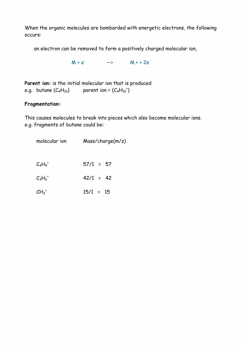
When the organic molecules are bombarded with energetic electrons, the following
occurs:
an electron can be removed to form a positively charged molecular ion,
M + e- —> M.+ + 2e-
Parent ion: is the initial molecular ion that is produced
e.g. butane (C4H10) parent ion = (C4H10+)
Fragmentation:
This causes molecules to break into pieces which also become molecular ions.
e.g. fragments of butane could be:
molecular ion Mass/charge(m/z)
C4H9+ 57/1 = 57
C3H6+ 42/1 = 42
CH3+ 15/1 = 15

Examples of Mass Spectra
Benzoic Acid
1. The peak with the highest m/z value is often the molecular ion:
C6H5COOH + = 122
2. The most abundant peak is called the base peak and is assigned an abundance of
100%:
C6H5CO + = 105
3. Other abundances are given smaller percentages, relative to the base peak:
C6H5 + = 77

Mass Spectra
Hexane
Mass Molecular Ion
86 C6H14+
71 C5H11+
15 CH3+
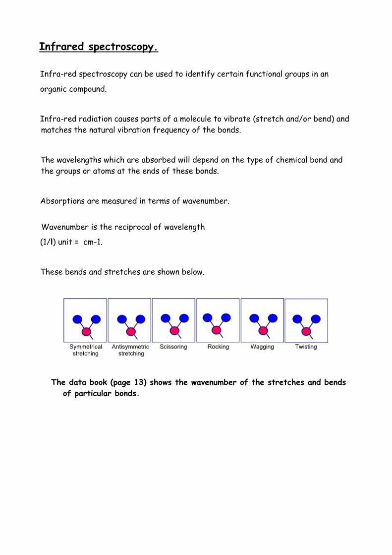
Infrared spectroscopy.
Infra-red spectroscopy can be used to identify certain functional groups in an
organic compound.
Infra-red radiation causes parts of a molecule to vibrate (stretch and/or bend) and
matches the natural vibration frequency of the bonds.
The wavelengths which are absorbed will depend on the type of chemical bond and
the groups or atoms at the ends of these bonds.
Absorptions are measured in terms of wavenumber.
Wavenumber is the reciprocal of wavelength
(1/l) unit = cm-1.
These bends and stretches are shown below.
The data book (page 13) shows the wavenumber of the stretches and bends
of particular bonds.

IR Spectrometer
An infra-red spectrometer passes radiation through a sample of the organic com-
pound and then to a detector.
An identical beam is passed through a reference blank cell and the detector com-
pares the intensities of radiation through the sample and reference cell.
Where groups absorb, infra-red radiation transmittance will be lower and a
trough is seen in the spectrum.
The main use of IR spectra, which can be obtained quickly and cheaply, is to iden-
tify the presence of functional groups and the carbon backbone type in un-
known organic compounds.
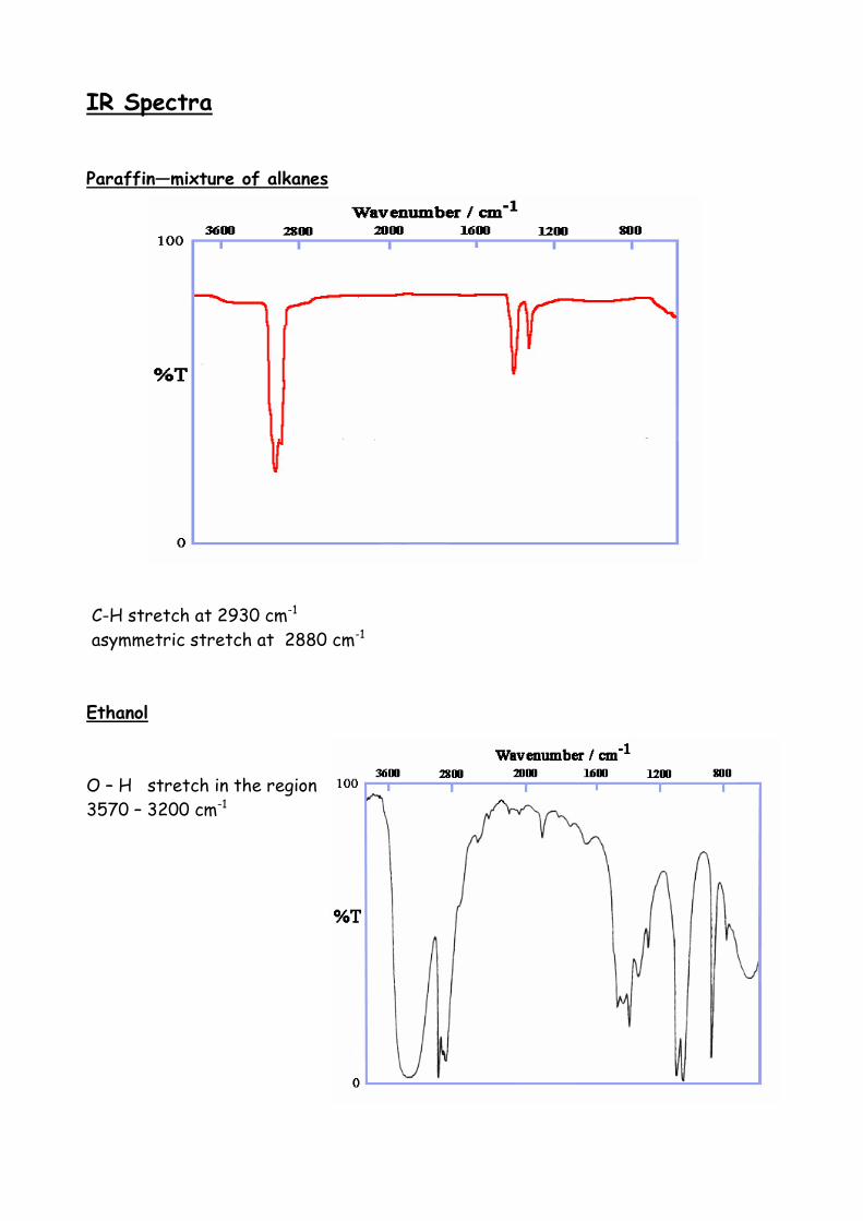
IR Spectra
Paraffin—mixture of alkanes
C-H stretch at 2930 cm-1
asymmetric stretch at 2880 cm-1
Ethanol
O – H stretch in the region
3570 – 3200 cm-1
Proton NMR Spectroscopy
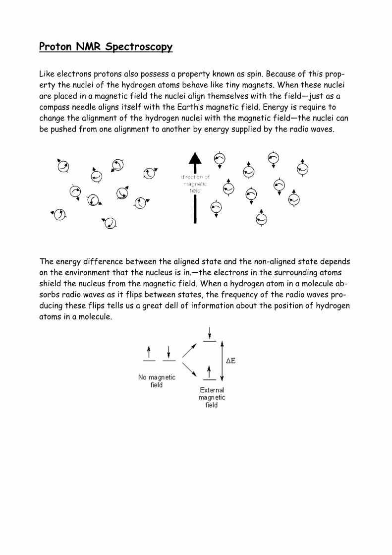
Like electrons protons also possess a property known as spin. Because of this prop-
erty the nuclei of the hydrogen atoms behave like tiny magnets. When these nuclei
are placed in a magnetic field the nuclei align themselves with the field—just as a
compass needle aligns itself with the Earth’s magnetic field. Energy is require to
change the alignment of the hydrogen nuclei with the magnetic field—the nuclei can
be pushed from one alignment to another by energy supplied by the radio waves.
The energy difference between the aligned state and the non-aligned state depends
on the environment that the nucleus is in.—the electrons in the surrounding atoms
shield the nucleus from the magnetic field. When a hydrogen atom in a molecule ab-
sorbs radio waves as it flips between states, the frequency of the radio waves pro-
ducing these flips tells us a great dell of information about the position of hydrogen
atoms in a molecule.
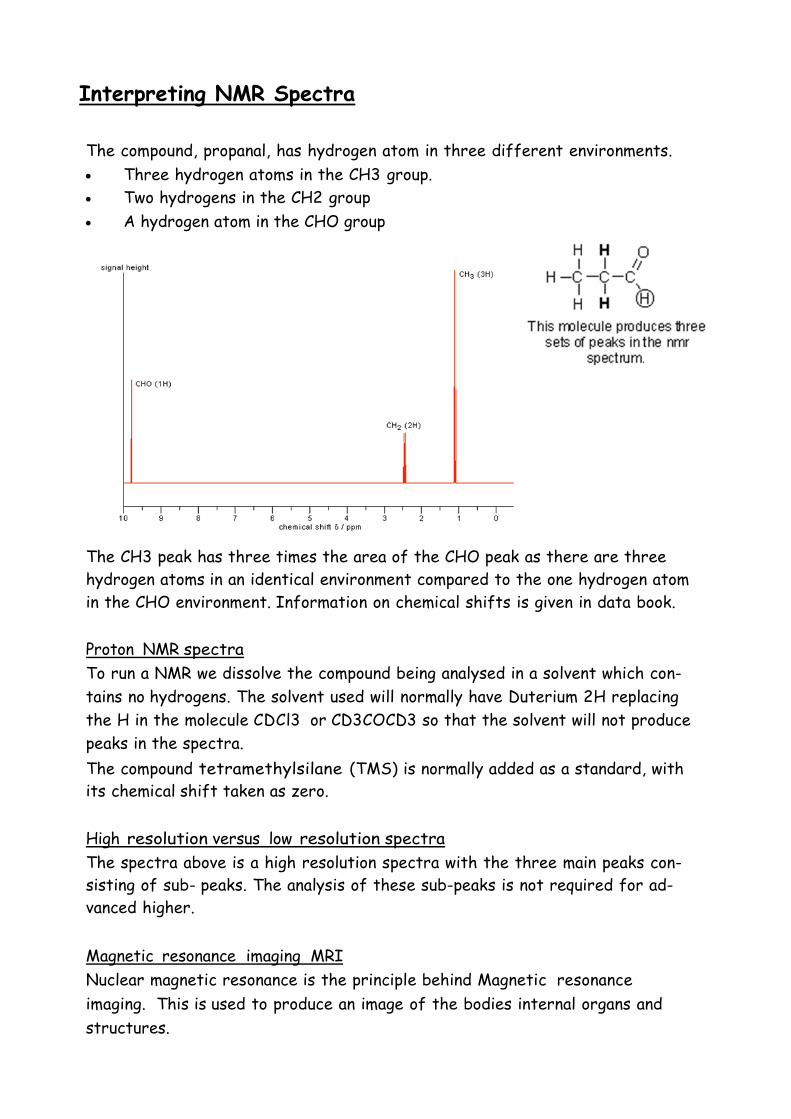
Interpreting NMR Spectra
The compound, propanal, has hydrogen atom in three different environments.
Three hydrogen atoms in the CH3 group.
Two hydrogens in the CH2 group
A hydrogen atom in the CHO group
The CH3 peak has three times the area of the CHO peak as there are three
hydrogen atoms in an identical environment compared to the one hydrogen atom
in the CHO environment. Information on chemical shifts is given in data book.
Proton NMR spectra
To run a NMR we dissolve the compound being analysed in a solvent which con-
tains no hydrogens. The solvent used will normally have Duterium 2H replacing
the H in the molecule CDCl3 or CD3COCD3 so that the solvent will not produce
peaks in the spectra.
The compound tetramethylsilane (TMS) is normally added as a standard, with
its chemical shift taken as zero.
High resolution versus low resolution spectra
The spectra above is a high resolution spectra with the three main peaks con-
sisting of sub- peaks. The analysis of these sub-peaks is not required for ad-
vanced higher.
Magnetic resonance imaging MRI
Nuclear magnetic resonance is the principle behind Magnetic resonance
imaging. This is used to produce an image of the bodies internal organs and
structures.
UV/Vis Spectroscopy
While many chemical compounds are coloured because they absorb visi-
ble light, most organic molecules appear colourless.
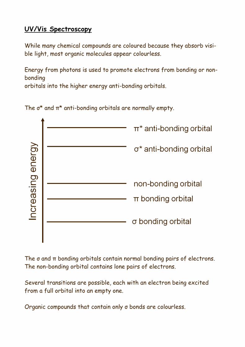
Energy from photons is used to promote electrons from bonding or non-
bonding
orbitals into the higher energy anti-bonding orbitals.
The σ* and π* anti-bonding orbitals are normally empty.
The σ and π bonding orbitals contain normal bonding pairs of electrons.
The non-bonding orbital contains lone pairs of electrons.
Several transitions are possible, each with an electron being excited
from a full orbital into an empty one.
Organic compounds that contain only σ bonds are colourless.
Pharmaceutical chemistry.
Drugs are the active ingredients in medicines that alter the biochemical
processes in the body and so affect the way in which your body works. Drugs
which have a beneficial effect are called medicines. The study of drugs and their
actions on the body is called pharmacology.
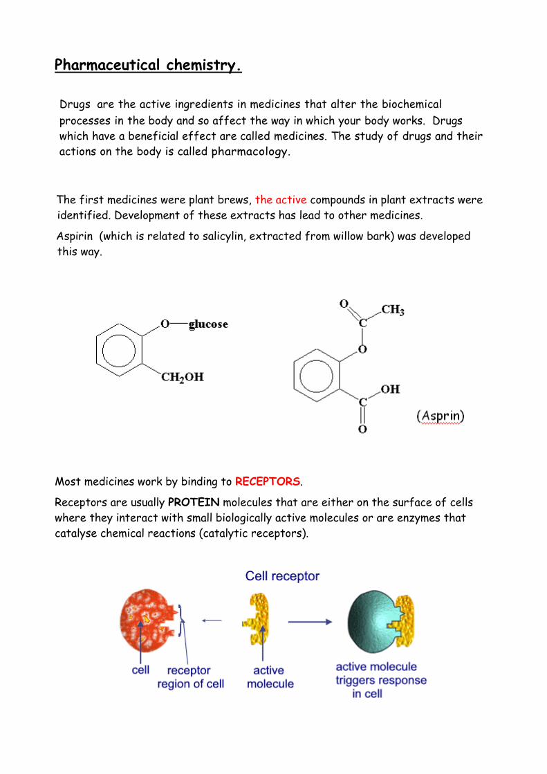
The first medicines were plant brews, the active compounds in plant extracts were
identified. Development of these extracts has lead to other medicines.
Aspirin (which is related to salicylin, extracted from willow bark) was developed
this way.
Most medicines work by binding to RECEPTORS.
Receptors are usually PROTEIN molecules that are either on the surface of cells
where they interact with small biologically active molecules or are enzymes that
catalyse chemical reactions (catalytic receptors).

Pharmacophore
The structural fragment of the molecule which confers pharmacological activity
on it is called the PHARMACOPHORE.
The shape of the pharmacophore complements that of the receptor site, allow-
ing it to fit into the receptor.
The functional groups on both are correctly positioned to interact and bind the
medicine to the receptor.
By comparing the structures of medicines with
similar pharmacological activity, the pharma-
cophore can be identified.
e.g.
morphine (highly addictive)
When the shape, dimensions and functional groups present have been identified,
the pharmacophore can be cropped, added to and manipulated by chemists to
produce compounds which are still analgesic but less addictive
Agonists and Antagonists
There are two types of drugs :
AGONISTS: which mimick the response of the body's active molecule
e.g. salbutamol
AND
ANTAGONISTS: which block the effect of the body's active molecule
e.g. anti-histamines, β-blockers
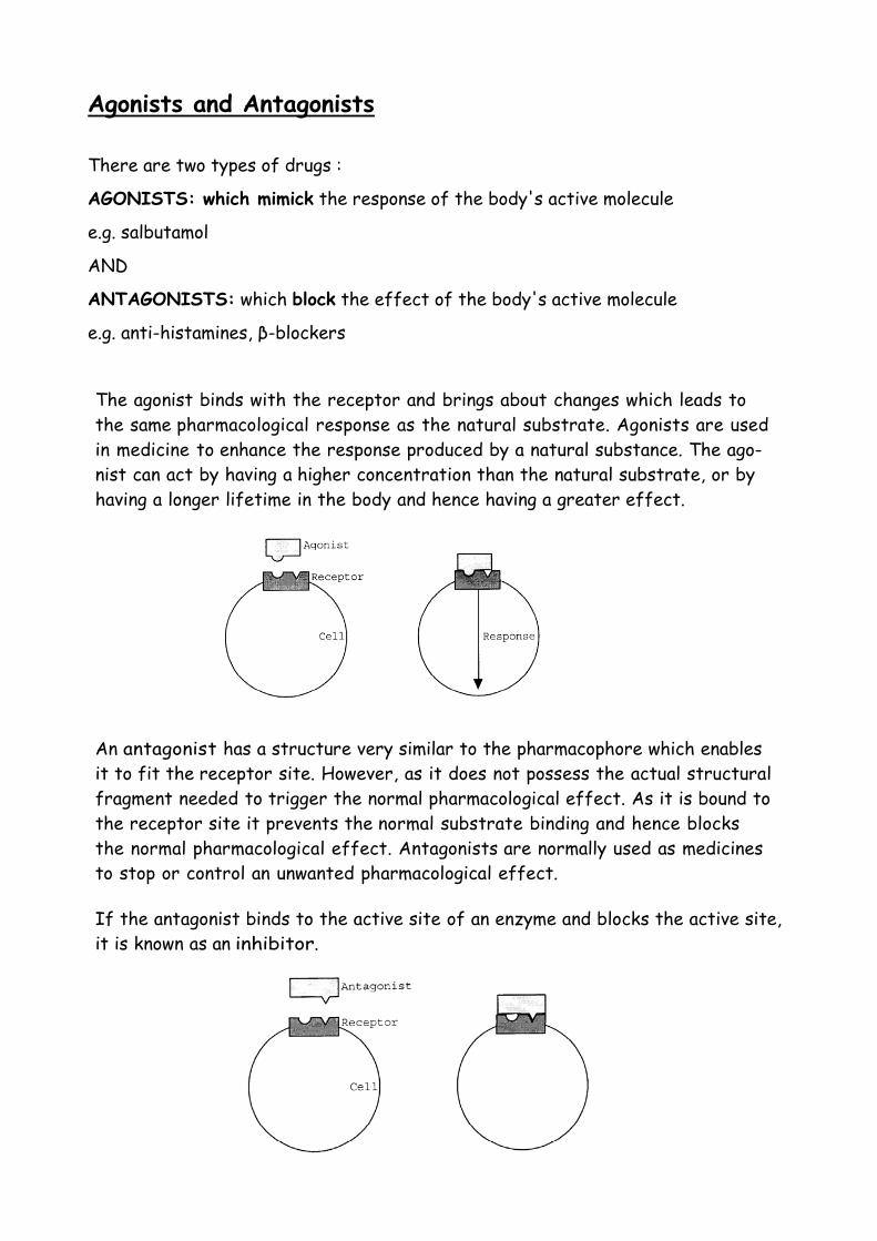
The agonist binds with the receptor and brings about changes which leads to
the same pharmacological response as the natural substrate. Agonists are used
in medicine to enhance the response produced by a natural substance. The ago-
nist can act by having a higher concentration than the natural substrate, or by
having a longer lifetime in the body and hence having a greater effect.
An antagonist has a structure very similar to the pharmacophore which enables
it to fit the receptor site. However, as it does not possess the actual structural
fragment needed to trigger the normal pharmacological effect. As it is bound to
the receptor site it prevents the normal substrate binding and hence blocks
the normal pharmacological effect. Antagonists are normally used as medicines
to stop or control an unwanted pharmacological effect. If the antagonist binds to the active site of an enzyme and blocks the active site,
it is known as an inhibitor.