Embed Size (px)
Citation preview

Rev. Sci. Tech. Off. Int. Epiz., 2016, 35 (3), ... - ...
No. 21122016-00096-EN 1/23
Leptospirosis in domestic animals in France: serological results from 1988 to 2007
This paper (No. 21122016-00096-EN) has been peer-reviewed, accepted, edited, and
corrected by authors. It has not yet been formatted for printing. It will be published in
December 2016 in issue 35 (3) of the Scientific and Technical Review
G. André-Fontaine
Laboratoire de bactériologie médicale et moléculaire des Leptospires,
École nationale vétérinaire, agroalimentaire et de l’alimentation,
Nantes, ONIRIS, Route de Gachet, CS 40706, 44307 Nantes Cedex
03, France
E-mail: [email protected]
Summary
Leptospirosis is a common infection in domestic animals. The
microscopic agglutination test (MAT) is used for serological
diagnosis. From 1988 to 2007, the Leptospira Medical and Molecular
Bacteriology Laboratory at the Nantes National College of Veterinary
Medicine, Food Science and Engineering used the MAT to test serum
samples from more than 40,000 cattle, 40,000 pigs, 20,000 horses and
9,500 dogs. Five Leptospira serogroups were prominent, with specific
variations within the four animal species: Icterohaemorrhagiae,
Australis, Sejroë, Grippotyphosa and Autumnalis. The prevalence and
incidence of each serogroup varied for each species over the 20-year
period: some serogroups were emergent during some years but
disappeared later. This study reports the complex epidemiological
features of leptospirosis.
Keywords
Cattle – Dog – Domestic animal – Horse – Leptospira – Leptospirosis
– Microscopic agglutination test – Pig – Serological diagnosis.

Rev. Sci. Tech. Off. Int. Epiz., 35 (3) 2
No. 21122016-00096-EN 2/23
Introduction
Pathogenic Leptospira species (spp.) were almost simultaneously
discovered in humans in several areas of the world, including Japan
(by Inada) and European countries, during World War I (1).
The first serodiagnostic tool for human leptospirosis was designed by
Martin and Pettit using agglutinating antibodies directed against the
lipopolysaccharide (LPS) of the infectious serovar in the sera of
patients (1). The microscopic agglutination test (MAT) is currently the
gold standard serological test used to detect leptospirosis in humans
and animals. However, more than 250 serovars have been described to
date and, therefore, use of the MAT requires the most geographically
relevant panel of serovars.
Approximately ten days after the onset of the disease, the MAT begins
to give positive results. Although not sensitive early in the disease
process, the MAT exhibits a high specificity: the detection of
agglutinating antibodies, whatever their blood concentration, is
indicative of previous exposure to Leptospira spp. Agglutinating
antibodies are evidence of an immune response, even if the animal has
never exhibited clinical signs. Many domestic animals are in close
contact with pathogenic Leptospira spp. that are present in the
environment, leading to the production of an agglutinating antibody
response. Therefore, a low level of agglutinating antibodies is usually
found in serum samples from unvaccinated, healthy animals. The
natural background of these anti-Leptospira antibodies is correlated
with the country, area, season and animal species. For example, the
serological background is lower in European countries compared with
South Asia. This background has to be taken into account when the
MAT is performed for diagnosis in a given geographical area.
During the 1980s, many reports highlighted the role of leptospirosis in
reproductive disorders and abortions in cattle in the United Kingdom
(UK) (2, 3). Using a micromethod of the MAT developed in the
Central Veterinary Laboratory (CVL, Weybridge, UK), a serological
survey of leptospirosis in 3,208 brucellosis-negative aborted cows was
implemented in the Loire Atlantique department (France) (4, 5). Since

Rev. Sci. Tech. Off. Int. Epiz., 35 (3) 3
No. 21122016-00096-EN 3/23
1987, the MAT has been used by the author’s laboratory for several
epidemiological surveys in domestic animals and feral species (6, 7, 8)
and is routinely used for leptospirosis diagnosis in domestic animals
(cattle, pigs, horses and dogs) (9).
Here, the results of the MAT performed for leptospirosis diagnoses in
domestic animals from January 1988 to June 2007 are synthesised to
highlight the epidemiological features of leptospirosis in four
domestic animal species (horses, cattle, pigs and dogs) in France over
a 20-year period. The most prevalent serogroups in each of the four
species are identified and the seroprevalence of each serogroup in
each year for each exposed species is reported.
Materials and methods
Strains
Since 1988, the panel of 21 live strains kindly provided in 1983 by the
CVL has been progressively replaced by reference strains provided by
the French National Reference Centre (NRC) at the Institut Pasteur.
To more closely match French epidemiological conditions, field
strains isolated in the author’s laboratory from clinical veterinary
samples were added to the reference panel after characterisation by the
NRC (G. Baranton):
– NANTES 16: Isolated in 1985 from a dead dog
– NANTES 19: Isolated in 1985 from a dead dog
– NANTES 32: Isolated in 1986 from dead puppies
– NANTES 35: Isolated in 1986 from an aborted calf
– NANTES 296: Isolated in 1994 from the liver of a dog with ascites
that was euthanised in a colony exhibiting leptospirosis disorders (10)
– NANTES 372: Isolated in 1995 from the kidney of another
euthanised dog in a colony exhibiting leptospirosis disorders (10)

Rev. Sci. Tech. Off. Int. Epiz., 35 (3) 4
No. 21122016-00096-EN 4/23
– NANTES 374: Isolated in 1995 from the kidney of a third
euthanised dog in a colony exhibiting leptospirosis disorders (10).
The strains of Leptospira spp. used for the MAT were cultivated at
29°C in EMJH (Ellinghausen, McCullough, Johnson, Harris) medium
that was enriched with an albuminous supplement (10% v/v) as
previously described (11).
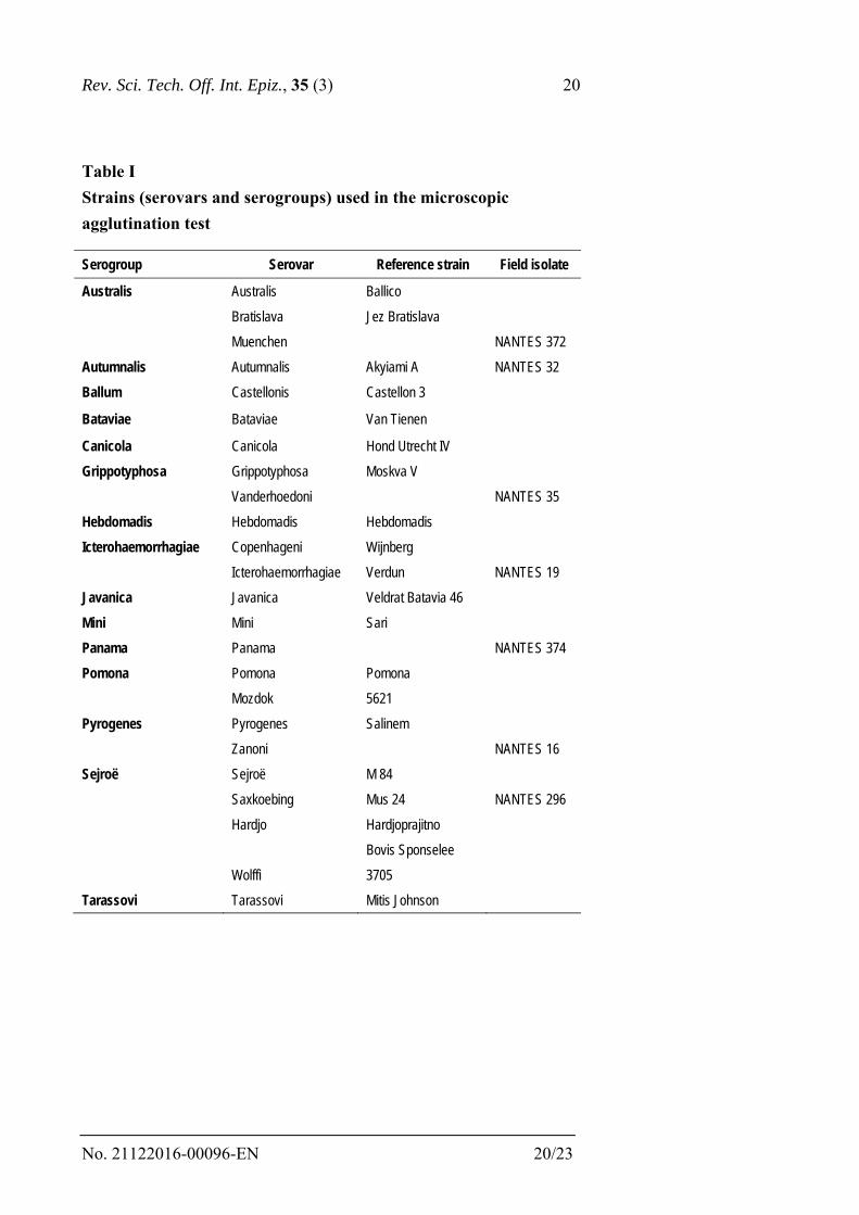
The strains used are shown in Table I.
Microscopic agglutination test
The standard micromethod on enzyme-linked immunosorbent assay
(ELISA) plates (previously developed at the CVL) was routinely used
on batches of samples received for leptospirosis diagnosis. Fresh
cultures were grown weekly and controlled by turbidimetric measures
using a Hach turbidimeter. Prior to its use in the MAT, each fresh
culture was filtered (0.8 µm) to remove the common ‘breeding nests’
of growing Leptospira. Cultures of each serovar were adjusted with
phosphate buffered saline (PBS; pH 7.2) to approximately 108 per ml
(100 Turbid Units). Serum dilutions and cultures were added to
ELISA plates by hand from 1984 to 2000. Thereafter, cultures were
automatically diluted and distributed by a Multiprobe 104 automate
(Packard).
Twenty-five microlitres of each culture and 25 µl of each serum
dilution were distributed in each well of the plates.
For each batch of analyses, several controls (different cultures and
samples from different animal species) were added, as described
below.
Culture controls
– Negative: 25 µl of PBS was mixed with 25 µl of each serovar
– Positive: 25 µl of a rabbit anti-serogroup specific serum was mixed
with 25 µl of each serovar belonging to this serogroup.

Rev. Sci. Tech. Off. Int. Epiz., 35 (3) 5
No. 21122016-00096-EN 5/23
Animal species controls
Sometimes the sera of a given animal species exhibited a paradoxical
effect by showing similar mild agglutination at each dilution, although
they were negative. To control for this effect, 25 µl of two pools of
serum from several animals of each species, one MAT negative and
one MAT positive, were mixed with 25 µl of each culture.
Cut-off
The serological background of each species was taken into account to
define the initial dilution used in the MAT. Environmental exposure to
Leptospira spp. is related to the lifestyle and normal behaviour of each
animal species. Clinical specificity and serological reactivity were
factors that were taken into account to define each specific initial
dilution.
– Dogs are highly receptive and usually exhibit acute disease. In a
naïve dog, agglutinating antibodies can be detected by the tenth day
after the onset of the disease (11). An earlier rise in the immune
response has clinical significance. To identify the start of this
response, the initial dilution of the serum was 1:20.
– Horses are potent serological responders to many bacteria and are
routinely exposed to environmental Leptospira spp. The initial
dilution of the serum was 1:100.
– Cattle and pigs are weakly susceptible to acute leptospirosis but
persistently exposed either at pasture or when housed. The initial
dilution of the serum was 1:50.
When the analyses were performed with the above technical
conditions, positive results at the cut-off level did not show clinical
significance or association with the observed disease. Positivity only
indicated agglutinating antibodies in the sera of the animals. The
clinical significance of the MAT results (as a diagnostic aid) could
only be estimated in view of several factors related to the animal
species and to the results themselves:

Rev. Sci. Tech. Off. Int. Epiz., 35 (3) 6
No. 21122016-00096-EN 6/23
– The animal: the age of the sampled animal, timing of the clinical
disorder (or economic loss in farm animals) and vaccination status are
necessary to reach a significant conclusion.
– The MAT titres: the level of antibodies can be high or low and is
matched to several serovars belonging to several serogroups. The
‘pattern’ of the reacting serovars (cross-agglutination) yields
significant information for the serum of a given species (11, 12).
Missing data generally reduce the usefulness of the test results as a
diagnostic aid to practitioners.
However, 20 years of results from the same laboratory are of interest
from an epidemiological point of view. Of course, this study was not a
prospective cohort study with randomised samples, which would have
allowed estimation of the prevalence and incidence of leptospirosis in
domestic animals (4, 13). However, ‘seroprevalence’ and
‘seroincidence’ were estimated in this group of samples from animals
suspected of having leptospirosis whose sera were sent for
serodiagnosis.
Sera
The blood samples sent by veterinary practitioners to the laboratory
were taken from animals suspected of having leptospirosis or herds
exhibiting symptoms or syndromes related to the disease.
Dogs are clinically very sensitive to acute leptospirosis, often
presenting with signs of fatal liver and kidney disease, whereas
infected farm animals exhibit milder symptoms but generate economic
losses related to leptospirosis-associated reproductive problems.
Several samples from farm animals belonging to the same herd are
recommended for simultaneous analysis. For example, the author’s
laboratory recommended collection of sera from aborting cows plus
other cows housed under the same conditions or, from pig farms
reporting reproductive disorders, sera from several sows.
For sport and companion animals (horses and dogs), sera were
submitted from individual cases of clinical disease (14, 15).

Rev. Sci. Tech. Off. Int. Epiz., 35 (3) 7
No. 21122016-00096-EN 7/23
Results
Numbers
Over the 20 years of serodiagnostic activity, 116,985 animal sera were
studied, from a minimum of 1,106 samples per year to a maximum of
9,978 samples. Some ‘peaks’ were related to an efficient and
temporary sensitisation of a regional veterinary association, for
example for cattle in 2002 or pigs in 1998.
More than 42,000 sera for each farm species were tested, and
approximately 22,000 horses and 9,500 dogs were individually
studied. Low and irregular numbers of samples from sheep, goats and
cats were analysed (data not shown).
Seroprevalence
Positive results are reported as ‘seroprevalence’. This estimation is
only based on the serological results performed in animals with
suspected leptospirosis.
A serum sample was defined as ‘MAT positive’ to one serogroup
when it showed agglutinating antibodies against one or several
serovars (Table I) belonging to this serogroup, at or above the
recommended cut-off dilution.
A positive animal often exhibited cross-reacting antibodies to several
serogroups of Leptospira. Consequently, the total number of positive
animals (n = 37,764) was different from the total number of positive
reactions to the major serogroups (n = 63,942), illustrating that many
serum samples reacted to more than one serogroup among those used
in the MAT. Most of the positive reactions were produced by just five
of the 15 serogroups (Table I), namely: Icterohaemorrhagiae (IH),
Grippotyphosa (GRIP), Sejroë (SEJ), Australis (AUS) and Autumnalis
(AUT) (Fig. 1). Therefore, this paper reports the results for these
major serogroups (with noted exceptions). High titres against IH or
AUS in pigs were generally associated with lower titres against
Pomona (POM) or Tarassovi, as previously shown by Plesko and
Lataste-Dorolle (16).

Rev. Sci. Tech. Off. Int. Epiz., 35 (3) 8
No. 21122016-00096-EN 8/23
Serogroup seroprevalence
For a given serogroup, significant differences were identified among
the different species. The most prevalent serogroup in domestic
animals was IH, with large differences between species, for example
cattle and dogs (Fig. 1). Similar differences were noticed for AUS,
whereas the seroprevalence of SEJ, GRIP and AUT was similar
among species. However, in the dog, it is necessary to take into
account the bias induced by the usual vaccination with IH and
Canicola (CAN) bacterins.
Serogroup seroincidence
High titres indicate recent infection by pathogenic Leptospira spp.,
whereas low titres may indicate an older immune response (or an early
sample). For the epidemiological statistical analysis, the highest titre
shown by one of the serovars belonging to a given serogroup was
attributed to this serogroup. Consequently, the field infectious serovar
generating this antibody response was said to belong to this serogroup,
even though this classification is not conclusive (17). Therefore, the
MAT results give a rough estimation of the ‘seroincidence’ of each
infectious serogroup. If high titres are related to the serogroup of the
strain that induced the enhanced serological response, serogroup
seroincidences may be compared. The most infectious serogroup in
French domestic animals (between 1987 and 2007) was IH (30.2% of
the high titres), followed by AUS (27.7%) and SEJ (21.9%).
Specific results
Cattle
In this species, over the 20-year analysis, 9,727 out of 42,982 samples
were positive. In herds suspected of having leptospirosis, the overall
mean seroprevalence (cut-off 1:100) was 22.63% (95% confidence
interval [CI]: 22.03–23.03) for the five studied serogroups (IH, GRIP,
AUS, SEJ, AUT).
The samples were collected from at least 11,310 herds. However, data
regarding the herds were missing for positive sera sampled during the

Rev. Sci. Tech. Off. Int. Epiz., 35 (3) 9
No. 21122016-00096-EN 9/23
years 1988, 1990 and 1992. Therefore, the herd seroprevalence (ratio
of infected herds to the total number of sampled herds) could not be
estimated.
Serogroup seroprevalence
For the 9,727 positive sera, the total number of positive results for the
five major serogroups was 11,510, showing that a positive animal
exhibited agglutinating antibodies cross-reacting with a mean of 1.18
serogroups. The most prevalent serogroup was SEJ (34.0% [95% CI:
33.4–34.6]), followed by GRIP (29.9% [95% CI: 29.3–30.5]), AUS
(27.6% [95% CI: 27.0–28.2]) and, to a lesser extent, IH (17.3% [95%
CI: 16.8–17.8]) and AUT (9.5% [95% CI: 9.1–9.8]).
These results only indicate exposure to the infection. Clinical
significance can only be established when high serological titres are
shown in animals suspected of disease.
Serogroup seroincidence
High titres, 1:400 or above, suggest a current immunological response.
This allows comparison of the seroincidence for each serogroup in the
positive cattle (except for 1991, when details about the 281 positive
cattle were missing). Among the 1,532 high serological titres
exhibited by the 9,727 positive animals, SEJ was the most infectious
(46% of the high titres), followed by GRIP (25%) and AUS (17%).
Annual dominant serogroup
Serogroup dominance varied during the study period. The dominant
serogroup in each year (with the exception of 1991) is shown in
Fig. 2. (Only the dominant serogroup is shown for each year; data on
the other four serogroups have not been included, except for 2000,
when the incidence of IH was the same as that of GRIP.) AUS was the
dominant serogroup in cattle between 1995 and 1997, again in 1999,
and between 2005 and 2007. IH appeared dominant only in 2004.
GRIP was the dominant serogroup between 1989 and 1993 and again
in 1998, but seroincidence decreased after the year 2000 and this

Rev. Sci. Tech. Off. Int. Epiz., 35 (3) 10
No. 21122016-00096-EN 10/23
serogroup was not serologically dominant in later years, in contrast to
SEJ.
Pigs
Over the 20-year analysis, 11,265 pigs were MAT positive out of
42,479 samples. The overall mean seroprevalence (cut-off 1:100) for
the six studied serogroups (IH, GRIP, AUS, SEJ, AUT, POM) was
26.5% (95% CI: 26.10–26.94).
Serogroup seroprevalence
Out of the 11,265 positive samples, the total number of positive
results against the six major serogroups was 15,651, indicating that a
positive animal exhibited agglutinating antibodies cross-reacting with
a mean of 1.39 serogroups. The most prevalent serogroup in positive
animals was IH (50.3% [95% CI: 49.6–51.0]), followed by AUS
(42.6% [95% CI: 42.0–43.2]) and, to a lesser extent, AUT (18.1%
[95% CI: 17.7–18.5]), SEJ (13.9% [95% CI: 13.4–14.4]), GRIP
(11.0% [95% CI: 10.6–11.4]) and POM (5.9% [95% CI: 5.6–6.2]).
The clinical significance of agglutinating antibodies can only be
correlated with an active infection when high serological titres are
present.
Serogroup seroincidence
High titres, 1:400 or above, indicate a current infection, allowing
comparative seroincidence estimation in the positive pigs. Of the
11,265 positive animals, the total number of high titres for the six
serogroups was 3,202, of which 42.3% were positive for IH, 32.4%
for AUS, 8.7% for AUT, 8.1% for SEJ, 6.9% for GRIP and 1.6% for
POM.
Annual dominant serogroup
Serogroup dominance varied during the study period (Fig. 3). A peak
of seroincidence generally was observed on pig farms from 1992 to
1999. Serogroups IH and AUS were the most prominent. The
Rev. Sci. Tech. Off. Int. Epiz., 35 (3) 11
No. 21122016-00096-EN 11/23
seroincidence of AUT decreased in 1998 then increased from 2003–
2007 (it only became the dominant serogroup in 2007); GRIP was
poorly represented in this species, and some POM cases were found in
1997–1999 and later in 2004.
Horses
Among the 22,018 horses analysed over the 20 years, 11,257 were
MAT positive. The overall mean serological seroprevalence (cut-off
1:200) for the five studied serogroups (IH, GRIP, AUS, SEJ, AUT)
was 51.9% [95% CI: 51.3–52.6].
Serogroup seroprevalence
For the 11,257 positive samples, the total number of positive results
for the five major serogroups was 18,719. A positive animal exhibited
agglutinating antibodies cross-reacting to a mean of 1.89 serogroups.
The most prevalent serogroups in positive animals were IH (52.6%
[95% CI: 52.0–53.2]) and AUS (52.0% [95% CI: 51.4–52.6]) and, to a
lesser extent, GRIP (24.2%, [95% CI: 23.7–24.7]), AUT (19.5%,
[95% CI: 19.1–19.9]) and SEJ (18.0% [95% CI: 17.6–18.4]).
As in other species, the clinical significance of agglutinating
antibodies can only be linked to an active infection if high serological
titres are present.
Serogroup seroincidence
High titres, 1:800 or above, indicate a current infection, allowing
comparison of seroincidence in positive horses. A total of 5,485
samples had high titres to the five serogroups, among which AUS and
IH were the most prominent serogroups (38.4% and 36.0%,
respectively), followed by GRIP (10.5%), SEJ and AUT (7.8% and
7.3%, respectively).
Annual dominant serogroup
Serogroup dominance varied during the study period (Fig. 4).
Serogroups AUS and IH were the most prominent: AUS was

Rev. Sci. Tech. Off. Int. Epiz., 35 (3) 12
No. 21122016-00096-EN 12/23
dominant in most years, but IH was sometimes more common (1998,
1999, and 2002–2004). The level of GRIP was generally low.
Dogs
Leptospirosis is often an acute and fatal disease in naïve dogs.
Therefore, vaccination is recommended in this species. In France,
between 1970 and 2007, only bivalent vaccines were available against
the virulent IH and CAN serogroups, to which dogs have been most
exposed. In contrast to other species, most dogs are vaccinated. The
study of the serological results in dogs had to take into account the IH
and CAN antibody background induced by vaccination (11, 18).
Therefore, the total seroprevalence (including IH and CAN) in 9,506
dogs clinically suspected of having leptospirosis was 73.2% (95% CI:
72.3–74.1).
Serogroup seroprevalence
As expected, serological responses against the vaccine serogroups IH
(80.5% [95% CI: 79.8–81.2]) and CAN (57.0% [95% CI: 56.4–57.6])
were the most prevalent (for a positive cut-off of 1:40) in the 6,956
positive dogs. These serogroups were followed by AUS (36.3% [95%
CI: 35.8–36.8]), SEJ (35.9% [95% CI: 35.4–36.4]), AUT (29.9%
[95% CI: 29.4–30.4]) and GRIP (24.0% [95% CI: 23.6–24.6]). These
results take into account that new field strains belonging to the SEJ
and AUS serogroups were added in 1994–1995 to the diagnostic panel
used in the MAT in dogs and, therefore, the seroprevalence for these
serogroups was only estimated for 14 years (6,535 positive dogs).
The 6,535 positive dogs exhibited 18,062 positive results, with each
positive dog showing reactivity to a mean of 2.59 serogroups.
The clinical significance of agglutinating antibodies can only be
linked to an acute infection when the serological titres are high, unless
the infection is fatal.
Serogroup seroincidence
Vaccine and non-vaccine serogroup results had to be differentiated.

Rev. Sci. Tech. Off. Int. Epiz., 35 (3) 13
No. 21122016-00096-EN 13/23
Vaccine serogroups IH and CAN:
Most of the agglutinating antibodies could have been induced by
current or past vaccinations. However, within six months after
vaccination, the titres decrease to less than 1:320. Comparison of
seroincidence between IH and CAN showed that 77.9% of the high
titres found in canine sera were IH antibodies and 22.1% were against
CAN.
Non-vaccine serogroups GRIP, AUT, AUS, and SEJ:
Within the non-vaccine serogroups, the highest incidence was found
for AUS (42.9%), followed by SEJ (26.3%), AUT (18.5%) and GRIP
(12.3%).
Annual prominent serogroups
Vaccine serogroups IH and CAN:
Despite annual variations, IH was always the most prominent, but
CAN sometimes achieved close values for incidence, for example in
1995 and 2006 (Fig. 5).
Non-vaccine serogroups GRIP, AUT, AUS and SEJ:
The non-vaccine serogroups AUT and GRIP were found in canine
serum at the beginning of the study period (with AUT the dominant
serogroup until 1994), and decreased later (Fig. 6). The annual
seroincidence of AUS and SEJ has been measured since 1994; AUS is
very infectious, although SEJ was most prominent in 1998 and 2000,
and AUT was most prominent in 1990.
Discussion
Leptospirosis is a global infectious disease of domestic animals. The
MAT is the gold standard for serological diagnosis of leptospirosis,
and the novel genospecies classification of the genus Leptospira has
not altered the efficiency of this test. The MAT detects agglutinating
antibodies against the outer membrane LPS shared by Leptospira spp.
Approximately ten days after the onset of the infection or vaccination,

Rev. Sci. Tech. Off. Int. Epiz., 35 (3) 14
No. 21122016-00096-EN 14/23
antibodies reach a detectable concentration in the serum of the host.
The MAT is performed with live leptospires and sometimes they auto-
agglutinate in the fresh cultures. It is essential to control each culture
by the addition of a negative control without serum. Moreover, it is
not unusual to observe a fresh culture with a good negative control but
exhibiting features of agglutination with the sera of a given species
and not for others. For these reasons, two species controls were added
for each batch for analysis: a negative and a positive pool of sera for
each species.
Whatever the serovar, each LPS shares many epitopes. Close serovars
sharing many similar epitopes are classified in the same serogroup.
However, some of these epitopes are also shared by other serogroups.
For a given strain, the immunological differentiation of the epitopes
depends on the general kinetics of the serological response
(immunoglobulin [Ig]M is less specific than IgG) and on the
specificity of the immune recognition of each host. For example, the
ratio of cross-reactivity between different serogroups was 1.89 in the
horse and 1.18 in cattle. These data highlight the fact that the horse is
a potent serological responder. The very high cross-reactivity ratio in
canine serum (2.59) reflects that, in France, most dogs are vaccinated
against IH and CAN. However, because protection by the usual
bacterins is serogroup specific, vaccinated dogs are not protected
against field infection, especially by a strain belonging to another
serogroup. The newly acquired infection triggers agglutinating
antibody production specific to the new LPS type but also mildly
boosts antibodies to some common epitopes shared by vaccine
serogroups (11). Newly infected vaccinated animals exhibit attenuated
symptoms of leptospirosis and have a better prognosis (personal data).
Serogroup CAN has been present in the bacterins since the beginning
of canine vaccination. In this study, the most seroprevalent serogroups
in the dog were IH (80.5%) and CAN (57.0%). These seroprevalence
values probably reflect the widely used vaccine. However, if these
serological titres were only due to vaccination, the serological profile
would have been similar for IH and CAN, and it was not. In the
environment, dogs are widely exposed to field strains, boosting their

Rev. Sci. Tech. Off. Int. Epiz., 35 (3) 15
No. 21122016-00096-EN 15/23
immune response and enhancing agglutinating antibodies. When only
dogs with high titres are taken into account, IH exposure (78%) is
significantly higher than that to CAN (22%), possibly because IH has
many maintenance hosts and reservoirs, whereas CAN is only
maintained in dogs. This may explain why, in a well-vaccinated
canine population, exposure to IH is higher than exposure to CAN.
These results are in accordance with the decrease of CAN throughout
Europe (19). However, a slight increase was shown in 2006 and 2007
(20). It is possible that the reduction in CAN cases led owners to
under-vaccinate their pets. Did the consequent reduction in general
canine immunity allow resurgence of CAN?
It is unclear why the prevalence of IH is low in cattle compared with
horses or pigs. Pigs may be more exposed to rats (the major reservoir
of IH) on intensive pig farms, but cattle are bred in more natural
conditions, close to those in which horses are kept. The difference
may be related to the specific nature of the bovine immune response
or to the prominent infecting serovar Hardjo (in serogroup SEJ),
which may inhibit IH infection in cattle.
The annual seroincidence of the serogroups estimated in this study
varied among the four domestic species.
The seroincidence of GRIP in cattle and dogs decreased from 1988 to
2000, but a peak was observed in 2009 in horses (A. Leon, personal
communication). However, some serogroups have remained
prominent over the 20-year period: IH and AUS have remained high
in all animal species, especially since 2011 in cattle and dogs (21) and
through 2014 in horses.
Annual fluctuations could be related either to a bias in sample
submission or to variations in exposure. Climate has a major effect on
wildlife reservoirs, which shed Leptospira spp. in their urine,
contaminating fresh water. However, an excessive amount of rain in
spring can cause the loss of voles and other small rodents, which may
be expected to result in a low density of the reservoir species for
GRIP. On the other hand, pathogenic Leptospira spp. are able to
survive and remain virulent for more than one year in fresh water (22).
Rev. Sci. Tech. Off. Int. Epiz., 35 (3) 16
No. 21122016-00096-EN 16/23
Excessive dryness can inactivate pathogenic Leptospira spp., but
drought also modifies the behaviour of reservoir species,
concentrating them near ponds or rivers.
The rapid evolution of Leptospira epidemiology, with decreases and
resurgence of serogroups, explains the use of a wide serogroup panel
in the MAT. It is necessary to modify, from time to time, the
serogroups included in the conventional bacterins. However, vaccine
companies may not be able to react sufficiently rapidly to these
epidemiological changes.
Acknowledgements
The author would like to acknowledge D. Pritchard and T. Little
(Central Veterinary Laboratory, Weybridge, UK), who kindly
provided in 1984 a panel of 21 live strains, their culture process and
their specific positive controls. She would also like to thank the
competent technicians who carefully performed the MAT and stored
the data in the laboratory and all of the practitioners who provided
serum samples with data.
References
1. Faine S., Adler B., Bolin C. & Perolat P. (1999). – Leptospira
and leptospirosis, 2nd Ed. MediSci Press, Melbourne, 272 pp.
2. Ellis W.A., O’Brien J.J., Neil S.D., Ferguson H.W. & Hanna
J. (1982). – Bovine leptospirosis: microbiological and serological
findings in aborted fetuses. Vet. Rec., 110 (7), 147–150.
doi:10.1136/vr.110.7.147.
3. Ellis W.A., O’Brien J.J. & Cassels J. (1981). – Role of cattle
in the maintenance of Leptospira interrogans serotype hardjo
infection in Northern Ireland. Vet. Rec., 108 (26), 555–557.
doi:10.1136/vr.108.26.555.

Rev. Sci. Tech. Off. Int. Epiz., 35 (3) 17
No. 21122016-00096-EN 17/23
4. André-Fontaine G., Ganière J.P., Quiniou M.A.,
Fourichon C., Protin P. & Menard M.F. (1988). – Prévalence des
anticorps antileptospires chez les bovins de Loire-Atlantique: II –
Évolution des titres sérologiques dans des exploitations infectées. Rec.
Méd. Vét., 164 (8–9), 617–622.
5. André-Fontaine G., Ganière J.P., Boukerrou A. &
Quiniou M.A. (1987). – Comparaison des prévalences des anticorps
antileptospires entre un échantillon de vaches ayant avorté et un
échantillon tiré au sort en Loire-Atlantique. Épidémiol. Santé Anim.,
11, 53–63.
6. Michel V., Ruvoen-Clouet N., Menard A., Sonrier C.,
Fillonneau C., Rakotovao F., Ganière J.P. & André-Fontaine G.
(2001). – Role of the coypu (Myocastor coypu) in the epidemiology of
leptospirosis in domestic animals and humans in France. Eur. J.
Epidemiol., 17 (2), 111–121. doi:10.1023/A:1017931607318.
7. Moinet M., Fournier-Chambrillon C., André-Fontaine G.,
Aulagnier S., Mesplède A., Blanchard B., Descarsin V., Dumas P.,
Dumas Y., Coïc C., Couzi L. & Fournier P. (2010). – Leptospirosis in
free-ranging endangered European mink (Mustela lutreola) and other
small carnivores (Mustelidae, Viverridae) from south-western France.
J. Wildl. Dis., 46 (4), 1141–1151. doi:10.7589/0090-3558-46.4.1141.
8. Aviat F., Blanchard B., Michel V., Blanchet B., Branger C.,
Hars J., Mansotte F., Brasme L., De Champs C., Bolut P., Mondot P.,
Faliu J., Rochereau S., Kodjo A. & André-Fontaine G. (2009). –
Leptospira exposure in the human environment in France: a survey in
feral rodents and in fresh water. Comp. Immunol. Microbiol. Infect.
Dis., 32 (6), 463–476. doi:10.1016/j.cimid.2008.05.004.
9. Baranton G. & Postic D. – Rapports annuels d’activité du
Centre national de référence des leptospires. Institut Pasteur, Paris.
Available at: www.pasteur.fr/fr/sante/centres-nationaux-reference/les-
cnr/leptospirose/rapports-d-activite (accessed on 13 December 2016).
Rev. Sci. Tech. Off. Int. Epiz., 35 (3) 18
No. 21122016-00096-EN 18/23
10. Adamus C., Buggin-Daubié M., Izembart A., Sonrier Pierre
C., Guigand L., Masson M.-T., André-Fontaine G. & Wyers M.
(1997). – Chronic hepatitis associated with Leptospiral infection in
vaccinated beagles. J. Comp. Pathol., 117 (4), 311–328.
doi:10.1016/S0021-9975(97)80079-5.
11. André-Fontaine G. (2013). – Diagnosis algorithm for
leptospirosis in dogs: disease and vaccination effects on the
serological results. Vet. Rec., 172 (19), 502–506.
doi:10.1136/vr.101333.
12. André-Fontaine G., Boudet R., De Coquet J., Reynal P.H.,
Ganière J.P. & Larrat M. (1992). – Contamination humaine et animale
par Leptospira interrogans australis. Méd. Mal. Infect., 22 (10), 880–
882. doi:10.1016/S0399-077X(05)80498-6.
13. André-Fontaine G., Nicholas D., Scalzo B., Keita A. &
Nanjiani I. (2010). – Prévalence sérologique de la leptospirose à
sérovar Hardjo chez les bovins femelles adultes en France en 2004.
Bull. GTV, 55, 67–74.
14. Léon A., Pronost S., Tapprest J., Foucher N., Blanchard B.,
André-Fontaine G., Laugier C., Fortier G. & Leclercq R. (2006). –
Identification of pathogenic Leptospira strains in tissues of a
premature foal by use of polymerase chain reaction analysis. J. Vet.
Diagn. Invest., 18 (2), 218–221. doi:10.1177/104063870601800216.
15. Muller A., Gau C., Chetboul V. & André-Fontaine G.
(1999). – Un cas de cholangio-hépatite associée à une leptospirose
chez un chien. Rec. Méd. Vét., 175 (1/2), 63–68.
16. Plesko I. & Lataste-Dorolle C. (1970). – Intertype immunity
relations of Leptospira strains belonging to the ‘Australis’ serogroup.
Biologia (Bratisl), 25 (6), 403–412.
17. Craig S.B., Graham G.C., Burns M.-A., Dohnt M.F.,
Smythe L.D. & McKay D.B. (2009). – A case of ‘original antigenic
sin’ or just a paradoxical reaction in leptospirosis? Ann. Trop. Med.
Parasitol., 103 (5), 467–470. doi:10.1179/136485909X435102.

Rev. Sci. Tech. Off. Int. Epiz., 35 (3) 19
No. 21122016-00096-EN 19/23
18. André-Fontaine G. (2006). – Canine leptospirosis: Do we
have a problem? Vet. Microbiol., 117 (1), 19–24.
doi:10.1016/j.vetmic.2006.04.005.
19. Ellis W.A. (2010). – Control of canine leptospirosis in
Europe: time for a change? Vet. Rec., 167 (16), 602–605.
doi:10.1136/vr.c4965.
20. Picardeau M. (2013). – Diagnosis and epidemiology of
leptospirosis. Méd. Mal. Infect., 43 (1), 1–9.
doi:10.1016/j.medmal.2012.11.005.
21. Ayral F.C., Bicout D.J., Pereira H., Artois M. & Kodjo A.
(2014). – Distribution of Leptospira serogroups in cattle herds and
dogs in France. Am. J. Trop. Med. Hyg., 91 (4), 756–759.
doi:10.4269/ajtmh.13-0416.
22. André-Fontaine G., Aviat F. & Thorin C. (2015). –
Waterborne leptospirosis: survival and preservation of the virulence of
pathogenic Leptospira spp. in fresh water. Curr. Microbiol., 71 (1),
136–142. doi:10.1007/s00284-015-0836-4.
__________
Rev. Sci. Tech. Off. Int. Epiz., 35 (3) 20
No. 21122016-00096-EN 20/23
Table I
Strains (serovars and serogroups) used in the microscopic
agglutination test
Serogroup Serovar Reference strain Field isolate
Australis Australis Ballico
Bratislava Jez Bratislava
Muenchen NANTES 372
Autumnalis Autumnalis Akyiami A NANTES 32
Ballum Castellonis Castellon 3
Bataviae Bataviae Van Tienen
Canicola Canicola Hond Utrecht IV
Grippotyphosa Grippotyphosa Moskva V
Vanderhoedoni NANTES 35
Hebdomadis Hebdomadis Hebdomadis
Icterohaemorrhagiae Copenhageni Wijnberg
Icterohaemorrhagiae Verdun NANTES 19
Javanica Javanica Veldrat Batavia 46
Mini Mini Sari
Panama Panama NANTES 374
Pomona Pomona Pomona
Mozdok 5621
Pyrogenes Pyrogenes Salinem
Zanoni NANTES 16
Sejroë Sejroë M 84
Saxkoebing Mus 24 NANTES 296
Hardjo Hardjoprajitno
Bovis Sponselee
Wolffi 3705
Tarassovi Tarassovi Mitis Johnson

Rev. Sci. Tech. Off. Int. Epiz., 35 (3) 21
No. 21122016-00096-EN 21/23
Fig. 1
Animal exposure to each serogroup (percentage of positive sera
among tested sera from each species)
Fig. 2
Dominant serogroup in positive cattle in each year (percentage of
sera with titre ≥ 1:400)
0%
10%
20%
30%
40%
50%
60%
70%
80%
90%
cattle pig horse dog
N=9727 N=11515 N=11257 N=6837
IH Grip Aut Aus Sej Pom/Can
0%
2%
4%
6%
8%
10%
12%
14%
16%
18%
20%
19881989199019911992199319941995199619971998199920002001200220032004200520062007
IH>/Bv+ GRIP>/Bv+ SEJ>/Bv+ AUS>/Bv+ AUT>/Bv+

Rev. Sci. Tech. Off. Int. Epiz., 35 (3) 22
No. 21122016-00096-EN 22/23
Fig. 3
Dominant serogroup in positive pigs in each year (percentage of
sera with titre ≥ 1:400)
Fig. 4
Dominant serogroup in positive horses in each year (percentage of
sera with titre ≥ 1:800)
0%
2%
4%
6%
8%
10%
12%
1988 1989 1990 1991 1992 1993 1994 1995 1996 1997 1998 1999 2000 2001 2002 2003 2004 2005 2006 2007
%IH>400Pc %Grip>400Pc %Sej>400Pc %Aus>400Pc %Aut>400Pc
0%5%10%15%20%25%30%35%40%
1988 1989 1990 1991 1992 1993 1994 1995 1996 1997 1998 1999 2000 2001 2002 2003 2004 2005 2006 2007
IH>800 Grip>800 Sej>800 Aus>800 Aut>800

Rev. Sci. Tech. Off. Int. Epiz., 35 (3) 23
No. 21122016-00096-EN 23/23
Fig. 5
Comparative annual seroincidence of Icterohaemorrhagiae (IN)
and Canicola (CAN) in positive dogs (percentage of sera with titre
≥ 1:320)
Fig. 6
Annual seroincidence of Grippotyphosa (GRIP), Autumnalis
(AUT), Australis (AUS) and Sejroë (SEJ) in positive dogs
(percentage of sera with titre ≥ 1:320)
0%
10%
20%
30%
40%
50%
60%
1988 1989 1990 1991 1992 1993 1994 1995 1996 1997 1998 1999 2000 2001 2002 2003 2004 2005 2006 2007
IH>320CN+ CAN>320CN+
0%
10%
20%
30%
40%
50%
60%
1988 1989 1990 1991 1992 1993 1994 1995 1996 1997 1998 1999 2000 2001 2002 2003 2004 2005 2006 2007
GRIP>CN+ AUT>CN+ AUS>CN+ Sej>CN+