Embed Size (px)
Citation preview

Leprosy• Hansen’s disease• M.leprae• Affects cooler parts of body - skin, mucous membranes
(e.g. nose), peripheral nervous system, eyes and testes. • Classification
- Lepromatous - TuberculoidThe form the disease takes depends on the person's
immune response to the infection.

Transmission
• Human to human• Lepromatous patients shed M.leprae from the nasal
mucosa• Minor routes- breast milk, materno-fetal• M.leprae can survive in environment up to 45 days• Multiplies very slowly, symptoms can take as long as 20
years to appear

Ridley & Joplings classification5 groups• TT (high resistance)• BT• BB• BL• LL (low resistance)Classification according to bacilliary index• Pauci-bacillary• Multi-bacillary

Tuberculoid leprosy
• Can be either one large red patch with well-defined raised borders or a large hypopigmented asymmetrical spot
• Lesions become dry and hairless • Loss of sensation may occur• Tender, thickened nerves with subsequent loss of
function are common • Spontaneous resolution may occur in a few years • or it may progress to borderline or rarely lepromatous
types

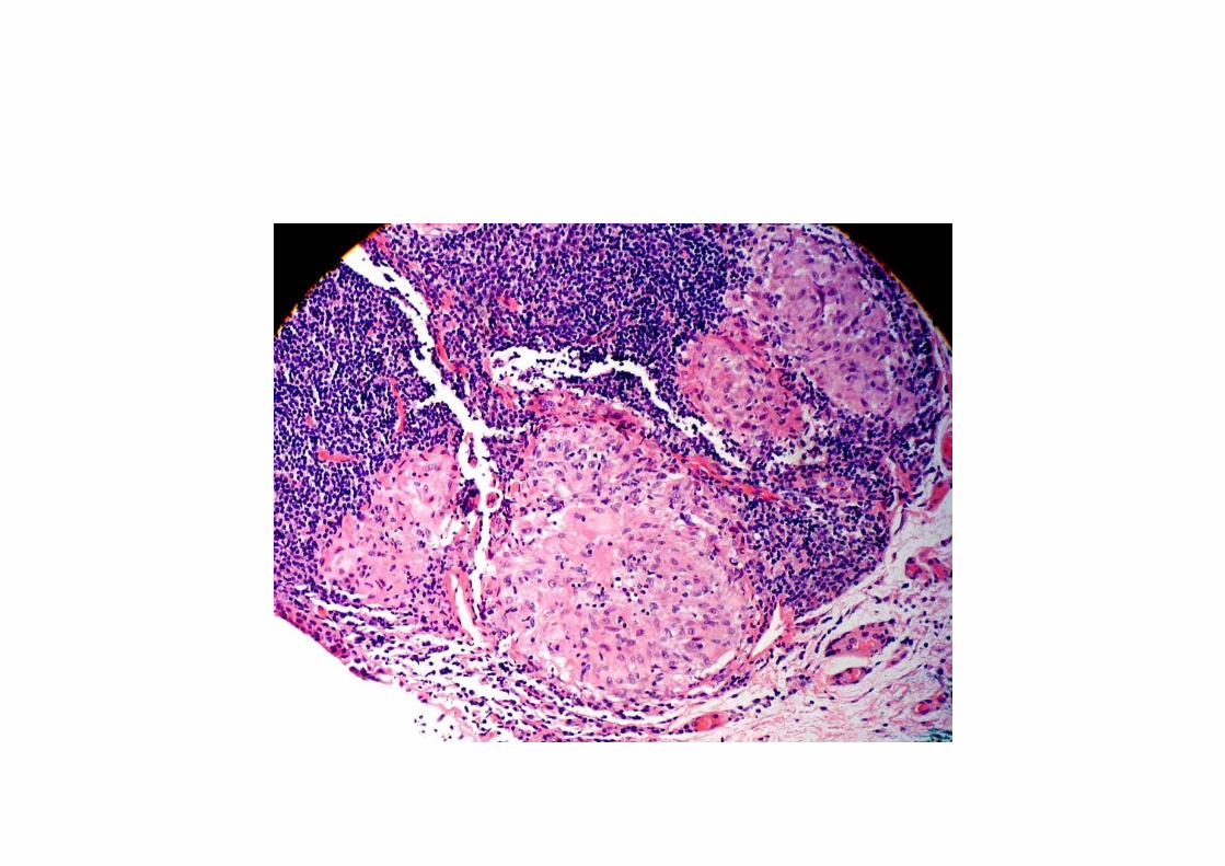
Skin biopsy
• collections of epithelioid cells surrounded by many groups of lymphocytes
• occasionally “Giant Cells”• such cell collections are present mainly around the
appendages of the skin such as the nerves and the hair follicles.
• inflammatory cell collection may spread even up to the epidermis.
• hair-follicles, sweat glands destroyed• Lepra bacilli


Lepromatous leprosy
• Numerous lesions - plaques, macules, papules and nodules
• Early symptoms include nasal stuffiness, discharge and bleeding, and swelling of the legs and ankles– Skin thickens over forehead (leonine facies),– eyebrows and eyelashes are lost, – nose becomes misshapen or collapses, ear lobes
thicken, – Eye involvement : photophobia , glaucoma and
blindness – Testicles shrivel causing sterility– Slow scarring of peripheral nerves resulting in nerve
thickening and sensory loss. Fingers and toes become deformed due to painless repeated trauma.

• M/E

• Diagnosis- split skin smear- biopsy & histopathology

Lepromin Test• Used for classifying leprosy on the basis of immune
response• CMI suppressed in LL but TT pt. have good immune
response• I/D inj of lepromin reveals delayed hypersensitivity
reaction in tuberculoid leprosy • Early Fernandez reaction---indurated area in 24-48 hrs• Late Mitsuda reaction—delayed granulomatous lesion
appearing after 3-4 weeks

Reactions in leprosyType I: Borderline reactions- two types
• Upgrading reaction-Occurs in BL pt. on treatment who upgrade towards tuberculoid type.
• Downgrading reaction-Occurs in BT pt. who downgrade towards lepromatous type
Type II : Erythema nodosum leprosum (ENL)---Occurs in lepromatous pt after treatmentTender cutaneous nodules, fever, iridocyclitis


SYPHILIS• T.pallidum• Coiled spiral filament 10um long• Cannot be cultured• Demonstrated by
- Dark ground illumination - Fluorescent antibody technique- Silver impregnation technique
Modes of transmission • Sexual• Person to person contact• Transfusion • Materno-foetal transmission
Stages• Primary• Secondary• Tertiary

Primary Syphilis• Chancre on penis, scrotum, vulva or cervix. Firm red
papule, erodes to create an ulcer, indurated- Hard Chancre
• 2-4 wk after acquiring infection • Heals without scarring even in absence of treatment • Ab test positive in 1-3 wk after appearance of chancre• Histologically– dense infiltrate of lympho, plasma cells &
macrophages- Perivascular aggregates of plasma cells- endarteritis obliterans

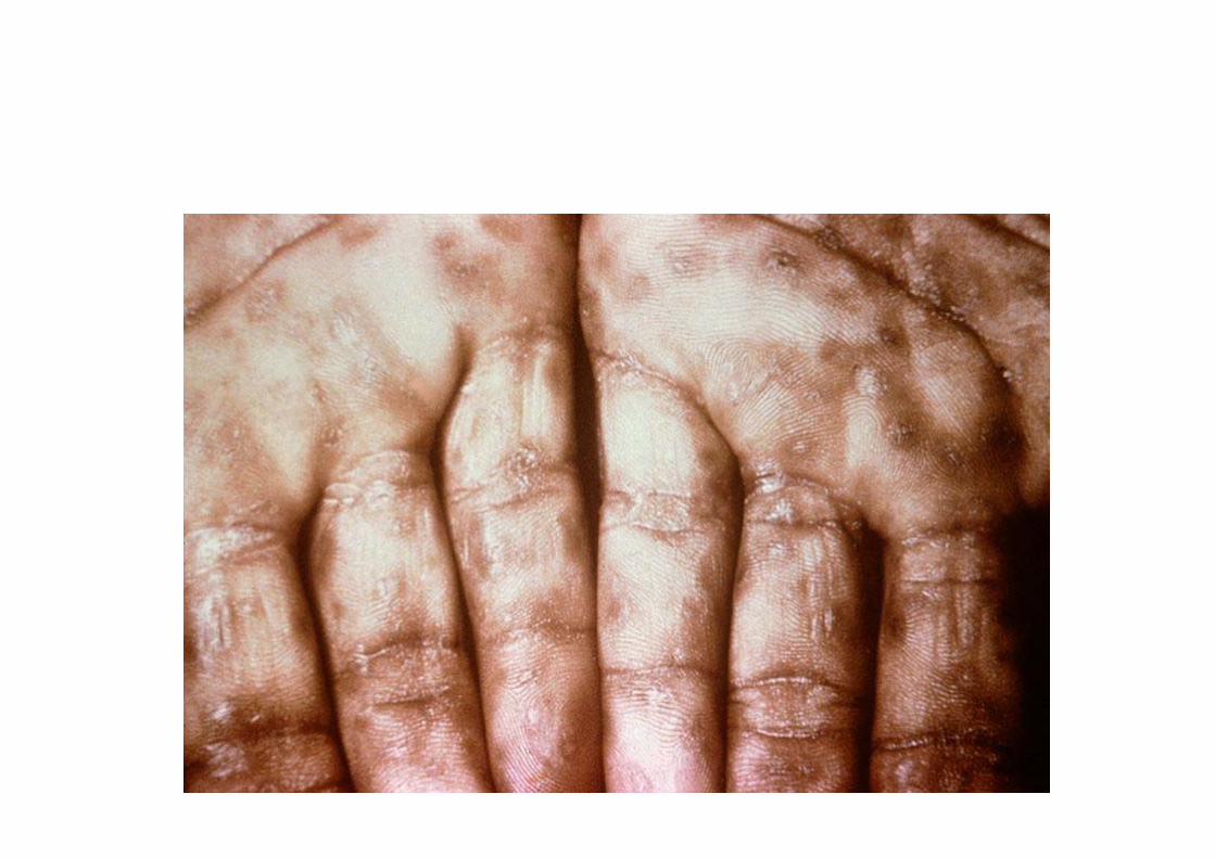
Secondary Syphilis• Mucocutaneous lesions & painless lymphadenopathy in
2-3 months after exposure• Mucous patches on mouth, pharynx, palms, soles.
Condyloma lata in anogenital region- red brown papular lesions
• Highly infective stage• Spirochetes easily demonstrated in mucocutaneous
lesions• Ab tests always positive• M/E

Tertiary Syphilis• 2-3 years following first exposure• Less infective• 2 types of lesionsSyphilitic gumma—solitary, localized, rubbery lesion with
central necrosis in testis, bone, brain, liver (hepar lobatum)M/E- granuloma containing central necrotic material, palisaded macrophages, mononuclear cells
Diffuse lesionsCVS- aortic aneurysmNeurosyphilis- meningovascular
tabes dorsalisgeneral paresis

Gumma

• Neurosyphilis• General paresis- results in personality changes,
emotional changes, hyperactive reflexes• Tabes dorsalis- a disorder of the spinal cord, often
results in a characteristic shuffling gait• Imaging of the brain usually shows atrophy.• Syphilitic aortitis- syphilis affects the ascending aorta
causing aortic dilation and regurgitation, massive hypertrophy of the left ventricle (over 1,000 grams)
• Heart is termed cor bovinum (cow's heart). • Tunica intima: tree bark appearance

Congenital Syphilis• Develop in fetus >16 wk gestation who is exposed to
maternal spirochaetemia• 3 types of lesions1. Child born dead2. Infantile type
- muco-cutaneous lesions- diffuse sloughing of epithelium, teem with spirochaetes- bony lesions- osteochondritis, perichondritis, saddle nose deformity, saber shin
3. Late type--- Hutchinson’s teeth- Interstitial keratitis- Deafness


Infectious Granulomatous Diseases
Examples of Diseases with Granulomatous Inflammations Disease Cause Tissue Reaction Tuberculosis Mycobacterium
tuberculosis Noncaseating tubercle (granuloma prototype): a focus of epithelioid cells, rimmed by fibroblasts, lymphocytes, histiocytes, occasional Langhans giant cell; caseating tubercle: central amorphous granular debris, loss of all cellular detail; acid-fast bacilli
Leprosy Mycobacterium leprae
Acid-fast bacilli in macrophages; non-caseating granulomas
Syphilis Treponema pallidum
Gumma: microscopic to grossly visible lesion, enclosing wall of histiocytes; plasma cell infiltrate; central cells are necrotic without loss of cellular outline
Cat-scratch disease
Gram-negative bacillus
Rounded or stellate granuloma containing central granular debris and recognizable neutrophils; giant cells uncommon
Bartonella henselae

ACTINOMYCOSIS
• Actinomycetes israelli• Gram positive, non acid fast• Commensal in oral cavity, alimentary tract & vagina• Endogenous in origin, never person to person• Four types
Cervicofacial actinomycosis• Commonest & have best prognosis• Infection enters from tonsils, carious tooth• Abscesses & sinuses• Discharging pus contains typical tiny yellow sulphar
granules.
ACTINOMYCOSIS
Thoracic actinomycosis• Aspiration of organism from oral cavity• Initially resembles pneumonia & subsequently spreads to
whole of lung, pleura, ribs & vertebrae
Abdominal actinomycosis• Common in appendix, caecum & liver• Results from swallowing of organism from oral cavity or
extension from thoracic cavity

ACTINOMYCOSIS
Pelvic actinomycosis• Complication of IUCD
Microscopy• Granuloma with central suppuration• Centre of abscess contains bacterial colony (sulphar
granule) characterized by radiating filaments with hyaline, eosinophilic club ends

Sarcoidosis• Systemic disease of unknown etiology characterized by
noncaseating granulomas• More common in women, 20-40 years
Etiology • Disease of disordered immune regulation in genetically
predisposed individuals exposed to certain environmental agents

• Immunologic factors- cell mediated response- accumulation of CD+4 T cells, CD4:CD8 ratio—5:1-15:1- T cell derived cytokines- IL-2, IFN
• accumulation of monocytes, macrophages and activated T-lymphocytes,
• increased production of mediators: TNF-alpha, IFN-gamma, IL-2 and IL-12, characteristic of a Th1 response
• Genetic factors- familial, racial clustering
• Environmental factors- Mycobacteria, Rickettesia

Morphology
Organs involved• Lungs• Hilar & mediastinal lymphadenopathy• Spleen/Hepatomegaly• Bone • Skin• Eye• Salivary glands• Muscles
M/E- non caseating granuloma- Schaumann bodies-laminated concretions- Asteroid bodies- stellate inclusions


Clinical features
• Symptoms are vague: shortness of breath, cough, chest pain, haemoptysis, fever, fatigue, weight loss, anorexia
• presents as a restrictive disease of the lungs, decrease in lung volume and decreased compliance, vital capacity
• Arthritis • Dry eyes, blurry vision • Skin lesions; range from rashes and nodules to
erythema nodosum.

Diagnosis• X-ray chest, CT scan • CT-guided biopsy, mediastinoscopy, • Bronchoscopy with biopsy • Special stains to rule out microorganisms, matter of
exclusion.• ACE- blood levels are used in diagnosis and monitoring
of sarcoidosis.• Serum calcium and 24-hour urine calcium

Prognosis• Progressive chronicity or periods of activity with
remissions• 65-70% recover with minimal or no residual
manifestations• 20% permanent loss of lung function• 10-15% die of cardiac/CNS manifestations