Embed Size (px)
Citation preview
Leica DMC2900
Digital microscope camera for easy, efficient documentation and presentation in industry and research
From Eye to Insight
�
�
3
Leica DMC2900 at a glance
• Digital camera with 3.1 megapixel resolution – optimal for
applications using standard microscopes
• Fast USB 3.0 interface for a direct connection between camera
and PC or notebook; also backward compatible with USB 2.0
at a lower live imaging speed
• Ideal for precise measurements, analyses, and documentation
• Color interpolation, image sharpening, and shading correction
are performed very rapidly by the camera hardware without
compromising live imaging speed
• Optimized control of the Leica DMC2900 using powerful
LAS software
• Fast live imaging speed of up to 30 frames per second
(XGA resolution), allowing authentic real-time sample
display on the monitor
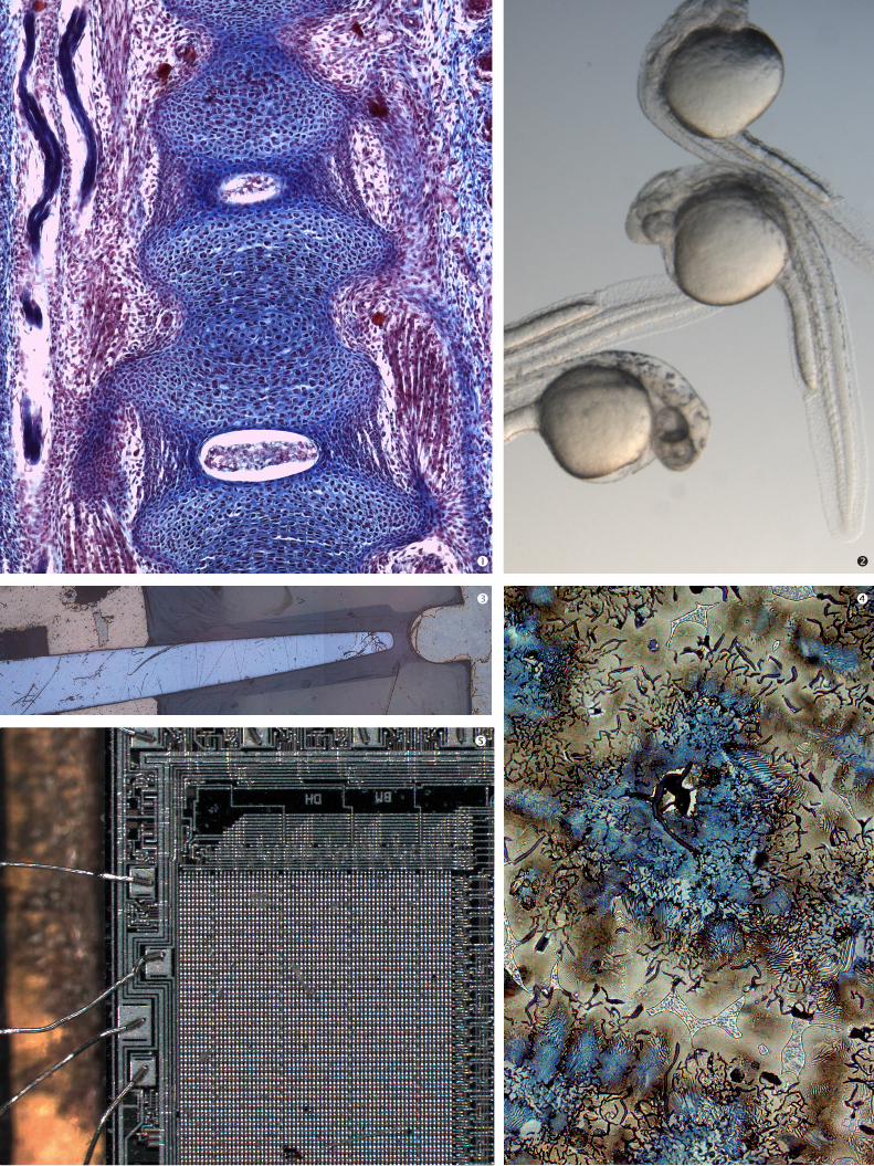
High Speed Imaging Leica DMC2900 with USB 3.0 interface for highest speed in image processing
Leica Microsystems' next generation digital camera features the speed of a USB 3.0 interface,
which allows easy image settings and precise documentation of microstructures – and can
even be used with modern notebooks. The Leica DMC2900, optimally controlled by the
user-friendly software Leica Application Suite (LAS), provides high quality images from all
industrial and research samples.
Professional imaging
The Leica DMC2900 digital color camera is optimized for real-
time imaging in standard microscopy applications. With up to
30 frames per second, its live preview function allows samples
to be easily positioned, perfectly focused, and even manipulated
on the computer screen. A reliable 1/2" CMOS sensor with
3 megapixel resolution ensures the optimal processing of standard
microscopic applications which require processor-intensive
series of single image captures.
The Leica DMC2900 is easily integrated with any microscope
system using the universal C-mount adapter.
USB 3.0 interface for a fast, secure connection
��
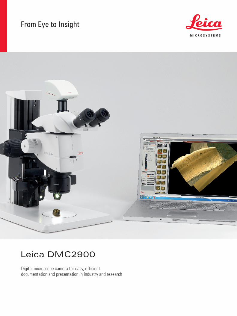
Examination of tissue sample (H&E staining) Examination of zebrafish embryosAssembled image of a metal sample (LAS-MultiStep)Structure of brass at 50x magnificationExamination of bonding on a chip
4
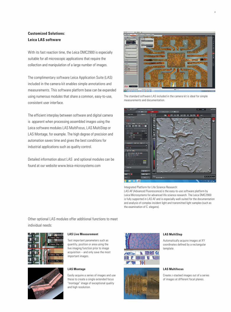
Customized Solutions:
Leica LAS software
With its fast reaction time, the Leica DMC2900 is especially
suitable for all microscopic applications that require the
collection and manipulation of a large number of images.
The complimentary software Leica Application Suite (LAS)
included in the camera kit enables simple annotations and
measurements. This software platform base can be expanded
using numerous modules that share a common, easy-to-use,
consistent user interface.
The efficient interplay between software and digital camera
is apparent when processing assembled images using the
Leica software modules LAS MultiFocus, LAS MultiStep or
LAS Montage, for example. The high degree of precision and
automation saves time and gives the best conditions for
industrial applications such as quality control.
Detailed information about LAS and optional modules can be
found at our website www.leica-microsystems.com
LAS Montage
Easily acquire a series of images and use these to create a single extended focus “montage” image of exceptional quality and high resolution.
LAS MultiStep
Automatically acquire images at XY coordinates defined by a rectangular template.
LAS Multifocus
Create z-stacked images out of a series of images at different focal planes.
LAS Live Measurement
Test important parameters such as quantity, position or area using the live imaging function prior to image acquisition – and only save the most important images.
The standard software LAS included in the camera kit is ideal for simple measurements and documentation.
Integrated Platform for Life Science Research:LAS AF (Advanced Fluorescence) is the easy-to-use software platform by Leica Microsystems for advanced life science research. The Leica DMC2900 is fully supported in LAS AF and is especially well-suited for the documentation and analysis of complex incident light and transmitted light samples (such as the examination of C. elegans).
Other optional LAS modules offer additional functions to meet
individual needs:

LIVE IMAGE SPEED
Image formats*2048 × 1536—Full frame1024 × 768—XGA
USB 2.0 USB 3.0 4 12 15 30
* 5 msec exposure time, in frames per second
COMPUTER
Min. computer configuration Intel Core 2 Duo 2.4 GHz, or faster2 GB RAM, high res. graphic card with
128 MB or 256 MB RAM, Direct X V9c or V10,USB 2.0 or USB 3.0 interface or free PCI Express slot
Software: Windows 7 and Windows 10 (LAS and LAS X)Windows 8 (LAS only)
TECHNICAL DATA AND OPERATING ENVIRONMENT
Power consumption ~ 4 W
Power supply via USB 3.0 cable
Housing Aluminum die cast
Size 112 × 74 × 68.4 mm
Weight 340 g
Operating temperature -5°C to +50°C
Relative humidity 10 % to 90 % non-condensing
DIGITAL CAMERA
Camera type Digital camera for microscopy with control software
Sensor Progressive scan CMOS, Micron (MT9T001)
Sensor type/size 6.55 mm × 4.92 mm (type 1/2")
Color filter RGB Bayer mosaic
Protective color filter UV/IR Filter
Shutter control Electronic rolling shutter/ Progressive scan readout
Number of pixels 3.1 megapixels, 2048 × 1536
Pixel size 3.2 µm × 3.2 µm
Color depth 30 bit
A/D converter 10 bit
Dynamic range Type > 55 dB/600:1
Readout noise σ < 1.8 LSB (10 bit) typical
Exposure time 0.1 msec – 2 sec
Gain control 1× – 4× / 0 – 12 dB
Shading correction yes, stored for all formats
Region of interest Freely adjustable in 2 pixel steps from 2 x 2 up to full resolution
ELECTRONIC INTERFACES
Optical C-mount
Recommended video adapter 0.5×/0.55×
Digital output connector USB 3.0 Micro-B, with screw holes
Leica DMC2900—Technical Data
ORDER NUMBER
12 730 466 Leica DMC2900 Camera (incl. USB 3.0 PCI Express card for computers with no USB 3.0 Interface, USB 3.0 cable 2.5 m, LAS Software)
Relative quantum efficiency of Leica DMC2900 (WB applied)
Light microscopes
C-mount
Stereo microscopes
Assembly Diagram
USB 3.0 cable, 2.5 m(USB 2.0 compatible)
LeicaSoftware
10 450 528 0.5× 11 541 544 0.55×
DMC290012 730 466
Wavelength (nm)
Rela
tive
Resp
onse
10ID
C22
010E
N •
© L
eica
Mic
rosy
stem
s (S
chw
eiz)
AG
• C
H-9
435
Heer
brug
g, 2
017
• Im
ages
, des
crip
tions
and
te
chni
cal d
ata
subj
ect t
o ch
ange
– w
e re
serv
e th
e rig
ht to
mak
e ch
ange
s w
ithou
t not
ice
•
LEIC
A an
d th
e Le
ica
logo
are
regi
ster
ed tr
adem
arks
of L
eica
Mic
rosy
stem
s IR
Gm
bH.
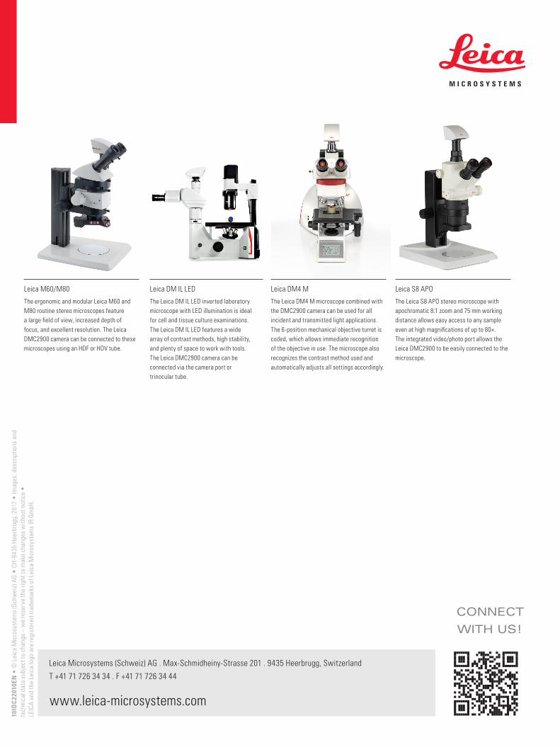
Leica M60/M80
The ergonomic and modular Leica M60 and M80 routine stereo microscopes feature a large field of view, increased depth of focus, and excellent resolution. The Leica DMC2900 camera can be connected to these microscopes using an HDF or HDV tube.
Leica DM IL LED
The Leica DM IL LED inverted laboratory microscope with LED illumination is ideal for cell and tissue culture examinations. The Leica DM IL LED features a wide array of contrast methods, high stability, and plenty of space to work with tools. The Leica DMC2900 camera can be connected via the camera port or trinocular tube.
Leica DM4 M
The Leica DM4 M microscope combined with the DMC2900 camera can be used for all incident and transmitted light applications. The 6-position mechanical objective turret is coded, which allows immediate recognition of the objective in use. The microscope also recognizes the contrast method used and automatically adjusts all settings accordingly.
Leica S8 APO
The Leica S8 APO stereo microscope with apochromatic 8:1 zoom and 75 mm working distance allows easy access to any sample even at high magnifications of up to 80×. The integrated video/photo port allows the Leica DMC2900 to be easily connected to the microscope.
CONNECT
WITH US!
Leica Microsystems (Schweiz) AG . Max-Schmidheiny-Strasse 201 . 9435 Heerbrugg, Switzerland
T +41 71 726 34 34 . F +41 71 726 34 44
www.leica-microsystems.com
10ID
C22
010E
N •
© L
eica
Mic
rosy
stem
s (S
chw
eiz)
AG
• C
H-9
435
Heer
brug
g, 2
017
• Im
ages
, des
crip
tions
and
te
chni
cal d
ata
subj
ect t
o ch
ange
– w
e re
serv
e th
e rig
ht to
mak
e ch
ange
s w
ithou
t not
ice
•
LEIC
A an
d th
e Le
ica
logo
are
regi
ster
ed tr
adem
arks
of L
eica
Mic
rosy
stem
s IR
Gm
bH.