Embed Size (px)
Citation preview

JOURNAL OF CLINICAL MICROBIOLOGY, Sept. 1980, p. 395-4010095-1 137/80/09-0395/07$02.00/0
Vol. 12, No. 3
Legionella pneumophila Serogroup Six: Isolation from Casesof Legionellosis, Identification by ImmunofluorescenceStaining, and Immunological Response to Infection
ROGER M. McKINNEY,'* HAZEL W. WILKINSON,' HERBERT M. SOMMERS,' BONNIE J. FIKES,'KAREN R. SASSEVILLE,' MARGARET M. YUNGBLUTH,2 AND JAMES S. WOLF'
Bureau of Laboratories, Center for Disease Control, Public Health Service, U.S. Department of Health andHuman Services, Atlanta, Georgia 30333,' and Departments of Pathology2 and Surgery,3 Northwestern
University Medical School, Chicago, Illinois 60611
Isolates of Legionella pneumophila that are serologically different from strainsof serogroups 1 through 5 were obtained from lung biopsy tissue or pleural fluidfrom three renal transplant recipients in Chicago, Ill. These strains were placedin a newly designated L. pneumophila serogroup, serogroup 6, on the basis offluorescent-antibody staining characteristics. An L. pneumophila strain obtainedfrom Bethesda, Md., one from Houston, Tex., and one from Oxford, England, alsobelong to this new serogroup. L. pneumophila serogroup 6 appears to be widelydistributed geographically.
Legionellosis is caused by a gram-negativebacillus (10) for which the name Legionellapneumophila has been proposed (2). Legionel-losis is manifested as Legionnaires disease, withpneumonia as the prominent feature, or as Pon-tiac fever, in which the absence of pneumonia istypical (8-10). Both forms of the disease areprobably acquired from environmental sources.
In early studies of L. pneumophila, Cherry etal. (3) prepared direct fluorescent-antibody(DFA) conjugates for five strains, and thesereagents were evaluated in staining tests withthe 10 strains of L. pneumophila that were avail-able to them at that time. Although some differ-ences in intensity of staining among the 10strains were observed, a conjugate prepared forthe Knoxville 1 strain stained all strains brightlywhen used at the appropriate dilution. The firstevidence of distinct serogroups of L. pneumo-phila came with isolation of Togus 1, which didnot stain with the Knoxville 1 conjugate (12).Shortly thereafter a total of four serogroupswere designated (11). Serogroups 1, 2, 3, and 4comprise strains staining with conjugates forKnoxville 1, Togus 1, Bloomington 2, and LosAngeles 1, respectively. Recently, a fifth sero-group was recognized of which there are pres-ently a number of environmental isolates andthe human isolate Cambridge 2 (A. C. England,R. M. McKinney, P. Skaliy, and G. W. Gorman,Ann. Intern. Med., in press).When serogroup 1 to 4 antigens were used in
the indirect immunofluorescence assay (IFA),Wilkinson et al. (20, 21) found that the humanantibody response to L. pneumophila infection
could be serogroup specific. That is, fourfoldrises in titer to a significant level often occurredagainst only one serogroup antigen. In manycases, however, the antibody response appearedto be against an antigen common to L. pneu-mophila serogroups 1 to 4, and in a few othercases, it appeared to be against a nonspecific,gram-negative bacterial antigen. The lattercross-reactions could be inhibited by prior mix-ing of the sera with a hot aqueous extract ofEscherichia coli 013:K92:H4, an antigen-block-ing fluid (20).
In this report we describe the DFA stainingcharacteristics of six L. pneumophila isolates ofa new serogroup (serogroup 6) and show theantibody response by IFA of two of the patientsfrom whom the isolates were obtained.
MATERIALS AND METHODSIsolation and characterization of the orga-
nisms. L. pneumophila strains Chicago 2, 3, and 4were isolated from human lung tissues from threerenal transplant patients with radiologically docu-mented pneumonia. Houston 2 was obtained as areference diagnostic culture from the Texas State Pub-lic Health Laboratory, Houston, Tex. Oxford 1 waskindly provided by R. J. Fallon. Bethesda 1 was kindlyprovided by the Microbiology Service of the NationalInstitute of Health Clinical Center, Bethesda, Md. Thesix strains were identified as L. pneumophila by pre-viously described growth requirements and character-istics (19) and by cellular fatty acid composition (14).Chicago 2 and Houston 2 were further characterizedas L. pneumophila by deoxyribonucleic acid related-ness measurements (2).The six strains of serogroup 1 listed in Table 1 were
among those studied by Cherry et al. (3). Togus 1 is395
Dow
nloa
ded
from
http
s://j
ourn
als.
asm
.org
/jour
nal/j
cm o
n 10
Dec
embe
r 20
21 b
y 45
.172
.99.
169.
396 McKINNEY ET AL.
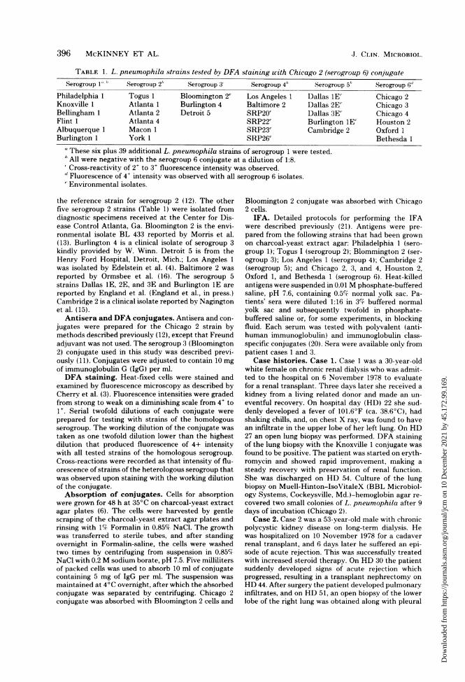
TABLE 1. L. pneumophila strains tested by DFA staining with Chicago 2 (serogroup 6) conjugateSerogroup 1" " Serogroup 2" Serogroup 3' Serogroup 4" Serogroup 5" ,Serogroup 6"
Philadelphia 1 Togus 1 Bloomington 2" Los Angeles 1 Dallas 1E' Chicago 2Knoxville 1 Atlanta 1 Burlington 4 Baltimore 2 Dallas 2E' Chicago 3Bellingham 1 Atlanta 2 Detroit 5 SRP20' Dallas 3E' Chicago 4Flint 1 Atlanta 4 SRP22" Burlington lE' Houston 2Albuquerque 1 Macon 1 SRP23" Cambridge 2 Oxford 1Burlington 1 York 1 SRP26" Bethesda 1
"These six plus 39 additional L. pneumophila strains of serogroup 1 were tested.h All were negative with the serogroup 6 conjugate at a dilution of 1:8.Cross-reactivity of 2+ to 3+ fluorescence intensity was observed.
"'Fluorescence of 4+ intensity was observed with all serogroup 6 isolates.Environmental isolates.
the reference strain for serogroup 2 (12). The otherfive serogroup 2 strains (Table 1) were isolated fromdiagnostic specimens received at the Center for Dis-ease Control Atlanta, Ga. Bloomington 2 is the envi-ronmental isolate BL 433 reported by Morris et al.(13). Burlington 4 is a clinical isolate of serogroup 3kindly provided by W. Winn. Detroit 5 is from theHenry Ford Hospital, Detroit, Mich.; Los Angeles 1was isolated by Edelstein et al. (4). Baltimore 2 wasreported by Ormsbee et al. (16). The serogroup 5strains Dallas lE, 2E, and 3E and Burlington lE arereported by England et al. (England et al., in press.)Cambridge 2 is a clinical isolate reported by Nagingtonet al. (15).
Antisera and DFA conjugates. Antisera and con-jugates were prepared for the Chicago 2 strain bymethods described previously (12), except that Freundadjuvant was not used. The serogroup 3 (Bloomington2) conjugate used in this study was described previ-ously (11). Conjugates were adjusted to contain 10 mgof immunoglobulin G (IgG) per ml.DFA staining. Heat-fixed cells were stained and
examined by fluorescence microscopy as described byCherry et al. (3). Fluorescence intensities were gradedfrom strong to weak on a diminishing scale from 4+ to1+. Serial twofold dilutions of each conjugate wereprepared for testing with strains of the homologousserogroup. The working dilution of the conjugate wastaken as one twofold dilution lower than the highestdilution that produced fluorescence of 4+ intensitywith all tested strains of the homologous serogroup.Cross-reactions were recorded as that intensity of flu-orescence of strains of the heterologous serogroup thatwas observed upon staining with the working dilutionof the conjugate.Absorption of conjugates. Cells for absorption
were grown for 48 h at 35°C on charcoal-yeast extractagar plates (6). The cells were harvested by gentlescraping of the charcoal-yeast extract agar plates andrinsing with 1% Formalin in 0.85% NaCi. The growthwas transferred to sterile tubes, and after standingovernight in Formalin-saline, the cells were washedtwo times by centrifuging from suspension in 0.85%-NaCI with 0.2 M sodium borate, pH 7.5. Five millilitersof packed cells was used to absorb 10 ml of conjugatecontaining 5 mg of IgG per ml. The suspension wasmaintained at 4°C overnight, after which the absorbedconjugate was separated by centrifuging. Chicago 2conjugate was absorbed with Bloomington 2 cells and
Bloomington 2 conjugate was absorbed with Chicago2 cells.
IFA. Detailed protocols for performing the IFAwere described previously (21). Antigens were pre-pared from the following strains that had been grownon charcoal-yeast extract agar: Philadelphia 1 (sero-group 1); Togus I (serogroup 2); Blommington 2 (ser-ogroup 3); Los Angeles 1 (serogroup 4); Cambridge 2(serogroup 5); and Chicago 2, 3, and 4, Houston 2,Oxford 1, and Bethesda 1 (serogroup 6). Heat-killedantigens were suspended in 0.01 M phosphate-bufferedsaline, pH 7.6, containing 0.5% normal yolk sac. Pa-tients' sera were diluted 1:16 in 3% buffered normalyolk sac and subsequently twofold in phosphate-buffered saline or, for some experiments, in blockingfluid. Each serum was tested with polyvalent (anti-human immunoglobulin) and immunoglobulin class-specific conjugates (20). Sera were available only frompatient cases 1 and 3.Case histories. Case 1. Case 1 was a 30-year-old
white female on chronic renal dialysis who was admit-ted to the hospital on 6 November 1978 to evaluatefor a renal transplant. Three days later she received akidney from a living related donor and made an un-eventful recovery. On hospital day (HD) 22 she sud-denly developed a fever of 101.6°F (ca. 38.6°C), hadshaking chills, and, on chest X ray, was found to havean infiltrate in the upper lobe of her left lung. On HD27 an open lung biopsy was performed. DFA stainingof the lung biopsy with the Knoxville 1 conjugate wasfound to be positive. The patient was started on eryth-romycin and showed rapid improvement, making asteady recovery with preservation of renal function.She was discharged on HD 54. Culture of the lungbiopsy on Muell-Hinton-IsoVitaleX (BBL Microbiol-ogy Systems, Cockeysville, Md.)-hemoglobin agar re-covered two small colonies of L. pneumophila after 9days of incubation (Chicago 2).Case 2. Case 2 was a 53-year-old male with chronic
polycystic kidney disease on long-term dialysis. Hewas hospitalized on 10 November 1978 for a cadaverrenal transplant, and 6 days later he suffered an epi-sode of acute rejection. This was successfully treatedwith increased steroid therapy. On HD 30 the patientsuddenly developed signs of acute rejection whichprogressed, resulting in a transplant nephrectomy onHD 44. After surgery the patient developed pulmonaryinfiltrates, and on HD 51, an open biopsy of the lowerlobe of the right lung was obtained along with pleural
J. CLIN. MICROBIOL.
Dow
nloa
ded
from
http
s://j
ourn
als.
asm
.org
/jour
nal/j
cm o
n 10
Dec
embe
r 20
21 b
y 45
.172
.99.
169.

L. PNEUMOPHILA SEROGROUP 6 397
fluid for culture and microscopy. The lung biopsy wasfelt to be histologically consistent with Pneumocystsicarinii but negative for L. pneumophila by culture.However, the pleural fluid was positive for L. pneu-mophila by culture (Chicago 3). Despite therapy witherythromycin and trimethoprim-sulfamethoxazole,signs and symptoms of infection persisted, and thepatient died on HD 59. Autopsy revealed organizingpneumocystis pneumonia, dissemianted candidiasis,and pulmonary cytomegalovirus infection. No histo-logical evidence of bacterial pneumonia was present.Case 3. Case 3 was a 44-year-old white male in
chronic renal failure from chronic pyelonephritis since1973. He had first received a cadaver kidney in 1976.Nine months later the kidney was removed because ofchronic rejection. On 20 June 1979 he received asecond cadaver kidney and did well until HD 19, whenhe began to complain of chills, malaise, pleuritic chestpain, and cough. He developed a fever of 101°F, anda chest X ray showed an infiltrate in the left lung. Hiscondition deteriorated rapidly, and later, on HD 21,he suffered a cardiorespiratory arrest while beingtransferred to the intensive care unit. He was revived,but sustained resultant irreversible ischemic cerebraldamage. On HD 22, an open lung biopsy was recom-mended and the patient was strated on erythromycin.On HD 25, a lung biopsy was performed. The chest Xray showed resolution of the left pulmonary infiltratewith erythromycin therapy by HD 25; however, thepatient remained comatose and flaccid, and he died onHD 64.A DFA stain of imprints from the lung biopsy
showed two to three bacilli per field when stained witheither the Knoxville 1 or the Togus 1 conjugate. Cul-ture on Mueller-Hinton-IsoVitaleX-hemoglobin agarrevealed several small colonies of L. pneumophilaafter 8 days of incubation (Chicago 4).
RESULTSDFA staining. As a stringent test of specific-
ity, the Chicago 2 conjugate was tested at a 1:8dilution against all strains of L. pneumophilaserogroups 1 through 5 listed in Table 1. The 45strains of serogroup 1, six strains of serogroup 2,six strains of serogroup 4, and five strains ofserogroup 5 were all negative in cellular stainingwith this conjugate. Stained flagella were ob-served in about 50% of the strains, but this didnot interfere with interpretation of cellular stain-ing.
Reciprocal cross staining between conjugatesand strains of serogroup 3 and serogroup 6 of 2+to 3+ fluorescence intensity was observed withthe Chicago 2 (group 6) and the Bloomington 2(group 3) conjugates at a 1:8 dilution. Therefore,the conjugates were tested further at the work-ing dilution (1:256) for cross-reactivity betweenthese two serogroups. Results are shown in Ta-ble 2. Significant cross-reactivity was observedwith both of the unabsorbed conjugates at theworking dilution. The cross-reactivity was com-pletely removed by absorption with cells of the
cross-reactive strain, but working dilutions werereduced in each case from 1:256 with the unab-sorbed conjugate to 1:32 with the absorbed con-jugate.
Cells of the six L. pneumophila serogroup 6strains were negative in DFA staining with con-jugates for serogroups 2 and 5 and producedweak, irregular fluorescent staining patternswith conjugates for serogroup 1. A smail per-centage of the cells fluoresced brightly with se-rogroup 4 conjugate.IFA. Thirteen serum samples drawn from
case 1 showed a diagnostic rise in titer (24-foldincrease to >128 during convalescence) againstall L. pneumophila antigens tested (Table 3).Titers obtained against the serogroup 6 antigenswith the polyvalent (anti-human immunoglob-ulin) conjugate, however, were 64-fold greaterthan those obtained against serogroup 1 to 5antigens 4 weeks after onset of illness, an indi-cation that this patient's antibody response toinfection was primarily serogroup 6 specific.Nine months after onset her serogroup 6 titerremained at an elevated level, 1,024. Similarlyserum drawn from case 3 approximately 5 weeksafter onset had a serogroup 6 titer that was atleast 32-fold greater than those obtained againstthe heterologous antigens. It is interesting thatthis patient's titer 13 days after onset had beenthe same (4,096) against all antigens tested, anindication that his initial antibody response wasagainst a common L. pneumophila antigen (20,21). Serogroup 6 strains were isolated from bothpatients: Chicago 2 from case 1 and Chicago 4from case 3.To determine which immunoglobulin class(es)
contributed to the titers obtained with thepolyvalent conjugate and to establish furtherthe specificity of the test, titers obtained withclass-specific conjugates and with blocking fluidwere compared. Blocking fluid has been usedsuccessfully to inhibit nonspecific titers in legi-onellosis patients' sera against a variety of gram-negative bacteria (20). Serogroup 6-specific andL. pneumophila common antigen responses ofcase 1 were in the IgG, IgM and IgA fractions,with maximal titers 22 days after onset of illnessfor the specific antigen and 14 days after onsetfor the common antigens (Fig. 1). Serogroup-specific titers remained elevated for at least 7weeks in the IgG and IgM fractions but not inthe IgA fraction, which had dropped to a titer of64 in 5 weeks. Case 3 also had maximal commonantigen responses within 2 weeks after onset inthe IgG, IgM, and IgA classes (Fig. 2). However,in contrast to case 1, his IgM serogroup-specificresponse also peaked at this time, whereas hismaximal IgG and IgA serogroup-specific re-
VOL. 12, 1980
Dow
nloa
ded
from
http
s://j
ourn
als.
asm
.org
/jour
nal/j
cm o
n 10
Dec
embe
r 20
21 b
y 45
.172
.99.
169.

398 McKINNEY ET AL.
TABLE 2. DFA staining intensities of homologous and cross-reactive antigens with serogroup 3 and 6conjugates
Conjugate Fluorescence intensity
Serogroup Working dilu- Bloomington Chicago 2,'Vaccine strain Absorbing strain tion< 2,h serogroup 3 serogroup 6
3 Bloomington 2 None 1:256 4 2Chicago 2 1:32 4
6 Chicago 2 None 1:256 1 4Bloomington 2 1:32 4
One twofold dilution lower than the maximum dilution that still gave 4+ (very bright) staining of all strainsof the homologous serogroup.'The other serogroup 3 strains, Burlington 4 and Detroit 5, were identical to Bloomington 2 in staining
reactions.' The other serogroup 6 strains, Chicago 3, Chicago 4, Houston 2, Oxford 1, and Bethesda 1, were identical to
Chicago 2 in staining reactions.
TABLE 3. IFA titers of sera from cases I and 3against L. pneumophila serogroup I to 6 antigens,usingpolyvalent anti-human immunoglobulin
conjugateSerum specimen IFA antigen titer
Day drawn be- Serogroup 6" SerogroupPatient fore (-) or after (Chicago 2) 1-5"
onset
Case 1 -4, -6, -21 128 C6414 4,096 51222, 26 16,384 51229 32,768 51235 16,384 25637, 45, 49 8,192 128254 2,048 64267 1,024 64
Case 3 2 C64 C6413 4,096 4,09637 32,768 1,02443' 16,384 1,024
"'Titers obtained against all other serogroup 6strains agreed within one doubling dilution.
"Titers obtained against serogroup 1 (shown intable) 2, 3, 4, and 5 antigens (see text for strains used)agreed within one doubling dilution for case 1. Case 3titers against serogroup 1 (shown) were higher thanthose serogroups 2, 3, and 5.
Autopsy specimen.
sponses were at 5 weeks postonset. Titers ob-tained when sera from both cases were dilutedin blocking fluid were within the acceptable one-tube variation of those obtained when the serawere diluted in phosphate-buffered saline, anindication that the titers were specific for Le-gionella antigens.
DISCUSSIONIn this report, we describe the isolation of a
new serogroup of L. pneumophila from cases ofLegionnaires disease. Although the isolated Chi-
65,536132,768
(9 16,384a- 8,192-O 4,096(D 2,048
w 1,024-F 512-<É 256-
128-64
-20 -10 o 10 20 30 40
DAYS POSTONSET
2,048- Poly-IgO IgGD1o24-O' zIgMDIgAtr 512- oIA , i\ _;
z256-
H 128- , CoI-o-CI *-*H 64-L / .* o.-O--O- 0o-0<
64- -<L L 64 *E .o-",_-20 -1o o 10 20 30 40 50 9months
DAYS POSTONSETFIG. 1. IFA titers of case I sera against L. pneu-
mophila (top) serogroup 6 (Chicago 2) and (bottom)serogroup I (Philadelphia 1) antigens with anti-hu-man immunoglobulin (Poly-Ig), anti-human IgG,anti-human IgM, and anti-human IgA conjugates.(Top) Specific response; (bottom) common antigenresponse.
cago 2, 3, and 4 were obtained on Mueller-Hin-ton-IsoVitaleX-hemoglobin agar, charcoal-yeast extract agar is currently the growth me-dium of choice because of its greater sensitivityfor primary isolation of L. pneumophila frominfected tissue (6). L. pneumophila strains Chi-cago 2, 3, and 4, Houston 2, Oxford 1, and Be-thesda 1 are serologically similar to each otherand distinct from serogroups 1 to 5 as shown byDFA staining tests. The cross-reactivity of thesestrains with the serogroup 3 conjugate is ofmoderate intensity, and strains of this new ser-ogroup could lead to confusion in interpretingdiagnostic staining with group 3 conjugate or
J. CLIN. MICROBIOL.
Dow
nloa
ded
from
http
s://j
ourn
als.
asm
.org
/jour
nal/j
cm o
n 10
Dec
embe
r 20
21 b
y 45
.172
.99.
169.

L. PNEUMOPHILA SEROGROUP 6 399
'-4 2,048-
C-1,024-
° 512-
& 256-W 128.F--
F- 641
Li L64_
10
le
-oe
10 20 30 40DAYS POSTONSET
FIG. 2. Case 3 sera tested as indicated for case I(see legend to Fig. 1). Symbols: e, anti-human im-munoglobulin; *, IgG; O, IgM; O, IgA.
with L. pneumophila polyvalent conjugates thatare currently in use. The Oxford 1 strain hasbeen reported as a clinical isolate of serogroup3 (5, 17). These authors recognized both serolog-ical difference and similarity between Oxford 1and Bloomington 2, and indeed the serogroup 6strains could be considered as variants of sero-
group 3. However, we believe that both DFAand IFA reactivities with the group of isolatesChicago 2, 3, and 4, Houston 2, Oxford 1, andBethesda 1 are sufficiently different from theserogroup 3 strains to warrant the designationof a new serogroup. We recommend that thesestrains be designated L. pneumophila serogroup6.Although appropriate absorption of conju-
gates for serogroups 6 and 3 renders them com-
pletely group specific, it is doubtful that ab-sorbed conjugates are necessary for diagnosticpurposes. A clear distinction can be made be-tween these two serogroups on the basis of in-tensity of fluorescence with working dilutions ofunabsorbed conjugates for the two serogroups.The curious pattern of staining of group 6
stains with conjugates for serogroups 1 and 4was first suspected to be due to a mixed culture.However, the staining pattern persisted in nu-
merous cultures from single-colony picks, andthe fact that it is common to all six strains ofserogroup 6 indicates that it is a characteristicof this serogroup. The observation of positive
DFA staining of lung biopsy material from cases1 and 3 with the Knoxville 1 (serogroup 1) con-jugate could be explained on the basis of thecharacteristic spotty staining pattern of sero-group 6 isolates with this conjugate. However,positive staining of biopsy material in case 3with Togus 1 (serogroup 2) conjugate is not asreadily explained since cross-reactivity was notobserved with serogroup 2 conjugate with any ofthe serogroup 6 strains from pure cultures oncharcoal-yeast extract plates. This phenomenonof L. pneumophila bacteria staining in tissuewith conjugate for a heterologous serogroup, butfailure to stain with the same heterologous ser-ogroup conjugate after isolation on artificial me-dia, was observed previously with the Togus 1isolate (12). The reason for the occasional obser-vation of serogroup cross-reactivity of this na-ture is not understood. In any case, experiencehas shown that it is necessary to use the homol-ogous serogroup conjugate to consistently revealL. pneumophila organisms in tissue. Retrospec-tive DFA staining of biopsy tissues from cases 1and 3 with serogroup 6 conjugate revealed manymore organisms and much more brightly flu-orescent organisms than were initially observedwith either the serogroup 1 or the serogroup 2conjugate.
Staining of flagella of L. pneumophila withlow dilutions of conjugates has been observedpreviously (18). The specificity of flagellar stain-ing among these organisms has not been studiedin detail, but it obviously has no relationship tocellular staining. Laboratorians should be awareof the possibility of common flagellar stainingwhich can be readily distinguished from theserogroup-specific cellular staining. The rela-tionship of the various staining patterns as seenin the DFA test and the common antigens asshown in the IFA have not yet been established.
In previous studies of the immune response tolegionellosis, diagnostic rises in titer during con-valescence were found that were specific forserogroup antigen 1, 2, 3, or 4 or that werespecific for antigens common in multiple-sero-group strains (20, 21). The IFA results in thisstudy support this concept. The patient fromwhom the Chicago 2 strain was isolated had arise in titer against all six serogroup 6 strains, toa level that was 64-fold greater than her titersagainst the serogroup 1 to 5 antigens. Althoughprimarily serogroup specific, this patient's anti-body response also occurred against a commonantigen as shown by the fact that initially herseroconversion was detected with the serogroup1 antigen used routinely at the time ofher illness.Similarly, the patient from whom the Chicago 4strain was isolated had both serogroup 6-specific
VOL. 12, 1980
Dow
nloa
ded
from
http
s://j
ourn
als.
asm
.org
/jour
nal/j
cm o
n 10
Dec
embe
r 20
21 b
y 45
.172
.99.
169.

400 MCKINNEY ET AL.
and common antigen titer rises, but the formerwas 32-fold greater than the latter.The sequence of antibody formation after on-
set of Legionnaires disease has not been studiedextensively due in part to a lack of sequentiallycollected specimens but also because of the scar-city of sera from culture-confirmed cases. In thepresent study two such cases were available.Case 1 had peak IgG, IgM, and IgA responses tothe Legionella common antigen(s) 14 days afteronset of Legionnaires disease, whereas her se-rogroup 6-specific IgG, IgM, and IgA titerspeaked in 22 days. In 5 weeks, her serogroup 6-specific IgA titer was below the level of diagnos-tic significance. In contrast, her serogroup 6-specific IgG and IgM titers declirled slowly to256 or 128, respectively, over the ensuing 8months. Case 3 also had maximal common an-tigen titers in all three immunoglobulin classes13 days after onset of Legionnaires disease. Un-like case 1, the IgM serogroup 6-specific responseof case 3 also peaked 13 days postonset. His IgGand IgA responses peaked at 5 weeks postonset,a time when the IgA level of case 1 had declined.The 3- to 5-week period to reach maximal anti-body synthesis found in this study of serogroup6 disease is similar to that found in Legionnairesdisease of serogroup 1 etiology (10). It is tempt-ing to speculate that the 2-week peak in thecommon antigen response represents an an-
amnestic response because of prior exposure tothese patients to L. pneumophila, but furtherdata are needed to test this hypothesis. The factthat the patients received immunosuppressivetherapy may have influenced their antibody re-
sponses to L. pneumophila infection.L. pneumophila has been studied previously
as a cause of Legionnaires disease in renal trans-plant recipients (1, 7). In the present study, wehave presented evidence that a new serogroupof L. pneumophila, serogroup 6, causes Legion-naires disease in renal transplant patients andthat the new group has a wide geographicaldistribution. Tests for L. pneumophila and le-gionellosis should include reagents for serogroup6.
LITERATURE CITED
1. Bock, B. V., B. D. Kirby, P. H. Edelstein, W. L.George, K. M. Snyder, M. L. Owens, C. M. Hatay-ama, C. E. Haley, R. P. Lewis, R. D. Meyer, and S.M. Finegold. 1978. Legionnaires' disease in renal-trans-plant recipients. Lancet i:410-413.
2. Brenner, D. J., A. G. Steigerwalt, and J. E. McDade.1979. Classification of the Legionnaires' disease bacte-rium. Legionella pneumophila, genus novium, speciesnova, of the family Legionellaceae familia nova. Ann.Intern. Med. 90:656-658.
3. Cherry, W. B., B. Pittman, P. P. Harris, G. A. Hebert,B. M. Thomason, L. Thacker, and R. E. Weaver.1978. Detection of Legionnaires disease bacteria by
J. CLIN. MICROBIOL.
direct immunofluorescence staining. J. Clin. Microbiol.8:329-338.
4. Edelstein, P. H., R. D. Meyer, and S. M. Finegold.1978. Isolation of a new serotype of Legionnaires' dis-ease bacterium. Lancet ii: 1172-1174.
5. Fallon, R. J., and W. H. Abraham. 1979. Legionnaires'disease caused by Legionella pneumophila serogroup3. Lancet ii:304.
6. Feeley, J. C., R. J. Gibson, G. W. Gorman, M. C.Langford, J. K. Rasheed, D. C. Mackel, and W. B.Baine. 1979. Charcoal-yeast extract agar: primary iso-lation medium for Legionella pneumophila. J. Clin.Microbiol. 10:437-441.
7. Foster, R. S., W. C. Winn, W. Marshall, and D. W.Gump. 1979. Legionnaires' disease following renaltransplantation. Transplant. Proc. 11:93-95.
8. Fraser, D. W., D. C. Deubner, D. L. Hill, and D. K.Gilliam. 1979. Nonpneumonic short-incubation-periodlegionellosis (Pontiac fever) in men who cleaned a steamturbine condenser. Science 205:690-691.
9. Glick, T. H., M. B. Gregg, B. Berman, G. Mallison, W.W. Rhodes, Jr., and I. Kassanoff. 1978. Pontiac fever.An epidemic of unknown etiology in a health depart-ment. I. Clinical and epidemiological aspects. Am. J.Epidemiol. 107:149-160.
10. MeDade, J. E., C. C. Shepard, D. W. Fraser, T. R.Tsai, M. A. Redus, W. R. Dowdle, and the Labo-ratory Investigation Team. 1977. Legionnaires' dis-ease. Isolation of a bacterium and demonstration of itsrole in other respiratory disease. N. Engl. J. Med. 297:1197-1203.
11. McKinney, R. M., L. Thacker, P. P. Harris, K. R.Lewallen, G. A. Hebert, P. H. Edelstein, and B. M.Thomason. 1979. Four serogroups of Legionnaires' dis-ease bacteria defined by direct immunofluorescence.Ann. Intern. Med. 90:621-624.
12. McKinney, R. M., B. M. Thomason, P. P. Harris, L.Thacker, K. R. Lewallen, H. W. Wilkinson, G. A.Hebert, and C. W. Moss. 1979. Recognition of a newserogroup of Legionnaires disease bacterium. J. Clin.Microbiol. 9:103-107.
13. Morris, G. K., C. M. Patton, J. C. Feeley, S. E. John-son, G. Gorman, W. T. Martin, P. Skaliy, G. F.Mallison, B. D. Politi, and D. C. Mackel. 1979. Iso-lation of the Legionnaires' disease bacterium from en-vironmental samples. Ann. Intern. Med. 4:664-667.
14. Moss, C. W., R. E. Weaver, S. B. Dees, and W. B.Cherry. 1977. Cellular fatty acid composition of isolatesfrom Legionnaires disease. J. Clin. Microbiol. 6:140-143.
15. Nagington, J., T. G. Wreghitt, and D. J. Smith. 1979.How many Legionnaires? Lancet ii:536-537.
16. Ormsbee, R. A., M. G. Peacock, G. L. Lattimer, L. A.Page, and B. Fiset. 1978. Legionnaires' disease: anti-genic peculiarities, strain differences, and antibiotic sen-sitivities of the agent. J. Infect. Dis. 138:260-264.
17. Taylor, A. G., and T. G. Harrison. 1979. Legionnaires'disease caused by Legionella pneumophila serogroup3. Lancet ii:47.
18. Thomason, B. M., F. W. Chandler, and D. G. Hollis.1979. Flagella on Legionnaires' disease bacteria: an in-terim report. Ann. Intern. Med. 91:224-225.
19. Weaver, R. E., and J. C. Feeley. 1979. Cultural andbiochemical characterization of Legionnaires' diseasebacterium, p. 20-25. In G. L. Jones and G. A. Hebert(ed.), "Legionnaires':" The disease, the bacterium andmethodology. Center for Disease Control, Atlanta.
20. Wilkinson, H. W., C. E. Farshy, B. J. Fikes, D. D.Cruce, and L. P. Yealy. 1979. Measure of immuno-globulin G-, M-, and A-specific titers against Legionellapneumophila and inhibition of titers against nonspecificgram-negative bacterial antigens in the indirect immu-
Dow
nloa
ded
from
http
s://j
ourn
als.
asm
.org
/jour
nal/j
cm o
n 10
Dec
embe
r 20
21 b
y 45
.172
.99.
169.

VOL. 12, 1980
nofluorescence test for legionellosis. J. Clin. Microbiol.10:685-689.
21. Wilkinson, H. W., B. J. Fikes, and D. D. Cruce. 1979.Indirect immunofluorescence test for serodiagnosis of
L. PNEUMOPHILA SEROGROUP6 401
Legionnaires disease: evidence for serogroup diversityof Legionnaires disease bacterial antigens and for mul-tiple specificity of human antibodies. J. Clin. Microbiol.9:379-383.
Dow
nloa
ded
from
http
s://j
ourn
als.
asm
.org
/jour
nal/j
cm o
n 10
Dec
embe
r 20
21 b
y 45
.172
.99.
169.