Embed Size (px)
Citation preview

LEDGF/p75 interacts with mRNA splicingfactors and targets HIV-1 integration tohighly spliced genesParmit Kumar Singh,1 Matthew R. Plumb,2 Andrea L. Ferris,3 James R. Iben,4 Xiaolin Wu,5
Hind J. Fadel,6 Brian T. Luke,7 Caroline Esnault,1 Eric M. Poeschla,8 Stephen H. Hughes,3
Mamuka Kvaratskhelia,2 and Henry L. Levin1
1Section on Eukaryotic Transposable Elements, Program inCellular Regulation andMetabolism, Eunice Kennedy ShriverNationalInstitute of Child Health and Human Development, National Institutes of Health, Bethesda, Maryland 20892, USA; 2Center forRetrovirus Research, College of Pharmacy, The Ohio State University, Columbus, Ohio 43210, USA; 3HIV Drug ResistanceProgram, National Cancer Institute, Frederick, Maryland 21702, USA; 4Program in Genomics of Differentiation, Eunice KennedyShriver National Institute for Child Health and Human Development, National Institutes of Health, Bethesda, Maryland 20892,USA; 5Leidos Biomedical Research, Inc., Frederick National Laboratory for Cancer Research, Frederick, Maryland 21702, USA;6Department ofMolecularMedicine, MayoClinic College ofMedicine, Rochester, Minnesota 55905, USA; 7Advanced BiomedicalComputing Center, Leidos Biomedical Research, Inc., Frederick National Laboratory for Cancer Research, Frederick, Maryland,21702, USA; 8Division of Infectious Diseases, University of Colorado School of Medicine, Aurora, Colorado 80045, USA
The host chromatin-binding factor LEDGF/p75 interacts with HIV-1 integrase and directs integration to activetranscription units. To understand how LEDGF/p75 recognizes transcription units, we sequenced 1 million HIV-1integration sites isolated from cultured HEK293T cells. Analysis of integration sites showed that cancer genes werepreferentially targeted, raising concerns about using lentivirus vectors for gene therapy. Additional analysis led tothe discovery that introns and alternative splicing contributed significantly to integration site selection. Thesecorrelations were independent of transcription levels, size of transcription units, and length of the introns. Multi-variate analysis with five parameters previously found to predict integration sites showed that intron density is thestrongest predictor of integration density in transcription units. Analysis of previously published HIV-1 integrationsite data showed that integration density in transcription units in mouse embryonic fibroblasts also correlatedstrongly with intron number, and this correlation was absent in cells lacking LEDGF. Affinity purification showedthat LEDGF/p75 is associated with a number of splicing factors, and RNA sequencing (RNA-seq) analysis ofHEK293T cells lacking LEDGF/p75 or the LEDGF/p75 integrase-binding domain (IBD) showed that LEDGF/p75contributes to splicing patterns in half of the transcription units that have alternative isoforms. Thus, LEDGF/p75interacts with splicing factors, contributes to exon choice, and directs HIV-1 integration to transcription units thatare highly spliced.
[Keywords: HIV-1; integration; mRNA splicing; LEDGF; p75; retrovirus]
Supplemental Material is available for this article.
Received June 18, 2015; revised version accepted October 9, 2015.
Gene therapy is being used with increasing success in thetreatment of genetic disorders and is showing particularpromise for immunotherapy of cancer. These therapiescommonly use retroviral vectors to stably integrate thecorrective/therapeutic sequences in the genomes of thepatient’s cells. The first successful retroviral gene thera-pies used vectors derived from the γ retroviruses to correctdisorders such as X-linked severe combined immunodefi-ciency (SCID-X1) (Hacein-Bey-Abina et al. 2003, 2010).However, there was a significant risk associated with
the use of γ retrovirus-based vectors because of their po-tential to activate, by integration, proto-oncogenes. Thiswas a problem for some of the SCID-X1 patients who hadvector-induced clonal proliferation of their T cells thatprogressed to leukemia (Hacein-Bey-Abina et al. 2003,2010).The γ retroviruses have a strong preference for integrat-
ing near-active enhancers (Wu et al. 2003; De Rijck et al.
Corresponding author: [email protected] is online at http://www.genesdev.org/cgi/doi/10.1101/gad.267609.115.
© 2015 Singh et al. This article is distributed exclusively by ColdSpring Harbor Laboratory Press for the first six months after the full-issue publication date (see http://genesdev.cshlp.org/site/misc/terms.xhtml). After six months, it is available under a Creative Commons Li-cense (Attribution-NonCommercial 4.0 International), as described athttp://creativecommons.org/licenses/by-nc/4.0/.
GENES & DEVELOPMENT 29:2287–2297 Published by Cold Spring Harbor Laboratory Press; ISSN 0890-9369/15; www.genesdev.org 2287
Cold Spring Harbor Laboratory Press on August 9, 2018 - Published by genesdev.cshlp.orgDownloaded from

2013; Gupta et al. 2013; Sharma et al. 2013; De Ravin et al.2014; LaFave et al. 2014). This bias increases the risk of ac-tivating proto-oncogenes and has led to the use of lentivi-rus vectors for recent gene therapy trials. HIV-1-basedvectors are thought to be less likely to causemalignanciesbecause integration is not directed to active enhancers (DeRijck et al. 2013). However, HIV-1 integration occursthroughout the bodies of active transcription units (Schro-der et al. 2002; Wu et al. 2003; Wang et al. 2007; Ferriset al. 2010), raising the concern that HIV integrations alsohave the potential to induce the expression of proto-on-cogenes. This concern was heightened by recent studiesof HIV-1 patients undergoing suppressive combinationanti-retroviral therapy, which indicated that integrationin specific genes can cause clonal expansion and persis-tence of the infected cells (Maldarelli et al. 2014; Wagneret al. 2014).
Structural and biochemical data show that HIV-1 inte-grase interacts with the host factor LEDGF/p75 (Cherepa-nov et al. 2003, 2004, 2005; Maertens et al. 2003; Llanoet al. 2004; Busschots et al. 2005; Shun et al. 2007), andthis interaction favors integration in active transcriptionunits, which is the portion of genes that are transcribed(Ciuffi et al. 2005; Llano et al. 2006a; Vandekerckhoveet al. 2006; Shun et al. 2007; Ferris et al. 2010; Wanget al. 2012; Koh et al. 2013). Alternative splicing of LEDGFproduces a full-length isoform (p75) and a truncated iso-form (p52). Both isoforms contain the chromatin-bindingdomains (PWWP andAT-hook elements), themajor deter-minants for high-affinity and site-specific binding to chro-matin (Llano et al. 2006b; Turlure et al. 2006; Meehanet al. 2009; Ferris et al. 2010; Gijsbers et al. 2010, 2011);however, only LEDGF/p75 contains the integrase-bindingdomain (IBD), and only this isoform mediates HIV-1integration.
Although it is clear that LEDGF/p75 helps direct HIV-1integration to active transcription units, there are impor-tant questions about the role of LEDGF/p75 that remainto be addressed. It is not clear how LEDGF/p75 recognizesactive transcription units (Schroder et al. 2002; Ferriset al. 2010; Wang et al. 2012). It is not known whetherLEDGF/p75 couples integration with transcription ormerely interacts with features of transcription units. Animportant reason these questions are unanswered is thatrelatively little is known about the cellular functions ofLEDGF/p75.
In order to understand integration targeting and itsbiological impact, we sought to measure integration atthe level of individual genes. To obtain the largest pos-sible number of integration events, we generated mapsof integration sites with a single-round HIV-1 vector inHEK293T cells. This system and improvements in se-quencing methods allowed us to map 961,274 indepen-dent integration sites; of the sites in transcription units,82% occurred in just 4000 genes. Importantly, the 1000transcription units with the highest numbers of integra-tion sites were highly enriched for cancer-associatedgenes. Analysis of the integration site densities per tran-scription unit (integration sites per kilobase) revealed astriking bias that favored transcription units with high
numbers of introns and those that produced multiplespliced mRNAs. Our analysis of published profiles ofHIV-1 integration demonstrated that LEDGF is requiredfor targeting highly spliced transcription units. Affin-ity purification of LEDGF/p75 coupled with tandemmass spectrometry (MS/MS) identified a variety of associ-ated splicing factors, including components of snRNPs,hnRNPs, and helicases. Biallelic deletion of the PSIP1region encoding the IBD of LEDGF/p75 resulted in signif-icant changes in the splicing patterns of the mRNAs from>5000 genes. Together, these results show that LEDGF/p75 specifically interacts with splicing machinery and isrequired for targeting HIV-1 integration to highly splicedgenes.
Results
Approximately 75% of HIV-1 integration occurs in RNApolymerase II (Pol II) transcription units (Schroder et al.2002; Wang et al. 2007). To improve our understandingof HIV-1 integration, we generated a high-density map ofHIV-1 integration sites in cultured human cells (Materialsand Methods). A single-round replication-defective viruswas used to infect HEK293T cells. Both the ligation reac-tions and PCR were performed in multiplexed format,which made it possible to generate complex integrationsite libraries that were directly sequenced without nestedPCR amplification or additional rounds of adaptor liga-tion. A total of 961,274 unique virus–host junctionswere obtained. This profile is large enough to provide inte-gration frequencies in individual transcription units thatare highly reproducible, as shown by pairwise compari-sons of eight independently generated sublibraries (R2 val-ues between 0.89 and 0.98) (Supplemental Table S1 forRefSeq genes).
We counted the total number of integrations withineach transcription unit and found that the number of inte-grations per transcription unit was high in a relativelysmall number of transcription units (Supplemental Fig.S1); 82% of all of the integration sites in transcriptionunits mapped to just 4000 transcription units. Analysisof the 1000 transcription units with the highest numberof total integration sites revealed significant enrichmentassociated with mRNA splicing and histone methylation(Table 1). An extended list of gene ontologies enriched inthis group of the top 1000 transcription units is given inSupplemental Table S2.
Recent identification of HIV-1 integration sites in pe-ripheral blood lymphocytes of infected patients undergo-ing antiretroviral therapy indicated that integration intosome transcription units may cause clonal expansion ofthe infected cells (Maldarelli et al. 2014; Wagner et al.2014). We asked what fraction of the HEK293T integra-tion sites was within transcription units that are knownto be associated with an increased risk of cancer. By eval-uating independently curated lists of cancer-associatedgenes, we found that the group of 1000 transcription unitswith the highest numbers of total integration sites includ-ed three to five times more cancer genes than would be
Singh et al.
2288 GENES & DEVELOPMENT
Cold Spring Harbor Laboratory Press on August 9, 2018 - Published by genesdev.cshlp.orgDownloaded from

predicted based on their genome-wide prevalence (Table1; Kandoth et al. 2013; Vogelstein et al. 2013; Forbeset al. 2014). These observations indicate that HIV-1 inte-gration favors cancer genes.To determine whether the high number of integration
sites in specific groups of transcription units such as can-cer-associated genes was due to targeted integration, wenormalized the number of integration sites in each tran-scription unit by the length. By analyzing the top 1000transcription units based on integration sites per kilobase,cancer-associated genes, histone methyltransferases, andsplicing-associated genes were still favored (SupplementalTables S3, S4). This shows that HIV-1 integration favorscancer-associated genes with enrichment levels as muchas fourfold.
LEDGF causes integration to favorthe 5′ end of transcription units
We examined how HIV-1 integration sites were dis-tributed within transcription units by dividing RefSeqtranscription units into 15 equal segments (bins) and tab-ulating the integration sites within each bin (Fig. 1A).Intergenic integration sites were displayed in 500-base-pair (bp) segments either upstream of or downstreamfrom transcription units, depending on whether the in-tegration sites were nearer to the 5′ or 3′ ends of transcrip-tion units. Seventy-two percent of the 961,274 integrationsites mapped within transcription units and the integra-tion sites had a strong 5′ bias. No such bias was observedin a matched random control (MRC) set of insertionsgenerated in silico (Materials and Methods) to simulaterandom insertions (Fig. 1B; Berry et al. 2006). Becausethe integration site libraries were generated fromMseI-di-gested DNA, this random library was designed so that thesites matched the distances toMseI sites seen in the actu-al integration site library. Forty-one percent of randomlygeneratedMRC insertions occurred in transcription units,a number similar to the fraction of the genome that lies
within the known Pol II transcription units. In theHEK293T library, there was an obvious reduction in thenumber of integrations in the region between 500 and1500 bp upstream of transcription units, which was notseen in the MRC library. (Fig. 1, A vs. B).The availability of previously published HIV-1 integra-
tion data in mouse embryonic fibroblasts (MEFs) allowedus to compare the data that we obtained from infectedHEK293T cells with data for HIV-1 integration in cellsfrom a different species to see whether there was thesame strong bias for the 5′ end of transcription units(Wang et al. 2012). The bias for preferential integrationnear the 5′ end of Pol II transcription units was apparentin the MEF cells (Fig. 1C) and, more importantly, was ab-sent in cells lacking LEDGF (Fig. 1D). Broadly speaking,the distribution of integration sites in MEF cells lackingLEDGF was similar to the MRC sites (Fig. 1B); however,there was a strong bias for integration in the extreme5′ end of transcription units. Very similar integration sitedistributionpatternswere seen in twoother independentlygenerated HIV-1 integration site libraries generated frominfected MEFs that did or did not express LEDGF (Supple-mental Figs. S2, S3; Wang et al. 2012; Koh et al. 2013).Efforts to identify the features of transcription units
that were responsible for preferential integration nearthe 5′ ends of transcription units included tests of whetherintrons might be involved. In transcription units thatlacked introns, integration favored the 3′ end of the tran-scription units (Fig. 1E). There was no such bias whenthe MRC insertions were mapped to intronless transcrip-tion units (Fig. 1F).
Integration is targeted to highly spliced transcriptionunits by a mechanism that requires LEDGF
The marked difference in the distribution of integra-tion sites in transcription units that have and lack intronsled us to ask whether transcription units with moremRNA splicing had higher levels of integration. We usedthe number of alternatively spliced transcripts producedper transcription unit as determined with RNA sequenc-ing (RNA-seq) data from our HEK293T cells as a measureof mRNA splicing (analyzed with Cufflinks) (Supplemen-tal Table S5). We observed a strong correlation betweenthe numbers of spliced isoforms per transcription unitand the density of integration sites (integration sites perkilobase) that was absent in the MRC (Fig. 2A). We alsoused, as an independentmeasure of splicing within a tran-scription unit, the total number of introns as annotated byRefSeq (Supplemental Table S6). We grouped all transcrip-tion units by the number of introns they contain and cal-culated the average integration site density (integrationsites per kilobase) for each group. This measure revealeda striking correlation between the total numbers ofintrons and the density of integration sites in the tran-scription unit (Fig. 2B). This relationship is absent in theMRC. We also observed a strong correlation between thenumber of introns and integration site density when weanalyzed integration sites obtained from HIV-1-infectedMEF cells (Fig. 2C; Wang et al. 2012). Importantly, these
Table 1. The 1000 transcription units with the highest totalnumber of integration sites in HEK293T cells are enriched withfunctions associated with histone methylation and cancer
NameFold
enrichmentP-value
(Bonferroni)
Histone-lysine N-methyltransferaseactivitya,b
8.51 2.1 × 10−09 (1.5 × 10−06)
Cancer-driving genesc 4.92 6.5 × 10⁻¹³
The Cancer GenomeAtlasd
5.00 1.5 × 10⁻¹³
Somatic mutations incancere
2.80 2.1 × 10⁻¹⁴
Alternative splicinga,b 1.61 2.1 × 10−47 (8.9 × 10−45)Splice varianta,b 1.61 1.6 × 10−47 (4.2 × 10−44)
aHuang et al. 2009a.bHuang et al. 2009b.cSet of 125 genes (Vogelstein et al. 2013).dSet of 127 genes (Kandoth et al. 2013).eSet of 507 genes (Futreal et al. 2004).
LEDGF directs integration to highly spliced genes
GENES & DEVELOPMENT 2289
Cold Spring Harbor Laboratory Press on August 9, 2018 - Published by genesdev.cshlp.orgDownloaded from

correlations were not seen in an analysis of integrationsites in MEF cells lacking LEDGF (Fig. 2D). The strongcorrelation between intron number and integration sitedensity was also seen in the two other independent setsof integration data generated in MEFs (SupplementalFigs. S4, S5; Wang et al. 2012; Koh et al. 2013).
One possible reason why the integration density corre-lated with the number of introns is that intronic sequenc-es could have higher numbers of integration sites perkilobase than exons. However, we found that the averageintegration density for all introns is 0.57 per kilobase, anumber that is very similar to the average integration den-sity in exons (0.54 per kilobase) (Supplemental Table S7).It is also possible that transcripts withmore introns have ahigher density of integration sites because they are largerand, as a result of an unknownmechanistic property, bet-ter able to recruit preintegration complexes. To test thispossibility, we compared two sets of 673 transcriptionunits that were matched to have equal lengths. The firstset of transcription units had 10 introns, and the size-matched transcription units in the other set had one,two, or three introns. The size-matched transcriptionunits with 10 introns had substantially more integrationsthan the transcription units with one, two, or three in-trons (Fig. 2E,F). Moreover, the distribution of integrationsites throughout the transcription units with one to threeintrons showed that the 5′ bias was much weaker com-
pared with the integration sites in transcription unitswith 10 introns (Figs. 1A,C, 2, F vs. E). The high level ofintegration and the 5′ bias seen in transcription unitswith 10 introns was not seen with the MRC sites (Fig.2G). Interestingly, the integration density in transcriptionunits with one, two, and three introns was similar to theMRC set for these transcription units, suggesting that,in this group, the integration was similar to what wouldoccur if integration were random (Fig. 2, cf. F and H).
Previous studies reported that HIV-1 integration favorshighly expressed genes (Schroder et al. 2002; Wang et al.2007; Ferris et al. 2010). We determined average integra-tion site densities from the HEK293T data for transcrip-tion units sorted by levels of transcription and observed,as reported by others, that integration site density in genescorrelates with the level of expression (Supplemental Fig.S6). However, this tendency was not responsible for thehigh integration densities that we observed in highlyspliced transcription units because transcripts with high-er numbers of introns have, on average, lower expressionlevels (Supplemental Fig. S7). Thus, the observed increasein the density of integration sites in transcripts with high-er numbers of introns is more pronounced when the inte-gration site densities are normalized for the average levelsof transcription (Supplemental Fig. S8).
The results presented above indicate that splicing is akey determinant of HIV-1 integration site selection.
0
0.6
1.2
1.8
1 3 5 7 9 11 13 15
Transcription Units
Bins
0
0.6
1.2
1.8
0
2
4
6
MR
C s
ite
s (
%)
0
2
4
6
Inte
gra
tio
n (
%)
Inte
gra
tio
n (
%)
x 1
02
MR
C S
ite
s (
%)
x 1
02
HEK293T Intronless
Transcription Units
MRC Intronless
Transcription Units
BA
C
E F
HEK293T MRC
–10 –8 –6 –4 –2 1 3 5 7 9 –10 –8 –6 –4 –2 1 3 5 7 9
Upstream (kb) Transcription Units Bins
Upstream (kb) Transcription Units Bins
1 3 5 7 9 11 13 15
Transcription Units
Bins
Inte
gra
tio
n (
%)
Inte
gra
tio
n (
%)
MEFs LEDGF+/+ MEFs LEDGF–/–D
0
2
4
6
0
2
4
6
–10 –8 –6 –4 –2 1 3 5 7 9
Upstream (kb) Transcription Units Bins
–10 –8 –6 –4 –2 1 3 5 7 9
Upstream (kb) Transcription Units Downstream (kb)
Downstream (kb) Downstream (kb)
Downstream (kb) Bins
Figure 1. Distribution of HIV-1 integrationsites within transcription units. Each tran-scription unit was divided into 15 equal parts(bins), andHIV-1 integration sites orMRC in-sertion sites were counted for each bin(shown as red bars). Outside of transcriptionunits, the genome was divided into 500-bpbins, and the HIV-1 integration sites orMRC insertion sites were counted in eachbin (shown as blue bars). The percentages ofall of theHIV-1 integration sites andMRC in-sertion sites are shown on the Y-axis. Thehorizontal arrow shows the direction of tran-scription. The green vertical lines separatethe transcription units from the upstreamand downstream regions to indicate that thebins within the transcription units are notequivalent in size to the 500-bp bins outsideof the transcription units. (A) Distributionof HIV-1 integration sites in humanHEK293T cells. (B) Distribution of MRC in-sertion sites in human transcription units.(C ) Distribution of HIV-1 integration sitesin transcription units in mouse fibroblastcells that contain the wild-type LEDGFgene. (D) Distribution of HIV-1 integrationsites in mouse fibroblast cells that lack theLEDGF gene. (E,F ) Distribution of HIV-1 in-tegration sites (E) and MRC insertion sites(F ) within intronless transcription units.
Singh et al.
2290 GENES & DEVELOPMENT
Cold Spring Harbor Laboratory Press on August 9, 2018 - Published by genesdev.cshlp.orgDownloaded from

With this knowledge, we asked whether levels of splicingmight explain thehighnumbers of integrations thatweob-served in cancer genes. The average number of introns pertranscription unit for all RefSeq genes is 8.6. Of the 1000transcription units with the highest integration site densi-ties, cancer genes had significantly higher numbers of in-trons, averaging 24.6, 17.7, and 18.4, depending on whichspecific set of cancer genes was analyzed (SupplementalTable S3). The average number of introns for the othertypes of genes in the top 1000HIV-1 targeted transcriptionunits was also high (29 for histonemethyltransferases and15 for alternative splicing factors) (Supplemental TableS3), indicating that genes with a high number of intronswere favored for HIV-1 integration.
LEDGF interacts with mRNA splicing machinery
Several lines of evidence demonstrate that mRNA splic-ing is highly coordinated with transcription and, in
some cases, functionally coupled to transcription (Crameret al. 1999; de la Mata et al. 2003; Reed 2003; Kornblihttet al. 2004; Listerman et al. 2006;Munoz et al. 2010; Brodyet al. 2011; David and Manley 2011; Moehle et al. 2014).As a result, it is possible that LEDGF/p75 not only tethersHIV-1 integrase to chromatin of active transcription unitsbut also interacts withmRNA splicing factors. Such an in-teraction could result in higher densities of integration inhighly spliced transcription units.A survey of the cellular binding partners of LEDGF
could reveal its role in biological processes. Therefore,we used MS/MS to identify cellular proteins from nu-clear extracts of HEK293T cells that interacted withGST-LEDGF/p75 or GST-LEDGF/p52. A total of 285 and293 proteins were detected in the GST-LEDGF/p75 andGST-LEDGF/p52 samples, respectively, thatwere not pre-sent in GST control fractions. To characterize the inter-acting partners, the biological processes of the proteinswere assigned using the UniProt ID mapping tool. One-
MR
C s
ite
s (
%)
MR
C s
ite
s (
%)
C
FE
HG
A
0.05
0.1
0.15
0.2
0.25
0
0.05
0.1
0.15
0.2
0.25
0
0
0.05
0.1
0.15
0.2
0.25
0
0.05
0.1
0.15
0.2
0.25
0
0.4
0.8
1.2
1.6
0 2 4 6Number of Alternative Transcripts
0
0.25
0.5
0 3 6 9 12 15
Number of Introns
HEK293T R2=0.89
MEFs LEDGF+/+ R2=0.96
MRC
HEK293T (1, 2, and 3 Introns)HEK293T (10 Introns)
MRC (1, 2, and 3 Introns) MRC (10 Introns)
Inte
gra
tio
n (
%)
Inte
gra
tio
n (
%)
Ave
rag
e I
nte
gra
tio
ns
pe
r k
b
Ave
rag
e I
nte
gra
tio
ns
pe
r k
b
D
B
Number of Introns
Number of Introns
0
0.25
0.5
0 3 6 9 12 15
0
0.4
0.8
1.2
0 3 6 9 12 15
HEK293T R2=0.97
MRC
MEFs LEDGF–/–
Ave
rag
e I
nte
gra
tio
ns
pe
r k
b
Ave
rag
e I
nte
gra
tio
ns
pe
r k
b
–10 –8 –6 –4 –2 1 3 5 7 9
Upstream (kb) Transcription Units Downstream (kb) Bins
–10 –8 –6 –4 –2 1 3 5 7 9
Upstream (kb) Transcription Units Downstream (kb) Bins
–10 –8 –6 –4 –2 1 3 5 7 9
Upstream (kb) Transcription Units Downstream (kb) Bins
–10 –8 –6 –4 –2 1 3 5 7 9
Upstream (kb) Transcription Units Downstream (kb) Bins
Figure 2. Integration density correlatesstrongly with the level of splicing. (A) Corre-lation between HIV-1 integration density andthe number of alternative transcripts pro-duced by transcription units in humanHEK293T cells. Each transcription unit wasassigned to a group based on the number of al-ternatively spliced transcripts that originatedfrom it. The X-axis shows the number of al-ternative transcripts per transcription unit,and the Y-axis shows the average HIV-1 inte-gration density for all transcription units thatproduce the same number of alternative tran-scripts. The blue diamonds represent data forHIV-1 integration sites in HEK293T, whereasthe red rectangles are for the MRC insertionsites. The vertical bar for each data point isthe standard error. R2 is the Pearson correla-tion. (B–D) Correlation between averageHIV-1 integration density and the numberof introns in the transcription units. TheX-axis shows the number of introns per tran-scription unit, and the Y-axis shows the aver-age HIV-1 integration density (integrationsites per kilobase) for all transcription unitsthat have the same number of introns. Theblue diamonds represent data for HIV-1 inte-grations, and the red rectangles are for theMRC insertion sites. (B) HIV-1 integrationsites in human HEK293T cells. C and D arebased on HIV-1 integration sites in mouse fi-broblast cells that either have or lack theLEDGF gene, respectively. (E–H) Distribu-tion of HIV-1 integration sites (E,F ) or MRCinsertion sites (G,H) within transcriptionunits that contain either 10 introns (E,G) orone to three introns (F,H), based on the datafrom HEK293T cells. For each transcriptionunit with 10 introns, a partner transcriptionunit of equal size was selected from the group
that contains transcription units with one to three introns, so the total length of all transcription units with 10 introns is equal to the totallength of all transcription units with one to three introns. As in Figure 1, each transcription unit was divided into 15 equal parts (shown inred), and HIV-1 integration sites or MRC sites were counted in each segment.
LEDGF directs integration to highly spliced genes
GENES & DEVELOPMENT 2291
Cold Spring Harbor Laboratory Press on August 9, 2018 - Published by genesdev.cshlp.orgDownloaded from
hundred-ninety-two proteins had at least one identifiedbiological process in each of the GST-LEDGF/p75 andGST-LEDGF/p52 samples. The most represented biologi-cal process in the LEDGF/p75 data set was mRNA pro-cessing (80 protein hits), and the second most abundantgroup was the 68 proteins that are involved in transcrip-tion (Supplemental Fig. S9). Of the LEDGF/p52-bindingpartners, 80 proteins are involved in transcription, and79 proteins are involved in mRNA processing (Supple-mental Fig. S10). These findings strongly suggest a rolefor LEDGF in mRNA processing and transcription.
To help determine which of the proteins identified inthe LEDGF/p75 and LEDGF/p52 data sets are involvedin mRNA processing, the proteins were cross-referencedagainst the Spliceosome Database (http://spliceosomedb.ucsc.edu), which identified a large number of splicing fac-tors (Table 2). The vastmajority of the splicing factors thatwere detected were found in both the LEDGF/p75 andLEDGF/p52 samples; however, the splicing factors DDX5and SNRPA were found only in the LEDGF/p75 data set.Two proteins, KMT2B and Men1, which are known to in-teract selectively with LEDGF/p75 but not with LEDGF/p52, yielded total spectrum counts of 26 and seven, re-spectively, in the LEDGF/p75 data set, whereas both pro-teins had a count of zero for GST and GST-LEDGF/p52(data not shown).
Our MS data showed that both GST-LEDGF/p75 andGST-LEDGF/p52 can interact with a number of cellularproteins that play a role in splicing. Because there werehigh spectral counts and because of the previous report ofinteractions with LEDGF/p52, we tested SRSF1, SF3B2,and hnRNP M for their ability to interact with bothLEDGF isoforms in affinity pull-down experiments. Theresults showed that all three proteins (SRSF1, SF3B2, andhnRNPM) bind to LEDGF/p75 and LEDGF/p52 (Fig. 3).
Deletion of the IBD of LEDGF/75 causedsubstantial changes in splicing patterns
Deletion of PSIP1, the gene encoding LEDGF in MEFcells, was previously reported to cause changes in splicingpatterns. The absence of LEDGFwas shown to alter inclu-sion of alternative exons in 90 genes (Pradeepa et al. 2012),and the changes in the splicing patterns of these geneswere attributed to the loss of the LEDGF/p52 isoform.To test the role of LEDGF/p75 in splicing, we performedRNA-seq on HEK293T cells that were altered withTALEN endonucleases to express LEDGF/p52 and a trun-cated form of LEDGF/p75 that lacked the IBD (Fadel et al.2014). We also analyzed a line of HEK293T cells that hadbeen modified by a TALEN-generated deletion so that itlacked both LEDGF/p75 and LEDGF/p52. For each cellline, four independent RNA samples were subjected toRNA-seq analysis, and the data were used to quantitatestatistically significant differences in the amounts ofspliced RNAs in the cells. The analysis included ∼11,000transcription units that produced two or more splicedmRNA products. When the RNA from the wild-typeHEK293T cells was compared with RNA from cells witha complete deletion of PSIP1, which encodes both
LEDGF/p75 and LEDGF/p52, there were significantchanges in the ratio of spliced products of 4305 transcrip-tion units (Table 3, Bayes factor >20). Analysis of theRNA species expressed by cells that produce LEDGF lack-ing the IBD showed that there were significant changes inspliced products of 5139 genes (Table 3, Bayes factor >20).These results show that LEDGF/p75 and the IBD contrib-ute significantly to the pattern of alternative splicing. Thechanges in alternative splicing seen in cells lacking the en-tire LEDGF protein verses just the IBD were very similar,indicating that, in the absence of an intact LEDGF/p75,LEDGF/p52 alone had little effect on the pattern of alter-nate splicing (Table 3, IBD vs. LEDGF).
Intron density of transcription units is thestrongest predictor of integration
To evaluate which features of transcription units bestpredict the level of integration, we developed regressionmodels using factors previously shown to correlate withintegration in genome-wide analyses (Schroder et al.2002; Berry et al. 2006;Wang et al. 2007; Craigie and Bush-man 2012). For each of the 21,188 transcription units an-alyzed, we tabulated values for intron density, histoneH3K4 trimethylation (H3K4me3), transcription level(FPKM [fragments per kilobase per million mapped frag-ments]), DNase I cleavage sites, and percent GC basepairs. The transcription units were grouped into sets of100 based on integration density, and, for each group oftranscription units, the values of each factor were aver-aged. Natural log values for FPKM, DNase I sites, andintegration density were used because this resulted inhigher correlations. An examination of each factor againstthe log(integration density) showed virtually no correla-tion of the integration density with the percent GCcontent (Supplemental Material). A weak, negative corre-lation was observed with log(DNAse1) (r =−0.387), andstrong correlations were obtained for log(FPKM) (r =0.808) and histone H3 trimethylation (r = 0.886), a histonemodification that is strongly associated with the level ofgene expression. The strongest correlation was with in-tron density (r = 0.903). This means that there is a stronglinear relationship between intron density and log(inte-grations density), and therefore it is the best single predic-tor of integration density in transcription units.
To identify the strongest predictor of integrationdensityin transcription units, we performed five-factormultivari-ate analysis.When all five factors are included inmultivar-iate analysis (Supplemental Material), the GC content isignoredbecause itdoesnotcontribute tothe fit.The impor-tance of the remaining four factors reflects the same order-ing as found in the individual correlations with log(integrationdensity).The introndensity is themost impor-tant factor, followedbyH3K4me3, log(FPKM), and then log(DNase1). By removing each factor individually and per-forming the multivariate fit, we found that the percentGC content and the log(DNase1) made small contribu-tions.Whenlog(FPKM)wasremoved, thebestmultivariatefit of the linear model included just intron density andH3K4me3 (r2 of 0.875). Finally, removing intron density
Singh et al.
2292 GENES & DEVELOPMENT
Cold Spring Harbor Laboratory Press on August 9, 2018 - Published by genesdev.cshlp.orgDownloaded from

hadby far the largest effect on themultivariate fit, decreas-ing the r2 to 0.812. All of these results are consistent withthe conclusion that, of the five factors considered, introndensity is thestrongestpredictorof log(integrationdensity)in transcription units, followed by H3K4me3.
Discussion
Over 2000 gene therapy clinical trials have been conduct-ed, and this number is growing rapidly (Kaufmann et al.2013). The majority of the gene therapies are designed totreat disorders of proliferating cells such as leukemia andlymphoma, in large part because of progress in developingprotocols to isolate haematopoietic stem cells, introduc-ing therapeutic genes into these cells, and transplantingthe altered stem cells back to the patient. Of particularnote are the recent successes in the treatment of B-cell leu-kemia by introducing chimeric antigen receptors (CARs)into T cells. The bulk of these clinical protocols use lenti-virus vectors to achieve stable gene transfer.The data we present here show that HIV-1 integration
disproportionately targets cancer genes and raises con-cerns about whether using lentivirus vectors for genetherapy could cause hyperplasia. Although our experi-ments were conducted in cultured cells, recent studiesshowed that integration sites in HeLa cells closely paral-leled integration sites in human CD34+ cells (Maldarelliet al. 2014). The possibility that lentivirus vectors couldtarget cancer genes and induce hyperplasia is consistentwith the observation that, in HIV-1-infected individuals,integration in certain oncogenes (MKL2 and BACH2)was associated with clonal expansion (Maldarelli et al.2014; Wagner et al. 2014; Cohn et al. 2015). These resultsfrom HIV-1-infected patients, taken together with our
Table 2. List of splicing factors from HEK293T cells that bindGST-LEDGF/p75 or GST-LEDGF/p52
Gene name Gene function GST P52 P75
HNRNPU hnRNP U 0 46 52SNRNP200 snRNP U5 0 54 48DDX21 DEAD-box helicase 21 0 47 44PRPF8 Component of U2/U12
spliceosomes0 47 43
HNRNPA2B1 hnRNP A2/B1 0 38 41DHX9 RNA helicase 0 51 39HNRNPM hnRNP M 0 32 37SF3B1 Splicing factor 3b 0 33 34DHX15 RNA helicase 0 30 34MATR3 Nuclear matrix protein 0 21 31NUMA1 Nuclear matrix protein 0 46 29SF3B3 Splicing factor 3b 0 29 29HNRNPR hnRNP R 0 25 28PABPC1 Poly(A)-binding protein 0 22 27HNRNPC hnRNP C 0 26 27SYNCRIP hnRNP 0 27 24HNRNPA1 hnRNPA 0 17 23SF3B2 Splicing factor 3b 0 24 21HNRNPL hnRNP L 0 13 18EEF1A1 Translation elongation
factor 1α10 22 16
RBMX RNA-binding motif protein 0 18 16U2SURP U2 snRNP-associated
protein0 16 16
HNRNPA3 hnRNA A3 0 15 15THOC1 Part of the TREX complexa 0 6 14HNRNPK hnRNP K 0 10 13PABPC4 Poly(A)-binding protein,
cytoplasmic 40 10 13
THOC2 Subset of TREX complexa 0 15 12ELAVL1 ELAV-like RNA-binding
protein 10 11 12
THRAP3 Thyroid hormone receptor-associated protein 3
0 9 12
HNRNPH1 hnRNP H1 0 11 12HNRNPH3 hnRNP H3 0 9 12HNRNPA0 hnRNP A0 0 7 12EFTUD2 A GTPase, component of
the spliceosomes0 12 11
RBM39 Member of the U2AF65familya
0 4 11
SPEN Spen family transcriptionalrepressor
0 13 9
EIF4A3 RNA helicasea 0 6 8SON Binds RNA and promotes
pre-mRNA splicing0 9 8
DDX17 DEAD-box helicase 17 0 2 8RALY hnRNP 0 6 8YBX1 Y-box-binding protein 1 0 7 8TRA2A Transformer 2α homologa 0 5 8SRSF7 SR splicing factor 7 0 7 7TRA2B Transformer 2β homologa 0 5 7BCLAF1 BCL2-associated
transcription factor 10 7 7
SF3A2 Splicing factor 3a 0 4 7TCERG1 Transcription elongation
regulator 1a0 4 6
SRSF1 SR splicing factor 1 0 7 6SRSF6 SR splicing factor 6 0 4 6HNRNPUL1 hnRNP U-like 1 0 5 6
Table 2. Continued
Gene name Gene function GST P52 P75
SRSF10 SR splicing factor 10 0 6 6SNRPB2 Component of U2 snRNPa 0 3 6DDX5 DEAD-box helicase 5a 0 0 6AQR Aquarius intron-binding
spliceosomal factor0 11 5
HNRNPUL2 hnRNP U-like 2 0 11 5CDC5L Cell division cycle 5-likea 0 9 5NCBP1 Component of the CBCa,b 0 4 5PTBP1 Polypyrimidine tract-
binding protein 1(hnRNP)
0 4 5
PABPN1 Poly(A)-binding protein,nuclear 1
0 2 5
SNRPA Binds with U1 snRNP 0 0 5PNN Pinin, desmosome-
associated protein0 7 0
XAB2 XPA-binding protein 2 0 7 0DDX23 DEAD-box polypeptide 23a 0 6 0
aRole in splicing.bNuclear cap-binding complex.Gene function was provided by the gene database of NCBI.Numbers are total spectral counts.
LEDGF directs integration to highly spliced genes
GENES & DEVELOPMENT 2293
Cold Spring Harbor Laboratory Press on August 9, 2018 - Published by genesdev.cshlp.orgDownloaded from

high-density integration site data, argue that patients whohave been treated with cells infected with lentivirus vec-tors should be carefully monitored for evidence of clonaloutgrowth of any of the infected cells and the possibilitythat this might lead to associated cancers.
In HEK293T cells, we showed that integration favoredthe 5′ regions of transcription units. A preference for the5′ ends of transcription unitswas also present in publisheddata sets, which allowed us to show that LEDGF was re-quired for this preference. In addition, integration densi-ties were low upstream of transcription start sites (Fig.1A,C). This is likely due to the low levels of nucleosomesupstream of start sites. The PWWP domain of LEDGF/p75binds to modified histone tails, and the prototype foamyvirus preintegration complex is known to preferentiallybind to bent DNA (Maertens et al. 2010; Pradeepa et al.2012; Eidahl et al. 2013). There is also evidence that HIV-1 integration favors regions that have nucleosomes (Pry-ciak and Varmus 1992; Wang et al. 2007). These factorsdirect integration to nucleosomes; conversely, regionsthat lack nucleosomes have low numbers of integrations.As expected, in cells lacking LEDGF, integrationupstreamof transcription units was significantly increased.
Structural, biochemical, and genetic evidence showsthat LEDGF/p75 interacts with HIV-1 integrase and helpsto direct the preintegration complex to transcription units(Cherepanov et al. 2003, 2004, 2005; Maertens et al. 2003;Busschots et al. 2005; Ciuffi et al. 2005; Llano et al. 2006a;Shun et al. 2007; Ferris et al. 2010; Wang et al. 2012; Kohet al. 2013). However, much less is known about the cellu-lar functions of LEDGF/p75. Previous studies of LEDGF/p52, the short isoform of LEDGF, revealed significant in-teractions with several splicing-associated factors, includ-ing SR proteins such as SRSF1 and proteins of thespliceosomal complex C (Ge et al. 1998; Pradeepa et al.
2012). In addition, LEDGF/p75 was previously reportedto interactwithNOVA1, anRNA-binding protein that reg-ulates splicing (Morchikh et al. 2013). Our proteomic ex-periments suggest that the LEDGF proteins have a role inmRNA processing. In particular, we found that LEDGF/p75 and LEDGF/p52 interact with many components ofthe splicing machinery, including U2 snRNP (SF3B1,SF3B2, and SF3B3), U2-associated proteins (PRPF8 andU2SURP), a factor of the U5 snRNP (SNRNP200), andmanyhnRNPsthat areassociatedwithalternativesplicing(Table 2).While a subset of these splicing factorswas previ-ously reported to interactwithLEDGF/p52 (Pradeepa et al.2012), our data identify a much wider range of splicing-as-sociated interactions. We note that some of the interac-tions with splicing machinery could be due to a smallnumber of direct contacts with large nucleoprotein com-plexes that are themselves held together by protein–pro-tein and protein nucleic acid interactions.
The interactions between LEDGF/p75 and splicingcomponents suggested that LEDGF may contribute to in-tron/exon choices. This idea is supported by our findingthat deletion of either the LEDGF IBD or all of LEDGFcaused widespread changes in splicing patterns. This re-sult provides new insight into the cellular function ofLEDGF; however, further study is necessary to determinehow LEDGF contributes to alternative splicing choices.
Detailed analyses of our data revealed a striking correla-tion between the transcription units that produce highlysplicedmRNAs and the density of HIV-1 integration. Inte-gration densities correlatedwith the numbers of introns ina transcript and the numbers of spliced RNAs produced bytranscription units. The correlation with the numbers ofintrons was supported by an analysis of several sets of pre-viously published integration site data. Significantly, wefound that integration in highly spliced transcription unitswas dependent on LEDGF. The high level of LEDGF/p75-mediated integration in highly spliced transcription units,taken together with LEDGF interactions with splicingcomponents, provides strong support for amodel in whichLEDGF/p75 interacts with splicing machinery, and this
Table 3. The impact of PSIP1 deletions on alternative splicing
Pairwisecomparisons ofRNA-seq fromHEK293T lines
Wild-typeIBDa
Wild-typefull
knockoutbIBD full
knockoutc
Total genes 11,395 10,860 10,853Genes with alteredspliced products(Bayes factor >20)
5139 4305 250
aNumber of genes with at least one spliced transcript thatchanges its proportion relative to all transcripts of the genewhen comparing mRNA from HEK293T cells expressingLEDGF lacking the IBD with HEK293T (wild-type) cells.bCompares mRNA from HEK293T cells lacking LEDGF/P52and LEDGF/P75 with HEK293T (wild-type) cells.cCompares mRNA from HEK293T cells lacking LEDGF/P52and LEDGF/P75 with HEK293T (wild-type) cells expressingLEDGF lacking the IBD.
GST-LEDGF/p52:
HEK 293T Nuclear Extract: + + +
GST:
GST-LEDGF/p75:
– – +
– + –
+ – –
Anti-SRSF1
Inp
ut
Lysa
te
1 2 3 4
A
GST-LEDGF/p52:
HEK 293T Nuclear Extract: + + +
GST:
GST-LEDGF/p75:
– – +
– + –
+ – –
Anti-hnRNP M
Inp
ut
Lysa
te
1 2 3 4
B
GST-LEDGF/p75:
HEK 293T Nuclear Extract: + + +
GST:
GST-LEDGF/p75:
– – +
– + –
+ – –
Anti-SF3B2
Inp
ut
Lysa
te
1 2 3 4
C
Figure 3. The interactions of representative splicing proteinswith LEDGF/p75 and LEDGF/p52. GST pull-down of HEK293Tnuclear lysate with GST, GST-LEDGF/p75, or GST–LEDGF/p52 followed by Western blotting to detect splicing factorsSRSF1 (A), hnRNP M (B), and SF3B2 (C ).
Singh et al.
2294 GENES & DEVELOPMENT
Cold Spring Harbor Laboratory Press on August 9, 2018 - Published by genesdev.cshlp.orgDownloaded from
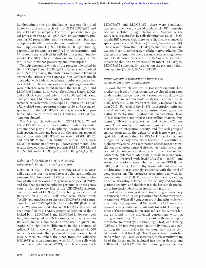
interaction helps to direct integration to highly splicedtranscription units (Fig. 4). This model is also supportedbymultivariate regression analyses that predictwhich fac-tors best correlate with the integration densities of tran-scription units. Considering the factors previously foundto correlate bestwith integration (transcription levels, his-tone H3K4 trimethylation, and DNase I cleavage sites) wefound intron density was the strongest predictor.The significance of the preferential integration of HIV-1
in highly spliced genes is underscored by our finding thatcancer-related genes are highly targeted by HIV-1. Thehigh number of introns in cancer-related genes indicatesthat the recognition of the splicing machinery byLEDGF/p75 directs HIV-1 integration to this collectionof medically relevant targets (Table 1). Therefore, thenumber of introns is a relevant issue when evaluatingwhether the integration of lentivirus vectors is linked toclonal expansion in gene therapy patients.
Materials and methods
The Supplemental Material contains detailed information aboutthe sequencing of integration sites, the oligonucleotides and barc-odes (Supplemental Tables S8, S9), the mapping of integrationsites in transcription units, analysis of alternatively spliced tran-scription units, reproducibility of RNA-seq data sets (Supplemen-tal Fig. S11), MS-based proteomics (Supplemental Table S10), themultivariate regression analyses (Supplemental Table S11), andanalysis of integration in cancer genes. All sequences of integra-tion sites and RNA-seq reads have been deposited with the Se-quence Read Archive accession number SRP065157.
Cell culture and virus production
HEK293T cells lacking LEDGF/p75 or the LEDGF IBD have beendescribed (Fadel et al. 2014).HEK293Tcells (wild type, LEDGF−/−,and IBD−/−) were maintained in DMEM supplemented with5%fetal bovine serum,5%newborncalf serum,and50U/mLpen-icillin plus 50 Ug/mL streptomycin. Replication-defective HIV-1virus was produced in wild-type HEK293T cells and quantifiedas described (Ferris et al. 2010).
Infections and nucleic acid isolation
HEK293T cells were infected with 0.5–1 µg of VSVg pseudotypedpNLNgoMIVR-Emodluc, and genomic DNA was isolated ∼4 d
following infection (Ferris et al. 2010). Total RNA was isolatedfrom uninfected cells using a Qiagen RNeasy minikit.The derivation of the PSIP1−/− and IBD−/− segment 293T cells
by site-specific gene editing has been described elsewhere (Fadelet al. 2014).
Analysis of spliced transcripts
Sequence read densities obtained by RNA-seq were analyzed byMISO as previously described (Katz et al. 2010). Briefly, read den-sities for each transcript relative to all reads for a gene were de-fined as ψ. Δψ values were the differences in the relative readdensities of a transcript between two RNA-seq experiments.ψ and Δψ values were evaluated statistically using the Bayes fac-tor, which quantifies the odds of differential regulation occurring.A Bayes factor of 20 indicates that the probability that a transcriptis differentially expressed between two data sets is 20 times great-er than if the densities occurred by chance.
Acknowledgments
P.K.S. dedicates this article to his parents, Shri Dhurandhar Singhand Smt. Sushila Devi. We thank Anthony Hickey for computa-tional assistance.We are grateful to Allen Kane for help in prepar-ing the figures. This research was supported by the IntramuralResearch Programs of the National Institutes of Health from theEunice Kennedy Shriver National Institute of Child Health andHuman Development (H.L.L.) and the National Cancer Institute(S.H.H.). This research also received support from theNational In-stitutesofHealth IntramuralAIDSTargetedAntiviralProgram(H.L.L. andS.H.H.).Thepresent studywasalsosupportedbyNationalInstitutes of Health grants AI062520 (to M.K.) and AI77344 (to E.M.P.). Funds from the National Cancer Institute under contractnumber HHSN261200800001E to the FrederickNational Labora-tory forCancer Research (B.T.L.) were also used. P.K.S.,M.R.P., A.L.F., andH.J.F. designed and performed experiments and analyzeddata. J.R.I., C.E., and X.W. provided computational expertise andanalysis. E.M.P., S.H.H., M.K., and H.L.L. designed, supervised,and analyzed experiments. B.T.L. and P.K.S. conducted themulti-variate analyses. P.K.S. and H.L.L. conceived the study and wrotethe paper. All authors contributed to editing of the paper.
References
BerryC,Hannenhalli S, Leipzig J, Bushman FD. 2006. Selection oftarget sites for mobile DNA integration in the human ge-nome. PLoS Comput Biol 2: e157.
Brody Y, Neufeld N, Bieberstein N, Causse SZ, Bohnlein EM,Neugebauer KM,Darzacq X, Shav-Tal Y. 2011. The in vivo ki-netics of RNA polymerase II elongation during co-transcrip-tional splicing. PLoS Biol 9: e1000573.
Busschots K, Vercammen J, Emiliani S, Benarous R, EngelborghsY, Christ F, Debyser Z. 2005. The interaction of LEDGF/p75with integrase is lentivirus-specific and promotes DNA bind-ing. J Biol Chem 280: 17841–17847.
Cherepanov P,MaertensG, Proost P, Devreese B, Van Beeumen J,Engelborghs Y, De Clercq E, Debyser Z. 2003. HIV-1 integraseforms stable tetramers and associates with LEDGF/p75 pro-tein in human cells. J Biol Chem 278: 372–381.
Cherepanov P, Devroe E, Silver PA, EngelmanA. 2004. Identifica-tion of an evolutionarily conserved domain in human lens ep-ithelium-derived growth factor/transcriptional co-activatorp75 (LEDGF/p75) that binds HIV-1 integrase. J Biol Chem279: 48883–48892.
Figure 4. Model for HIV-1 integration in highly spliced tran-scription units. RNA Pol II transcription and splicing of intronsare concurrent. Splicing factors (SF) interact with LEDGF/p75,which in turn binds HIV-1 integrase (IN). These interactionshelp direct HIV-1 integration to transcription units with a largenumber of introns.
LEDGF directs integration to highly spliced genes
GENES & DEVELOPMENT 2295
Cold Spring Harbor Laboratory Press on August 9, 2018 - Published by genesdev.cshlp.orgDownloaded from

Cherepanov P, Ambrosio AL, Rahman S, Ellenberger T, Engel-man A. 2005. Structural basis for the recognition betweenHIV-1 integrase and transcriptional coactivator p75. ProcNatl Acad Sci 102: 17308–17313.
Ciuffi A, Llano M, Poeschla E, Hoffmann C, Leipzig J, Shinn P,Ecker JR, Bushman F. 2005. A role for LEDGF/p75 in targetingHIV DNA integration. Nat Med 11: 1287–1289.
Cohn LB, Silva IT, Oliveira TY, Rosales RA, Parrish EH, LearnGH, Hahn BH, Czartoski JL, McElrath MJ, Lehmann C,et al. 2015. HIV-1 integration landscape during latent and ac-tive infection. Cell 160: 420–432.
Craigie R, Bushman FD. 2012. HIVDNA integration.Cold SpringHarb Perspect Med 2: a006890.
Cramer P, Caceres JF, Cazalla D, Kadener S, Muro AF, Baralle FE,Kornblihtt AR. 1999. Coupling of transcription with alterna-tive splicing: RNA Pol II promoters modulate SF2/ASF and9G8 effects on an exonic splicing enhancer. Mol Cell 4:251–258.
David CJ, Manley JL. 2011. The RNA polymerase C-terminaldomain: a new role in spliceosome assembly. Transcription2: 221–225.
de laMataM, Alonso CR, Kadener S, Fededa JP, BlausteinM, Pel-isch F, Cramer P, Bentley D, Kornblihtt AR. 2003. A slowRNA polymerase II affects alternative splicing in vivo. MolCell 12: 525–532.
De Ravin SS, Su L, Theobald N, Choi U, Macpherson JL, Poi-dinger M, Symonds G, Pond SM, Ferris AL, Hughes SH,et al. 2014. Enhancers are major targets for murine leukemiavirus vector integration. J Virol 88: 4504–4513.
De Rijck J, de Kogel C, Demeulemeester J, Vets S, El Ashkar S,Malani N, Bushman FD, Landuyt B, Husson SJ, Busschots K,et al. 2013. The BET family of proteins targets moloney mu-rine leukemia virus integration near transcription start sites.Cell Rep 5: 886–894.
Eidahl JO, Crowe BL, North JA, McKee CJ, Shkriabai N, Feng L,PlumbM, Graham RL, Gorelick RJ, Hess S, et al. 2013. Struc-tural basis for high-affinity binding of LEDGF PWWP tomononucleosomes. Nucleic Acids Res 41: 3924–3936.
Fadel HJ, Morrison JH, Saenz DT, Fuchs JR, Kvaratskhelia M,Ekker SC, Poeschla EM. 2014. TALEN knockout of thePSIP1 gene in human cells: analyses of HIV-1 replicationand allosteric integrase inhibitor mechanism. J Virol 88:9704–9717.
Ferris AL, Wu X, Hughes CM, Stewart C, Smith SJ, Milne TA,Wang GG, Shun MC, Allis CD, Engelman A, et al. 2010.Lens epithelium-derived growth factor fusion proteins redi-rect HIV-1 DNA integration. Proc Natl Acad Sci 107:3135–3140.
Forbes SA, Beare D, Gunasekaran P, Leung K, Bindal N, Boutsela-kis H, Ding M, Bamford S, Cole C, Ward S, et al. 2014. COS-MIC: exploring the world’s knowledge of somatic mutationsin human cancer. Nucleic Acids Res 43: D805–D811.
Futreal PA, Coin L, Marshall M, Down T, Hubbard T, Wooster R,Rahman N, Stratton MR. 2004. A census of human cancergenes. Nat Rev Cancer 4: 177–183.
Ge H, Si Y, Wolffe AP. 1998. A novel transcriptional coactivator,p52, functionally interacts with the essential splicing factorASF/SF2. Mol Cell 2: 751–759.
Gijsbers R, Ronen K, Vets S, Malani N, De Rijck J, McNeely M,Bushman FD, Debyser Z. 2010. LEDGF hybrids efficiently re-target lentiviral integration into heterochromatin. Mol Ther18: 552–560.
Gijsbers R, Vets S, De Rijck J, Ocwieja KE, Ronen K, Malani N,Bushman FD, Debyser Z. 2011. Role of the PWWP domainof lens epithelium-derived growth factor (LEDGF)/p75 cofac-
tor in lentiviral integration targeting. J Biol Chem 286:41812–41825.
Gupta SS, Maetzig T, Maertens GN, Sharif A, RotheM,Weidner-Glunde M, Galla M, Schambach A, Cherepanov P, Schulz TF.2013. Bromo and ET domain (BET) chromatin regulators serveas co-factors for murine leukemia virus integration. J Virol 87:12721–12736.
Hacein-Bey-Abina S, Von Kalle C, Schmidt M, McCormack MP,WulffraatN, Leboulch P, LimA,OsborneCS, PawliukR,Mor-illon E, et al. 2003. LMO2-associated clonal T cell prolifera-tion in two patients after gene therapy for SCID-X1. Science302: 415–419.
Hacein-Bey-Abina S, Hauer J, Lim A, Picard C, Wang GP, BerryCC, Martinache C, Rieux-Laucat F, Latour S, BelohradskyBH, et al. 2010. Efficacy of gene therapy for X-linked severecombined immunodeficiency. N Engl J Med 363: 355–364.
Huang DW, Sherman BT, Lempicki RA. 2009a. Systematic andintegrative analysis of large gene lists using DAVID bioinfor-matics resources. Nat Protoc 4: 44–57.
Huang DW, Sherman BT, Lempicki RA. 2009b. Bioinformaticsenrichment tools: paths toward the comprehensive functionalanalysis of large gene lists. Nucleic Acids Res 37: 1–13.
Kandoth C, McLellan MD, Vandin F, Ye K, Niu B, Lu C, Xie M,Zhang Q, McMichael JF, Wyczalkowski MA, et al. 2013. Mu-tational landscape and significance across 12 major cancertypes. Nature 502: 333–339.
Katz Y, Wang ET, Airoldi EM, Burge CB. 2010. Analysis and de-sign of RNA sequencing experiments for identifying isoformregulation. Nat Methods 7: 1009–1015.
Kaufmann KB, Büning H, Galy A, Schambach A, Grez M. 2013.Gene therapy on the move. EMBO Mol Med 5: 1642–1661.
Koh Y, Wu X, Ferris AL,Matreyek KA, Smith SJ, Lee K, KewalRa-mani VN, Hughes SH, Engelman A. 2013. Differential effectsof human immunodeficiency virus type 1 capsid and cellularfactors nucleoporin 153 and LEDGF/p75 on the efficiencyand specificity of viral DNA integration. J Virol 87: 648–658.
Kornblihtt AR, de la Mata M, Fededa JP, Munoz MJ, Nogues G.2004. Multiple links between transcription and splicing.RNA 10: 1489–1498.
LaFave MC, Varshney GK, Gildea DE, Wolfsberg TG, BaxevanisAD, Burgess SM. 2014. MLV integration site selection is driv-en by strong enhancers and active promoters. Nucleic AcidsRes 42: 4257–4269.
Listerman I, Sapra AK, Neugebauer KM. 2006. Cotranscriptionalcoupling of splicing factor recruitment and precursor messen-ger RNA splicing inmammalian cells.Nat StructMol Biol 13:815–822.
Llano M, Vanegas M, Fregoso O, Saenz D, Chung S, Peretz M,Poeschla EM. 2004. LEDGF/p75 determines cellular traffick-ing of diverse lentiviral but not murine oncoretroviral inte-grase proteins and is a component of functional lentiviralpreintegration complexes. J Virol 78: 9524–9537.
Llano M, Saenz DT, Meehan A, Wongthida P, Peretz M, WalkerWH, Teo W, Poeschla EM. 2006a. An essential role forLEDGF/p75 in HIV integration. Science 314: 461–464.
LlanoM, VanegasM,HutchinsN, ThompsonD, Delgado S, Poes-chla EM. 2006b. Identification and characterization of thechromatin-binding domains of the HIV-1 integrase interactorLEDGF/p75. J Mol Biol 360: 760–773.
Maertens G, Cherepanov P, Pluymers W, Busschots K, De ClercqE, Debyser Z, Engelborghs Y. 2003. LEDGF/p75 is essential fornuclear and chromosomal targeting of HIV-1 integrase in hu-man cells. J Biol Chem 278: 33528–33539.
Singh et al.
2296 GENES & DEVELOPMENT
Cold Spring Harbor Laboratory Press on August 9, 2018 - Published by genesdev.cshlp.orgDownloaded from
Maertens GN, Hare S, Cherepanov P. 2010. The mechanism ofretroviral integration fromX-ray structures of its key interme-diates. Nature 468: 326–329.
Maldarelli F,WuX, Su L, Simonetti FR, ShaoW, Hill S, Spindler J,Ferris AL, Mellors JW, Kearney MF, et al. 2014. HIV latency.Specific HIV integration sites are linked to clonal expansionand persistence of infected cells. Science 345: 179–183.
MeehanAM, SaenzDT,Morrison JH,Garcia-Rivera JA, PeretzM,LlanoM, Poeschla EM. 2009. LEDGF/p75 proteins with alter-native chromatin tethers are functional HIV-1 cofactors. PLoSPathog 57: e1000522.
Moehle EA, Braberg H, Krogan NJ, Guthrie C. 2014. Adventuresin time and space: splicing efficiency and RNA polymeraseII elongation rate. RNA Biol 11: 313–319.
MorchikhM,NaughtinM, Di Nunzio F, Xavier J, Charneau P, Ja-cob Y, Lavigne M. 2013. TOX4 and NOVA1 proteins are part-ners of the LEDGF PWWP domain and affect HIV-1replication. PLoS One 8: e81217.
Munoz MJ, de la Mata M, Kornblihtt AR. 2010. The carboxy ter-minal domain of RNA polymerase II and alternative splicing.Trends Biochem Sci 35: 497–504.
Pradeepa MM, Sutherland HG, Ule J, Grimes GR, Bickmore WA.2012. Psip1/Ledgf p52 binds methylated histone H3K36 andsplicing factors and contributes to the regulation of alterna-tive splicing. PLoS Genet 8: e1002717.
Pryciak PM, Varmus HE. 1992. Nucleosomes, DNA-binding pro-teins, and DNA sequence modulate retroviral integration tar-get site selection. Cell 69: 769–780.
ReedR. 2003. Coupling transcription, splicing andmRNAexport.Curr Opin Cell Biol 15: 326–331.
Schroder AR, Shinn P, Chen H, Berry C, Ecker JR, Bushman F.2002. HIV-1 integration in the human genome favors activegenes and local hotspots. Cell 110: 521–529.
Sharma A, Larue RC, PlumbMR,Malani N, Male F, Slaughter A,Kessl JJ, Shkriabai N, Coward E, Aiyer SS, et al. 2013. BETproteins promote efficient murine leukemia virus integrationat transcription start sites. Proc Natl Acad Sci 110: 12036–12041.
ShunMC, Raghavendra NK, Vandegraaff N, Daigle JE, Hughes S,Kellam P, Cherepanov P, Engelman A. 2007. LEDGF/p75functions downstream from preintegration complex forma-tion to effect gene-specific HIV-1 integration. Genes Dev 21:1767–1778.
Turlure F, Maertens G, Rahman S, Cherepanov P, Engelman A.2006. A tripartite DNA-binding element, comprised of the nu-clear localization signal and two AT-hook motifs, mediatesthe association of LEDGF/p75 with chromatin in vivo.Nucle-ic Acids Res 34: 1653–1665.
Vandekerckhove L, Christ F, VanMaele B, De Rijck J, Gijsbers R,Van den Haute C, Witvrouw M, Debyser Z. 2006. Transientand stable knockdown of the integrase cofactor LEDGF/p75reveals its role in the replication cycle of human immunode-ficiency virus. J Virol 80: 1886–1896.
Vogelstein B, PapadopoulosN,VelculescuVE, Zhou S,Diaz LA Jr,Kinzler KW. 2013. Cancer genome landscapes. Science 339:1546–1558.
Wagner TA, McLaughlin S, Garg K, Cheung CY, Larsen BB,Styrchak S, Huang HC, Edlefsen PT, Mullins JI, Frenkel LM.2014. HIV latency. Proliferation of cells with HIV integratedinto cancer genes contributes to persistent infection. Science345: 570–573.
Wang GP, Ciuffi A, Leipzig J, Berry CC, Bushman FD. 2007. HIVintegration site selection: analysis by massively parallel pyro-sequencing reveals associationwith epigeneticmodifications.Genome Res 17: 1186–1194.
Wang H, Jurado KA, Wu X, Shun MC, Li X, Ferris AL, Smith SJ,Patel PA, Fuchs JR, Cherepanov P, et al. 2012. HRP2 deter-mines the efficiency and specificity of HIV-1 integration inLEDGF/p75 knockout cells but does not contribute to the an-tiviral activity of a potent LEDGF/p75-binding site integraseinhibitor. Nucleic Acids Res 40: 11518–11530.
Wu XL, Li Y, Crise B, Burgess SM. 2003. Transcription start re-gions in the human genome are favored targets for MLV inte-gration. Science 300: 1749–1751.
LEDGF directs integration to highly spliced genes
GENES & DEVELOPMENT 2297
Cold Spring Harbor Laboratory Press on August 9, 2018 - Published by genesdev.cshlp.orgDownloaded from

10.1101/gad.267609.115Access the most recent version at doi: 29:2015, Genes Dev.
Parmit Kumar Singh, Matthew R. Plumb, Andrea L. Ferris, et al. integration to highly spliced genesLEDGF/p75 interacts with mRNA splicing factors and targets HIV-1
Material
Supplemental
http://genesdev.cshlp.org/content/suppl/2015/11/06/29.21.2287.DC1
References
http://genesdev.cshlp.org/content/29/21/2287.full.html#ref-list-1
This article cites 57 articles, 25 of which can be accessed free at:
License
Commons Creative
.http://creativecommons.org/licenses/by-nc/4.0/at Creative Commons License (Attribution-NonCommercial 4.0 International), as described
). After six months, it is available under ahttp://genesdev.cshlp.org/site/misc/terms.xhtmlsix months after the full-issue publication date (see This article is distributed exclusively by Cold Spring Harbor Laboratory Press for the first
ServiceEmail Alerting
click here.right corner of the article or
Receive free email alerts when new articles cite this article - sign up in the box at the top
© 2015 Singh et al.; Published by Cold Spring Harbor Laboratory Press
Cold Spring Harbor Laboratory Press on August 9, 2018 - Published by genesdev.cshlp.orgDownloaded from


![LEDGF/p75 is Dispensable for Hematopoiesis but Essential ...nid]/documents/... · While LEDGF/p75 is dispensable for the hematopoietic reconstitution, it is essential for the initiation](https://img.dokumen.tips/doc/110x75/5ad9b8c97f8b9a137f8c8a87/ledgfp75-is-dispensable-for-hematopoiesis-but-essential-niddocumentswhile.jpg)