Embed Size (px)
Citation preview
[Collapse]
[Collapse]
[Expand]
Spiegel and Casseri: Deformato foetu liber singularis(Dedication dated 1626).
Historic - Semi-diagramaticsagittal section of humanuterus containing an embryo ofabout five weeks
Uterine and placentalvasculature
Lecture - Placenta DevelopmentFrom Embryology
Contents
IntroductionThis lecture is an introduction to the development and functions of the placenta.
The placenta (Greek, plakuos = flat cake) named on the basis of this organs appearance. The placenta a mateno-fetal organ which beginsdeveloping at implantation of the blastocyst and is delivered with the fetus at birth. Only recently have we begun to understand the manydifferent functions this organ carries out in addition to its role in embryonic nutrition. This lecture follows on the concepts of cardiovasculardevelopment covered in the previous lecture.
The placenta and placental blood at birth has recently been seen as a new source for stem cells in bone marrow replacement therapy in manydiseases. (More? Stem Cells)
Lecture ObjectivesUnderstanding of placental villi developmentUnderstanding of placental structureUnderstanding of placental functionsBrief understanding of placental abnormalities
Lecture Resources
Movies
Week 2 - ImplantPage | Play
Week 2 - BilaminarPage | Play
References
Hill, M.A. (2014). UNSWEmbryology (14th ed.) RetrievedSeptember 1, 2014, from
http://php.med.unsw.edu.au/embryology
Placenta Links: Introduction | Lecture - Placenta | Practical - Placenta |Implantation | Villi Development | Trophoblast | Maternal Decidua | Endocrine |Cord | Membranes | Abnormalities | Stage 13 | Stage 22 | Histology | Vascular Beds| Blood Vessel Development | Stem Cells | Category:Placenta
Lecture Archive:
Moore, K.L., Persaud, T.V.N. &Torchia, M.G. (2011). The developinghuman: clinically orientedembryology (9th ed.). Philadelphia:Saunders.
The following chapter links only work with a UNSW connection.
Chapter 7 - Placenta and Fetal Membranes (http://er.library.unsw.edu.au/er/cgi-bin/eraccess.cgi?url=http://www.mdconsult.com/books/page.do?eid=4-u1.0-B978-1-4377-2002-0..00007-2&isbn=978-1-4377-2002-0&uniqId=330028653-2#4-u1.0-B978-1-4377-2002-0..00007-2)
Schoenwolf, G.C., Bleyl, S.B.,Brauer, P.R. & Francis-West, P.H.(2009). Larsen's human embryology(4th ed.). New York; Edinburgh:Churchill Livingstone.
The following chapter links only work with a UNSW connection.
Chapter 2 - Second Week: Becoming Bilaminar and Fully Implanting(http://er.library.unsw.edu.au/er/cgi-bin/eraccess.cgi?url=http://www.mdconsult.com/books/linkTo?type=bookPage&isbn=978-0-443-06811-9&eid=4-u1.0-B978-0-443-06811-9..10002-8)Chapter 13 - Development of the Vasculature (http://er.library.unsw.edu.au/er/cgi-bin/eraccess.cgi?url=http://www.mdconsult.com/books/linkTo?type=bookPage&isbn=978-0-443-06811-9&eid=4-u1.0-B978-0-443-06811-9..10013-2)
ECHO360 Recording
NutritionHistiotrophic nutrition describes early placental development and the form of intital transfer of nutrition from maternal to embryo.Haemotrophic nutrition describes the later blood-borne nutrition.
Fetal Membranes

Villi Stages
Primary villi
Week 2 - first stage of chorionic villi development,trophoblastic shell cells (syncitiotrophoblasts andcytotrophoblasts) form finger-like extensions intomaternal decidua.
Secondary villi
Week 3 - second stage of chorionic villidevelopment, extraembryonic mesodermgrows into villi, covers entire surface ofchorionic sac.
Tertiary villi
Week 4 - third stage of chorionic villi development, mesenchymedifferentiates into blood vessels and cells, forms arteriocapillary network,fuse with placental vessels, developing in connecting stalk. Basal regionwill form chorionic plate.
Chorionoic Villi Location
Originally cover entire chorionic surface and become restricted to decidua basalis region forming 2 regions:
Week 4 - Carnegie stage 11

Frondosum - "leafy" where villi are mainly locatedCapsularis - smooth chorion, where villi are absent or not abundant
Week 5 - Carnegie stage 14Stage 14 - Lateral View
Mobile(http://embryology.med.unsw.edu.au/embryology/Slides/Embryo_Stages/Stage14/bf18/leaflet.html) |Desktop(http://embryology.med.unsw.edu.au/embryology/Slides/Embryo_Stages/Stage14/bf18/stage14bf18.html)| Original
Stage 14 | Embryo Slides
Stage 14 - Ventral View
Mobile(http://embryology.med.unsw.edu.au/embryology/Slides/Embryo_Stages/Stage14/bf21/leaflet.html)Desktop(http://embryology.med.unsw.edu.au/embryology/Slides/Embryo_Stages/Stage14/bf21/stage14bf21.html)| Original
Stage 14 | Embryo SlidesWeek 7 - Carnegie stage 18)

Early placental structure
Fetus in utero, between fifthand sixth months
Villi Terms
primary villi - week 2, first stage of chorionic villi development, trophoblastic shell cells (syncitiotrophoblasts and cytotrophoblasts)form finger-like extensions into maternal decidua.secondary villi - week 3, second stage of chorionic villi development, extraembryonic mesoderm grows into villi, covers entiresurface of chorionic sac.tertiary villi third stage of chorionic villi development, mesenchyme differentiates into blood vessels and cells, forms arteriocapillarynetwork, fuse with placental vessels, developing in connecting stalkstem villi - or anchoring villi, cytotrophoblast cells attached to maternal tissue.branched villi - or terminal villi, grow from sides of stem villi, region of main exchange, surrounded by maternal blood in intervillousspaces.terminal villi - not active outgrowths caused by proliferation of the trophoblast. Passive protrusions induced by capillary coiling due to growth of the fetal capillarieswithin the mature intermediate villi (third trimester).chorionic plate - region of membrane at the base of the villi through which placental arteries and vein passes.
Placenta anchoring villi
Villi first trimester
Villi at term
Villi at term
Placenta at BirthPlacenta (Greek, plakuos = flat cake)embryonic/maternal organvillous chorion/decidua basaliscontinuous with amniotic and chorionic sacks
Dimensions
at birth - discoid up to 20cm diameter and 3 cm thick (term) and weighs 500-600 gmShapes - accessory placenta, bidiscoid, diffuse, horseshoematernal and embryonic surface, both delivered at parturition
retention may cause uterine hemorrhage
Fetal Surface
umbilical cord attachment - cord 1-2 cm diameter, 30-90cm longcovered with amniotic membrane and attached to chorionic plateumbilical vessels branch into chorionic vessels which anastomose
Maternal Surface
Cotyledons - form cobblestone appearance, originally placental septa formed grooves.covered with maternal decidua basalis
Placental ClassificationClassification of placenta is on the basis of histological (microscopic) structural organization and layers between fetal and maternal circulation, giving 3 main groups:
Haemochorial - placenta where the chorion comes in direct contact with maternal blood (human)

Dog placenta (E35-38)
Mouse placenta
Endotheliochorial - maternal endometrial blood vessels are bare to their endothelium and these comes in contact with the chorion. (dogs, cats)Epitheliochorial - maternal epithelium of the uterus comes in contact with the chorion.considered as primitive (pigs, cows)
The presence of these three differing types of placenta have also been used to describe the pattern mammalian evolution. See also PlacentalLayers
Placental TypesDiscoid in humans, mice, insectivores, rabbits, rats, and monkeys.Zonary in dogs, cats, bears and seals.Cotyledenary in cows, deer, goat, and giraffe.Diffuse in horses, pigs, camels, lemurs, opossums, kangaroos, and whales
Links: Comparative Placentation (http://placentation.ucsd.edu/homefs.html)
Chorionoic Villi Trimester Development
Trimester 1 and 2 - immature intermediate villi, developmental steps towards the stem villi.Trimester 3 - mature intermediate villi develop during the last trimester, produce numerous terminal villi.
Terminal villi are not active outgrowths caused by trophoblast proliferation, passive protrusions induced by capillary coiling dueto excessive longitudinal growth of the fetal capillaries within the mature intermediate villi.Capillary bed arrangement in the terminal villi can vary from simple U-like loops to branched network, due to capillary elongation and sprouting.(Data fromPMID 2327595)
Placental Cord Blood Vessels
initially in the connecting stalk (then umbilical cord)anastomose in chorion
extend maternally - toward chorionic villiextend embryonically - to the sinus venosus and dorsalaorta
Arteries - paired, carry deoxygenated blood (from dorsal aorta) and waste products to theplacental villiVeins - paired initially then only left at end of embryonic period, carry oxygenated blood to theembryo (sinus venosus)
Placental Function4 layers separate maternal and fetal blood: syncitiotrophoblast, cytotrophoblast, villi connective tissue and fetal capillary endothelium3 main functions: metabolism, transport and endocrine
Placental Metabolism

Fetal blood flow liver andbrain
Trophoblast cell hCG
Uterine and placentalvasculature
Placenta spiral arteryconversion
Synthesizes: glycogen, cholesterol, fatty acids
provides nutrient and energy
Placental Transport
gases and nutrition
oxygen, carbon dioxide, carbon monoxidewater, glucose, vitaminshormones, mainly steroid not proteinelectrolytesmaternal antibodieswaste products - urea, uric acid, bilirubindrugs and their metabolites (fetal drug addiction)infectious agents (cytomegalovirus, rubella, measles, microorganisms)
Placental Endocrine
Human chorionic gonadotrophin (hCG) - like leutenizing hormone, supports corpus luteumHuman chorionic somatommotropin (hCS) (or placental lactogen) - hormone level increases in maternal blood through pregnancy,decreases maternal insulin sensitivity (raising maternal blood glucose levels and decreasing maternal glucose utilization) aiding fetalnutrition ("anti-insulin" function)Human chorionic thyrotropin (hCT) - Peptide placental hormone, similar to anterior pituitary released thyroid stimulating hormone(TSH), which along with human chorionic gonadotrophin (hCG) is thought to act on maternal thyroid. There is little recent researchpublished on this hormone, its level and activities.Human chorionic corticotropin (hCACTH) - placental hormone thought to have corticotropin (ACTH)-like activity, increasingmaternal cortisol levels.Steroid Hormones
progestins - progesterone, support of the endometrium and suppress uterine smooth muscle contractility.estrogens - estriol, stimulate growth of the myometrium and mammary gland development.both hormones support maternal endometrium
Relaxin - Humans high levels early in pregnancy than at birth promotes angiogenesis probably plays a role in development of theuterus/ placenta than in the birth process
Fetal PlacentaTrophoblast cells are the major source of placental hormones.
Placental growth hormone (PGH) is mainly expressed in the syncytiotrophoblast cells (PGH differs from pituitary derived growth hormoneby 13 amino acids). extravillous cytotrophoblast - arise from anchoring villi invade the uterine spiral arteries, generating fibrinoid materialand endovascular trophoblastic cells. syncytiotrophoblast
Fetal Blood Vessels At least 2 phases of development during pregnancy driven by vascular endothelial growth factor (VEGF):
1. Initially cytotrophoblasts are the cellular stimulus to vasculogenesis and angiogenesis.2. Later Hofbauer (lacental villi macrophages of mesenchymal origin) and stromal cells take over the stimulation of blood vessel development.
Placenta Human chorionic gonadotrophin (hCG) After implantation cells within the developing placenta (syncitiotrophoblasts) synthesize and secrete Human chorionicgonadotrophin (hCG) into the maternal bloodstream. The main function of serum hCG is to maintain the corpus luteum in the maternal ovary and therefore maintain the earlypregnancy, that is block the menstrual cycle. Later the placenta itself supports the pregnancy.
Maternal PlacentaFibrinoid - said to exist as 2 forms of extracellular matrix:
1. Fibrin-type fibrinoid is a maternal blood-clot product which replaces degenerative syncytiotrophoblast2. Matrix-type fibrinoid is secreted by invasive extravillous trophoblast cells.
Fibrinoid layer (Nitabuch's layer) is thought to act to prevent excessively deep implantation.
Decidualization - process of endometrial stromal cells (fibroblast-like) change in morphology (polygonal cells) and protein expression andsecretion (specific decidual proteins: prolactin, insulin-like growth factor binding protein-1, tissue factor, interleukin-15, and VEGF).
1. Estrogen and progesterone - receptive phase, luminal and glandular epithelial cells change in preparation for blastocyst adplantation.2. Human Chorionic gonadotropin - luminal epithelium endoreplication leading to epithelial plaque formation.3. Human Chorionic gonadotropin - trophoblast invasion and decidualization of human stromal fibroblasts.
Artery Dilatation - due to extravillous trophoblast cells invading uterine wall and maternal spiral arteries replacing both smooth musclewith fibrinoid material and part of vessel endothelium. There is also a proliferation of maternal blood vessels.
Other changes
Endoreplication - rounds of nuclear DNA replication without intervening cell or nuclear division (mitosis).Cytokines - of maternal origin also act on placental development.Natural Killer (NK) cells - 30% of all the decidual cells towards the end of the first trimester of pregnancy. These lymphocytes arepresent in the maternal decidua in large numbers (70%, normal circulating blood lymphocytes 15%) close to the extravilloustrophoblast cells. Have a cytolytic potential against virus-infected and tumor-transformed cells.
Placental AbnormalitiesPlacenta Accreta - abnormal adherence, with absence of decidua basalis. The incidence of placenta accreta also significantly increases in women with previouscesarean section compared to those without a prior surgical delivery.Placenta Increta - occurs when the placenta attaches deep into the uterine wall and penetrates into the uterine muscle, but does not penetrate the uterine serosa.

Placental abnormalities
Historic model of placentaprevia
Hydatidiform mole pathology
Placenta increta accounts for approximately 15-17% of all cases.Placenta Percreta - placental villi penetrate myometrium and through to uterine serosa.Placenta Previa - In this placenatal abnormality, the placenta overlies internal os of uterus, essentially covering the birth canal. This condition occurs in approximately1 in 200 to 250 pregnancies. In the third trimester and at term, abnormal bleeding can require cesarian delivery and can also lead toAbruptio Placenta. Ultrasound screening programs during 1st and early 2nd trimester pregnancies now include placental localization.Diagnosis can also be made by transvaginal ultrasound.Vasa Previa - (vasa praevia) placental abnormality where the fetal vessels lie within the membranes close too or crossing the innercervical os (opening). This occurs normally in 1:2500-5000 pregnancies and leads to complications similar too those for PlacentaPrevia.Type II is defined as the condition where the fetal vessels are found crossing over the internal os connecting either a bilobedplacenta or a succenturiate lobe with the main placental mass. Some recent evidence of successful in utero laser ablation of type IIvasa previa at 22.5 weeks of gestation.Abruptio Placenta - a retroplacental blood clot formation, abnormal hemorrhage prior to delivery.Chronic Intervillositis - (massive chronicintervillositis, chronic histiocytic intervillositis) Rare placental abnormality and pathologydefined by inflammatory placental lesions, mainly in the intervillous space (IVS), with a maternal infiltrate of mononuclear cells(monocytes, lymphocytes, histiocytes) and intervillous fibrinoid deposition.
Hydatidiform mole - placental tumor with no embryo development. Several forms of hydatidiform mole: partial mole, complete moleand persistent gestational trophoblastic tumor. Many of these tumours arise from a haploid sperm fertilizing an egg without a femalepronucleus (the alternative form, an embryo without sperm contribution, is called parthenogenesis). The tumour has a "grape-like"placental appearance without enclosed embryo formation. Following a first molar pregnancy, there is approximately a 1% risk of asecond molar pregnancy.
Links: Placenta - Abnormalities
Placental Cord Abnormalities
There are few abnormalities associated with umbilical cord development, other that abnormally short or long cords, which in most cases donot cause difficulties. In some cases though, long cords can wrap around limbs or the fetus neck, which can then restrict blood flow or lead totissue or nerve damage, and therefore effect develoment.
Cord knotting - can also occur (1%) in most cases these knots have no effect, in some cases of severe knotting this can prevents the passage of placental blood.Cord torsion - Rare event where even without knot formation can also affect placental blood flow, even leading to fetal demise.
Links: Placental Abnormalities (http://embryology.med.unsw.edu.au/Notes/placenta2.htm) | WebPath - umbilical cord knot 1 (http://www-medlib.med.utah.edu/WebPath/PLACHTML/PLAC010.html) | WebPath - umbilical cord knot 2 (http://www-medlib.med.utah.edu/WebPath/PLACHTML/PLAC028.html) | WebPath - Pseudoknot of umbilical cord, gross (http://www-medlib.med.utah.edu/WebPath/PLACHTML/PLAC073.html) | WebPath - Torsion of umbilical cord, gross (http://www-medlib.med.utah.edu/WebPath/PLACHTML/PLAC012.html) | WebPath - Torsion of umbilical cord, with fetal demise, gross (http://www-medlib.med.utah.edu/WebPath/PLACHTML/PLAC011.html)
Placental Infections
Malaria (plasmodium falciparum) Listeria maternal-fetal barrier
Several infective agents may cross into the placenta from the maternal circulation, as well as enter the embry/fetal circulation. The variety of bacterial infections thatcan occur during pregnancy is as variable as the potential developmental effects, from virtually insignificant to a major developmental, abortive or fatal in outcome.Pregnant women have an increased susceptibility to malaria infection. Malarial infection of the placenta by sequestration of the infected red blood cells leading to lowbirth weight and other effects. There are four types of malaria caused by the protozoan parasite Plasmodium falciparum (main), Plasmodium vivax, Plasmodium ovale,Plasmodium malariae). This condition is common in regions where malaria is endemic with women carrying their first pregnancy (primigravida).
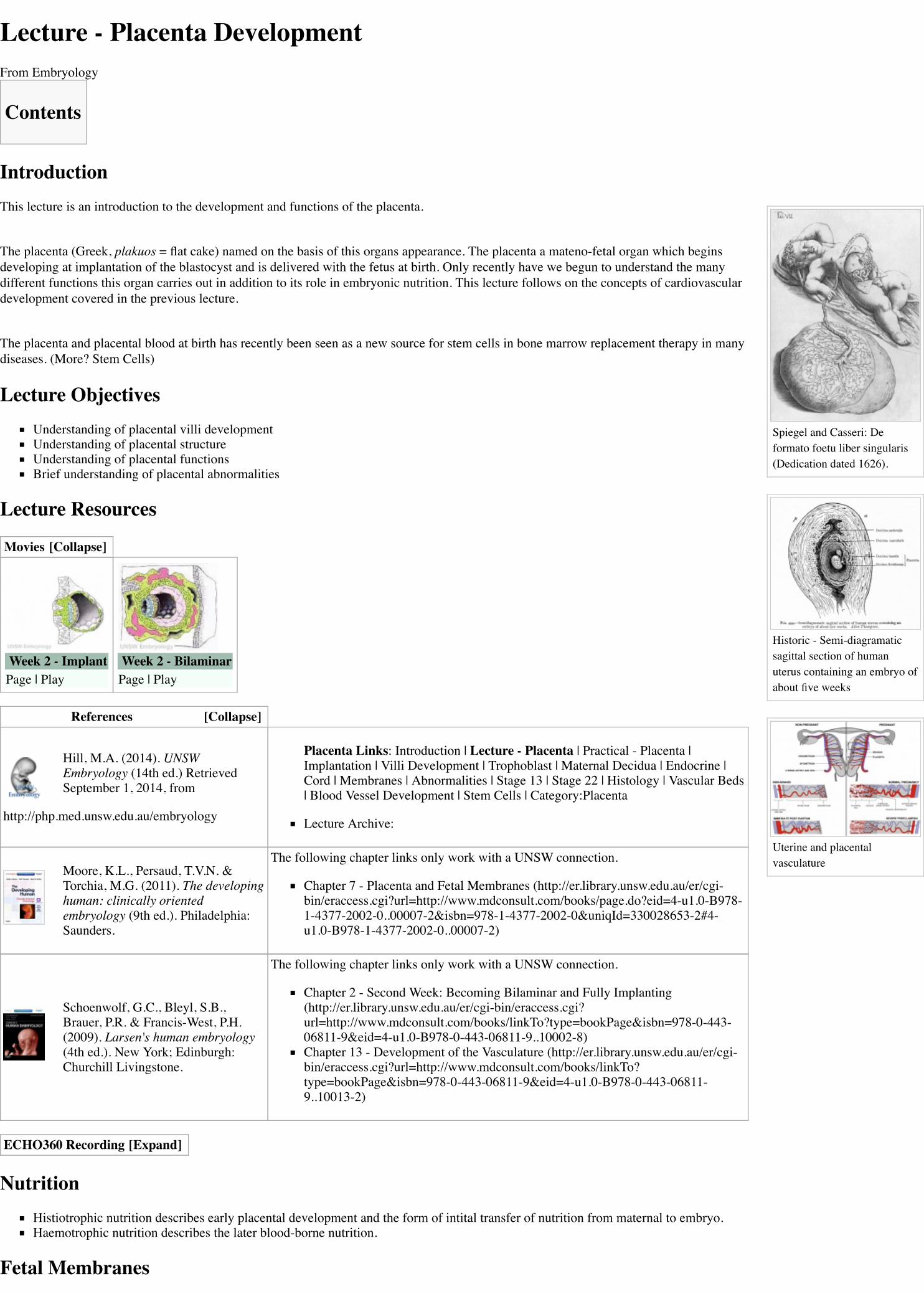
Placental Pathology
MH - content in this section is not examinable.
Chronic Villitis - can occur following placental infection leading to maternal inflammation of the villous stroma, often with associated intervillositis. The inflammationcan lead to disruption of blood flow and necrotic cell death.Massive Chronic Intervillositis (MCI) - maternal blood-filled space is filled with CD68-positive histiocytes and an increase in fibrin, occuring more commonly in thefirst trimester.Meconium Myonecrosis - prolonged meconium exposure leads to toxic death of myocytes of placental vessels (umbilical cord or chorionic plate).Neuroblastoma - a fetal malignancy that leads to an enlarged placenta, with tumor cells in the fetal circulation and rarely in the chorionic villi.Thrombophilias - (protein C or S deficiency, factor V Leiden, sickle cell disease, antiphospholipid antibody) can generate an increased fibrin/fibrinoid deposition in thematernal or intervillous space, this can trap and kill villi.
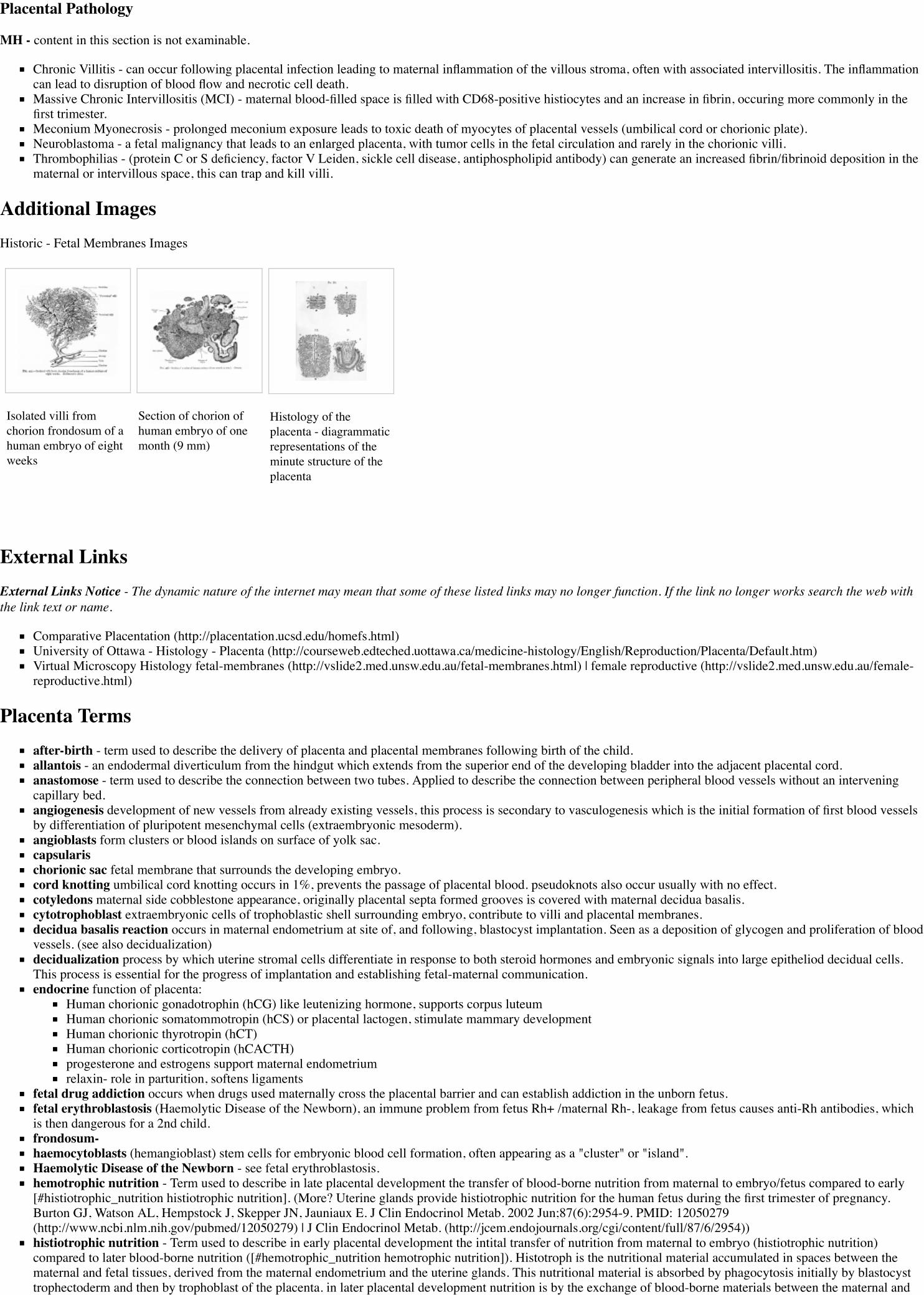
Additional ImagesHistoric - Fetal Membranes Images
Isolated villi fromchorion frondosum of ahuman embryo of eightweeks
Section of chorion ofhuman embryo of onemonth (9 mm)
Histology of theplacenta - diagrammaticrepresentations of theminute structure of theplacenta
External LinksExternal Links Notice - The dynamic nature of the internet may mean that some of these listed links may no longer function. If the link no longer works search the web withthe link text or name.
Comparative Placentation (http://placentation.ucsd.edu/homefs.html)University of Ottawa - Histology - Placenta (http://courseweb.edteched.uottawa.ca/medicine-histology/English/Reproduction/Placenta/Default.htm)Virtual Microscopy Histology fetal-membranes (http://vslide2.med.unsw.edu.au/fetal-membranes.html) | female reproductive (http://vslide2.med.unsw.edu.au/female-reproductive.html)
Placenta Termsafter-birth - term used to describe the delivery of placenta and placental membranes following birth of the child.allantois - an endodermal diverticulum from the hindgut which extends from the superior end of the developing bladder into the adjacent placental cord.anastomose - term used to describe the connection between two tubes. Applied to describe the connection between peripheral blood vessels without an interveningcapillary bed.angiogenesis development of new vessels from already existing vessels, this process is secondary to vasculogenesis which is the initial formation of first blood vesselsby differentiation of pluripotent mesenchymal cells (extraembryonic mesoderm).angioblasts form clusters or blood islands on surface of yolk sac.capsularischorionic sac fetal membrane that surrounds the developing embryo.cord knotting umbilical cord knotting occurs in 1%, prevents the passage of placental blood. pseudoknots also occur usually with no effect.cotyledons maternal side cobblestone appearance, originally placental septa formed grooves is covered with maternal decidua basalis.cytotrophoblast extraembryonic cells of trophoblastic shell surrounding embryo, contribute to villi and placental membranes.decidua basalis reaction occurs in maternal endometrium at site of, and following, blastocyst implantation. Seen as a deposition of glycogen and proliferation of bloodvessels. (see also decidualization)decidualization process by which uterine stromal cells differentiate in response to both steroid hormones and embryonic signals into large epitheliod decidual cells.This process is essential for the progress of implantation and establishing fetal-maternal communication.endocrine function of placenta:
Human chorionic gonadotrophin (hCG) like leutenizing hormone, supports corpus luteumHuman chorionic somatommotropin (hCS) or placental lactogen, stimulate mammary developmentHuman chorionic thyrotropin (hCT)Human chorionic corticotropin (hCACTH)progesterone and estrogens support maternal endometriumrelaxin- role in parturition, softens ligaments
fetal drug addiction occurs when drugs used maternally cross the placental barrier and can establish addiction in the unborn fetus.fetal erythroblastosis (Haemolytic Disease of the Newborn), an immune problem from fetus Rh+ /maternal Rh-, leakage from fetus causes anti-Rh antibodies, whichis then dangerous for a 2nd child.frondosum-haemocytoblasts (hemangioblast) stem cells for embryonic blood cell formation, often appearing as a "cluster" or "island".Haemolytic Disease of the Newborn - see fetal erythroblastosis.hemotrophic nutrition - Term used to describe in late placental development the transfer of blood-borne nutrition from maternal to embryo/fetus compared to early[#histiotrophic_nutrition histiotrophic nutrition]. (More? Uterine glands provide histiotrophic nutrition for the human fetus during the first trimester of pregnancy.Burton GJ, Watson AL, Hempstock J, Skepper JN, Jauniaux E. J Clin Endocrinol Metab. 2002 Jun;87(6):2954-9. PMID: 12050279(http://www.ncbi.nlm.nih.gov/pubmed/12050279) | J Clin Endocrinol Metab. (http://jcem.endojournals.org/cgi/content/full/87/6/2954))histiotrophic nutrition - Term used to describe in early placental development the intital transfer of nutrition from maternal to embryo (histiotrophic nutrition)compared to later blood-borne nutrition ([#hemotrophic_nutrition hemotrophic nutrition]). Histotroph is the nutritional material accumulated in spaces between thematernal and fetal tissues, derived from the maternal endometrium and the uterine glands. This nutritional material is absorbed by phagocytosis initially by blastocysttrophectoderm and then by trophoblast of the placenta. in later placental development nutrition is by the exchange of blood-borne materials between the maternal and

fetal circulations, hemotrophic nutrition. (More? Uterine glands provide histiotrophic nutrition for the human fetus during the first trimester of pregnancy. Burton GJ,Watson AL, Hempstock J, Skepper JN, Jauniaux E. J Clin Endocrinol Metab. 2002 Jun;87(6):2954-9. PMID: 12050279(http://www.ncbi.nlm.nih.gov/pubmed/12050279) | J Clin Endocrinol Metab. (http://jcem.endojournals.org/cgi/content/full/87/6/2954))Hofbauer cells - placental villi macrophages of mesenchymal origin with potentially additional functions (vasculogenesis/angiogenesis, villi remodeling, regulation ofstromal water content) to their macrophage role.Human chorionic gonadotrophin- (hCG) like leutenizing hormone, supports corpus luteumHuman chorionic somatommotropin - (hCS) or placental lactogen - hormone level increases in maternal blood through pregnancy, decreases maternal insulinsensitivity (raising maternal blood glucose levels and decreasing maternal glucose utilization) aiding fetal nutrition.Human chorionic thyrotropin- (hCT) placental derived hormone equivilant to thyroidHuman chorionic corticotropin- (hCACTH) placental derived hormone equivilant to corticotropin (ACTH) from the pituitary.methyldopa - (alpha methyldopa) A central alpha agonist used to lower blood pressure. Used as an antihypertensive drug to lower blood pressure in pre-eclampsia,acting by either a direct or indirect central vasodilatory mechanism. A recent study suggests this drug may have a direct effect on placental and/or endothelial cellfunction in pre-eclampsia patients, altering angiogenic proteins. Drug commercial brandname (USA) "Aldomet", also available in combination with other drugs:methyldopa and chlorothiazide "Aldochlor", methyldopa and hydrochlorothiazide "Aldoril". (More? Placenta Abnormalities - Pre-eclampsia(http://embryology.med.unsw.edu.au/Notes/placenta2.htm#Pre-eclampsia) | Medline Plus - Methyldopa(http://www.nlm.nih.gov/medlineplus/druginfo/meds/a682242.html) | Effect of antihypertensive therapy with alpha methyldopa on levels of angiogenic factors inpregnancies with hypertensive disorders. Khalil A, Muttukrishna S, Harrington K, Jauniaux E. PLoS ONE. 2008 Jul 23;3(7):e2766. PMID: 18648513(http://www.ncbi.nlm.nih.gov/pubmed/18648513))maternal antibodies- antibodies from the mother's immune system that are capable of crossing placental barrier. They can provide immune protection to the embryo,but may also participate in immune disease (fetal erythroblastosis).maternal sinusoids- placental spaces around chorionic villi that are filled with maternal blood. Closest maternal/fetal exchange site.Nitabuch's layer (fibrinoid layer) layer formed at maternal/fetal interface during placentation and is thought to act to prevent excessively deep conceptus implantation.Fibrin-type fibrinoid (maternal blood-clot product) and matrix-type fibrinoid (secreted by invasive extravillous trophoblast cells).placenta- (Gk. plakuos= flat cake) describes its typical mature discoid shape (20cm diameter and 3 cm thick at term,weighs 500-600 gm).placenta accreta- abnormal, adherence with absence of decidua basalis.placental arteries- paired, carry deoxygenated blood and waste from the embryo (dorsal aorta->internal iliacs->PA)placental blood- blood found within the placental vessels. Obviously part of the fetal blood, but can be collected at birth for theraputic use containing blood stem cells(see cord blood banks).placental blood vessels- form initially in the connecting stalk (then umbilical cord), anastomose in chorioni and extend maternally toward chorionic villi, extendembryonically to the sinus venosus and dorsal aorta.placental layers- 4 layers separate maternal and fetal blood: syncitiotrophoblast, cytotrophoblast, villi connective tissue, and fetal capillary endothelium.placenta percreta- abnormal, villi penetrate myometrium.placenta previa- placenta overlies internal os of uterus, abnormal bleeding, may require cesarian delivery.placental veins- paired initially then usually only one left at end of embryonic period, carry oxygenated blood to the embryo (sinus venosus)primary villi- develop week 2, consist of trophoblastic shell cells both syncitiotrophoblasts and cytotrophoblasts. Form finger-like extensions into the maternalendometrium.protein hormone- usually a protein distributed in the blood that binds to membrane receptors on target cells in different tissues. Do not easliy cross placental barrier.relaxin- hormone.secondary villi- develop week 3, extraembryonic mesoderm grows into villi, initially covers entire surface of chorionic sac.sinus venosus- cavity into which all major embryonic paired veins supply (vitelline, placental, cardinal)syncitiotrophoblast- extraembryonic cells of trophoblastic shell surrounding embryo, outside the cytotrophoblast layer, involved with implantation of the blastocyst byeroding extracellular matrix surrounding maternal endometrial cells at site of implantation, also contribute to villi. (dark staining, multinucleated)tertiary villi- develop week 4, mesenchyme within secondary villi differentiates into blood vessels and cells, forms arteriocapillary network, fuse with placental vesselsdeveloping in connecting stalk.trophoblast-umbilical cord- fetal attachment cord 1-2 cm diameter, 30-90cm long, covered with amniotic attached to chorionic plate, umbilical vessels (artery, vein) branch intochorionic vessels. Vessels anastomose within the placenta.vasculogenesis formation of first blood vessels by differentiation of pluripotent mesenchymal cells (extraembryonic mesoderm) followed by angiogenesis which is thedevelopment of new vessels from already existing vessels.villi- initially outgrowth of the trophoblastic shell which involve other tissues with development. Develop in sequence (primary, secondary, tertiary) with mature villibeing stem por branched type.virus- small infectious agent able to cross placental barrier. Can infect embryo and cause developmental abnormalities. (e.g. cytomegalovirus, rubella, measles)vitelline- Blood vessels cover entire surface of yolk sac and connect to embryo through yolk stalk
Arteries- arises from dorsal aorta and contribute to adult GIT arteries.Veins- empties into sinus venosus and contribute to the adult portal system.
waste products products of cellular metabolism and cellular debris, e.g.- urea, uric acid, bilirubinWharton's jelly placental cord (umbilical cord) gelatinous connective tissue composed of myofibroblast-like stromal cells, collagen fibers, and proteoglycans.Increases in volume (myxomatous, connective tissue embedded in mucus) at parturition to assist closure of placental blood vessels. Matrix cells from Wharton's jellyhave recently been identified as a potential source of stem cells. This placental cord substance is named after Thomas Wharton (1614-1673) an English physician andanatomist who first described it.
2014 Course: Week 2 Lecture 1 Lecture 2 Lab 1 | Week 3 Lecture 3 Lecture 4 Lab 2 | Week 4 Lecture 5 Lecture 6 Lab 3 | Week 5 Lecture 7 Lecture 8 Lab 4 | Week 6Lecture 9 Lecture 10 Lab 5 | Week 7 Lecture 11 Lecture 12 Lab 6 | Week 8 Lecture 13 Lecture 14 | Lab 7 | Week 9 Lecture 15 Lecture 16 Lab 8 | Week 10 Lecture 17Lecture 18 Lab 9 | Week 11 Lecture 19 Lecture 20 Lab 10 | Week 12 Lecture 21 Lecture 22 Lab 11 | Week 13 Lecture 23 Lecture 24 Lab 12 | Student Projects -Group 1 | Group 2 | Group 3 | Group 4 | Group 5 | Group 6 | Group 7 | Group 8 | Moodle (http://moodle.telt.unsw.edu.au/course/view.php?id=9262)
Retrieved from ‘https://php.med.unsw.edu.au/embryology/index.php?title=Lecture_-_Placenta_Development&oldid=142205’Categories: Placenta 2014 Science-Undergraduate
This page was last modified on 26 August 2014, at 14:39.This page has been accessed 10,613 times.