Embed Size (px)
Citation preview

Lecture and Lab exercise 5 Tissues
Part I

Question
• What is a tissue?

Tissues
• A tissue is composed of specialized cells of the same type that perform a common function in the body.

Question
• What are the four major tissue types found in the human body?

Types of Tissues
• The tissues of the human body can be categorized into four major types.
• Epithelial Tissue• Connective Tissue• Muscular Tissue• Nervous Tissue

Question
• What is the function of the Epithelial Tissue?

Function of Epithelial Tissues
• cover body surfaces and lines body cavities.• protects, absorbs molecules, absorbs
nutrients, and removes impurities out of the body.
• are described as avascular.

Epithelial cells
• Epithelial cells must create a layer that prevents leakage from one side of the sheet to the other because they cover the surface of organs and line the body cavities.

Question
• What is the function of the basement membrane? What is the basement membrane composed of?

Basement membrane
• The basement membrane is composed of carbohydrates and proteins that anchors the epithelium to underlying connective tissue.

Epithelium
Basement membrane
Connective Tissue

Question
• How are epithelial tissues classified?

Epithelial tissues are classified according to
• Cell shapes. Epithelial cells vary in shape depending on their function.
• Arrangement of cells in layers. The cells are arranged in one or more layers depending on function


Question
• What are the basic types of epithelia?

Types of Epithelial
• Simple squamous• Simple cuboidal• Simple columnar• Pseudostratified ciliated columnar• Stratified squamous• Transitional epithelium• Glandular epithelia

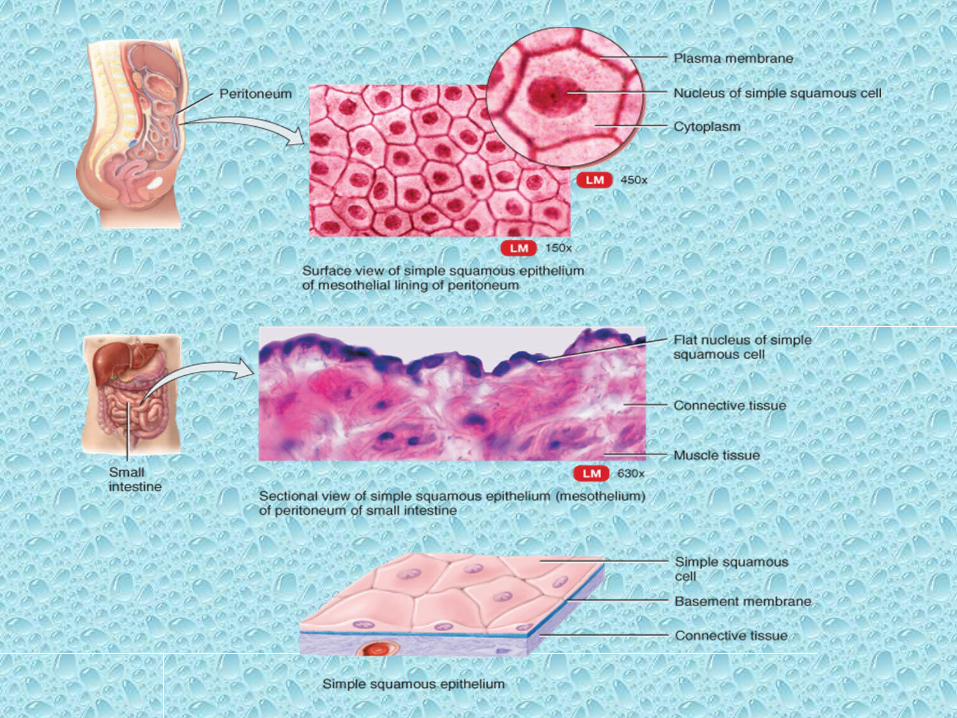
Simple epithelium
Simple epithelium is a single layer of cells that functions in diffusion, osmosis, filtration, secretion, or absorption. Secretion is the production and release of substances such as mucus, sweat, or enzymes. Absorption is the intake of substances, such as digested food from the intestinal tract.

Stratified epithelium
• Stratified epithelium (stratum = layer) consists of two or more layers of cells that protect underlying tissues in locations where there is considerable wear and tear

Pseudostratified epithelium• Pseudostratified epithelium (soo -dō-STRA-te-′
fīd; pseudo- = false) appears to have multiple layers of cells because the cell nuclei lie at different levels and not all cells reach the surface, but it is actually a simple epithelium because all its cells rest on the basement membrane.
• Cells that do extend to the surface may contain cilia; others (goblet cells) secrete mucus.

Squamous cells
• Squamous cells (SKWĀ-mus = flat) are thin, which allows substances to pass rapidly through them.

Keratinized and Nonkeratinized Stratified Squamous Epithelium


Cuboidal Cells• are as tall as they are wide and are shaped like cubes.
• They may have microvilli at their apical surface and function in either secretion or absorption

Columnar cells
• Columnar cells are much taller than they are wide and protect underlying tissues.
• Their apical surface may have cilia or microvilli, and they often are specialized for secretion and absorption.

Question
• What is the function of cilia?• What is the function of microvilli?• Where are these structures found in your
body?

Cilia
• Is involved in movement.• The ciliated cells that line our respiratory tract
sweep debris trapped within mucus back up the throat. This helps keep the lungs clean.
• Ciliated cells found in the oviduct move an egg along the oviduct toward the uterus.

Microvilli
• Are found in the small intestine• Increase the surface area for the absorption of
nutrients.

Simple columnar Epithelial
Nonciliated simple columnar Ciliated simple columnar


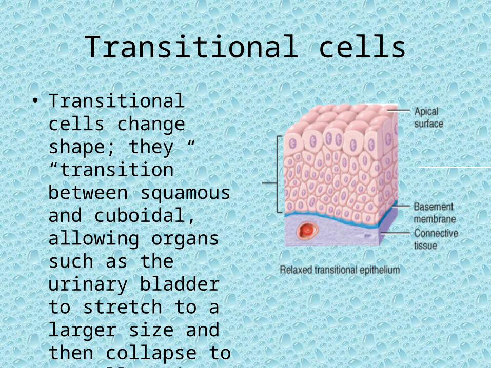
Transitional cells
• Transitional cells change shape; they “transition” between squamous and cuboidal, allowing organs such as the urinary bladder to stretch to a larger size and then collapse to a smaller size


Question
• What type of epithelial tissue is found in the walls of the urinary bladder to provide it with the ability to distend?

Glandular Epithelial Tissues
Glandular epithelia secrets a product wither into ducts or into the blood. • Glands– Exocrine glands
• Secrete into ducts to exterior of body• Salivary glands, mammary glands and sweat glands
– Endocrine glands• Secrete into the blood to carry chemical messages
throughout the body• Thyroid gland, and parathyroid gland

Exocrine glands secrete fluids into ducts

Endocrine cells secrete hormones into the blood

Cell Junctions• The epithelial cells of a tissue are connected
by cell junctions.• These junctions help a tissue perform its
particular function.

Cell junctions
• Junctions: hold epithelial cells together– Tight junctions• Allow epithelia cells to form a layer that prevents
leakage from one side of the sheet to the other
– Adhesion junctions/spot desmosomes• Keep cells anchored to one another, but the layer of
cells can still bend and stretch.
– Gap junctions• Allow the passage of small molecules and ions from
one cell to the other.
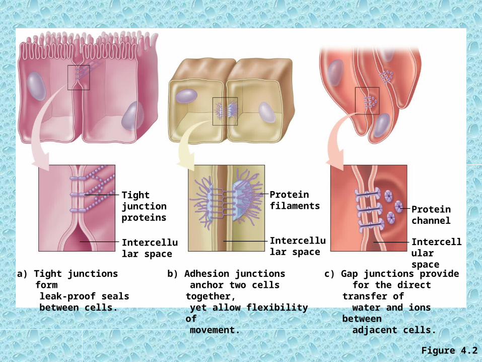
Figure 4.2
Tight junction proteins
Intercellular space
Protein filaments
Intercellular space
Intercellular space
Protein channel
a) Tight junctions form leak-proof seals between cells.
b) Adhesion junctions anchor two cells together, yet allow flexibility of movement.
c) Gap junctions provide for the direct transfer of water and ions between adjacent cells.
Questions
• What are the three types of junctions between cells, and how do they differ in structure and function?
• Why would you expect cell junctions more often in epithelia than in other types of tissues?
• Which type of cell junction would you expect to find between muscle cells in the heart wall? Why?

Activity 1.
• complete Box 5.1 Organs, Tissues, and Functions

Box 5.1 Organs, Tissues, and Functions
Organ Tissue Function of Tissue
Heart
Kidney
Eye
Lung
Stomach
Esophagus

Activity 1
• Examine the following photomicrographs and describe them.
• Also include their function and location.
.

Simple squamous epithelium

Stratified squamous epithelium

Simple Cuboidal Epithelium

Simple Columnar Epithelium

Pseudostratified Ciliated Columnar Epithelium