Embed Size (px)
Citation preview

Lecture 7: Basic Optics, EM and visualization
รวบรวมและเรียบเรียงโดยอ. วรรณพงษ์ เตรียมโพธ์ิ
ภาควิชาฟิสิกส์ คณะวิทยาศาสตร์ ม. มหิดล

Many of the following figures or pictures are copiedfrom open sources at the Web or Else. I do not claimany intellectual property for the following materials.

Topics
0. Nature of Science and physics1. Mechanics2. Temperature and Heat3. Fluid4. Waves5. Sound and hearing6. Optics and visualization7. basic electromagnetism8. basic quantum mechanics9. atomic physics10. basic nuclear physics and radioactivity

MRI and RF (Radio Frequency)
• Used to produce images of soft tissues, fluid, fat and bone.
• Does this by producing a map which depends on the density of hydrogen in the body.
• Uses strong superconducting magnet with a magnetic field strength 40,000 x that of the Earth’s.
• It is used to diagnose many problems e.g. helps identify tumours.
Diagram of an MRI scanner

Pulse Oximetry

What is the electromagnetic spectrum?

RADIOWAVES

The electromagnetic spectrum consists of all the
different wavelengths of lectromagnetic radiation, including light, radio waves, and X-rays.

Radio Waves
• In 1887, Heinrich Hertz demonstrated the reality of Maxwell's electromagnetic waves by experimentally generating radio waves in his laboratory.
• Their frequencies range from 300GHz to as low as 3kHz.
• Radiofrequency is a rate of oscillation in the range of radio waves, it refers to electrical rather than mechanical oscillations.
• Radiofrequency energy is used in medicine e.g. MRI and RFA.
Heinrich Hertz

Micro Waves
In 1888, Heinrich Hertz was the first to demonstrate the existence of radio waves by building a spark gap radio transmitter that produced 450 MHz microwaves.
Microwaves have typically 300GHz to 300MHz
Heinrich Hertz

Microwave Heat Therapy
Hyperthermia therapy is a type of medical treatment in which body tissue is exposed to slightly higher temperatures to damage and kill cancer cells or to make cancer cells more sensitive to the effects of radiation and certain anti-cancer drugs.
Cancer cells being targeted
by microwaves


Infrared 0.1 cm to 0.00007 cm
Heat!

Visible Light
• Visible light covers the range of
• wavelengths from 400 to 700 nm.
• Our eyes are sensitive only to this
• small portion of the electromagnetic
• spectrum.

Visible Light
• Can be detected by the human eye.
• Wavelengths range from 750-400nm.
• In the 17th Century, Isaac Newton explained the optical spectrum in his book ‘Opticks’. He divided the spectrum into seven named colours: ROYGBIV.
• The actual concept of a visible ‘spectrum’was defined in the early 19th century when light outside the visible range was discovered e.g. Johann Ritter with Ultraviolet Light.
Sir Isaac Newton

Endoscopy/ Keyhole Surgery
Allows us to look inside the human body through a narrow, flexible scope.
It is mostly used to diagnose problems in the oesophagus, stomach and intestines, including ulcers, bleeding and tumours.
Typically optical fibres are used to transfer light to the end of the endoscope and a miniature video camera records the image, and viewed on a video screen.
Endoscope inside the body

Ultraviolet
• Ultraviolet radiation
• has wavelengths of
• 10 to 310 nm (about the
• size of a virus).
• Young, hot stars
• produce a lot of
• ultraviolet light and
• bathe interstellar
• space with this
• energetic light.

Ultraviolet

Ultraviolet light in treatment of Psoriasis and Vitiligo
Psoriasis Vitiligo

Ultraviolet light in Dental Care
Ultraviolet light hardening a patient’s filling
Ultraviolet light waves produce free radicals that activate the catalyst and speed up polymerisation of the composite resin.
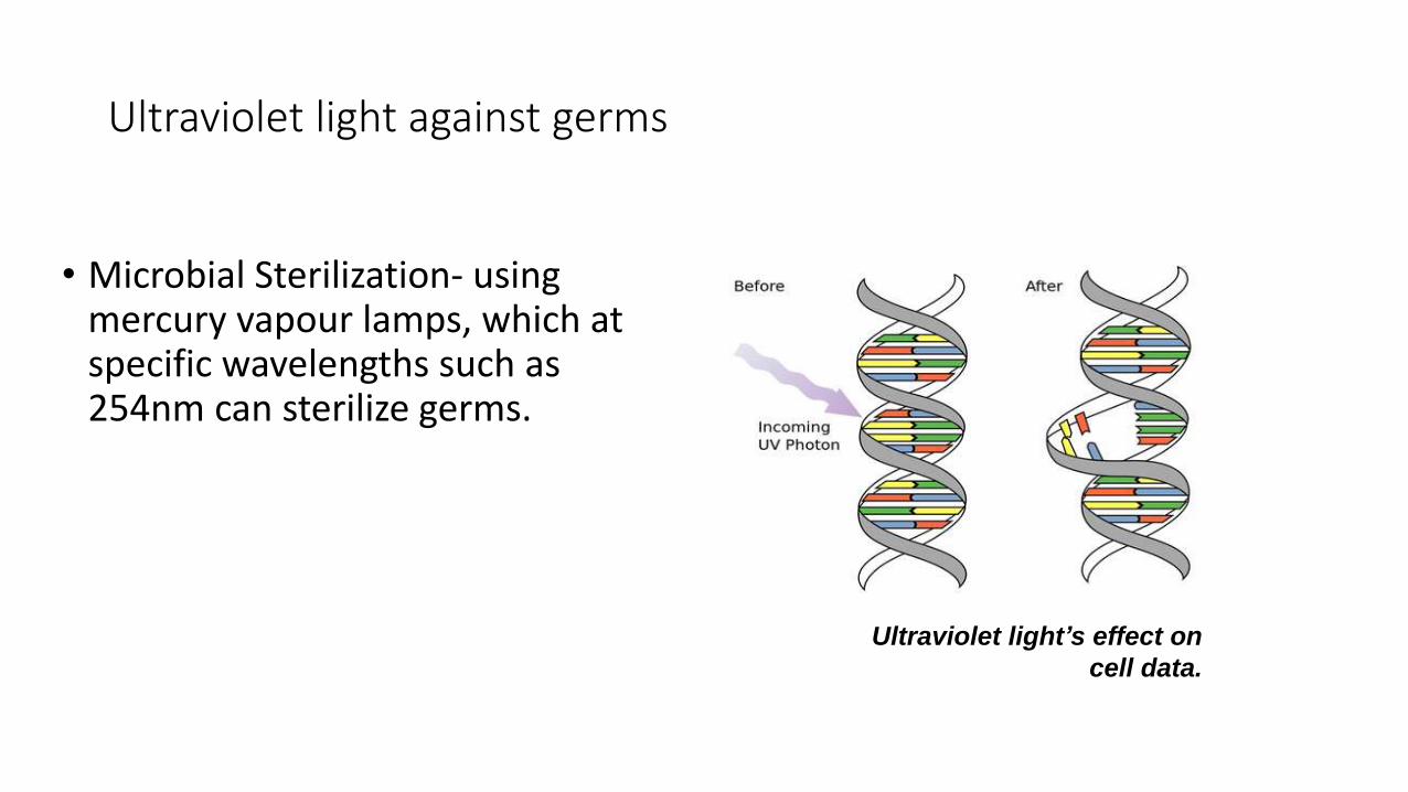
Ultraviolet light against germs
• Microbial Sterilization- using mercury vapour lamps, which at specific wavelengths such as 254nm can sterilize germs.
Ultraviolet light’s effect on
cell data.

Discovery of X-Rays
They were discovered serendipitously by German Physicist Wilhelm Roentgen in 1895.
Roentgen was working with electron beams in discharge tubes.
In the early days many patients and doctors developed radiation sickness since they were shining x-rays in all directions for large amounts of time.
Wilhelm Roentgen

X-Rays and the Body• Visible light photons (0-3 MeV) are
absorbed by the atoms that make up body tissue (hence we are not see through!)
• X-ray photons have much more energy (1-100KeV) .
• A larger atom is more likely to absorb an X-ray photon in this way, because larger atoms have greater energy differences between
• Smaller atoms, where the electron orbitals are separated by relatively low jumps in energy, are less likely to absorb X-ray photons.
• Soft tissue does not absorb x-ray radiation well but it still possible, that is why you don’t only see bones.

X-Ray Machine
• The heart of the x-ray machine is the electrode pair. A Cathode (heated filament) and the Anode (made of Tungsten)
• The Cathode source accelerates electrons to a high speed and these electrons then collide with the Tungsten .
X-ray machine

CT-Scan Setup

CT Scan

Gamma Rays
• EM Radiation high frequency
• High energy photon- kill cancer cells
• Produced by decay from high energy states of atomic nuclei
• Discovered in 1900 by Paul Villard. (right)
Paul Villard

Positron Emission Tomography (PET)
• Nuclear medical imaging
• Gamma Rays- positron annihilation
• 3D image
• Non- Invasive
Image of a PET Scanner

History of PET Scans
• David E. Kuhl, Luke Chapman and Roy Edwards in the late 1950s.
• University of Pennsylvania
• First demonstration at Massachusettes General Hospital.
PET scans at University of
Pennsylvania