Embed Size (px)
Citation preview

Leading Edge
Perspective
Plugging in to Human Memory:Advantages, Challenges, and Insightsfrom Human Single-Neuron Recordings
Rodrigo Quian Quiroga1,*1Centre for Systems Neuroscience, University of Leicester, Leicester, UK
*Correspondence: [email protected]
https://doi.org/10.1016/j.cell.2019.10.016
We describe single-neuron recordings in the human hippocampal formation, performed in epilepticpatients for clinical reasons, and highlight their advantages, challenges, and limitations comparedwith non-invasive recordings in humans and invasive recordings in animals. We propose a unifiedframework to explain different findings—responses to novel stimuli, spatial locations, and specificconcepts—linking the rodent and human literature regarding the function of the hippocampal for-mation. Moreover, we propose a model of how memories are encoded in this area, suggestingthat the context-independent, invariant coding by concept cellsmay provide a uniquely human neu-ral mechanism underlying memory representations.
In the 1980s, zoologist Hans Kummer reported a now very
famous observation of a female Hamadrya baboon grooming
with a young male hiding behind a big rock but keeping part
of the body visible to her own male, who was feeding several
meters away, unaware of the situation (Whiten and Byrne,
1988). This and similar types of behaviors have been offered
as evidence that non-human primates have a ‘‘theory of
mind’’; that is, the ability to understand other subjects’
thoughts. However, this interpretation has been disputed by
behavioralists (see comments in Whiten and Byrne (1988),
who argue that the animal may have learned to act this way
without truly understanding why; that means, without neces-
sarily wondering what her male was thinking. The problem is
that we cannot get into the animal’s head or simply ask her
why she hid behind the rock. In fact, pinning down the real mo-
tives or thoughts of animals is difficult and requires well-
controlled paradigms (one such study showed decades later
that non-human primates do indeed have a theory of mind [Kru-
penye et al., 2016]). The same applies to memory, particularly
to episodic memory (i.e., the memory of our experiences)
because we cannot interrogate animals about their thoughts
and recollections.
A key challenge in neuroscience is to understand how the
firing of neurons underlies behavior. However, advances in
this area face some very basic limitations. On one hand, non-
invasive recording techniques—e.g., electroencephalography
(EEG), magnetoencephalography (MEG), and fMRI—are used
with human subjects for obvious ethical reasons, but, although
these methods have provided insights into the activation of
brain areas during different tasks, they can only offer an indirect
and vague measure of the activity of individual neurons (Logo-
thetis, 2008). On the other hand, invasive recordings provide
direct access to study the firing of multiple neurons but can
usually only be performed in animals, and, as the story of Kum-
mer illustrates, the lack of direct verbal feedback limits our un-
derstanding of what is going on in the animal’s brain. Moreover,
the types of experiments and questions that can be addressed
with animals are limited because they need extensive
reward-driven training to perform different tasks, far from the
natural conditions of how these behaviors occur in real-life sit-
uations.
In very particular cases, however, it is possible to perform
invasive recordings in human subjects for clinical reasons.
This is the case with patients suffering from epilepsy refractory
to medication, who are implanted with intracranial electrodes to
determine the seizure-originating area and evaluate the possi-
bility of its surgical resection (Rey et al., 2015a), offering the
extraordinary opportunity to record the activity of multiple sin-
gle neurons in awake and behaving human subjects performing
different tasks. (Single-cell recordings are also performed dur-
ing deep brain stimulation [DBS], and we refer to Engel et al.
[2005] for a review of these studies, which will not be covered
here.)
Single-Neuron Recordings in HumansThe first recordings of individual neurons in the human brain
were performed in the 1950s, using a glass pipette attached
to a micromanipulator during epilepsy surgery (Ward and
Thomas, 1955). Later on, in the 1970s, a procedure was devel-
oped to record from multiple microwires that were inserted
through hollow-depth intracranial electrodes protruding a
few millimeters from their end (Babb et al., 1973), a design
that it is still used today (Figures 1A–1C). Contacts placed
along the depth electrodes allow recording of intracranial elec-
troencephalographic (iEEG) data used for clinical assessment
of the patients, whereas the microwires provide recordings of
multiple single neurons and local field potentials (LFPs) (Fig-
ures 1D and 1E). Recording sites often cover the medial
Cell 179, November 14, 2019 ª 2019 Elsevier Inc. 1015

Figure 1. Human Single-Neuron Recordings(A) Electrodes used for intracranial recordings. The intracranial EEG (iEEG) contacts are used to localize the epileptic activity, whereas local field potentials (LFPs)and spiking activity are recorded from the microwires protruding from the electrode tip.(B and C) Computed tomography (CT) (B) and CT fused with MRI (C) of one electrode implanted in the hippocampal formation.(D) Raw data recorded from the microwires, where spikes are hardly visible (inset) because of the presence of large-amplitude, low-frequency activity. From theraw data, LFPs are obtained by low-pass filtering, and spikes are visualized after high-pass filtering (the marked segment correspond to the inset in the raw data).(E) Spikes are represented in a feature space (in this case, the first two wavelet coefficients) in which the classification of the spike shapes of the different neurons(i.e., spike sorting) is done. Clusters with relatively few spikes (the ones in red, green, and cyan) are typically difficult to identify because they tend to be mergedwith larger clusters (the one in blue).
temporal lobe (MTL; the hippocampal formation and its sur-
rounding cortex) because of the involvement of this area in
different forms of epilepsy (Niedermeyer, 1993). Subjects
remain with the electrodes implanted for about a week and
are monitored to record a sufficient number of spontaneous
seizures to evaluate an eventual surgical resection of the
epileptic focus.
Advantages
Advantages Comparedwith Non-invasive Recordings in Humans.
Compared with non-invasive studies, the key advantage of
single-neuron recordings is the possibility of having access to
the activity of individual neurons, which can be measured only
indirectly with non-invasive methods. Let us illustrate this with
two concrete cases. First, it is common that MTL neurons
respond sparsely to very few pictures (Quian Quiroga et al.,
2007). Because of a general lack of topographic organization in
the MTL (i.e., responses are not spatially clustered, and nearby
1016 Cell 179, November 14, 2019
neurons fire to completely different stimuli; De Falco et al.,
2016), there is not a common and localized activation that can
be observed at the more macroscopic level of fMRI or EEG/
MEG recordings, and, therefore, these responses are only iden-
tified at the single-neuron level. Second, besides providing infor-
mation about neuronal responses that cannot be seen with non-
invasive methods, single-neuron recordings can also validate
and provide further mechanistic evidence of fMRI and EEG/
MEG findings. For example, fMRI studies have consistently
shown the presence of preferential responses to scenes in the
parahippocampal place area (PPA) (Epstein and Kanwisher,
1998). However, fMRI recordings cannot distinguish between
different mechanisms that can produce these responses: (1)
each PPA neuron may respond sparsely to one or relatively
few scenes, giving category scene responses when averaging
the activity of neighboring neurons constituting each voxel; (2)
PPA neurons may be tuned to visual features that are more

prevalent in scene images, showing scene responses in the
population average but some visual feature tuning, rather than
scene selectivity, at the single-neuron level; (3) PPA neurons
may be scene-selective, responding preferentially to pictures
of scenes. Analysis of about 2,000 human MTL neurons clearly
showed that the latter was the case; parahippocampal neurons
had a tendency to respond to scenes (Mormann et al., 2017;
Figure 2A) with much broader category tuning compared with
the selective responses found for known people (Quian Quiroga
et al., 2007).
Advantages Compared with Invasive Recordings in Animals.
Compared with recordings with animals, a first obvious
advantage is that, if the ultimate goal is to understand the hu-
man brain (although this need not necessarily be the case),
then, by performing recordings directly in humans, we can
avoid the potentially false assumption of similar brain func-
tioning in animal models and humans. Another advantage of
human single-neuron recordings is the possibility of communi-
cating with and getting direct feedback from the subjects,
which allows us to perform experiments that cannot be
done in other animals. This is particularly the case when
studying internally generated top-down activations, such as
those arising during memory recall (Gelbard-Sagiv et al.,
2008; Ison et al., 2015), imagery (Kreiman et al., 2000), or
voluntary control of the neuron’s firing based on the subject’s
thoughts (Cerf et al., 2010).
Another key advantage is the possibility of directly explaining
the experiments to the subjects without the need of extensive
reward-driven training and the ensuing potential caveats of over-
training effects that could influence the neurons’ responses after
months of performing the same task. Moreover, communication
with the subjects permits tuning the experiments according to
their background and interests. For example, Figure 2B shows
the responses of a neuron in a subject interested in mathematics
that fired to different equations and math-related stimuli,
whereas Figure 2C shows a neuron’s responses to ‘‘Mr. T,’’ a
character in the film Rocky III, in a subject who was a fan of
this movie. The rationale for presenting equations (among other
things) to the first subject and characters from the Rocky films to
the second was that we expected to find more responses to
personally relevant things, as was shown to be the case from
analysis of a large number of responses (Viskontas et al., 2009).
Disadvantages and Limitations
Limitations Arising from Performing Recordings with Patients.
Human single-neuron recordings offer clear advantages but
have also several limitations, mainly because of recording con-
straints and the fact that these experiments are performed with
patients in a clinical environment.
A major limitation is the availability of patients. Most hospitals
performing these recordings have relatively few implantations a
year, and it may take several years to get a sufficient number
of neurons to have statistically sound results. Moreover, the
time to perform experiments with each patient is also limited,
and it is not always possible to consider all of the control exper-
iments that one would like to do. In addition, recordings are per-
formed in a clinical environment, which can be very noisy, and
there is relatively little time to sort out technical issues compared
with a standard laboratory environment.
The fact that recordings are done in patients with epilepsy
also raises the concern that the obtained results may reflect
different aspects of this pathology rather than normal brain
functioning. This is, however, very unlikely given that similar
types of responses have been obtained in recordings close to
the seizure-originating area and in more distant areas, including
the non-seizure-originating hemisphere (Mormann et al., 2008).
Moreover, results are similar for patients with different types of
epilepsy involving different pathophysiological mechanisms
(Niedermeyer, 1993). In addition, epileptic activity is, in principle,
expected to produce an increase in neural excitability and con-
nectivity, which would give a global increase in the neurons’
responsiveness, contrary to the very high selectivity observed
in these recordings (Quian Quiroga et al., 2007).
Results could also be attributed to effects of the medication
taken by the patients. This is particularly a concern when consid-
ering, for example, the relatively late onset of MTL neuron re-
sponses compared with response onsets in animals (Mormann
et al., 2008). However, different patients have different medica-
tions and dosages, and, furthermore, medication is gradually
tapered down during the time the patient is in the hospital to in-
crease the chances of recording seizures. Because similar re-
sults are obtained in different patients and at different days of
the intervention, the effects of medication in the MTL responses
can be ruled out.
Limitations Arising from the Location of Intracranial Recordings.
A caveat of human single neuron recordings is their limited
coverage compared with non-invasive techniques. The location
of the intracranial electrodes is always determined by clinical
criteria. Consequently, scientist do not have—and should not
have—a say in decisions about the implantation of the elec-
trodes, which may not necessarily cover the key areas involved
in the processes under study. Moreover, as with chronic record-
ings, electrodes cannot be externally moved to search for
responsive neurons, and millimeter variations in the electrode
implantation can mean the difference between obtaining and
not obtaining single-neuron recordings. However, because the
electrodes are fixed, there are no potential biases as there can
be with acute recordings: moving the electrodes and targeting
easily identifiable neurons with high firing rates can lead to
sparsely firing neurons being overlooked (Shoham et al., 2006).
Recording sites typically include the MTL because of the
involvement of this area in different forms of epilepsy (Nieder-
meyer, 1993). This is ideal to study memory processes, given
the well-documented role of the MTL in declarative memory
(Squire and Zola-Morgan, 1991). However, the study of MTL
neurons provides only a limited picture of memory functions,
which should ideally also consider interactions with the dien-
cephalon (Aggleton and Brown, 1999) and neocortical areas (Ei-
chenbaum, 2017; Fletcher and Henson, 2001; Sekeres et al.,
2018). This is particularly important to study memory consolida-
tion and the interplay of these areas in the coding of episodic and
semantic memories (Moscovitch et al., 2005; Squire and Zola-
Morgan, 1991). It is, however, possible to extract some informa-
tion about neocortical activations from the iEEG contacts of the
depth electrodes (Figure 1). In this respect, of particular interest
is analysis of high-frequency oscillations, which correlate with
local neuronal activity (Fisch et al., 2009; Lachaux et al., 2012;
Cell 179, November 14, 2019 1017

Figure 2. Examples of Single-Neuron Responses in the Human MTL(A) Pictures of landmark scenes (highlighted box) that elicited responses of a neuron in the parahippocampal cortex. The second and third rows correspond to theraster plot and the peristimulus time histogram, respectively. Dashed vertical lines show the time of picture onset and offset, 1 s apart.(B) Images of equations and math-related stimuli (highlighted) that elicited responses in a neuron in the entorhinal cortex in a subject interested in mathematics.(C) Images of ‘‘Mr. T,’’ including his written name (highlighted), that elicited responses in a neuron in the amygdala in a subject who was fan of the film Rocky III.Due to copyright issues, in this and the following figures some of the pictures were replaced by copyright-free pictures of the same persons.
Rich and Wallis, 2017; Watson et al., 2018) although, of course,
not giving single-neuron resolution, as with implanted micro-
wires (e.g., to estimate stimulus selectivity; Rey et al., 2014).
1018 Cell 179, November 14, 2019
Another caveat is that it is not possible to give a precise loca-
tion of the microwires used for human recordings. Animal
studies, particularly with rodents, have offered clear evidence

Figure 3. Single-Neuron Responses Identified after Spike Sorting(A) Spike sorting from an electrode in the amygdala. From the detected spikes, we observe responses to all 5 pictures depicted. However, after spike sorting, werather see that there is multiunit (blue) responding mainly to actress Sandra Bullock (stimulus 86) and a single unit (green) responding to Kris Kristofferson (stim.62) and Kenny Rogers (stim. 59), both actors and country singers, and to actor Mel Gibson (stim 69), who was costar with Kristofferson in the movie Payback. Theother single unit (red) responded only to actress Jennifer Lopez (stim. 49). For space reasons, the responses to only 5 of 105 pictures presented are shown (butthere were no significant responses to the pictures not included in the figure).
(legend continued on next page)
Cell 179, November 14, 2019 1019

of distinct roles of substructures within the hippocampus (see,
e.g., Treves and Rolls [1994]). But with human single-cell record-
ings, it is difficult to delineate the hippocampal substructures in
the co-registered MRI scans (Wisse et al., 2017), and it is also
very difficult to visualize the microwires in the CT scans (Figures
1B and 1C) (and even if we could, we would not know which
microwire is which).
Limitations Arising from the Number of Recorded Neurons.
Another major limitation is given by the number of microwires im-
planted for single-neuron recordings. MTL neurons tend to show
very sparse responses, firing to relatively few stimuli (Mormann
et al., 2008; Quian Quiroga et al., 2007). Consequently, it is diffi-
cult to trigger the neurons’ responses (that is why we use the
‘‘screening sessions’’ described below), and it is very unlikely
to record simultaneously from two or more neurons responding
to a particular stimulus, although it is still possible to infer prop-
erties at the population level by using statistical arguments (e.g.,
Waydo et al. [2006]). Moreover, the number of identified units can
be increased using optimal spike sorting algorithms (see next
section) (Rey et al., 2015b) and, particularly, new electrode de-
signs. Spectacular advances have been made in the design of
electrodes used for animal studies (e.g., Jun et al. [2017]), but
we have essentially been using the same type of electrodes for
human single-cell recordings since the 1970s (Babb et al.,
1973), in spite of the fact that progress in this area is likely to
have a large effect on the recording conditions.
Stability of the Recordings, Consolidation, and Long-
Term Representations
Using chronic recordings in animals, it has been shown that it is
possible to record from the same neurons during several days
(Dhawale et al., 2017; Okun et al., 2016). Tracking neurons
over days is indeed critical for human MTL recordings because
it allows us to assess the stability/plasticity of the responses
and study consolidation mechanisms. In particular, there has
been a long ongoing dispute about the role of theMTL inmemory
coding. Supporters of the standard consolidation model (Squire
et al., 2015; Squire and Zola-Morgan, 1991) argue that the MTL
encodes memories only during learning and not after their
consolidation in the neocortex, whereas supporters of the multi-
ple trace theory (Nadel and Moscovitch, 1997; Sekeres et al.,
2018) argue that the MTL continues to remain critical for
(episodic) memory after learning, thus providing a long-term rep-
resentation. Evidence backing one or the other theory comes
mainly from two sources: behavioral studies in patients with le-
sions, which, because of the variability of the precise location
and extent of the lesions, have provided mixed results (Mosco-
vitch et al., 2005), and non-invasive (fMRI) recordings (Mosco-
vitch et al., 2005), but this technique cannot directly assess, at
the single-cell level, the stability and plasticity of neuronal repre-
sentations.
Tracking neurons in time is a challenging task with recordings
in epileptic patients (and patients typically have the electrodes
implanted for no longer than a week). This is because, in the clin-
(B) Spike sorting from an electrode in the hippocampus. In this case, we do not obone multiunit (blue) and 5 single units were identified, with one of them (in red) firingpicture of Stonehenge (stim 23). Note that the multiunit does not have clear respoThe responses to 5 of 24 pictures presented are shown, but there were no signifi
1020 Cell 179, November 14, 2019
ical environment where the experiments are performed, the
signal and noise conditions can change abruptly, and electrodes
may also move; for example, when the patients have seizures
with abrupt contractions. In spite of this limitation, first results
show that it is at least possible to track neurons producing similar
responses on consecutive days (Niediek et al., 2016). However, it
is still difficult to quantitatively assess the stability and plasticity
of the neural representations—i.e., what the neurons fire to—
across days because different responses could arise from
different neurons being recorded. To address this issue, it is
important to perform continuous 24/7 recordings and track the
neurons’ properties (e.g., spike shape, firing characteristics) to
check their identity, ensuring that any changes in the neurons’
properties are relatively smooth (Harris et al., 2016). In this
respect, it should be noted that, although it is difficult to record
from the same neurons over several days in humans, it is, how-
ever, possible to infer whether responses are created de novo
during the task or whether they reflect a long-term representa-
tion. The latter seems to be the case, given that MTL responses
are observed during passive viewing the first time the patient
sees a picture of a particular person (or place, animal, etc.),
meaning that the neuron was already encoding this person
before the experiment took place (Pedreira et al., 2010; Rey
et al., 2015a).
Silent Neurons—Screening Sessions
The spikes fired by the neurons are metabolically expensive
(Attwell and Laughlin, 2001), and it has been argued that, similar
to the dark matter problem in physics, a large proportion of neu-
rons may remain silent most of the time and are therefore not
observed with extracellular recordings (Shoham et al., 2006).
Figure 3 shows two hippocampal recordings where the activity
of different neurons was identified after spike sorting. In both
cases, we observe units with very sparse responses that fired
only to one or a few of the pictures shown and remained nearly
silent during the rest of the recording. These neurons are diffi-
cult to detect for two reasons: first, their responses are masked
by the firing of other nearby neurons recorded from the same
electrode (Harris et al., 2016; Rey et al., 2015a), and second,
the neurons cannot be detected unless the right stimulus
is shown.
With respect to the first problem, it is challenging to separate
clusters with relatively very few spikes from other, much larger
clusters, but current spike-sorting algorithms can deal with
sparsely firing neurons (Figure 1E; Rey et al., 2015b). Note that
an optimal separation of the neurons recorded from a single
electrode is important not only to identify sparse responses
that would not be observed otherwise (Figure 3B; Rey et al.,
2015a) (and that cannot be detected with non-invasive record-
ings) but also to avoidmisinterpretations about coding principles
underlying the firing of these neurons (Figure 3A); for example,
without proper sorting, it would be difficult to assess the neu-
rons’ very high selectivity (Quian Quiroga et al., 2007) and their
tendency to fire to related concepts (De Falco et al., 2016).
serve any response for the detected spikes, but after spike sorting, we see thatto musician George Harrison (stim 9) and another one (in green) firing only to a
nses and masks the responses of the single units before spike sorting is done.cant responses to the other pictures.

Concerning the second problem, we do not know a priori
which pictures trigger the neurons’ responses. As illustrated in
Figures 2B and 2C, based on interactions with the patients, we
know about their interests and we therefore tend to use pictures
of things that are familiar to them. Besides tuning the stimulus set
based on the patient’s interests, screening sessions can be per-
formed in which a large number of pictures is shown repeatedly
and in pseudorandom order to determine which of the pictures
trigger neuronal activations, and then use these specific pictures
in follow-up experiments. This way, responses in the screening
sessions were used to study how the firing of MTL neurons cor-
relates with conscious perception (Quian Quiroga et al., 2014,
2008), internally generated processes (Cerf et al., 2010), working
memory (Kornblith et al., 2017; Reddy et al., 2006), and rapid for-
mation of associations (Ison et al., 2015), among other functions.
The screening sessions also provide valuable data to estimate
the coding properties of MTL neurons, such as their degree of
selectivity (Waydo et al., 2006).
Memory Coding in the Human MTLThe seminal study of patient H.M. showed the critical role of the
MTL in declarative memory (i.e., memories of facts and experi-
ences; Scoville and Milner, 1957). Investigations in patients
with similar lesions (Moscovitch et al., 2005; Nadel and Mosco-
vitch, 1997), evidence from animal studies (Squire and Zola-Mor-
gan, 1991), and imaging studies in normal subjects (Paller and
Wagner, 2002) have provided further support of the role of the
MTL in the coding and consolidation of episodic memories (Se-
keres et al., 2018; Squire et al., 2015) but cannot address how
neurons in the human MTL underlie memory functions. In this
section, we describe three lines of research with human single-
neuron recordings that have shown the involvement of MTL neu-
rons in memory and propose a unified framework to explain
these responses.
Recognition Memory and Novelty Responses
Episodic memories are based on single experiences, and, there-
fore, many studies have focused on how the MTL responds to
novel stimuli. Imaging studies in humans (Paller and Wagner,
2002) and electrophysiology studies in monkeys (Brown and
Xiang, 1998) have established that the MTL is involved in the
encoding of novel items using recognition memory paradigms
(Bird, 2017) and that such activations can predict later recall (Pal-
ler andWagner, 2002). Implementing a similar approach with hu-
man intracranial recordings, it has been shown thatMTL neurons
respond to novel stimuli (Fried et al., 1997; Heit et al., 1988).
Further studies showed that, in contrast to the very selective re-
sponses to familiar persons (Quian Quiroga et al., 2007), re-
sponses to novel stimuli are not selective because a relatively
large proportion of MTL neurons (�20%) change their firing in
response to most novel stimuli (Rutishauser et al., 2006; Rutish-
auser et al., 2008; Viskontas et al., 2006). Interestingly, a more
recent study showed that the precise timing of MTL neurons’
firing, occurring at specific phases of local theta oscillations,
signaled whether novel items would later be recognized (Rutish-
auser et al., 2010), a finding in line with other studies showing
correlations between the precise timing of the neurons’ firing
and the phase of LFPs in specific frequency bands (Quian Quir-
oga and Panzeri, 2009).
Concept Cells
Several advances, including the use of screening sessions with
stimulus sets tuned for each subject to maximize the chance of
getting responses (Figures 2B and 2C) and the use of an
advanced spike-sorting algorithm to identify nearly silent neu-
rons (Figure 3), led to the finding of MTL neurons with very sparse
and invariant responses. Figure 4 shows two neurons from a
recording in the amygdala that were separated after spike sort-
ing. The first one fired to one of the experimenters performing re-
cordingswith the patient and to his name (Arne) presented on the
screen and pronounced by a computer-synthetized voice (but
not to the other 95 pictures and names shown). That means
that the neuron responded to the concept ‘‘Arne’’ but not to
the details of the visual or auditory stimuli used. The second
neuron responded to actor Michael Douglas, but in this case,
the response to the written name was not significant. In fact,
about 20%–30% of amygdala neurons responding to the picture
of a person also responded to his/her name, whereas about 50%
did so in the hippocampus, which shows a higher degree of
abstraction in this area, going beyond a specific sensory modal-
ity. More generally, there is an increase of abstraction and multi-
modal invariance along the anatomically hierarchical structure of
the MTL; about half of the neurons in the parahippocampal cor-
tex, at the bottom of this hierarchy, show visual invariance but no
multisensory responses, whereas more than 70% of the neurons
show visual invariance and about half multisensory responses in
the entorhinal cortex and the hippocampus, at the top of this hi-
erarchy (Quian Quiroga, 2012, 2009).
Concept cells can be characterized as neurons in the human
MTL that (1) respond very selectively to specific and well-known
concepts (like a famous person or place), (2) have a high degree
of multimodal invariance (i.e., responding to different pictures of
the same person, irrespective of the varying details of the pic-
tures used, and even to the person’s written or spoken name);
and (3) are not modulated by context (see below). Several
studies have further characterized the properties of these neu-
rons, and it has been proposed that they are involved in declar-
ative memory (Quian Quiroga, 2012), in line with the well-estab-
lished role of the MTL for this function (Squire and Zola-Morgan,
1991). This is supported by the facts that (1) concept cells have a
relatively late latency of responses (�300 ms; Mormann et al.,
2008), much later than what would be expected for sensory pro-
cessing; (2) they fire to personally relevant concepts (Viskontas
et al., 2009; namely, those that tend to be stored in memory);
(3) they show a high degree of invariance (Quian Quiroga et al.,
2009, 2005), which is in agreement with the fact that we tend
to conceptualize and forget irrelevant details; (4) they have
high selectivity (Quian Quiroga et al., 2007), which, as shown
by theoretical studies, is ideal for memory functions, such as
creating new associations (Marr, 1971); (5) their function is
beyond sensory processing because their firing can be triggered
by different stimulus modalities (Quian Quiroga et al., 2009) or
internal processes in the absence of external stimulation (Gel-
bard-Sagiv et al., 2008); and (6) they respond to the subjective
attribution of meaning by the subjects (i.e., how they will eventu-
ally store in memory what they believe they saw), mostly with all-
or-none responses, irrespective of the sensory features of the
stimuli (Quian Quiroga et al., 2014, 2008).
Cell 179, November 14, 2019 1021

Figure 4. Concept CellsTwo neurons identified from a microwire in the amygdala of a patient after spike sorting. The first neuron (top) responded to 3 different pictures of one of theresearchers performing experiments with the patient and to his name (Arne) written on the screen (stim 27) and pronounced by a synthetized voice (soundwaveform of stim 5). The second neuron (bottom) responded to the pictures of actor Michael Douglas. In this case, there were no strong responses to his written(stim 59) and pronounced name (stim 10). The neurons did not respond to any of the other 90 pictures and names presented in this experiment.
Spatial Memory
Since the discovery of neurons that fire at specific locations
(‘‘place cells’’) in the rodent hippocampus (O’Keefe and Dostrov-
sky, 1971), many studies have described spatially tuned neu-
rons, including grid cells in the medial entorhinal cortex (Hafting
et al., 2005), and have suggested a crucial role of the hippocam-
pal formation in spatial navigation (Moser et al., 2017).
The exquisitely complex structure of the hippocampus is
similar in rodents and humans (Strange et al., 2014), and a large
number of fMRI studies have also shown the involvement of the
human hippocampus in spatial navigation (e.g., Burgess et al.
[2002] andMaguire et al. [2000], amongmany others). Moreover,
using a virtual navigation task with human single-neuron record-
ings, it has been shown that place cells are present in the human
hippocampus as well (Ekstrom et al., 2003). Imaging studies (Do-
eller et al., 2010) and iEEG recordings analyzing activity in the
theta frequency band (Kunz et al., 2019) have also reported a
6-fold rotational symmetry in the entorhinal cortex, as expected
from the geometrical structure of grid cells, and single-neuron
recordings with epileptic patients later confirmed that grid cells
are also present in the human entorhinal cortex as well as in
the cingulate cortex and the hippocampus (Jacobs et al., 2013).
A General Framework for MTL Memory Function
Several different functions have been attributed to the hippo-
campal formation, and onemay wonder whether we are perhaps
asking too much of this area and most findings could
be explained as different manifestations of the same general
principle. Within this view, how could we reconcile the finding
of spatially tuned neurons in rodents (and humans) with the
1022 Cell 179, November 14, 2019
episodic memory function attributed to this area? Several
studies have shown that place cells in rodents remap following
physical changes in the environment and that they are also
modulated by non-spatial factors according to the specific
task performed by the animal (Eichenbaum and Cohen, 2014; Ei-
chenbaum et al., 1999; Moser et al., 2017), indicating that the
role of these neurons goes beyond spatial processing. Although
grid cells also realign with physical changes in the environment
(Fyhn et al., 2007), the geometrical structure of their fields has
been considered to provide a more invariant spatial representa-
tion (Moser et al., 2017). However, more recent studies have
shown that grid cells encode cognitive aspects as well, with their
precise place of firing being modulated by reward location (Boc-
cara et al., 2019; Butler et al., 2019; Quian Quiroga, 2019).
The modulations produced by cognitive factors of the spatial
representations by place and grid cells provide contextual infor-
mation about the experience of the animals in the environment,
which is in line with the memory function attributed to this area
based on human studies. Therefore, to merge the rodent and hu-
man literature regarding the function of the hippocampal forma-
tion, it has been argued that this area has a general ‘‘relational
memory’’ role, linking together the elements of experiences
(Eichenbaum and Cohen, 2014; Eichenbaum et al., 1999). Within
this framework, spatial location is one of several components
that constitute a memory. This spatial representation is behav-
iorally critical in rodents because of the importance of knowing
their precise location and routes to reach safety–hence the prev-
alence of spatially tuned neurons in their hippocampal formation.
Furthermore, rodents acquire information about the environment

through exploration, whereas primates rely mainly on vision and
eye movements to explore and navigate their surroundings (Ek-
strom, 2015; Rolls andWirth, 2018). However, although theymay
be represented differently at the neuronal level (Discussion), ro-
dents also have notions of concepts (e.g., cat, cheese, etc.), and
humans also have spatial representations that enrich their mem-
ories and help avoid interference; for example, the context of my
office helps me remember a conversation I had there with a
colleague, which I do not confuse with another one we have
had at a conference dinner. A particular location in space, repre-
sented by place cells or by other spatially tuned neurons, can
then be seen as a concept that is associated with different
experiences.
Novel stimuli activate a relatively large proportion of neurons.
These responses tend to be weaker compared with the ones to
familiar stimuli (Quian Quiroga et al., 2007), suggesting that the
initial modulations in response to novel stimuli may get stronger
for stimuli that become familiar. For example, a set of neurons
will fire to a group of persons we meet for the first time at a party,
but only a few of them will consolidate a more stable representa-
tion of one of these persons that we happen to meet often again
and get to know better. More generally, we can postulate a
similar mechanism for the observation of hippocampal activa-
tions in declarative memory tasks. MTL neurons that are not
already recruited in strong assemblies may offer a temporary
and malleable representation to perform these tasks. For
example, if a subject is asked to remember a set of faces, words,
or images, as in standard recognition memory paradigms, these
stimuli will modulate the activity of MTL neurons, producing an
activation that could be observed non-invasively with fMRI and
EEG (Paller and Wagner, 2002). However, the representation of
these stimuli is labile, and the involved neurons could soon be re-
cruited to encode something else after the experiments are
done, unless the stimuli is rehearsed over and over again,
becoming familiar and triggering specific memories (e.g., the
memory of doing the experiment). This simple mechanism can
offer an adaptive and temporary code that is able to deal with
different hippocampus-dependent tasks and form long-term
representations.
Coding of Associations in the Human MTLEpisodic memory relies on the fast formation of associations (Ei-
chenbaum, 2004; Quian Quiroga, 2012; Wallenstein et al., 1998);
for example, the memory of seeing a celebrity in the subway in-
volves making a link between these concepts. Concept cells
represent familiar concepts (Viskontas et al., 2009)—concepts
we form memories about—to encode meaningful associations.
Moreover, a very sparse representation, as the one by concept
cells, is ideal for the fast encoding of new associations required
for episodic memory (Marr, 1971; McClelland et al., 1995).
Furthermore, we tend to forget irrelevant details and remember
concepts, which is exactly the type of information encoded by
these neurons.
Encoding of New Associations
The hypothesis that concept cells are involved in memory was
tested using a pair association task, in which, for each person
to whom a neuron initially responded (as determined from previ-
ous screening sessions), an association with an arbitrary place
was created by showing an artificial image (created with Photo-
shop) of the person in the place. Neurons initially firing to a per-
son showed a significant increase in firing to the presentation of
the associated place (without the person) but not to other places
that were associated with other persons (the associations also
worked the other way around; neurons initially firing to a place
started firing to the person associated with it and not to other
persons) (Ison et al., 2015). Figure 5A shows the normalized re-
sponses of these neurons, which, for the preferred stimulus
(the one the neurons originally fired to), showed a decrease after
learning due to repetition suppression, as described in previous
studies (Pedreira et al., 2010; Rey et al., 2015a). In contrast, for
the non-preferred associated stimulus, there was a marked in-
crease in the neurons’ responses after learning. Moreover,
Figure 5B shows that, after learning, the responses to the asso-
ciated stimuli were similar in different tasks and conditions.
When aligning trials to the time of learning, we observed that
the increase in the response to the non-preferred associated
stimulus was relatively abrupt and happened at the exact time
of learning the associations, which sometimes was after a single
presentation (Figure 5C). The fact that such rapid learning was
observed is very relevant because episodic memories, like
remembering seeing a person in a place, are typically formed
by single unique experiences.
Long-Term Coding of Associations
A somewhat puzzling result from the previous study was the fact
that about 40% of the neurons initially firing to a concept
expanded their tuning to start firing to the associated one (Ison
et al., 2015). The problem is that, with such a high probability
of firing to associated concepts, the neurons should end up re-
sponding to most concepts (because, directly or indirectly,
they are all somehow related to each other), which is incompat-
ible with the very high selectivity of these neurons (QuianQuiroga
et al., 2007). It could, however, be the case that many neurons
encode the associations during the task but only a few of them
will continue to do so afterward if the associations remain rele-
vant and are later remembered.
What, then, is the chance of neurons encoding such associa-
tions in the long term? This issue was addressed by evaluating
the probability of the neurons to respond to associated concepts
in the screening sessions (De Falco et al., 2016), in which no
memory task was performed, and the neurons’ activities
reflected what they code for rather than temporary task-related
activations. For this, after performing the experiments, the pa-
tients were asked to rank how much the concepts eliciting re-
sponses (and other concepts for comparison) were related to
each other. The left bars in Figure 5D show that, as illustrated
in Figure 3 with the example of a neuron firing to 3 related actors,
when neurons fired to more than one concept, these concepts
tended to be associated. In other words, these neurons encode
long-term associations; associations that were already mean-
ingful to the subject and were not created by the task (passive
viewing of the pictures), supporting the notion of a permanent
role of the MTL in encoding of episodic memories, as proposed
by the multiple trace theory (Nadel and Moscovitch, 1997).
Because it is not possible to ask patients to rank how much
each of the �100 concepts presented are associated with
each other (which would give about 5,000 comparisons), a
Cell 179, November 14, 2019 1023

Figure 5. Formation and Long-Term Coding of Associations in the Human Hippocampal Formation(A) Average responses to the preferred (P) pictures and the associated non-preferred (NP) pictures before and after learning a pair association. Shaded areasrepresent SEM. The preferred pictures show a decrease in response because of repetition suppression, whereas the non-preferred ones show an increase afterlearning, encoding the association.(B) Differential activity index (that is, the normalized difference between the P and NP responses) before learning (BL; task 1) and after learning (AL; tasks 2–5).A clear difference between preferred and non-preferred responses was present before learning, which was reduced by a factor of 5.5, on average after learning (p< 10�6), because of the increase of the non-preferred responses. After learning, there were no significant differences in the differential activity index betweentasks 2–5, suggesting that (after learning), the responses were not task dependent. Task 1/2, picture presentations before/after learning; task 3, testing of theassociations; task 4, recall; task 5, final passive viewing of the pictures without a memory task.(C) Normalized learning (behavioral) curve and neural responses to the associated pictures, aligned to the time of learning (trial 0). Note the correlation betweenboth curves and the step increase in the neural responses (and behavior) at the time of learning the associations.(D) Mean association score for pairs of pictures to which the neuron fired and other picture pairs, based on the patients’ scores (left) and on an internet searchassociation metric (middle), andmean association score between pictures eliciting responses in nearby neurons, showing a non-topographic organization (right).Values are Z score normalized.(E) Probability of responses to pairs of pictures as a function of their degree of association using the internet search metric. Error bars show SEM.(F) Normalized activation for responsive and non-responsive neurons (0 corresponds to baseline activity and 1 to the maximum response for each neuron),showing a nearly binary code.(A)–(C) were adapted from Ison et al. (2015). (D) and (E) were adapted from De Falco et al. (2016). (F) was adapted from Rey et al. (2018).
metric of association was used based on the number of hits ob-
tained when doing an internet search of each pair of concepts
together, normalized by the number of hits obtained when
searching for each concept on its own (De Falco et al., 2016);
e.g., a Google search for ‘‘Bill Clinton’’ and ‘‘Hillary Clinton’’ gives
many more hits than a search for ‘‘Bill Clinton’’ and ‘‘Jennifer
Aniston,’’ because the first two are more related to each other.
The middle bars in Figure 5D show the result obtained using
this associationmetric, where we again observe that the neurons
tend to encodemeaningful associations. However, this tendency
was not as large as when asking the patients for their own asso-
ciation scores, which are based on subjective evaluations that
are not necessarily shared by other web users. In other words,
1024 Cell 179, November 14, 2019
the neurons reflect idiosyncratic rather than universal associa-
tions, suggesting that they encode episodic memories from
personal experiences. In line with this view, the coding of asso-
ciations was specific to particular concepts (e.g., an actor and a
place) and not to other concepts corresponding to the same
broad semantic categories (other actors and other places) (De
Falco et al., 2016). Moreover, when having a response to a
concept, the probability of finding a response to another one
was calculated as a function of its association with the first.
Figure 5E shows that, as expected, the probability of finding a
second response increased with the degree of association with
the first but saturated at about 4% for highly associated con-
cepts, ten times less than the 40% probability when learning

new associations, showing that only a fraction of the neurons
consolidate the associations in the long term.
Non-topographic Organization
In Figure 4, we show two nearby neurons that fired to allegedly
unrelated concepts: Arne andMichael Douglas. This observation
was quantified by evaluating the association metric for concepts
to which nearby neurons (separated after spike sorting) fired,
and it was found that the things nearby neurons fired to were
not related to each other (right bars in Figure 5D). Contrasting
with the topographically organized information in visual neocor-
tical areas—i.e., with nearby neurons firing to similar things
(Tanaka, 1996; see section 3.1 in Quian Quiroga, 2016)—MTL
neurons show a non-topographic organization, as found with
place cells in the rat hippocampus (Muller et al., 1987; Redish
et al., 2001). Such lack of spatial organization is ideal for episodic
memory, to quickly form associations between any two items
(of any category) without the need of establishing connections
between distant areas.
Binary Responses
In Figure 5A the response to the associated pictures was not as
large as the one to the preferred pictures to which the neurons
originally fired. It is therefore possible that MTL neurons prefer-
entially encode one concept and respond less strongly to
others according to their similarity (or degree of association)
with the first. Alternatively, the difference in the neurons’ re-
sponses to the preferred and associated pictures may only be
present during learning but not in the long term. To address
this issue, the pictures eliciting the neurons’ firing (as deter-
mined from the screening sessions) were shown several times,
and, for neurons responding to more than one picture, the re-
sponses to them was compared. In most cases (�80%), it
was not possible to distinguish the pictures eliciting responses
from the neuron’s firing (Rey et al., 2018). Interestingly, the 20%
of cases where the differences were significant corresponded
to less-associated concepts; that is, associations that were
not well consolidated, as those studied in the pair association
learning task described above (Ison et al., 2015). So, differ-
ences in the neurons’ responses are observed for less consol-
idated (temporary) associations and tend not to be present for
the most consolidated ones. In line with this, and in contrast
to the graded tuning typically found in the neocortex (Tanaka,
1996), Figure 5E shows that responses were mostly binary.
That is, if the neuron responded to a set of pictures, it did so
with the same strength, and if it did not respond, the firing
was mostly indistinguishable from baseline (Rey et al., 2018).
The finding of such binary coding shows that (associated) con-
cepts can only be distinguished from each other at the assem-
bly level. Moreover, such binary coding also has implications for
memory functions, increasing the network capacity, robustness
to noise, ease of readout, and avoidance of interference (Treves
and Rolls, 1994).
A Simple Memory Model with Concept CellsHaving reviewed concept cells, let us now discuss how these
neurons encode episodic memories. Concept cells do not act
in isolation but are part of cell assemblies representing familiar
concepts (Quian Quiroga, 2012). Each of these assemblies, on
its own, does not represent any particular memory or context,
but the associations between these assemblies encode specific
memories, which may be further enriched by representations
in the neocortex. For example, we may have an assembly of
concept cells encoding a particular friend and another one en-
coding our favorite cafe in town, and the memory of meeting
our friend at the cafe is given by having an association between
both assemblies (and perhaps some others related to this
encounter), which produces a coactivation of both concept rep-
resentations when retrieving the memory. We therefore propose
that the main function of concept cells is to (1) form and retrieve
meaningful associations and (2) point to and coactivate neocor-
tical sensory representations.
Coding of Associations with Partially Overlapping
Assemblies
Following the example presented in Figure 3A, let us consider a
hippocampal cell assembly representing Kris Kristofferson
(Figure 6). Different pictures of him activate similar (but different)
representations in the neocortex that initially ignite different sub-
sets of the MTL cell assembly but then rapidly activate most of
the assembly representing Kristofferson through pattern
completion. This activation of the same assembly (or most of
it) by different pictures of a person is the neural substrate under-
lying the ‘‘unitization’’ observed at the behavioral level; i.e., the
fact that different pictures of the same concept convey the
same meaning for memory functions (Graf and Schacter, 1989).
A first function of this assembly of concept cells is to act as a
pointer to coactivate neocortical representations related to Kris-
tofferson (Teyler and DiScenna, 1986) (solid blue arrows in
Figure 6), such as how his face looks, the sound of his voice,
etc., as well as related semantic information; e.g., the fact that
he is a country singer and an actor. A second function is to bring
about related information coded by associated MTL assemblies
(filled arrows in Figure 6), such as the fact that he acted with Mel
Gibson in the film Payback or that he sang a song with his wife,
Rita Coolidge. The association between two concepts in the
MTL is encoded with neurons firing to both of them, thus having
a partial overlap of their assemblies (as described above, of
about 4%), which is low enough to distinguish the concepts
from each other but, at the same time, large enough to encode
meaningful associations that may eventually lead to temporary
coactivations (or sequential activations to go from one concept
to the other, as in the flow of consciousness). The firing to an
associated concept can be generated very rapidly (Ison et al.,
2015) through Hebbian synaptic plasticity (Hebb, 1949), consid-
ering that there aremany instances in which the related concepts
appear or are recalled together (e.g., when watching or remem-
bering the movie or a song), thus generating the overlap. Given
the finding of binary responses at the single-neuron level, the de-
gree of overlap between the assemblies gives a distance metric
of how associated with each other two concepts are.
We can then argue that these associations between concepts
constitute the skeleton of episodic memories; the association
between Kristofferson and Gibson (together with a few other
ones) may provide a rough representation of having watched
the movie Payback, whereas the one with Coolidge will be
the substrate of the memory of having heard them sing a
country song. Furthermore, memories are also enriched by
details encoded in neocortical representations that MTL
Cell 179, November 14, 2019 1025
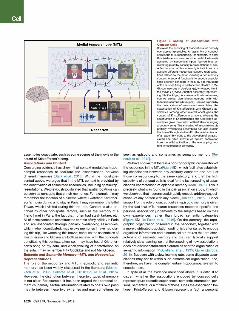
Figure 6. Coding of Associations with
Concept CellsShown is the encoding of associations via partiallyoverlapping assemblies. An assembly of conceptcells in the MTL responding, for example, to actorKris Kristofferson (neurons joined with blue lines) isactivated by neocortical inputs (curved blue ar-rows) triggered by sensory representations of him.A first function of this assembly is to link and co-activate different neocortical sensory representa-tions related to the actor, creating a rich memorycontent. A second function is to encode associa-tions between concepts in the MTL. For this, someof the neurons firing to Kristofferson also fire to MelGibson (neurons in blue/orange), who faced him inthe movie Payback. Another assembly represent-ing Rita Coolidge, his ex-wife, with whom he sangcountry songs, also shares neurons with Kris-tofferson (neurons in blue/pink). Context is given bythe coactivation of associated assemblies: thecoactivation of Kristofferson’s with Gibson’s as-semblies (among other related ones) gives thecontext of Kristofferson in a movie, whereas thecoactivation of Kristofferson’s and Coolidge’s as-semblies gives the context of Kristofferson singinga country song. The encoding of associations viapartially overlapping assemblies can also sustainthe flow of thoughts in theMTL: the initial activationof an assembly leads to the activation of an asso-ciated one (filled arrows) via pattern completionfrom the initial activation of the overlapping neu-rons encoding both concepts.
assemblies coactivate, such as some scenes of the movie or the
sound of Kristofferson’s song.
Associations and Context
Converging evidence has shown that context modulates hippo-
campal responses to facilitate the discrimination between
different memories (Stark et al., 2018). Within the model pre-
sented above, we argue that in the MTL context is provided by
the coactivation of associated assemblies, including spatial rep-
resentations. We previously postulated that spatial locations can
be seen as concepts that enrich memories. For example, I may
remember the location of a cinema where I watched Kristoffer-
son’s movie during a holiday in Paris, I may remember the Eiffel
Tower, which I visited during this trip, etc. Context is also en-
riched by other non-spatial factors, such as the memory of a
friend I met in Paris, the fact that I often had steak tartare, etc.
All of these concepts constitute the context of my holiday in Paris
and are associated through partially overlapping assemblies,
which, when coactivated, may evoke memories I have had dur-
ing this trip, like watching this movie, because the assemblies of
Kristofferson and Gibson are both associated with the concepts
constituting this context. Likewise, I may have heard Kristoffer-
son’s song on my sofa, and when thinking of Kristofferson on
the sofa, I may remember Rita Coolidge and not Mel Gibson.
Episodic and Semantic Memory—MTL and Neocortical
Representations
The role of the neocortex and MTL in episodic and semantic
memory has been widely discussed in the literature (Mosco-
vitch et al., 2005; Sekeres et al., 2018; Squire et al., 2015).
However, the distinction between these two types of memory
is not clear. For example, it has been argued that personal se-
mantics (namely, factual information related to one’s own past)
may lie between these two extremes and may sometimes be
1026 Cell 179, November 14, 2019
seen as episodic and sometimes as semantic memory (Re-
noult et al., 2012).
We have shown that there is a non-topographic organization of
the responses in the MTL (Figure 5D), which facilitates establish-
ing associations between any arbitrary concepts and not just
those corresponding to the same category, and that the high
selectivity of concept cells is ideal for the fast encoding of asso-
ciations characteristic of episodic memory (Marr, 1971). This is
precisely what was found in the pair association study, in which
we observed that neurons could rapidly encode arbitrary associ-
ations (of any person with any place) (Ison et al., 2015). Further
support for the role of concept cells in episodic memory is given
by the fact that MTL neuron responses matched specific and
personal association judgements by the subjects based on their
own experiences rather than broad semantic categories
(Figure 5D; De Falco et al., 2016). On the contrary, the topo-
graphic organization observed in the neocortex, together with
a more distributed population coding, is better suited to encode
organized information and hierarchical structures that are char-
acteristic of semantic memory and that can typically support
relatively slow learning, so that the encoding of new associations
does not disrupt established hierarchies and the organization of
semantic information (McClelland et al., 1995; Quian Quiroga,
2016). But even with a slow learning rate, some disparate asso-
ciations may not fit within such hierarchical organization, and,
therefore, we have the complementary hippocampal system to
encode them.
In spite of all the evidence mentioned above, it is difficult to
discern whether the associations encoded by concept cells
represent pure episodic experiences, semantic information, per-
sonal semantics, or a mixture of these. Does the association be-
tween Kristofferson and Gibson represent a fact, a personal

experience, or both? In our view, and based on the data pre-
sented above, it might be more plausible to argue that the MTL
is the substrate to form and encode disparate associations
that mainly support episodic memory, in contrast with more
ordered and topographically organized associations supporting
semantic memory in the neocortex. Such associations in the
MTL would then constitute the skeleton of long-term episodic
memories, in line with the multiple memory trace theory and
the dramatic effect on episodic memory produced by lesions
in this area (Moscovitch et al., 2005). With this framework, the
long-term coding of disparate associations by the MTL allows
‘‘jumps’’ in a memory narrative, like the transitions between
scenes in a movie, and, not surprisingly, it has been reported
that patients with MTL lesions are quite limited in their recall
and imagination and are able to provide only fractional accounts
that are supported by neocortical structures (Hassabis et al.,
2007), as when remembering a few isolated scenes from amovie
but not the movie plot—the details of the scenes are encoded in
the neocortex and the link between them in the MTL.
Memory Formation, Consolidation, and Forgetting
MTL neurons that are already recruited in consolidated assem-
blies represent concepts that are very relevant to the subject.
But memories are far from stable representations or engravings
in a wax tablet, as Plato saw them, and can be formed, consol-
idated, and, to a large extent, forgotten (Hardt et al., 2013; Ri-
chards and Frankland, 2017). At the neuronal level, we could
argue that the stability of the assemblies representing specific
concepts is maintained by the coactivation of the neurons that
form them if the concepts are revisited frequently enough. If
this is not the case, then the assemblies become labile, and their
neurons can be recruited to encode other memories that may
become more relevant.
The largest proportion ofMTL neuron responseswas to exper-
imenters who were initially unknown to the patients and per-
formed recordings with them (Figure 4A; Viskontas et al.,
2009). For the patients, the experimenters were recently known
but, at the same time, very salient because they interacted
with them very frequently while they remained in the hospital dur-
ing the intracranial recordings. However, the experimenters kept
no contact with the patients afterward, and although we cannot
track the neurons over long periods of time, it is reasonable to as-
sume that, after some time without contact, these neurons are
now encoding other concepts.
Memories ‘‘fight’’ to recruit MTL neurons. Novel concepts
initially recruit neurons with relatively labile responses. As famil-
iarity increases, these responses get stronger, and neurons
representing a new concept connect with each other, forming
a stable assembly. But as the concept becomes more familiar,
it has more associations related to it, thus recruiting neurons
initially responding to other concepts that start firing to the first
one, which explains the tendency to find responses to very
familiar items (Viskontas et al., 2009). Memories that are period-
ically revisited form relatively stable assemblies, whereas those
that are not have more labile representations with neurons that
have weaker connections with each other and that can be easily
co-opted to encode new memories. This simple competition
mechanism may be the neural basis of forgetting episodic infor-
mation. Going back to the example of the responses to experi-
menters, it may be the case that, after a few years, the patient
will still recognize them or feel that they are familiar. But recogni-
tion is performed in the neocortex, not in the hippocampus, and
the lack of a hippocampal representation would mean that the
subject has forgotten episodic memories related to them.
When creating a new association, about 40% of the neurons
encoding a concept initially respond to the associated one, but
only about 4% of the neurons may consolidate this information
and keep encoding the association in the long term, if the asso-
ciation is further revisited and it is well established and remem-
bered. Unfortunately, we could not directly track this consolida-
tion process (responses to the associated items remained at the
same levels after learning), but such a decay in the encoding of
associated itemswas, in fact, observed in another study in which
the neurons started firing in anticipation of the presentation of a
stimulus triggering their responses after about a dozen trials,
but—probably because of a weaker association established by
showing a sequence of consecutive pictures compared with
showing simultaneously a pair of items (a person in a place), as
in Ison et al. (2015)—this anticipatory associative response grad-
ually decreased over time as the session progressed (Reddy
et al., 2015).
Comparison with Other SpeciesAlthough neurons representing high-level features have been
described in monkeys and rodents, neurons like concept cells
have so far not been reported in other animals. In this section,
we describe key differences compared with findings in other
species—based on the level of abstraction and multimodal
invariance of these neurons, the latency of their responses,
and their context-independent representation—and postulate
that concept cellsmay support our uniquememory and cognitive
abilities.
Multimodal Invariance
Along the monkey ventral visual pathway, there is an increase in
selectivity to complex features and visual invariance (Logothetis
and Sheinberg, 1996; Tanaka, 1996). At the end of this sensory
processing pathway, neurons in the anterior medial face patch
(AM) in the monkey temporal lobe respond to relatively few
faces (Tsao et al., 2006), apparently showing a coding similar
to the one of concept cells. However, a recent study demon-
strated that these neurons, rather than being activated by spe-
cific individuals, respond to complex visual features, according
to the projection of the faces onto specific feature axes (Chang
and Tsao, 2017). Another study in the monkey hippocampus
replicated the protocol used to find concept cells—showing
very familiar faces, such as those of other monkeys in the col-
ony, pictures of researchers interacting with the animals,
etc.—but did not find neurons with such a degree of selectivity
andmultimodal invariance (Sliwa et al., 2016). Likewise, another
study performed recordings in the rat hippocampus while the
animals interacted with other rats and showed that, although
the presence of conspecifics altered the firing of hippocampal
neurons, no cell responded selectively to individual rats (von
Heimendahl et al., 2012).
Response Latencies
Neurons in high-level visual areas in monkeys (see Table 1 in
Mormann et al. 2008) and humans (Davidesco et al., 2014;
Cell 179, November 14, 2019 1027

Jacques et al., 2016; Liu et al., 2009) have similar response la-
tencies, about 100–150 ms after stimulus onset. From high-level
visual areas, there are direct connections to the MTL (Suzuki,
1996), and although the latency of hippocampal responses in
monkeys is about 150 ms (Jutras and Buffalo, 2010; Rolls
et al., 1989, 2005; Yanike et al., 2004), in humans it is about
300 ms (Mormann et al., 2008; Quian Quiroga et al., 2009; Rey
et al., 2018). So, the MTL response latencies in monkeys have
the values expected from direct feedforward inputs from visual
areas (Thorpe and Fabre-Thorpe, 2001) but in humans are about
double and shortly preceded by a theta LFP deflection (Rey
et al., 2014, 2018). This longer latency in humans could be attrib-
uted to much further neocortical processing—possibly involving
the prefrontal cortex to sustain specific stimulus-induced activa-
tions (Goldman-Rakic, 1995) according to the context and task
at hand (Eichenbaum, 2017; Fletcher and Henson, 2001)—to
merge information from different sensory modalities and extract
a high-level ‘‘conceptual meaning’’ of the stimulus (Quian Quir-
oga, 2012); for example, to abstract that a glass of water should
be taken as ‘‘water’’ irrespective of the glass.
Context Modulations
The responses of neurons in the monkey hippocampus are, to
a large extent, modulated by the task (Baraduc et al., 2019;
Cahusac et al., 1989; Miyashita et al., 1989; Rolls and Wirth,
2018; Rolls et al., 2005), whereas in humans, concept cells
show a more abstract, context-independent representation. For
example, a concept cell fires to a particular person irrespective
of whether the subject is passively looking at pictures of the per-
son in a screening session, seeing morphed versions of it (Quian
Quiroga et al., 2014), performing a pair association task, seeing
the person on his/her own or in a specific location, or when recall-
ing him/her (Figure 5C; Ison et al., 2015; Quian Quiroga, 2019).
In the rodent hippocampal formation, there are neurons encod-
ing the spatial location of the animals, most notably place cells in
the hippocampus and grid cells in the entorhinal cortex (Moser
et al., 2017). These neurons also showsomedegree of abstraction
because, in openarenas, they fire to specific locations irrespective
of the trajectory of the animal. However, a key difference from
concept cells is that, as in the monkey hippocampus, these neu-
rons tend to remap and be modulated by context; that is, they
change their firing when cues in the environment or the specific
tasks performed by the animal are altered (Eichenbaum and Co-
hen, 2014; Eichenbaumet al., 1999;Moser et al., 2017). Therefore,
both in the rodent and themonkey hippocampus, neurons show a
‘‘conjunctive coding,’’ being modulated by the task and context,
whichcanbeseenasa logical ‘‘AND’’ function; thefiringof theneu-
rons is triggered by a particular location (or an object) AND in a
particular task. This representation tends to orthogonalize mem-
ories and might be ideal to avoid interference if enough neurons
are available. This might be the case for animals raised in the lab,
performing just a handful of tasks in their life, but it might not apply
to the richness of humanmemory. On the contrary, human hippo-
campal responses seem to be better described by an ‘‘OR’’ func-
tion because they fire in the same way to a particular concept in
one OR another condition or task. This gives an explicit represen-
tationof themeaningof the stimulus, devoidof context anddetails,
that facilitates establishing high-level relationships between con-
cepts and might be ideal for generalization and fast learning
1028 Cell 179, November 14, 2019
when changing context, building associations in a high-level con-
ceptual space that is also supported by neocortical activations.
Back to the example of Figure 6, in the MTL, the context of
Kristofferson as an actor in the movie Payback is given by coac-
tivation of the assembly firing to Mel Gibson (among other asso-
ciations), and the context of Kristofferson as a country singer is
given by coactivation of the assembly coding Rita Coolidge.
The key difference with rodents andmonkeys is that the ‘‘Kristof-
ferson neurons’’ fire in the same way in both contexts. In other
words, the coding of specific associations and context, likely en-
forced by activations of the prefrontal cortex (Eichenbaum,
2017), is not represented at the single-neuron level but given
by the coactivation of invariant assemblies representing the con-
cepts that are part of a specificmemory. Episodic memory, then,
seems to be implemented with different coding strategies in ro-
dents, monkeys, and humans—something that could also be
attributed to different types of neocortical inputs, considering
the larger neocortex in humans and themuch longer time for pro-
cessing incoming stimuli before reaching the hippocampus.
Are Concept Cells Uniquely Human?
Animals clearly have notions of concepts. For a rat, a cat is a cat,
no matter what: seen in front view, in profile, or even when hear-
ing its meow.What seems to be lacking though, is an explicit and
context-independent representation of such concepts at the sin-
gle-neuron level in memory areas. This abstract representation
has so far not been found in animals. Why?
One can first argue that more experiments are needed to rule
out that other species, and particularly monkeys, lack concept
cells. In particular, the way animals are trained and the fact
that animals in the lab perform only a few tasks and have rela-
tively limited experiences may impose conjunctive representa-
tions, whereas real-life experiences force generalizations and
perhaps other type of coding. Future experiments could indeed
show that neurons like concept cells may also exist to some
extent in monkeys but perhaps not with the same level of
abstraction as in humans. One could also argue that the abstract
representations by concept cells are just the end result of
elaborated processing in the much larger and refined human
neocortex. Besides this, a major obvious difference between hu-
mans and other species is our refined use of language. Language
allow us to exchange information and communicate elaborate
thoughts, to talk about our past and plan our future (without lan-
guage, we can only refer to things at hand in our immediate pre-
sent). Language facilitates shared knowledge and culture, but
another key advantage of language is that it reinforces abstrac-
tions—to think in terms of concepts detached of meaningless
details and circumstances. Every noun, every verb, and every
adjective is in itself an abstraction, a representation of meaning
upon which we construct our high-level thoughts. It therefore
seems reasonable to postulate that, after tens and perhaps hun-
dreds of thousands of years of evolution, concept cells may have
developed together with language, reinforcing abstractions and
providing the machinery to facilitate our cognitive abilities.
Conclusions and Future ChallengesSingle-neuron recordings in the humanMTL give unique insights
into memory function, allowing the possibility of asking subjects
about their thoughts and recollections while directly recording

from neurons involved in these tasks. These recordings have,
however, several limitations because of the availability of pa-
tients, limitations in covering the areas involved in these
functions, the number of recorded neurons, the stability of the
recordings, etc. Therefore, they should not be seen as a replace-
ment technique but, rather, as complementary to the findings of
non-invasive studies in humans and invasive studies in animals.
Future developments may, however, overcome some of these
limitations. In particular, optimal spike-sorting algorithms and
new electrode designs (considering that these have remained
basically unchanged for decades) should facilitate recording
from hundreds of neurons simultaneously, which would not
only increase the yield of responsive neurons but also encourage
population analyses to further understand complex brain pro-
cesses. Moreover, the use of continuous 24/7 recordings will
permit tracking neurons across days to evaluate the stability
and plasticity of the neural representations and study consolida-
tion processes and the activity of neurons during sleep. In
addition, although the coverage of single-neuron recordings is
relatively limited, further studies may provide a better under-
standing of the relationship between single-neuron, LFP, and
iEEG activity; for example, to use high-frequency activity as a
proxy of neural firing and complement MTL single-neuron find-
ings with those inferred from high-frequency patterns measured
in the neocortex.
Compared with non-invasive human studies, single-neuron
recordings can validate and complement these works and give
information that it is not possible to obtain with non-invasive
tools, such as the finding of sparsely firing neurons like concept
cells (Figures 1, 2, 3, and 4), or an understanding of the neuronal
activity underlying the scene-selective responses observed with
fMRI recordings (Figure 2A). A better understanding of what can
and cannot be done with each recording technique should lead
to a better andmore comprehensive interpretation of results with
different recordings. In particular, the finding of context modula-
tions of MTL responses with fMRI recordings in humans (Stark
et al., 2018) is a description at the population level that does
not imply context modulations at the single-neuron level, as
found in other species. In fact, we have argued that human
MTL neurons are largely context-independent and that context
is provided by coactivation of different assemblies (which can
give different fMRI responses).
We have described how single-neuron recordings allow the
study of memory mechanisms, showing that associations are
encoded by partially overlapping assemblies of concept cells
in the MTL, which can be formed very rapidly to link initially
disparate concepts. We postulated that this simple model
constitutes the basis of episodic memories, which are further
enriched by the coactivation of more detailed and hierar-
chically organized information in the neocortex. We have
further argued that episodic and semantic memories could
be seen as the coding of disparate (episodic) and organized
(semantic) associations that interact to store our memories.
We have also proposed a unified framework explaining re-
sponses to novel items, spatial representations, and specific
concepts, linking the rodent and human literature about the
function of the hippocampal formation in spatial navigation
and memory.
The study of the different aspects that encode our experiences
requires new experimental paradigms that may give further in-
sights into the role of the MTL and neocortex in memory forma-
tion, consolidation, storage, and recall. Performing experiments
with humans, and particularly being able to record the neurons’
activity directly while the subjects perform memory tasks, cries
out for a paradigm shift, focusing on ‘‘real-life’’ memory experi-
ments, exploiting to the maximum the possibility of obtaining
complex and detailed behavioral feedback that can be corre-
lated with the neuronal responses.
Research lines of particular interest are those that tap into
human brain mechanisms and may explain our unique cognitive
abilities. Compared with other animals, there are some similar-
ities, such as the finding of responses to familiar and relatively
complex items (e.g., particular faces or places) and the coding
of associations in the hippocampal formation. However, some
key differences are also noticeable: the highly selective,
invariant, and multimodal responses by concept cells have so
far been found only in humans; response latencies in the human
MTL are about double compared with monkeys, showing further
neocortical processing before reaching this area; and, in
contrast to what is found in rodents and monkeys, responses
in the human MTL seem to be mostly context independent,
which highlights the interesting possibility that a completely
different coding principle may underlie human thoughts and
memories. Future work, ideally developed in parallel in humans
and other animals, should provide a better characterization of
the neuronal machinery storing memories in different species;
a major goal of this discussion is to encourage animal physiolo-
gists to further seek this type of neuron and better characterize
whether and to what degree animals may have analogous
explicit abstract representations. Modeling studies may also
offer further mechanistic insights into the advantages and ca-
veats a representation such as the one by concept cells may
have for memory and cognitive functions.
There is no doubt that other animals, and particularly mon-
keys, our closest relatives, are very intelligent, but they do not
solve integrals or wonder about the origin of the universe. It is
currently unclear what key component of the human brain gives
rise to the unique intelligence of our species. In this respect, we
postulate that the explicit abstract representation by concept
cells may provide the machinery to facilitate high-level thoughts,
which may have evolved together with the abstractions facili-
tated by the use of language.
REFERENCES
Aggleton, J.P., and Brown, M.W. (1999). Episodic memory, amnesia, and the
hippocampal-anterior thalamic axis. Behav. Brain Sci. 22, 425–444, discus-
sion 444–489.
Attwell, D., and Laughlin, S.B. (2001). An energy budget for signaling in the grey
matter of the brain. J. Cereb. Blood Flow Metab. 21, 1133–1145.
Babb, T.L., Carr, E., and Crandall, P.H. (1973). Analysis of extracellular firing
patterns of deep temporal lobe structures in man. Electroencephalogr. Clin.
Neurophysiol. 34, 247–257.
Baraduc, P., Duhamel, J.R., andWirth, S. (2019). Schema cells in themacaque
hippocampus. Science 363, 635–639.
Bird, C.M. (2017). The role of the hippocampus in recognition memory. Cortex
93, 155–165.
Cell 179, November 14, 2019 1029

Boccara, C.N., Nardin, M., Stella, F., O’Neill, J., and Csicsvari, J. (2019). The
entorhinal cognitive map is attracted to goals. Science 363, 1443–1447.
Brown, M.W., and Xiang, J.-Z. (1998). Recognition memory: neuronal sub-
strates of the judgement of prior occurrence. Prog. Neurobiol. 55, 149–189.
Burgess, N., Maguire, E.A., and O’Keefe, J. (2002). The human hippocampus
and spatial and episodic memory. Neuron 35, 625–641.
Butler, W.N., Hardcastle, K., and Giocomo, L.M. (2019). Remembered reward
locations restructure entorhinal spatial maps. Science 363, 1447–1452.
Cahusac, P.M.B., Miyashita, Y., and Rolls, E.T. (1989). Responses of hippo-
campal formation neurons in the monkey related to delayed spatial response
and object-place memory tasks. Behav. Brain Res. 33, 229–240.
Cerf, M., Thiruvengadam, N., Mormann, F., Kraskov, A., Quian Quiroga, R.,
Koch, C., and Fried, I. (2010). On-line, voluntary control of human temporal
lobe neurons. Nature 467, 1104–1108.
Chang, L., and Tsao, D.Y. (2017). The code for facial identity in the primate
brain. Cell 169, 1013–1028.e14.
Davidesco, I., Zion-Golumbic, E., Bickel, S., Harel, M., Groppe, D.M., Keller,
C.J., Schevon, C.A., McKhann, G.M., Goodman, R.R., Goelman, G., et al.
(2014). Exemplar selectivity reflects perceptual similarities in the human fusi-
form cortex. Cereb. Cortex 24, 1879–1893.
De Falco, E., Ison, M.J., Fried, I., andQuianQuiroga, R. (2016). Long-term cod-
ing of personal and universal associations underlying the memory web in the
human brain. Nat. Commun. 7, 13408.
Dhawale, A.K., Poddar, R., Wolff, S.B., Normand, V.A., Kopelowitz, E., and Ol-
veczky, B.P. (2017). Automated long-term recording and analysis of neural
activity in behaving animals. eLife 6, e27702.
Doeller, C.F., Barry, C., and Burgess, N. (2010). Evidence for grid cells in a
human memory network. Nature 463, 657–661.
Eichenbaum, H. (2004). Hippocampus: cognitive processes and neural repre-
sentations that underlie declarative memory. Neuron 44, 109–120.
Eichenbaum, H. (2017). Prefrontal-hippocampal interactions in episodic mem-
ory. Nat. Rev. Neurosci. 18, 547–558.
Eichenbaum, H., and Cohen, N.J. (2014). Can we reconcile the declarative
memory and spatial navigation views on hippocampal function? Neuron 83,
764–770.
Eichenbaum, H., Dudchenko, P., Wood, E., Shapiro, M., and Tanila, H. (1999).
The hippocampus, memory, and place cells: is it spatial memory or a memory
space? Neuron 23, 209–226.
Ekstrom, A.D. (2015). Why vision is important to how we navigate. Hippocam-
pus 25, 731–735.
Ekstrom, A.D., Kahana, M.J., Caplan, J.B., Fields, T.A., Isham, E.A., Newman,
E.L., and Fried, I. (2003). Cellular networks underlying human spatial naviga-
tion. Nature 425, 184–188.
Engel, A.K., Moll, C.K.E., Fried, I., and Ojemann, G.A. (2005). Invasive record-
ings from the human brain: clinical insights and beyond. Nat. Rev. Neurosci. 6,
35–47.
Epstein, R., and Kanwisher, N. (1998). A cortical representation of the local
visual environment. Nature 392, 598–601.
Fisch, L., Privman, E., Ramot, M., Harel, M., Nir, Y., Kipervasser, S., Andelman,
F., Neufeld, M.Y., Kramer, U., Fried, I., andMalach, R. (2009). Neural ‘‘ignition’’:
enhanced activation linked to perceptual awareness in human ventral stream
visual cortex. Neuron 64, 562–574.
Fletcher, P.C., and Henson, R.N. (2001). Frontal lobes and human memory:
insights from functional neuroimaging. Brain 124, 849–881.
Fried, I., MacDonald, K.A., andWilson, C.L. (1997). Single neuron activity in hu-
man hippocampus and amygdala during recognition of faces and objects.
Neuron 18, 753–765.
Fyhn, M., Hafting, T., Treves, A., Moser, M.-B., and Moser, E.I. (2007). Hippo-
campal remapping and grid realignment in entorhinal cortex. Nature 446,
190–194.
1030 Cell 179, November 14, 2019
Gelbard-Sagiv, H., Mukamel, R., Harel, M., Malach, R., and Fried, I. (2008).
Internally generated reactivation of single neurons in human hippocampus
during free recall. Science 322, 96–101.
Goldman-Rakic, P.S. (1995). Cellular basis of working memory. Neuron 14,
477–485.
Graf, P., and Schacter, D. (1989). Unitization and grouping mediate dissocia-
tions in memory for new associations. J. Exp. Psychol. 15, 930–940.
Hafting, T., Fyhn, M., Molden, S., Moser, M.-B., and Moser, E.I. (2005). Micro-
structure of a spatial map in the entorhinal cortex. Nature 436, 801–806.
Hardt, O., Nader, K., and Nadel, L. (2013). Decay happens: the role of active
forgetting in memory. Trends Cogn. Sci. 17, 111–120.
Harris, K.D., Quian Quiroga, R., Freeman, J., and Smith, S.L. (2016). Improving
data quality in neuronal population recordings. Nat. Neurosci. 19, 1165–1174.
Hassabis, D., Kumaran, D., Vann, S.D., and Maguire, E.A. (2007). Patients with
hippocampal amnesia cannot imagine new experiences. Proc. Natl. Acad. Sci.
USA 104, 1726–1731.
Hebb, D.O. (1949). The Organization of Behavior (John Wiley & Sons).
Heit, G., Smith, M.E., and Halgren, E. (1988). Neural encoding of individual
words and faces by the human hippocampus and amygdala. Nature 333,
773–775.
Ison, M.J., Quian Quiroga, R., and Fried, I. (2015). Rapid encoding of new
memories by individual neurons in the human brain. Neuron 87, 220–230.
Jacobs, J., Weidemann, C.T., Miller, J.F., Solway, A., Burke, J.F., Wei, X.-X.,
Suthana, N., Sperling, M.R., Sharan, A.D., Fried, I., and Kahana, M.J. (2013).
Direct recordings of grid-like neuronal activity in human spatial navigation.
Nat. Neurosci. 16, 1188–1190.
Jacques, C., Witthoft, N., Weiner, K.S., Foster, B.L., Rangarajan, V., Hermes,
D., Miller, K.J., Parvizi, J., and Grill-Spector, K. (2016). Corresponding ECoG
and fMRI category-selective signals in human ventral temporal cortex. Neuro-
psychologia 83, 14–28.
Jun, J.J., Steinmetz, N.A., Siegle, J.H., Denman, D.J., Bauza, M., Barbarits, B.,
Lee, A.K., Anastassiou, C.A., Andrei, A., Aydın, C., et al. (2017). Fully integratedsilicon probes for high-density recording of neural activity. Nature 551,
232–236.
Jutras, M.J., and Buffalo, E.A. (2010). Recognition memory signals in the
macaque hippocampus. Proc. Natl. Acad. Sci. USA 107, 401–406.
Kornblith, S., Quian Quiroga, R., Koch, C., Fried, I., and Mormann, F. (2017).
Persistent single-neuron activity during working memory in the human medial
temporal lobe. Curr. Biol. 27, 1026–1032.
Kreiman, G., Koch, C., and Fried, I. (2000). Imagery neurons in the human
brain. Nature 408, 357–361.
Krupenye, C., Kano, F., Hirata, S., Call, J., and Tomasello, M. (2016). Great
apes anticipate that other individuals will act according to false beliefs. Sci-
ence 354, 110–114.
Kunz, L., Maidenbaum, S., Chen, D., Wang, L., Jacobs, J., and Axmacher, N.
(2019). Mesoscopic neural representations in spatial navigation. Trends Cogn.
Sci. 23, 615–630.
Lachaux, J.P., Axmacher, N., Mormann, F., Halgren, E., and Crone, N.E.
(2012). High-frequency neural activity and human cognition: past, present
and possible future of intracranial EEG research. Prog. Neurobiol. 98, 279–301.
Liu, H., Agam, Y., Madsen, J.R., and Kreiman, G. (2009). Timing, timing, timing:
fast decoding of object information from intracranial field potentials in human
visual cortex. Neuron 62, 281–290.
Logothetis, N.K. (2008).What we can do andwhat we cannot dowith fMRI. Na-
ture 453, 869–878.
Logothetis, N.K., and Sheinberg, D.L. (1996). Visual object recognition. Annu.
Rev. Neurosci. 19, 577–621.
Maguire, E.A., Gadian, D.G., Johnsrude, I.S., Good, C.D., Ashburner, J.,
Frackowiak, R.S., and Frith, C.D. (2000). Navigation-related structural change
in the hippocampi of taxi drivers. Proc. Natl. Acad. Sci. USA 97, 4398–4403.
Marr, D. (1971). Simplememory: a theory for archicortex. Philos. Trans. R. Soc.
Lond. B Biol. Sci. 262, 23–81.

McClelland, J.L., McNaughton, B.L., and O’Reilly, R.C. (1995). Why there are
complementary learning systems in the hippocampus and neocortex: insights
from the successes and failures of connectionist models of learning and mem-
ory. Psychol. Rev. 102, 419–457.
Miyashita, Y., Rolls, E.T., Cahusac, P.M.B., Niki, H., and Feigenbaum, J.D.
(1989). Activity of hippocampal formation neurons in the monkey related to a
conditional spatial response task. J. Neurophysiol. 61, 669–678.
Mormann, F., Kornblith, S., Quian Quiroga, R., Kraskov, A., Cerf, M., Fried, I.,
and Koch, C. (2008). Latency and selectivity of single neurons indicate hierar-
chical processing in the human medial temporal lobe. J. Neurosci. 28,
8865–8872.
Mormann, F., Kornblith, S., Cerf, M., Ison, M.J., Kraskov, A., Tran, M., Knieling,
S., Quian Quiroga, R., Koch, C., and Fried, I. (2017). Scene-selective coding by
single neurons in the human parahippocampal cortex. Proc. Natl. Acad. Sci.
USA 114, 1153–1158.
Moscovitch, M., Rosenbaum, R.S., Gilboa, A., Addis, D.R., Westmacott, R.,
Grady, C., McAndrews, M.P., Levine, B., Black, S., Winocur, G., and Nadel,
L. (2005). Functional neuroanatomy of remote episodic, semantic and spatial
memory: a unified account based onmultiple trace theory. J. Anat. 207, 35–66.
Moser, E.I., Moser, M.-B., and McNaughton, B.L. (2017). Spatial representa-
tion in the hippocampal formation: a history. Nat. Neurosci. 20, 1448–1464.
Muller, R.U., Kubie, J.L., and Ranck, J.B., Jr. (1987). Spatial firing patterns of
hippocampal complex-spike cells in a fixed environment. J. Neurosci. 7,
1935–1950.
Nadel, L., and Moscovitch, M. (1997). Memory consolidation, retrograde
amnesia and the hippocampal complex. Curr. Opin. Neurobiol. 7, 217–227.
Niedermeyer, E. (1993). Epileptic seizure disorders. In Electroencephalog-
raphy, F. Lopes da Silva and E. Niedermeyer, eds. (Williams and Wilkins),
pp. 461–564.
Niediek, J., Bostrom, J., Elger, C.E., and Mormann, F. (2016). Reliable analysis
of single-unit recordings from the human brain under noisy conditions: tracking
neurons over hours. PLoS ONE 11, e0166598.
O’Keefe, J., and Dostrovsky, J. (1971). The hippocampus as a spatial map.
Preliminary evidence from unit activity in the freely-moving rat. Brain Res.
34, 171–175.
Okun, M., Lak, A., Carandini, M., and Harris, K.D. (2016). Long term recordings
with immobile silicon probes in the mouse cortex. PLoS ONE 11, e0151180.
Paller, K.A., and Wagner, A.D. (2002). Observing the transformation of experi-
ence into memory. Trends Cogn. Sci. 6, 93–102.
Pedreira, C., Mormann, F., Kraskov, A., Cerf, M., Fried, I., Koch, C., and Quian
Quiroga, R. (2010). Responses of human medial temporal lobe neurons are
modulated by stimulus repetition. J. Neurophysiol. 103, 97–107.
Quian Quiroga, R. (2016). Neuronal codes for visual perception and memory.
Neuropsychologia 83, 227–241.
QuianQuiroga, R., and Panzeri, S. (2009). Extracting information from neuronal
populations: information theory and decoding approaches. Nat. Rev. Neuro-
sci. 10, 173–185.
Quian Quiroga, R., Kraskov, A., Koch, C., and Fried, I. (2009). Explicit encoding
of multimodal percepts by single neurons in the human brain. Curr. Biol. 19,
1308–1313.
Quian Quiroga, R., Kraskov, A., Mormann, F., Fried, I., and Koch, C. (2014).
Single-cell responses to face adaptation in the human medial temporal lobe.
Neuron 84, 363–369.
Quian Quiroga, R. (2012). Concept cells: the building blocks of declarative
memory functions. Nat. Rev. Neurosci. 13, 587–597.
QuianQuiroga, R. (2019). Neural representations across species. Science 363,
1388–1389.
Quian Quiroga, R., Reddy, L., Kreiman, G., Koch, C., and Fried, I. (2005).
Invariant visual representation by single neurons in the human brain. Nature
435, 1102–1107.
Quian Quiroga, R., Reddy, L., Koch, C., and Fried, I. (2007). Decoding visual
inputs from multiple neurons in the human temporal lobe. J. Neurophysiol.
98, 1997–2007.
Quian Quiroga, R., Mukamel, R., Isham, E.A., Malach, R., and Fried, I. (2008).
Human single-neuron responses at the threshold of conscious recognition.
Proc. Natl. Acad. Sci. USA 105, 3599–3604.
Reddy, L., Quian Quiroga, R., Wilken, P., Koch, C., and Fried, I. (2006). A sin-
gle-neuron correlate of change detection and change blindness in the human
medial temporal lobe. Curr. Biol. 16, 2066–2072.
Reddy, L., Poncet, M., Self, M.W., Peters, J.C., Douw, L., van Dellen, E., Claus,
S., Reijneveld, J.C., Baayen, J.C., and Roelfsema, P.R. (2015). Learning of
anticipatory responses in single neurons of the human medial temporal lobe.
Nat. Commun. 6, 8556.
Redish, A.D., Battaglia, F.P., Chawla, M.K., Ekstrom, A.D., Gerrard, J.L., Lipa,
P., Rosenzweig, E.S., Worley, P.F., Guzowski, J.F., McNaughton, B.L., and
Barnes, C.A. (2001). Independence of firing correlates of anatomically proxi-
mate hippocampal pyramidal cells. J. Neurosci. 21, RC134.
Renoult, L., Davidson, P.S., Palombo, D.J., Moscovitch, M., and Levine, B.
(2012). Personal semantics: at the crossroads of semantic and episodic mem-
ory. Trends Cogn. Sci. 16, 550–558.
Rey, H.G., Fried, I., and Quian Quiroga, R. (2014). Timing of single-neuron and
local field potential responses in the human medial temporal lobe. Curr. Biol.
24, 299–304.
Rey, H.G., Ison, M.J., Pedreira, C., Valentin, A., Alarcon, G., Selway, R., Ri-
chardson, M.P., and Quian Quiroga, R. (2015a). Single-cell recordings in the
human medial temporal lobe. J. Anat. 227, 394–408.
Rey, H.G., Pedreira, C., and Quian Quiroga, R. (2015b). Past, present and
future of spike sorting techniques. Brain Res. Bull. 119 (Pt B), 106–117.
Rey, H.G., De Falco, E., Ison, M.J., Valentin, A., Alarcon, G., Selway, R.,
Richardson, M.P., and Quian Quiroga, R. (2018). Encoding of long-term asso-
ciations through neural unitization in the human medial temporal lobe. Nat.
Commun. 9, 4372.
Rich, E.L., andWallis, J.D. (2017). Spatiotemporal dynamics of information en-
coding revealed in orbitofrontal high-gamma. Nat. Commun. 8, 1139.
Richards, B.A., and Frankland, P.W. (2017). The persistence and transience of
memory. Neuron 94, 1071–1084.
Rolls, E.T., and Wirth, S. (2018). Spatial representations in the primate hippo-
campus, and their functions in memory and navigation. Prog. Neurobiol. 171,
90–113.
Rolls, E.T., Miyashita, Y., Cahusac, P.M.B., Kesner, R.P., Niki, H., Feigen-
baum, J.D., and Bach, L. (1989). Hippocampal neurons in the monkey with
activity related to the place in which a stimulus is shown. J. Neurosci. 9,
1835–1845.
Rolls, E.T., Xiang, J., and Franco, L. (2005). Object, space, and object-space
representations in the primate hippocampus. J. Neurophysiol. 94, 833–844.
Rutishauser, U., Mamelak, A.N., and Schuman, E.M. (2006). Single-trial
learning of novel stimuli by individual neurons of the human hippocampus-
amygdala complex. Neuron 49, 805–813.
Rutishauser, U., Schuman, E.M., and Mamelak, A.N. (2008). Activity of human
hippocampal and amygdala neurons during retrieval of declarative memories.
Proc. Natl. Acad. Sci. USA 105, 329–334.
Rutishauser, U., Ross, I.B., Mamelak, A.N., and Schuman, E.M. (2010). Human
memory strength is predicted by theta-frequency phase-locking of single neu-
rons. Nature 464, 903–907.
Scoville, W.B., and Milner, B. (1957). Loss of recent memory after bilateral hip-
pocampal lesions. J. Neurol. Neurosurg. Psychiatry 20, 11–21.
Sekeres, M.J., Winocur, G., andMoscovitch, M. (2018). The hippocampus and
related neocortical structures in memory transformation. Neurosci. Lett. 680,
39–53.
Shoham, S., O’Connor, D.H., and Segev, R. (2006). How silent is the brain: is
there a ‘‘dark matter’’ problem in neuroscience? J. Comp. Physiol. A Neuroe-
thol. Sens. Neural Behav. Physiol. 192, 777–784.
Cell 179, November 14, 2019 1031

Sliwa, J., Plante, A., Duhamel, J.R., andWirth, S. (2016). Independent neuronal
representation of facial and vocal identity in the monkey hippocampus and
inferotemporal cortex. Cereb. Cortex 26, 950–966.
Squire, L.R., and Zola-Morgan, S. (1991). The medial temporal lobe memory
system. Science 253, 1380–1386.
Squire, L.R., Genzel, L., Wixted, J.T., and Morris, R.G. (2015). Memory consol-
idation. Cold Spring Harb. Perspect. Biol. 7, a021766.
Stark, S.M., Reagh, Z.M., Yassa, M.A., and Stark, C.E.L. (2018). What’s in a
context? Cautions, limitations, and potential paths forward. Neurosci. Lett.
680, 77–87.
Strange, B.A., Witter, M.P., Lein, E.S., and Moser, E.I. (2014). Functional orga-
nization of the hippocampal longitudinal axis. Nat. Rev. Neurosci. 15, 655–669.
Suzuki, W.A. (1996). Neuroanatomy of the monkey entorhinal, perirhinal and
parahippocampal cortices: organization of cortical inputs and interconnec-
tions with amygdala and striatum. Semin. Neurosci. 8, 3–12.
Tanaka, K. (1996). Inferotemporal cortex and object vision. Annu. Rev. Neuro-
sci. 19, 109–139.
Teyler, T.J., and DiScenna, P. (1986). The hippocampal memory indexing
theory. Behav. Neurosci. 100, 147–154.
Thorpe, S.J., and Fabre-Thorpe, M. (2001). Neuroscience. Seeking categories
in the brain. Science 291, 260–263.
Treves, A., and Rolls, E.T. (1994). Computational analysis of the role of the hip-
pocampus in memory. Hippocampus 4, 374–391.
Tsao, D.Y., Freiwald, W.A., Tootell, R.B., and Livingstone, M.S. (2006). A
cortical region consisting entirely of face-selective cells. Science 311,
670–674.
Viskontas, I.V., Knowlton, B.J., Steinmetz, P.N., and Fried, I. (2006). Differ-
ences in mnemonic processing by neurons in the human hippocampus and
parahippocampal regions. J. Cogn. Neurosci. 18, 1654–1662.
1032 Cell 179, November 14, 2019
Viskontas, I.V., Quian Quiroga, R., and Fried, I. (2009). Humanmedial temporal
lobe neurons respond preferentially to personally relevant images. Proc. Natl.
Acad. Sci. USA 106, 21329–21334.
von Heimendahl, M., Rao, R.P., and Brecht, M. (2012). Weak and nondiscrimi-
native responses to conspecifics in the rat hippocampus. J. Neurosci. 32,
2129–2141.
Wallenstein, G.V., Eichenbaum, H., and Hasselmo, M.E. (1998). The hippo-
campus as an associator of discontiguous events. Trends Neurosci. 21,
317–323.
Ward, A.A., and Thomas, L.B. (1955). The electrical activity of single units in the
cerebral cortex of man. Electroencephalogr. Clin. Neurophysiol. 7, 135–136.
Watson, B.O., Ding, M., and Buzsaki, G. (2018). Temporal coupling of field
potentials and action potentials in the neocortex. Eur. J. Neurosci. 48,
2482–2497.
Waydo, S., Kraskov, A., Quian Quiroga, R., Fried, I., and Koch, C. (2006).
Sparse representation in the human medial temporal lobe. J. Neurosci. 26,
10232–10234.
Whiten, A., and Byrne, R.W. (1988). Tactical deception in primates. Behav.
Brain Sci. 11, 233–273.
Wisse, L.E.M., Daugherty, A.M., Olsen, R.K., Berron, D., Carr, V.A., Stark,
C.E.L., Amaral, R.S.C., Amunts, K., Augustinack, J.C., Bender, A.R., et al.;
Hippocampal Subfields Group (2017). A harmonized segmentation protocol
for hippocampal and parahippocampal subregions: Why do we need one
and what are the key goals? Hippocampus 27, 3–11.
Yanike, M., Wirth, S., and Suzuki, W.A. (2004). Representation of well-learned
information in the monkey hippocampus. Neuron 42, 477–487.