Embed Size (px)
Citation preview

Leading Edge
Perspective
Disease-Associated Microglia: A UniversalImmune Sensor of Neurodegeneration
AleksandraDeczkowska,1,5 Hadas Keren-Shaul,1,2,5 AssafWeiner,1,5 MarcoColonna,3 Michal Schwartz,4,* and IdoAmit1,*1Department of Immunology, Weizmann Institute of Science, Rehovot, Israel2Life Science Core Facility, Weizmann Institute of Science, Rehovot, Israel3Department of Pathology and Immunology, Washington University School of Medicine, St. Louis, MO, USA4Department of Neurobiology, Weizmann Institute of Science, Rehovot, Israel5These authors contributed equally*Correspondence: [email protected] (M.S.), [email protected] (I.A.)https://doi.org/10.1016/j.cell.2018.05.003
Amajor challenge in the field of neurodegenerative diseases and brain aging is to identify the body’sintrinsic mechanism that could sense the central nervous system (CNS) damage early and protectthebrain fromneurodegeneration.Accumulatingevidencesuggests thatdisease-associatedmicro-glia (DAM), a recently identified subset of CNS resident macrophages found at sites of neurodegen-eration,might play such a protective role. Here, we propose thatmicroglia are endowedwith a dedi-cated sensory mechanism, which includes the Trem2 signaling pathway, to detect damage withinthe CNS in the form of neurodegeneration-associated molecular patterns (NAMPs). Combiningdata from transcriptional analysis of DAM at single-cell level and from human genome-wide associ-ation studies (GWASs), we discuss potential function of different DAM pathways in the diseasedbrain and outline how manipulating DAM may create new therapeutic opportunities.
IntroductionThe immune system activity was long considered detrimental inthe context of CNS diseases. Due to cellular heterogeneity,impossible to dissect with available research tools, the rolesof different immune cell subsets in neurodegeneration onsetand progression were poorly understood (Keren-Shaul et al.,2017). In this context, microglia, the resident macrophages ofthe brain and spinal cord, have been widely viewed as a homo-geneous population of cells involved in CNS development andimportant for steady-state brain maintenance, which, neverthe-less, ‘‘lose control’’ and become destructive to the CNS underneurodegenerative conditions (Crotti and Ransohoff, 2016; He-neka et al., 2015). Recently, comprehensive single-cell RNAanalysis of CNS immune cells in neurodegenerative conditionsdiscovered disease-associated microglia (DAM), a subset ofmicroglia showing a unique transcriptional and functionalsignature (Keren-Shaul et al., 2017). DAM are associated withexpression of genes—many of which were found in humangenome-wide association studies (GWASs) as linked to Alz-heimer’s disease (AD) and other neurodegenerative condi-tions—including TREM2, a receptor required for DAM activa-tion (Keren-Shaul et al., 2017; Lambert et al., 2013; Yehet al., 2017). Studies in mouse models of neurodegenerativediseases with deficiency in TREM2 demonstrated that itssignaling is essential for microglia to detect and respond toneurodegeneration cues (Lee et al., 2018; Wang et al., 2015).Emergence of DAM as players in brain disease may shed a
new light on the conflicting views regarding the detrimental orbeneficial role of microglia that have arisen in recent years(Aguzzi et al., 2013). Further investigation is urgently needed to
better understand the sensorymechanisms, signaling pathways,and regulatory checkpoints of DAM, as well as their involvementin brain diseases. We propose that microglia display a dedicatedsensory mechanism to detect neural tissue damage in theform of neurodegeneration-associated molecular patterns(NAMPs), a model analogical to the peripheral immune system’spathogen- and damage-associated stress signals (PAMPs andDAMPs). Further, using the analogy to pathogen sensing, whichinitiates innate immune responses outside the brain, deep mo-lecular profiling of DAM cells, and comparison to humanGWAS studies, we speculate on the role of DAM in CNS pathol-ogies. Finally, we discuss the therapeutic potential of modulatingDAM function in CNS diseases.
The Discovery and Molecular Characterization ofDisease-Associated MicrogliaComprehensive single-cell RNA sequencing (RNA-seq) analysisof AD, amyotrophic lateral sclerosis (ALS), and aging identifieda new and rare subset of microglia, DAM, conserved in miceand humans (Keren-Shaul et al., 2017). DAM are characterizedmolecularly as immune cells expressing typical microglialmarkers, Iba1, Cst3, and Hexb, coincident with downregulationof ‘‘homeostatic’’ microglial genes, including P2ry12, P2ry13,Cx3cr1, CD33, and Tmem119 (Butovsky et al., 2014). DAMfurther display upregulation of genes involved in lysosomal,phagocytic, and lipid metabolism pathways, including severalknown AD risk factors, such as Apoe, Ctsd, Lpl, Tyrobp, andTrem2 (Lambert et al., 2013).DAM were initially identified in a mouse model of AD that
expresses five human familial AD mutations (5XFAD);
Cell 173, May 17, 2018 ª 2018 Elsevier Inc. 1073

however, their characteristics were since then further validatedin other amyloid-b (Ab) AD mouse models, such as the PS2APPand APP/PS1 (Ajami et al., 2018; Friedman et al., 2018; Holtmanet al., 2015; Kamphuis et al., 2016; Krasemann et al., 2017;Mrdjen et al., 2018; Ofengeim et al., 2017). Importantly, DAMwere primarily detected in the CNS regions affected by the dis-ease, but not in other regions. In 5XFAD and APP/PS1 modelsof AD, DAM co-localized with Ab plaques in the cortex andwere absent from the cerebellum, where Ab plaques do notoccur (Keren-Shaul et al., 2017; Mrdjen et al., 2018). Furtherinvestigation had led to the discovery of DAM or a DAM-likephenotype in tauopathy models Tau P301L and Tau P301S(Friedman et al., 2018; Leyns et al., 2017) and other neurodegen-erative diseases, such as ALS (Chiu et al., 2013; Friedman et al.,2018; Holtman et al., 2015; Keren-Shaul et al., 2017; Krasemannet al., 2017; Spiller et al., 2018) and multiple sclerosis (MS; (Kra-semann et al., 2017), as well as in aging (Holtman et al., 2015;Keren-Shaul et al., 2017; Mrdjen et al., 2018; Olah et al., 2018),and in mouse models of severe neurodegeneration, such asCK-p25 (Mathys et al., 2017). Markers of a DAM signaturewere also observed in human AD postmortem brains (Friedmanet al., 2018; Keren-Shaul et al., 2017). These compelling obser-vations led us to propose that the DAM phenotype is a commonsignature of microglial response to CNS pathology, irrespectiveof the disease etiology.
Analysis of microglia from 5XFAD and APP/PS1 mice defi-cient in TREM2 revealed that DAM differentiation is a sequentialtwo-step process (Figure 1). The stage 1 DAM transition isrequired for further activation of the stage 2 DAM program(Friedman et al., 2018; Keren-Shaul et al., 2017). While transi-tion from stage 1 to stage 2 DAM requires a TREM2 signal,
the transition from homeostatic microglia to stage 1 DAMis TREM-independent, and factors mediating this step arecurrently unknown. TREM2-independent signaling initiatesthe marked downregulation of homeostatic microglia genes(Cx3cr1, P2ry12) and is associated with upregulation of theTREM2 regulators/adaptors Tyrobp and Apoe. On the otherhand, the TREM2-dependent signaling involves upregulationof the lysosomal, phagocytic, and lipid metabolism pathways(such as Lpl, Cst7, and Axl). How does TREM2 signaling pro-mote phenotypic transition from stage 1 to stage 2 DAM?Several hypotheses can be proposed: (1) TREM2 may sustainmicroglia activation induced by other receptors in stage 1, or(2) TREM2 signaling may initiate a stage-2-specific transcrip-tional program. Recent data support the first hypothesis:TREM2 has been shown to have pro-proliferative and pro-sur-vival function through PI-3K, b-catenin, and mTOR pathways(Otero et al., 2009; Peng et al., 2010; Ulland et al., 2017), sug-gesting a role for TREM2 in sustaining microglia activation andsurvival rather than promoting additional transcriptional pro-grams. However, further studies are needed to resolve themechanisms of DAM regulation. Future experiments usingsingle-cell RNA analysis will verify whether the transcriptionalprofiles of DAM in other disease models are identical withDAM signature found initially in 5XFAD and mSOD1(ALS)mouse models or whether there are various disease-specificsubtypes of DAM.
DAM: Sensors of Neurodegeneration-AssociatedMolecular PatternsThe identification of sensors andmolecular mechanisms of path-ogen detection was a turning point in immunology (Janeway,
Figure 1. A Two-Step Model of DAM InductionUnknown signals promote transition from homeostatic to stage 1 DAM, while TREM2 signaling is required for stage 2 induction. Each stage is characterized by aunique transcriptional signature.
1074 Cell 173, May 17, 2018

1989; Lemaitre et al., 1996; Poltorak et al., 1998). After the initialdiscovery of Toll-like receptor 4 (TLR-4) it became apparent thatvarious stress signals known as PAMPs and DAMPs are recog-nized via pattern recognition receptors (PRRs) to initiate a de-fense response (Janeway, 1989; Matzinger, 1994). PRRs detectshared features of whole groups of various pathogens and stresssignals (patterns), enabling a relatively small number of sensorsto cover a broad spectrum of dangers. For example, diverseGram-negative bacteria are recognized by TLR-4, a receptorsensitive to lipopolysaccharide (LPS) in the outer bacterial mem-brane; stimulated TLR-4 then elicits responses to protect thetissue and organism from bacterial threats. In addition, evenvarious immune stimuli engaging different receptors can inducea similar protective response via converging intracellularsignaling; for example, both single-stranded RNA (recognizedby TLR-7/8, RIG-I, and more) and unmethylated CpG motifs(recognized by TLR-9) elicit interferon type I. Response to suchtriggers is protective against the pathogen or spread of damagebut can simultaneously lead to dysregulation of tissue function(Matzinger and Kamala, 2011).We propose that induction of the DAM phenotype, shared
across various neurodegenerative diseases, is a specific caseof this fundamental principle. Accordingly, neurodegeneration-associated molecular patterns (NAMPs), danger signals thatare commonly present in various CNS conditions, are recog-nized by a battery of receptors constitutively expressed onmicroglia and trigger their transition into DAM, whose primaryfunction is to contain and remove the damage (Figure 2). The
NAMPs include danger molecules present on apoptotic bodiesof dying neural cells, myelin debris, lipid degradation products,extracellular protein aggregates typical for neurodegenerativediseases and aging (e.g., Ab), and other known danger signalsspecifically relevant in the CNS environment (e.g., extracellularpurines).DAM appear under conditions of accumulation of neuronal
apoptotic bodies and myelin debris (ALS, AD, aging, demyelin-ation) (Keren-Shaul et al., 2017; Krasemann et al., 2017; Polianiet al., 2015; Wang et al., 2015). Further, injection of labeledapoptotic neurons into the brain of naive mice elicits emergenceof phagocytic microglia, which engulf cell debris and show tran-scriptomic changes identical with DAM cells (ApoEhi Clec7ahi
Tmem119low P2Y12low). This effect is largely abrogated by an-nexin V-mediated blockade of phosphatidylserine (PtdSer),which is exposed on the surface of apoptotic cells (Krasemannet al., 2017; Wang et al., 2015), suggesting that appearance ofcell death in the CNS precedes emergence of DAM and thatPtdSer can be important for their induction. In vitro studies usingthe NFAT-GFP reporter cell line identified TREM2, a key player inDAM phenotype induction, as a direct receptor of several dam-age-related signals, including PtdSer and glycolipids sphingo-myelin and sulfatide, released from damaged myelin (Polianiet al., 2015; Wang et al., 2015). Accordingly, injection ofapoptotic neurons to mice deficient in Trem2 largely abrogatedinduction of the DAM phenotype but, interestingly, did not inhibitphagocytic activity of microglia surrounding the injection site(Krasemann et al., 2017). Similarly, microglia deficient in
Figure 2. A Model for the Universal Disease-Associated Microglia PhenotypeMicroglia are equipped with a mechanism to sense neuronal damage (NAMPs) via a specific set of receptors (including TREM2, purinergic receptors, andpossibly others) in analogy to the peripheral immune system pathogen- and damage-associated stress signals (PAMPs and DAMPs) detected via PRRs.
Cell 173, May 17, 2018 1075

TREM2 engulfed apoptotic cells as efficiently as their wild-typecounterparts in vitro (Wang et al., 2015), highlighting the possi-bilty, that TREM2 and DAM display additional roles in neurode-generation beyond engulfment of debris and misfolded proteins.
The Tyro3/Axl/Mer (TAM) receptor tyrosine kinases, with theirbound protein ligands (Gas6 and Pros1), also recognize PtdSeron apoptotic cell membranes (Lemke, 2017). Axl is dramaticallyupregulated in DAM compared to homeostatic microglia in the5xFAD model of AD and mSOD1 model of ALS and in microgliafrom pathologically affected areas of the Thy1-SNCAA53T modelof neurodegeneration (Fourgeaud et al., 2016; Keren-Shaul et al.,2017). TAM receptors are involved in regulation of clearance ofapoptotic cells in parallel to repressing inflammation in thecontext of neurogenesis and laser-induced lesions; futureresearch will determine the role of TAM as a NAMPs sensor inthe degenerating CNS.
In AD brains, DAM associate with Ab plaques, and TREM2wasfound to bind Abwith nanomolar affinity in vitro (Zhao et al., 2018;Zhong et al., 2018). In addition, TREM2 signalingwas activated inTREM2-NFAT reporter cells by an array of negatively chargedphospholipids, including phosphatidylinositol, phosphatidylcho-line, and possibly others, which associate with Ab and accumu-late in the brain during Abdeposition (Ahyayauch et al., 2012; Na-garathinam et al., 2013; Wang et al., 2015). Moreover, TREM2was found to bind lipoproteins (ApoE, LDL, CLU/apoj), whichform complexes with Ab aggregates, facilitating their uptake bymicroglia (Song et al., 2017; Terwel et al., 2011; Yeh et al.,2016). This broad spectrum of ligands makes TREM2 a bonafide PRR of CNS damage. Importantly, rare human variants ofTREM2 associated with higher risk of neurodegenerative dis-ease showed impaired binding of many of these ligands in vitroand in vivo (Song et al., 2017, 2018; Wang et al., 2015; Yehet al., 2016; Zhao et al., 2018).
Induction of stage 1 DAM is associated with decreasedexpression of a purinergic receptor, P2Y12, which recognizesextracellular ADP, another universal danger signal in the CNS(Keren-Shaul et al., 2017; Rodrigues et al., 2015). Purine micro-gradients are created by dying cells in the CNS under physiolog-ical conditions, upon tissue injury or hyperactivity of neuronalnetworks (e.g., in epilepsy), and serve as ‘‘find me’’ signals toinduce microglial chemotaxis toward the stressed region andphagocytosis of cell debris (Abiega et al., 2016; Davalos et al.,2005). The role of extracellular ADP, UDP, and other nucleotidesin DAM induction remains unexplored, but based on a broadliterature on these factors as danger signals in the brains, theyare likely to be involved in shaping DAM responses in neurode-generative diseases (Rodrigues et al., 2015).
NAMP-dependent induction of DAM is only one aspect of thecomplex mechanisms of neurodegenerative diseases that spansloss of blood-brain barrier integrity, alterations in the peripheralimmune system, and dysregulated function of other brain cells(Mosher and Wyss-Coray, 2014). Microglia, as sentinels, maysense these changes in their environment. In this context, itis interesting to consider how death of a specific subtypes ofCNS cells could modulate the DAM phenotype; for instance,Parkinson’s disease is associated with death of dopaminergicneurons only. Microglia express neurotransmitter receptors(such as GABA, glutamate, adrenaline, and dopamine) and can
respond to them by adopting a range of immune phenotypes de-pending on the neurotransmitter involved and the specific recep-tor activated (Lee, 2013; Pocock and Kettenmann, 2007).Similar to the peripheral immune system, where immune acti-
vation may have deleterious consequences to the surroundingtissue if it is not counterbalanced by negative regulators, micro-glia are equipped with a set of restraining mechanisms—forexample, CX3CR1 and CD200 molecules—which upon interac-tion with their ligands promote microglial homeostatic stateupon challenge (Biber et al., 2007). Thereby these inhibitorycheckpoints can possibly compete with activating NAMPsignaling and transition to DAM (Deczkowska et al., 2018).Accordingly, expression of CX3CR1 and CD200 molecules isdecreased in stage 1 DAM, which may suggest that blockingcertain restraining mechanisms may be a prerequisite for acqui-sition of the DAM phenotype (Keren-Shaul et al., 2017). In addi-tion, evidence exists that mice deficient in CX3CR1 show dimin-ished Ab pathology, but loss of CX3CR1 was shown to have avarying effect on cognitive ability and neuronal death, suggestingthat sensitizing microglia to NAMPs present in AD brain maystimulate phagocytic activity but may also be associated withpotentially detrimental side effects (Lee et al., 2010; Wolfet al., 2013).A recent study proposed that TDP-43 is a neurodegeneration-
related factor that may inhibit phagocytic function of DAM(Spiller et al., 2018). In this study, in amodel of TDP-43-mediatedALS-like neurodegeneration, microglia undergo only minorphenotypic changes, but blocking the expression of pathologicalTDP-43 in this model causes microglial activation. Depleting mi-croglia using CSF1R inhibitor in the early recovery phase sup-pressed the ability to restore the motor function, demonstratingthe protective role of microglia in this context. During late stageof the disease and the recovery stage after the pathologicalTDP-43 had been removed, the microglia showed DAM-liketranscriptional profiles, including elevated expression of ApoE,Lpl, Ctsb, and Itgax, but microglial activation and clearance ofTDP-43 took place only after its expression had been stopped.In this regard, it would be important to find out whether in thismodel DAM occur as a subset of microglia involved in the recov-ery process and whether suppression or perturbation of DAMactivity is common in neurodegenerative conditions.Another important aspect of neurodegenerative diseases that
is often not reproduced in animal models is aging (Galatro et al.,2017), a process that impacts various facets of CNS function,including decreased phagocytic activity (Safaiyan et al., 2016)and lowered threshold of microglial activation by immune stimuliknown as ‘‘microglia priming’’ (Deczkowska et al., 2017; Niraulaet al., 2017; Perry and Holmes, 2014). As chromatin analysisshowed that microglia are ‘‘prepared’’ for quick transition toDAM upon need, aging-related dysregulation of transcriptionfactors and chromatin modifiers in microglia may hamper theirtransformation to DAM in old age (Deczkowska et al., 2017;Keren-Shaul et al., 2017). In summary, identification of specificNAMPs and their receptors, as well as their positive and negativeregulators alongside development of pharmacological agonistsand antagonists, would greatly advance our understanding of auniversal molecular basis for CNS disease and possibly revealnew therapeutic opportunities.
1076 Cell 173, May 17, 2018
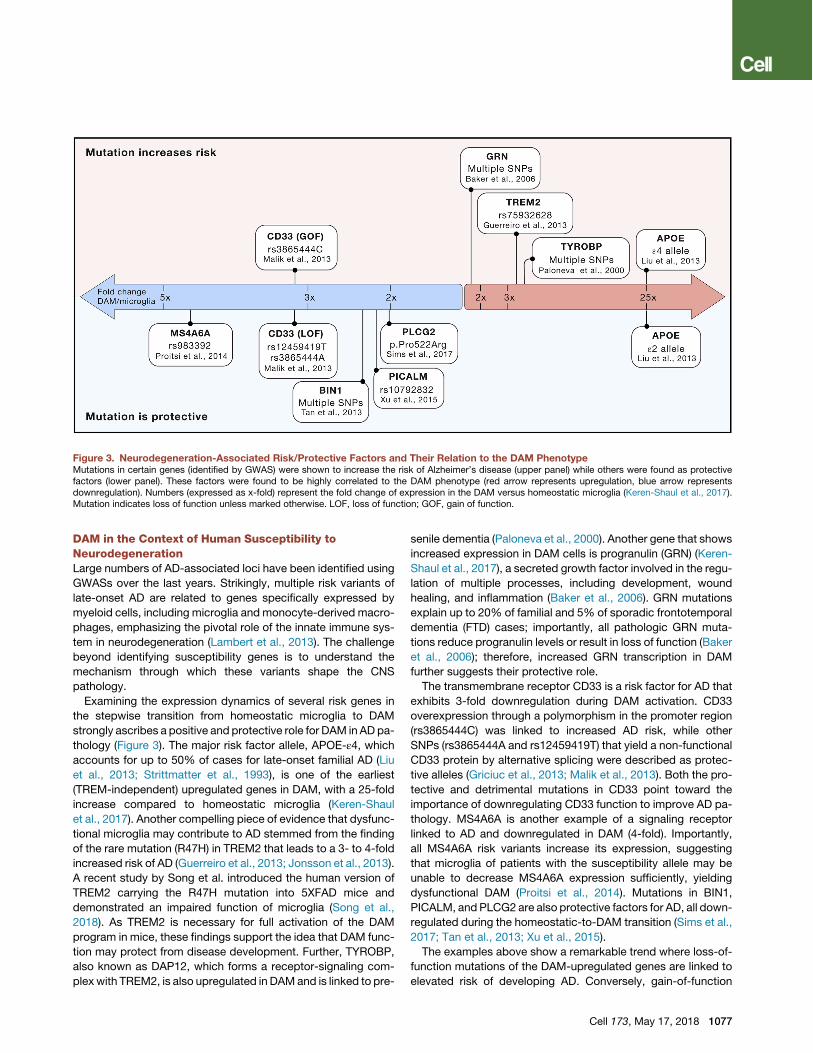
DAM in the Context of Human Susceptibility toNeurodegenerationLarge numbers of AD-associated loci have been identified usingGWASs over the last years. Strikingly, multiple risk variants oflate-onset AD are related to genes specifically expressed bymyeloid cells, including microglia and monocyte-derived macro-phages, emphasizing the pivotal role of the innate immune sys-tem in neurodegeneration (Lambert et al., 2013). The challengebeyond identifying susceptibility genes is to understand themechanism through which these variants shape the CNSpathology.Examining the expression dynamics of several risk genes in
the stepwise transition from homeostatic microglia to DAMstrongly ascribes a positive and protective role for DAM in AD pa-thology (Figure 3). The major risk factor allele, APOE-ε4, whichaccounts for up to 50% of cases for late-onset familial AD (Liuet al., 2013; Strittmatter et al., 1993), is one of the earliest(TREM-independent) upregulated genes in DAM, with a 25-foldincrease compared to homeostatic microglia (Keren-Shaulet al., 2017). Another compelling piece of evidence that dysfunc-tional microglia may contribute to AD stemmed from the findingof the rare mutation (R47H) in TREM2 that leads to a 3- to 4-foldincreased risk of AD (Guerreiro et al., 2013; Jonsson et al., 2013).A recent study by Song et al. introduced the human version ofTREM2 carrying the R47H mutation into 5XFAD mice anddemonstrated an impaired function of microglia (Song et al.,2018). As TREM2 is necessary for full activation of the DAMprogram in mice, these findings support the idea that DAM func-tion may protect from disease development. Further, TYROBP,also known as DAP12, which forms a receptor-signaling com-plex with TREM2, is also upregulated in DAMand is linked to pre-
senile dementia (Paloneva et al., 2000). Another gene that showsincreased expression in DAM cells is progranulin (GRN) (Keren-Shaul et al., 2017), a secreted growth factor involved in the regu-lation of multiple processes, including development, woundhealing, and inflammation (Baker et al., 2006). GRN mutationsexplain up to 20% of familial and 5% of sporadic frontotemporaldementia (FTD) cases; importantly, all pathologic GRN muta-tions reduce progranulin levels or result in loss of function (Bakeret al., 2006); therefore, increased GRN transcription in DAMfurther suggests their protective role.The transmembrane receptor CD33 is a risk factor for AD that
exhibits 3-fold downregulation during DAM activation. CD33overexpression through a polymorphism in the promoter region(rs3865444C) was linked to increased AD risk, while otherSNPs (rs3865444A and rs12459419T) that yield a non-functionalCD33 protein by alternative splicing were described as protec-tive alleles (Griciuc et al., 2013; Malik et al., 2013). Both the pro-tective and detrimental mutations in CD33 point toward theimportance of downregulating CD33 function to improve AD pa-thology. MS4A6A is another example of a signaling receptorlinked to AD and downregulated in DAM (4-fold). Importantly,all MS4A6A risk variants increase its expression, suggestingthat microglia of patients with the susceptibility allele may beunable to decrease MS4A6A expression sufficiently, yieldingdysfunctional DAM (Proitsi et al., 2014). Mutations in BIN1,PICALM, and PLCG2 are also protective factors for AD, all down-regulated during the homeostatic-to-DAM transition (Sims et al.,2017; Tan et al., 2013; Xu et al., 2015).The examples above show a remarkable trend where loss-of-
function mutations of the DAM-upregulated genes are linked toelevated risk of developing AD. Conversely, gain-of-function
Figure 3. Neurodegeneration-Associated Risk/Protective Factors and Their Relation to the DAM PhenotypeMutations in certain genes (identified by GWAS) were shown to increase the risk of Alzheimer’s disease (upper panel) while others were found as protectivefactors (lower panel). These factors were found to be highly correlated to the DAM phenotype (red arrow represents upregulation, blue arrow representsdownregulation). Numbers (expressed as x-fold) represent the fold change of expression in the DAM versus homeostatic microglia (Keren-Shaul et al., 2017).Mutation indicates loss of function unless marked otherwise. LOF, loss of function; GOF, gain of function.
Cell 173, May 17, 2018 1077
mutations in DAM-downregulated genes increase susceptibilityto AD. Taken together, accumulating evidence from human ge-netics studies provide strong evidence to propose that theDAM program is a protective innate immune response againstAD pathology.
Using mice deficient in genes involved in the DAM pheno-type, as well as in-depth analysis of human patients carryingmutations in DAM-related genes, will illuminate the role ofDAM in specific disease contexts. For example, CX3CR1deficiency alters microglial activation and reduces Ab deposi-tion in APP/PS1, R1.40, and CRND8 AD mouse models (Leeet al., 2010; Liu et al., 2010) but exacerbates the diseasepathology in a tau-driven AD model (Bhaskar et al., 2010).TREM2 deletion in P301S-tau model exacerbated the diseaseoutcome (Jiang et al., 2016), while studies of TREM2 defi-ciency in models of Ab pathology (5XFAD or APP/PS1) showedconflicting results, suggesting that the outcome of DAMpresence may depend on the disease stage (Jay et al.,2017). Recent study used a humanized TREM2 mouse modelcrossed to a 5XFAD mouse and showed that the risk factormutation R47H decreases microglial interaction with Abplaques in AD animals, but the study did not evaluate the ADpathology in terms of plaque burden or cognitive ability(Song et al., 2018).
DAM Biology and Therapeutics: Challenges andOpportunitiesAccumulating evidence shows that microglia have developeda set of sensory mechanisms to detect any deviation fromhomeostasis in the CNS. Similar to the resident macrophagesin other tissues, microglial response to danger-associatedtriggers, NAMPs, might be counterbalanced by co-occurringinhibitory signals. In principle, the DAM response arises as aprotective mechanism aiming to contain and remove theneuronal damage. The universal disease-associated subsetof microglia, found in various CNS conditions, seems to pro-vide a central piece in the neurodegenerative disease puzzle(Aguzzi et al., 2013); nevertheless, many questions remain tobe answered.
For example, microglial phagocytosis and complementcascade were linked to synaptic pruning, a physiological partof CNS development (Schafer et al., 2012; Stevens et al.,2007). Analysis of mouse models and human tissue suggeststhat in AD, this developmental mechanism is aberrantly andexcessively re-activated; microglia take up viable synapses,leading to memory loss (Hong et al., 2016). Microglia depletionusing CSF1R blockade in Ab models leads to an increasedplaque size but protects against synapse loss and rescuesbehavioral deficits (Dagher et al., 2015; Olmos-Alonso et al.,2016; Spangenberg et al., 2016). In addition, microglia deple-tion also prevented neuronal loss in animal models of ALSand tau propagation in a model of tau pathology (Asai et al.,2015; Martınez-Muriana et al., 2016). In all these cases, thetreatment led to ablation of all microglia without distinction be-tween the disease-associated and homeostatic; specific rolesof DAM in the process of synapse clearance and neuronalloss under neurodegenerative conditions need to be furtherexplored. As molecular markers best correlated with AD-asso-
ciated cognitive impairment are tau pathology and synapseloss (Jack et al., 2010), these questions must be thoroughlystudied before designing DAM-boosting strategies for AD.In addition, it has been shown that activation of microglia
through the NLRP3 inflammasome promotes Ab accumulation.The inflammasome is known to activate a unique inflammatorypathway that leads to release of IL-1b (Heneka et al., 2013)and the inflammasome component ASC, which forms large poly-meric protein complexes called ASC specks that directly asso-ciate with Ab plaque cores, facilitating plaque deposition (Vene-gas et al., 2017). These results suggest that microglial detectionof Ab may actually amplify Ab deposition. However, TREM2signaling was proposed to inhibit pro-inflammatory signaling(Hamerman et al., 2005; Turnbull et al., 2006). It is possible thatAb induces protective DAM early on during disease progression,whereas at late stages, when the accumulation of plaquesactivates the inflammasome inmicroglia, DAMbecome dysregu-lated and accelerate the disease. This hypothesis is supportedby a recent study on TREM2-deficient APP/PS1 mice, showinga disease stage-dependent effect of TREM2 deletion on Abdeposition and inflammatory conditions in the brain (Jayet al., 2017).In light of these observations, boosting DAM activity in early
disease stages, or prophylactically before the disease occurs atall, may present a promising strategy to fight any hallmark ofneurodegeneration as soon as it appears, preventing accumu-lation of the CNS damage (McDade and Bateman, 2017). Thelink between protective gene variants in AD and transcriptomicDAM signature (Figure 3) supports this direction of futureresearch. The effect of DAM presence in later stages of neuro-degeneration is much less understood, and this importantaspect cannot be elucidated without animal models moreaccurately reflecting human diseases. Further, as other factorslike gender, microbiome, and systemic exposure to immunesignals can affect microglia in steady state and were proposedto influence the risk of neurodegenerative disease occurrenceand progression, these are also likely to modulate DAM (Knue-sel et al., 2014; May, 2016; Quigley, 2017; Thion et al., 2018;Wendeln et al., 2018).AD is a heterogeneous disease in which multiple detrimental
factors contribute to cognitive loss and disease escalation.Therefore, targeting any single factor (e.g., Ab, tau, neuroinflam-mation etc.), even if successful, is not sufficient tomodify the dis-ease, and thus, there is a desperate need for new, more compre-hensive approaches (McDade andBateman, 2017). Boosting theintrinsic protective mechanism may constitute a more compre-hensive therapy that is independent of the disease etiology.Such a holistic approach has worked effectively in various typesof cancers (Pardoll, 2012; Sharma and Allison, 2015) and hascreated the basis for using immune checkpoint blockade in AD(Baruch et al., 2016).The discovery of DAM created an opportunity to develop a
therapy targeting the universal and intrinsic mechanism offighting against neuronal death shared across multiple neurode-generative conditions. Intensive research will be needed to bet-ter understand the DAM induction mechanisms and their effecton various aspects of CNS pathology to make effective use oftheir promising therapeutic potential.
1078 Cell 173, May 17, 2018

ACKNOWLEDGMENTS
We thank Tal Wiesel and Tal Bigdary for artwork. A.D. is supported by Steven
and Eden Romick. M.C. is supported by NIH RF1 AG05148501 and the Cure
Alzheimer’s Fund. M.S. is supported by the Advanced European Research
Council (ERC-2016-ADG 741744), Israel Science Foundation-Legacy Heritage
Biomedical Science Partnership research (1354/15), Israel Science Founda-
tion (991/16), Consolidated Anti-Aging Foundation Chicago (2016-2017), and
Adelis Foundation (2018-2021). M.S. holds the Maurice and Ilse Katz Profes-
sorial Chair in Neuroimmunology. I.A. is supported by the Chan Zuckerberg
Initiative (CZI); the HHMI International Scholar award; the European Research
Council Consolidator Grant (ERC-COG) 724471-HemTree2.0; an MRA
Established Investigator Award (509044); the Israel Science Foundation
(703/15); the Ernest and Bonnie Beutler Research Program of Excellence in
Genomic Medicine; the Helen and Martin Kimmel award for innovative inves-
tigation; a Minerva Stiftung research grant; the Israeli Ministry of Science,
Technology, and Space; the David and Fela Shapell Family Foundatio; the
NeuroMac DFG/Transregional Collaborative Research Center Grant; an Inter-
national Progressive MS Alliance/NMSS PA-1604-08459; an Adelis Founda-
tion grant; and the Abramson Family Center for Young Scientists. I.A. is the
incumbent of the Alan and Laraine Fischer Career Development Chair.
DECLARATION OF INTERESTS
A patent application has been filed related to this work.
REFERENCES
Abiega, O., Beccari, S., Diaz-Aparicio, I., Nadjar, A., Laye, S., Leyrolle, Q.,
Gomez-Nicola, D., Domercq, M., Perez-Samartın, A., Sanchez-Zafra, V.,
et al. (2016). Neuronal hyperactivity disturbs ATP microgradients, impairs mi-
croglial motility, and reduces phagocytic receptor expression triggering
apoptosis/microglial phagocytosis uncoupling. PLoS Biol. 14, e1002466.
Aguzzi, A., Barres, B.A., and Bennett, M.L. (2013). Microglia: scapegoat, sabo-
teur, or something else? Science 339, 156–161.
Ahyayauch, H., Raab, M., Busto, J.V., Andraka, N., Arrondo, J.L., Masserini,
M., Tvaroska, I., and Goni, F.M. (2012). Binding of b-amyloid (1-42) peptide
to negatively charged phospholipid membranes in the liquid-ordered state:
modeling and experimental studies. Biophys. J. 103, 453–463.
Ajami, B., Samusik, N., Wieghofer, P., Ho, P.P., Crotti, A., Bjornson, Z., Prinz,
M., Fantl, W.J., Nolan, G.P., and Steinman, L. (2018). Single-cell mass
cytometry reveals distinct populations of brain myeloid cells in mouse neuro-
inflammation and neurodegeneration models. Nat. Neurosci. 21, 541–551.
Asai, H., Ikezu, S., Tsunoda, S., Medalla, M., Luebke, J., Haydar, T., Wolozin,
B., Butovsky, O., Kugler, S., and Ikezu, T. (2015). Depletion of microglia and
inhibition of exosome synthesis halt tau propagation. Nat. Neurosci. 18,
1584–1593.
Baker, M., Mackenzie, I.R., Pickering-Brown, S.M., Gass, J., Rademakers, R.,
Lindholm, C., Snowden, J., Adamson, J., Sadovnick, A.D., Rollinson, S., et al.
(2006). Mutations in progranulin cause tau-negative frontotemporal dementia
linked to chromosome 17. Nature 442, 916–919.
Baruch, K., Deczkowska, A., Rosenzweig, N., Tsitsou-Kampeli, A., Sharif,
A.M., Matcovitch-Natan, O., Kertser, A., David, E., Amit, I., and Schwartz, M.
(2016). PD-1 immune checkpoint blockade reduces pathology and improves
memory in mouse models of Alzheimer’s disease. Nat. Med. 22, 135–137.
Bhaskar, K., Konerth, M., Kokiko-Cochran, O.N., Cardona, A., Ransohoff,
R.M., and Lamb, B.T. (2010). Regulation of tau pathology by the microglial
fractalkine receptor. Neuron 68, 19–31.
Biber, K., Neumann, H., Inoue, K., and Boddeke, H.W. (2007). Neuronal ‘On’
and ‘Off’ signals control microglia. Trends Neurosci. 30, 596–602.
Butovsky, O., Jedrychowski, M.P., Moore, C.S., Cialic, R., Lanser, A.J., Ga-
briely, G., Koeglsperger, T., Dake, B., Wu, P.M., Doykan, C.E., et al. (2014).
Identification of a unique TGF-b-dependent molecular and functional signature
in microglia. Nat. Neurosci. 17, 131–143.
Chiu, I.M., Morimoto, E.T., Goodarzi, H., Liao, J.T., O’Keeffe, S., Phatnani,
H.P., Muratet, M., Carroll, M.C., Levy, S., Tavazoie, S., et al. (2013). A neuro-
degeneration-specific gene-expression signature of acutely isolated microglia
from an amyotrophic lateral sclerosis mouse model. Cell Rep. 4, 385–401.
Crotti, A., and Ransohoff, R.M. (2016). Microglial physiology and pathophysi-
ology: insights from genome-wide transcriptional profiling. Immunity 44,
505–515.
Dagher, N.N., Najafi, A.R., Kayala, K.M.N., Elmore, M.R., White, T.E., Me-
deiros, R., West, B.L., and Green, K.N. (2015). Colony-stimulating factor 1
receptor inhibition prevents microglial plaque association and improves cogni-
tion in 3xTg-AD mice. J. Neuroinflammation 12, 139.
Davalos, D., Grutzendler, J., Yang, G., Kim, J.V., Zuo, Y., Jung, S., Littman,
D.R., Dustin, M.L., and Gan, W.-B. (2005). ATP mediates rapid microglial
response to local brain injury in vivo. Nat. Neurosci. 8, 752–758.
Deczkowska, A., Amit, I., and Schwartz, M. (2018). Microglial immune check-
point mechanisms. Nat. Neurosci. Published online May 7, 2018. https://doi.
org/10.1038/s41593-018-0145-x.
Deczkowska, A., Matcovitch-Natan, O., Tsitsou-Kampeli, A., Ben-Hamo, S.,
Dvir-Szternfeld, R., Spinrad, A., Singer, O., David, E., Winter, D.R., Smith,
L.K., et al. (2017). Mef2C restrains microglial inflammatory response and is
lost in brain ageing in an IFN-I-dependent manner. Nat. Commun. 8, 717.
Fourgeaud, L., Traves, P.G., Tufail, Y., Leal-Bailey, H., Lew, E.D., Burrola, P.G.,
Callaway, P., Zagorska, A., Rothlin, C.V., Nimmerjahn, A., and Lemke, G.
(2016). TAM receptors regulate multiple features of microglial physiology. Na-
ture 532, 240–244.
Friedman, B.A., Srinivasan, K., Ayalon, G., Meilandt, W.J., Lin, H., Huntley,
M.A., Cao, Y., Lee, S.H., Haddick, P.C.G., Ngu, H., et al. (2018). Diverse Brain
Myeloid Expression Profiles Reveal Distinct Microglial Activation States and
Aspects of Alzheimer’s Disease Not Evident in Mouse Models. Cell Rep. 22,
832–847.
Galatro, T.F., Holtman, I.R., Lerario, A.M., Vainchtein, I.D., Brouwer, N., Sola,
P.R., Veras, M.M., Pereira, T.F., Leite, R.E.P., Moller, T., et al. (2017). Tran-
scriptomic analysis of purified human cortical microglia reveals age-associ-
ated changes. Nat. Neurosci. 20, 1162–1171.
Griciuc, A., Serrano-Pozo, A., Parrado, A.R., Lesinski, A.N., Asselin, C.N., Mul-
lin, K., Hooli, B., Choi, S.H., Hyman, B.T., and Tanzi, R.E. (2013). Alzheimer’s
disease risk gene CD33 inhibits microglial uptake of amyloid beta. Neuron
78, 631–643.
Guerreiro, R., Wojtas, A., Bras, J., Carrasquillo, M., Rogaeva, E., Majounie, E.,
Cruchaga, C., Sassi, C., Kauwe, J.S., Younkin, S., et al.; Alzheimer Genetic
Analysis Group (2013). TREM2 variants in Alzheimer’s disease. N. Engl. J.
Med. 368, 117–127.
Hamerman, J.A., Tchao, N.K., Lowell, C.A., and Lanier, L.L. (2005). Enhanced
Toll-like receptor responses in the absence of signaling adaptor DAP12. Nat.
Immunol. 6, 579–586.
Heneka, M.T., Kummer, M.P., Stutz, A., Delekate, A., Schwartz, S., Vieira-
Saecker, A., Griep, A., Axt, D., Remus, A., Tzeng, T.-C., et al. (2013). NLRP3
is activated in Alzheimer’s disease and contributes to pathology in APP/PS1
mice. Nature 493, 674–678.
Heneka, M.T., Carson, M.J., El Khoury, J., Landreth, G.E., Brosseron, F.,
Feinstein, D.L., Jacobs, A.H., Wyss-Coray, T., Vitorica, J., Ransohoff, R.M.,
et al. (2015). Neuroinflammation in Alzheimer’s disease. Lancet Neurol. 14,
388–405.
Holtman, I.R., Raj, D.D., Miller, J.A., Schaafsma, W., Yin, Z., Brouwer, N., Wes,
P.D., Moller, T., Orre, M., Kamphuis, W., et al. (2015). Induction of a common
microglia gene expression signature by aging and neurodegenerative condi-
tions: a co-expression meta-analysis. Acta Neuropathol. Commun. 3, 31.
Hong, S., Beja-Glasser, V.F., Nfonoyim, B.M., Frouin, A., Li, S., Ramakrishnan,
S., Merry, K.M., Shi, Q., Rosenthal, A., Barres, B.A., et al. (2016). Complement
andmicroglia mediate early synapse loss in Alzheimermousemodels. Science
352, 712–716.
Jack, C.R., Jr., Knopman, D.S., Jagust, W.J., Shaw, L.M., Aisen, P.S., Weiner,
M.W., Petersen, R.C., and Trojanowski, J.Q. (2010). Hypothetical model of
Cell 173, May 17, 2018 1079

dynamic biomarkers of the Alzheimer’s pathological cascade. Lancet Neurol.
9, 119–128.
Janeway, C.A., Jr. (1989). Approaching the asymptote? Evolution and revolu-
tion in immunology. Cold Spring Harb. Symp. Quant. Biol 54, 1–13.
Jay, T.R., Hirsch, A.M., Broihier, M.L., Miller, C.M., Neilson, L.E., Ransohoff,
R.M., Lamb, B.T., and Landreth, G.E. (2017). Disease progression-dependent
effects of TREM2 deficiency in a mouse model of Alzheimer’s disease.
J. Neurosci. 37, 637–647.
Jiang, T., Zhang, Y.-D., Chen, Q., Gao, Q., Zhu, X.-C., Zhou, J.-S., Shi, J.-Q.,
Lu, H., Tan, L., and Yu, J.-T. (2016). TREM2modifies microglial phenotype and
provides neuroprotection in P301S tau transgenic mice. Neuropharmacology
105, 196–206.
Jonsson, T., Stefansson, H., Steinberg, S., Jonsdottir, I., Jonsson, P.V., Snae-
dal, J., Bjornsson, S., Huttenlocher, J., Levey, A.I., Lah, J.J., et al. (2013).
Variant of TREM2 associated with the risk of Alzheimer’s disease. N. Engl. J.
Med. 368, 107–116.
Kamphuis, W., Kooijman, L., Schetters, S., Orre, M., and Hol, E.M. (2016).
Transcriptional profiling of CD11c-positivemicroglia accumulating around am-
yloid plaques in a mouse model for Alzheimer’s disease. Biochim. Biophys.
Acta 1862, 1847–1860.
Keren-Shaul, H., Spinrad, A., Weiner, A., Matcovitch-Natan, O., Dvir-Sztern-
feld, R., Ulland, T.K., David, E., Baruch, K., Lara-Astaiso, D., Toth, B., et al.
(2017). A Unique Microglia Type Associated with Restricting Development of
Alzheimer’s Disease. Cell 169, 1276–1290.
Knuesel, I., Chicha, L., Britschgi, M., Schobel, S.A., Bodmer, M., Hellings, J.A.,
Toovey, S., and Prinssen, E.P. (2014). Maternal immune activation and
abnormal brain development across CNS disorders. Nat. Rev. Neurol. 10,
643–660.
Krasemann, S., Madore, C., Cialic, R., Baufeld, C., Calcagno, N., El Fatimy, R.,
Beckers, L., O’Loughlin, E., Xu, Y., Fanek, Z., et al. (2017). The TREM2-APOE
Pathway Drives the Transcriptional Phenotype of Dysfunctional Microglia in
Neurodegenerative Diseases. Immunity 47, 566–581.
Lambert, J.C., Ibrahim-Verbaas, C.A., Harold, D., Naj, A.C., Sims, R., Bellen-
guez, C., DeStafano, A.L., Bis, J.C., Beecham, G.W., Grenier-Boley, B.,
et al.; European Alzheimer’s Disease Initiative (EADI); Genetic and Environ-
mental Risk in Alzheimer’s Disease; Alzheimer’s Disease Genetic Consortium;
Cohorts for Heart and Aging Research in Genomic Epidemiology (2013).
Meta-analysis of 74,046 individuals identifies 11 new susceptibility loci for
Alzheimer’s disease. Nat. Genet. 45, 1452–1458.
Lee, M. (2013). Neurotransmitters and microglial-mediated neuroinflamma-
tion. Curr. Protein Pept. Sci. 14, 21–32.
Lee, S., Varvel, N.H., Konerth, M.E., Xu, G., Cardona, A.E., Ransohoff, R.M.,
and Lamb, B.T. (2010). CX3CR1 deficiency alters microglial activation and re-
duces beta-amyloid deposition in two Alzheimer’s disease mouse models.
Am. J. Pathol. 177, 2549–2562.
Lee, C.Y.D., Daggett, A., Gu, X., Jiang, L.L., Langfelder, P., Li, X., Wang, N.,
Zhao, Y., Park, C.S., Cooper, Y., et al. (2018). Elevated TREM2 Gene Dosage
ReprogramsMicroglia Responsivity and Ameliorates Pathological Phenotypes
in Alzheimer’s Disease Models. Neuron 97, 1032–1048.
Lemaitre, B., Nicolas, E., Michaut, L., Reichhart, J.-M., and Hoffmann, J.A.
(1996). The dorsoventral regulatory gene cassette spatzle/Toll/cactus controls
the potent antifungal response in Drosophila adults. Cell 86, 973–983.
Lemke, G. (2017). Phosphatidylserine is the signal for TAM receptors and their
ligands. Trends Biochem. Sci. 42, 738–748.
Leyns, C.E.G., Ulrich, J.D., Finn, M.B., Stewart, F.R., Koscal, L.J., Remolina
Serrano, J., Robinson, G.O., Anderson, E., Colonna, M., and Holtzman, D.M.
(2017). TREM2 deficiency attenuates neuroinflammation and protects against
neurodegeneration in a mousemodel of tauopathy. Proc. Natl. Acad. Sci. USA
114, 11524–11529.
Liu, Z., Condello, C., Schain, A., Harb, R., and Grutzendler, J. (2010). CX3CR1
in microglia regulates brain amyloid deposition through selective protofibrillar
amyloid-b phagocytosis. J. Neurosci. 30, 17091–17101.
Liu, C.-C., Liu, C.C., Kanekiyo, T., Xu, H., and Bu, G. (2013). Apolipoprotein E
and Alzheimer disease: risk, mechanisms and therapy. Nat. Rev. Neurol. 9,
106–118.
Malik, M., Simpson, J.F., Parikh, I., Wilfred, B.R., Fardo, D.W., Nelson, P.T.,
and Estus, S. (2013). CD33 Alzheimer’s risk-altering polymorphism, CD33
expression, and exon 2 splicing. J. Neurosci. 33, 13320–13325.
Martınez-Muriana, A., Mancuso, R., Francos-Quijorna, I., Olmos-Alonso, A.,
Osta, R., Perry, V.H., Navarro, X., Gomez-Nicola, D., and Lopez-Vales, R.
(2016). CSF1R blockade slows the progression of amyotrophic lateral scle-
rosis by reducing microgliosis and invasion of macrophages into peripheral
nerves. Sci. Rep. 6, 25663.
Mathys, H., Adaikkan, C., Gao, F., Young, J.Z., Manet, E., Hemberg, M., De
Jager, P.L., Ransohoff, R.M., Regev, A., and Tsai, L.H. (2017). Temporal
Tracking of Microglia Activation in Neurodegeneration at Single-Cell Resolu-
tion. Cell Rep. 21, 366–380.
Matzinger, P. (1994). Tolerance, danger, and the extended family. Annu. Rev.
Immunol. 12, 991–1045.
Matzinger, P., and Kamala, T. (2011). Tissue-based class control: the other
side of tolerance. Nat. Rev. Immunol. 11, 221–230.
May, M. (2016). Sex on the brain: unraveling the differences between women
and men in neurodegenerative disease. Nat. Med. 22, 1370–1372.
McDade, E., and Bateman, R.J. (2017). Stop Alzheimer’s before it starts. Na-
ture 547, 153–155.
Mosher, K.I., and Wyss-Coray, T. (2014). Microglial dysfunction in brain aging
and Alzheimer’s disease. Biochem. Pharmacol. 88, 594–604.
Mrdjen, D., Pavlovic, A., Hartmann, F.J., Schreiner, B., Utz, S.G., Leung, B.P.,
Lelios, I., Heppner, F.L., Kipnis, J., and Merkler, D. (2018). High-dimensional
single-cell mapping of central nervous system immune cells reveals distinct
myeloid subsets in health, aging, and disease. Immunity 48, 380–395.
Nagarathinam, A., Hoflinger, P., Buhler, A., Schafer, C., McGovern, G., Jeffrey,
M., Staufenbiel, M., Jucker, M., and Baumann, F. (2013). Membrane-anchored
Ab accelerates amyloid formation and exacerbates amyloid-associated
toxicity in mice. J. Neurosci. 33, 19284–19294.
Niraula, A., Sheridan, J.F., and Godbout, J.P. (2017). Microglia priming with
aging and stress. Neuropsychopharmacology 42, 318–333.
Ofengeim, D., Mazzitelli, S., Ito, Y., DeWitt, J.P., Mifflin, L., Zou, C., Das, S.,
Adiconis, X., Chen, H., and Zhu, H. (2017). RIPK1 mediates a disease-associ-
ated microglial response in Alzheimer’s disease. Proc. Natl. Acad. Sci. U.S.A.
114, E8788–E8797.
Olah, M., Patrick, E., Villani, A.C., Xu, J., White, C.C., Ryan, K.J., Piehowski, P.,
Kapasi, A., Nejad, P., Cimpean, M., et al. (2018). A transcriptomic atlas of aged
human microglia. Nat. Commun. 9, 539.
Olmos-Alonso, A., Schetters, S.T., Sri, S., Askew, K., Mancuso, R., Vargas-
Caballero, M., Holscher, C., Perry, V.H., and Gomez-Nicola, D. (2016). Phar-
macological targeting of CSF1R inhibits microglial proliferation and prevents
the progression of Alzheimer’s-like pathology. Brain 139, 891–907.
Otero, K., Turnbull, I.R., Poliani, P.L., Vermi, W., Cerutti, E., Aoshi, T., Tassi, I.,
Takai, T., Stanley, S.L., Miller, M., et al. (2009). Macrophage colony-stimulating
factor induces the proliferation and survival of macrophages via a pathway
involving DAP12 and b-catenin. Nat. Immunol. 10, 734–743.
Paloneva, J., Kestila, M., Wu, J., Salminen, A., Bohling, T., Ruotsalainen, V.,
Hakola, P., Bakker, A.B., Phillips, J.H., Pekkarinen, P., et al. (2000). Loss-of-
function mutations in TYROBP (DAP12) result in a presenile dementia with
bone cysts. Nat. Genet. 25, 357–361.
Pardoll, D.M. (2012). The blockade of immune checkpoints in cancer immuno-
therapy. Nat. Rev. Cancer 12, 252–264.
Peng, Q., Malhotra, S., Torchia, J.A., Kerr, W.G., Coggeshall, K.M., and Hum-
phrey, M.B. (2010). TREM2- and DAP12-dependent activation of PI3K requires
DAP10 and is inhibited by SHIP1. Sci. Signal. 3, ra38.
Perry, V.H., and Holmes, C. (2014). Microglial priming in neurodegenerative
disease. Nat. Rev. Neurol. 10, 217–224.
1080 Cell 173, May 17, 2018

Pocock, J.M., and Kettenmann, H. (2007). Neurotransmitter receptors on mi-
croglia. Trends Neurosci. 30, 527–535.
Poliani, P.L.,Wang, Y., Fontana, E., Robinette, M.L., Yamanishi, Y., Gilfillan, S.,
and Colonna, M. (2015). TREM2 sustains microglial expansion during aging
and response to demyelination. J. Clin. Invest. 125, 2161–2170.
Poltorak, A., He, X., Smirnova, I., Liu, M.-Y., Van Huffel, C., Du, X., Birdwell, D.,
Alejos, E., Silva, M., Galanos, C., et al. (1998). Defective LPS signaling in
C3H/HeJ and C57BL/10ScCr mice: mutations in Tlr4 gene. Science 282,
2085–2088.
Proitsi, P., Lee, S.H., Lunnon, K., Keohane, A., Powell, J., Troakes, C., Al-Sar-
raj, S., Furney, S., Soininen, H., K1oszewska, I., et al.; AddNeuroMed Con-
sortium (2014). Alzheimer’s disease susceptibility variants in the MS4A6A
gene are associated with altered levels of MS4A6A expression in blood. Neu-
robiol. Aging 35, 279–290.
Quigley, E.M.M. (2017). Microbiota-brain-gut axis and neurodegenerative dis-
eases. Curr. Neurol. Neurosci. Rep. 17, 94.
Rodrigues, R.J., Tome, A.R., and Cunha, R.A. (2015). ATP as a multi-target
danger signal in the brain. Front. Neurosci. 9, 148.
Safaiyan, S., Kannaiyan, N., Snaidero, N., Brioschi, S., Biber, K., Yona, S., Ed-
inger, A.L., Jung, S., Rossner, M.J., and Simons, M. (2016). Age-related myelin
degradation burdens the clearance function of microglia during aging. Nat.
Neurosci. 19, 995–998.
Schafer, D.P., Lehrman, E.K., Kautzman, A.G., Koyama, R., Mardinly, A.R., Ya-
masaki, R., Ransohoff, R.M., Greenberg, M.E., Barres, B.A., and Stevens, B.
(2012). Microglia sculpt postnatal neural circuits in an activity and comple-
ment-dependent manner. Neuron 74, 691–705.
Sharma, P., and Allison, J.P. (2015). Immune checkpoint targeting in cancer
therapy: toward combination strategies with curative potential. Cell 161,
205–214.
Sims, R., van der Lee, S.J., Naj, A.C., Bellenguez, C., Badarinarayan, N., Ja-
kobsdottir, J., Kunkle, B.W., Boland, A., Raybould, R., Bis, J.C., et al.; ARUK
Consortium; GERAD/PERADES, CHARGE, ADGC, EADI (2017). Rare coding
variants in PLCG2, ABI3, and TREM2 implicate microglial-mediated innate
immunity in Alzheimer’s disease. Nat. Genet. 49, 1373–1384.
Song, W., Hooli, B., Mullin, K., Jin, S.C., Cella, M., Ulland, T.K., Wang, Y.,
Tanzi, R.E., and Colonna, M. (2017). Alzheimer’s disease-associated TREM2
variants exhibit either decreased or increased ligand-dependent activation.
Alzheimers Dement 13, 381–387.
Song, W.M., Joshita, S., Zhou, Y., Ulland, T.K., Gilfillan, S., and Colonna, M.
(2018). Humanized TREM2mice reveal microglia-intrinsic and-extrinsic effects
of R47H polymorphism. J. Exp. Med. 215, 745–760.
Spangenberg, E.E., Lee, R.J., Najafi, A.R., Rice, R.A., Elmore, M.R., Blurton-
Jones, M., West, B.L., and Green, K.N. (2016). Eliminating microglia in
Alzheimer’s mice prevents neuronal loss without modulating amyloid-b pathol-
ogy. Brain 139, 1265–1281.
Spiller, K.J., Restrepo, C.R., Khan, T., Dominique, M.A., Fang, T.C., Canter,
R.G., Roberts, C.J., Miller, K.R., Ransohoff, R.M., Trojanowski, J.Q., and
Lee, V.M. (2018). Microglia-mediated recovery from ALS-relevant motor
neuron degeneration in a mouse model of TDP-43 proteinopathy. Nat. Neuro-
sci. 21, 329–340.
Stevens, B., Allen, N.J., Vazquez, L.E., Howell, G.R., Christopherson, K.S.,
Nouri, N., Micheva, K.D., Mehalow, A.K., Huberman, A.D., Stafford, B., et al.
(2007). The classical complement cascade mediates CNS synapse elimina-
tion. Cell 131, 1164–1178.
Strittmatter, W.J., Saunders, A.M., Schmechel, D., Pericak-Vance, M., En-
ghild, J., Salvesen, G.S., and Roses, A.D. (1993). Apolipoprotein E: high-avidity
binding to beta-amyloid and increased frequency of type 4 allele in late-onset
familial Alzheimer disease. Proc. Natl. Acad. Sci. USA 90, 1977–1981.
Tan, M.S., Yu, J.T., and Tan, L. (2013). Bridging integrator 1 (BIN1): form, func-
tion, and Alzheimer’s disease. Trends Mol. Med. 19, 594–603.
Terwel, D., Steffensen, K.R., Verghese, P.B., Kummer, M.P., Gustafsson,
J.-A., Holtzman, D.M., and Heneka, M.T. (2011). Critical role of astroglial apoli-
poprotein E and liver X receptor-a expression for microglial Ab phagocytosis.
J. Neurosci. 31, 7049–7059.
Thion, M.S., Low, D., Silvin, A., Chen, J., Grisel, P., Schulte-Schrepping, J.,
Blecher, R., Ulas, T., Squarzoni, P., and Hoeffel, G. (2018). Microbiome Influ-
ences Prenatal and Adult Microglia in a Sex-Specific Manner. Cell 172,
500–516.
Turnbull, I.R., Gilfillan, S., Cella, M., Aoshi, T., Miller, M., Piccio, L., Hernandez,
M., and Colonna, M. (2006). Cutting edge: TREM-2 attenuates macrophage
activation. J. Immunol. 177, 3520–3524.
Ulland, T.K., Song, W.M., Huang, S.C.-C., Ulrich, J.D., Sergushichev, A.,
Beatty, W.L., Loboda, A.A., Zhou, Y., Cairns, N.J., and Kambal, A. (2017).
TREM2 maintains microglial metabolic fitness in Alzheimer’s disease. Cell
170, 649–663. e613.
Venegas, C., Kumar, S., Franklin, B.S., Dierkes, T., Brinkschulte, R., Tejera, D.,
Vieira-Saecker, A., Schwartz, S., Santarelli, F., Kummer, M.P., et al. (2017).
Microglia-derived ASC specks cross-seed amyloid-b in Alzheimer’s disease.
Nature 552, 355–361.
Wang, Y., Cella, M., Mallinson, K., Ulrich, J.D., Young, K.L., Robinette, M.L.,
Gilfillan, S., Krishnan, G.M., Sudhakar, S., Zinselmeyer, B.H., et al. (2015).
TREM2 lipid sensing sustains the microglial response in an Alzheimer’s dis-
ease model. Cell 160, 1061–1071.
Wendeln, A.-C., Degenhardt, K., Kaurani, L., Gertig, M., Ulas, T., Jain, G.,
Wagner, J., Hasler, L.M., Wild, K., Skodras, A., et al. (2018). Innate immune
memory in the brain shapes neurological disease hallmarks. Nature 556,
332–338.
Wolf, Y., Yona, S., Kim, K.-W., and Jung, S. (2013). Microglia, seen from the
CX3CR1 angle. Front. Cell. Neurosci. 7, 26.
Xu, W., Tan, L., and Yu, J.-T. (2015). The role of PICALM in Alzheimer’s dis-
ease. Mol. Neurobiol. 52, 399–413.
Yeh, F.L., Wang, Y., Tom, I., Gonzalez, L.C., and Sheng, M. (2016). TREM2
binds to apolipoproteins, including APOE and CLU/APOJ, and thereby facili-
tates uptake of amyloid-beta by microglia. Neuron 91, 328–340.
Yeh, F.L., Hansen, D.V., and Sheng, M. (2017). TREM2, Microglia, and Neuro-
degenerative Diseases. Trends Mol. Med 23, 512–533.
Zhao, Y., Wu, X., Li, X., Jiang, L.-L., Gui, X., Liu, Y., Sun, Y., Zhu, B., Pina-
Crespo, J.C., and Zhang, M. (2018). TREM2 is a receptor for b-amyloid that
mediates microglial function. Neuron 97, 1023–1031.
Zhong, L., Wang, Z., Wang, D., Wang, Z., Martens, Y.A., Wu, L., Xu, Y., Wang,
K., Li, J., Huang, R., et al. (2018). Amyloid-beta modulates microglial re-
sponses by binding to the triggering receptor expressed on myeloid cells 2
(TREM2). Mol. Neurodegener. 13, 15.
Cell 173, May 17, 2018 1081

![Physics Department Sites - Shaul Hanany Eli Zeldov[3]phsites.technion.ac.il/.../Tom-Leviant-PHpresentation.pdf · 2018. 5. 9. · Tom Leviant[1] Amit Keren[1] Shaul Hanany[2] Eli](https://img.dokumen.tips/doc/110x75/60b6a9834e63a47f3f648aa5/physics-department-sites-shaul-hanany-eli-zeldov3-2018-5-9-tom-leviant1.jpg)