Embed Size (px)
Citation preview
J A C C : C A R D I O V A S C U L A R I M A G I N G V O L . 6 , N O . 5 , 2 0 1 3
© 2 0 1 3 B Y T H E A M E R I C A N C O L L E G E O F C A R D I O L O G Y F O U N D A T I O N I S S N 1 9 3 6 - 8 7 8 X / $ 3 6 . 0 0
P U B L I S H E D B Y E L S E V I E R I N C . h t t p : / / d x . d o i . o r g / 1 0 . 1 0 1 6 / j . j c m g . 2 0 1 3 . 0 1 . 0 0 9
O R I G I N A L R E S E A R C H
Layer-Specific Quantification of MyocardialDeformation by Strain EchocardiographyMay Reveal Significant CAD in Patients WithNon–ST-Segment Elevation AcuteCoronary SyndromeSebastian I. Sarvari, MD,*†‡ Kristina H. Haugaa, MD, PHD,*†‡ Wasim Zahid, MD,*†‡Bjørn Bendz, MD, PHD,* Svend Aakhus, MD, PHD,* Lars Aaberge, MD, PHD,*Thor Edvardsen, MD, PHD*†‡
Oslo, Norway
O B J E C T I V E S Our objective was to assess whether patients with significant coronary artery disease
(CAD) had reduced endocardial function assessed by layer-specific strain compared with patients
without significant CAD.
B A C K G R O U N D The left ventricular (LV) wall of the heart comprises 3 myocardial layers. The
endocardial layer is most susceptible to ischemic injury.
M E T H O D S Seventy-seven patients referred to coronary angiography due to suspected non–ST-
segment elevation-acute coronary syndromes (NSTE-ACS) were prospectively included. Coronary
occlusion was found in 28, significant stenosis in 21, and no stenosis in 28 patients. Echocardiography
was performed 1 to 2 h before angiography. Layer-specific longitudinal and circumferential strains were
assessed from endocardium, mid-myocardium, and epicardium by 2-dimensional (2D) speckle-tracking
echocardiography (STE). Territorial longitudinal strain (TLS) was calculated based on the perfusion
territories of the 3 major coronary arteries in a 16-segment LV model, whereas global circumferential
strain (GCS) was averaged from 6 circumferential LV segments in all 3 layers.
R E S U L T S Patients with significant CAD hadworse function in all 3 myocardial layers assessed by TLS and
GCS compared with patients without significant CAD. Endocardial TLS (mean –14.0 � 3.3% vs. –19.2 � 2.2%;
p � 0.001) and GCS (mean –19.3 � 4.0% vs. –24.3 � 3.4%; p � 0.001) were most affected. The absolute
differences between endocardial and epicardial TLS and GCS were lower in patients with significant CAD
(∆2.4 � 3.6% and ∆6.7 � 3.8%, respectively) than in those without significant CAD (∆5.3 � 2.1% and ∆10.4 �
3.0%; p � 0.001). This reflects a pronounced decrease in endocardial function in patients with significant
CAD. A receiver-operating characteristic curve analysis showed that endocardial and mid-myocardial TLS
were superior to identify significant CAD compared with epicardial TLS (p � 0.05), wall motion score
index (p � 0.01), and ejection fraction (EF) (p � 0.001).
C O N C L U S I O N S Assessment of layer-specific strain by 2D-STE might identify NSTE-ACS patients
with significant CAD. Endocardial function was more affected in patients with significant CAD compared
with epicardial function and EF. (J Am Coll Cardiol Img 2013;6:535–44) © 2013 by the American College
of Cardiology Foundation
S(ieqpeiau(nth
r(sitcL
ud
ds
A
A
c
G
longitudinal strain
J A C C : C A R D I O V A S C U L A R I M A G I N G , V O L . 6 , N O . 5 , 2 0 1 3
M A Y 2 0 1 3 : 5 3 5 – 4 4
Sarvari et al.
Layer-Specific Strain and CAD
536
The clinical presentation of coronary artery
disease (CAD) varies from silent ischemia,stable angina pectoris to acute coronary syn-dromes (ACS) and death. ACSs comprise non-
T–segment elevation-acute coronary syndromeNSTE-ACS) and ST-segment elevation myocardialnfarction (STEMI). The presence of ST-segmentlevation typically represents coronary occlusion re-uiring acute reperfusion therapy. On the other hand,atients with suspected NSTE-ACS are a more het-rogeneous group. Coronary occlusion and/or signif-cant stenosis may or may not be present, and coronaryngiography and revascularization therapy might bennecessary in as many as one-third of these patients1). Therefore, a better selection of patients with realeed for coronary angiography and revascularizationherapy could reduce both complication rates andealthcare costs associated with this procedure.
The left ventricular (LV) wall of theheart comprises an endocardial, a mid-myocardial, and an epicardial layer. Ofthese 3 layers, the endocardium is mostsusceptible to ischemic injury (2–5). Care-ful evaluation of this layer might increasethe diagnostic accuracy of CAD.
Two-dimensional (2D) speckle-trackingechocardiography (STE) is a semi-automatedquantitative technique for assessment ofstrain, a measure of cardiac function basedon grayscale images. Strain is a measureof deformation, an intrinsic mechanicalproperty, and measures myocardial systolicfunction more directly compared withconventional cavity-based echocardio-graphic measures (6–8). Strain echocardi-ography has been introduced as an accu-
ate tool for the assessment of global and regional9) LV myocardial function and has been demon-trated to be more sensitive and accurate in thedentification of coronary artery occlusion in pa-ients with NSTE-ACS (10,11) compared withonventional echocardiographic measures of systolicV function. Recent software allows separate eval-
From the *Department of Cardiology, Oslo University Hospital, Rikshos-pitalet, Oslo, Norway; †Institute for Surgical Research, Oslo UniversityHospital, Rikshospitalet, Oslo, Norway; and the ‡University of Oslo,Oslo, Norway. This work was supported by the South-Eastern NorwayRegional Health Authority, the Norwegian Research Council, and the Ingerand John Fredriksen Foundation. Dr. Sarvari has received honoraria fromToshiba for a lecture at one occasion. All other authors have reported thatthey have no relationships relevant to the contents of this paper to disclose.
se
rain
nt
Manuscript received October 18, 2012; revised manuscript receivedDecember 18, 2012, accepted January 23, 2013.
ation of endocardial, mid-myocardial, and epicar-ial myocardial deformation.The aim of this study was to evaluate myocardial
eformation in 3 myocardial layers in patients withuspected NSTE-ACS.
M E T H O D S
This study was conducted in a single tertiary coro-nary care center. Seventy-seven patients with sus-pected NSTE-ACS (12,13) referred to our hospitalfor coronary angiography were prospectively in-cluded in the study. Exclusion criteria were: age�18 years and/or a history of previous myocardialinfarction, percutaneous coronary intervention andopen chest surgery, left bundle branch block, severevalvular dysfunction, atrial fibrillation with heartrate �100 beats/min, sustained severe arrhythmia,and/or any condition that might interfere with thepatient’s ability to comply. Electrocardiograms(ECGs) were evaluated by experienced cardiologistsat admission. ECGs were described as ischemic if STdepression or T-wave changes were present. Allpatients received medical treatment according tocurrent guidelines. Echocardiography was per-formed 1 to 2 h prior to coronary angiography andwithin 48 h after the last episode of chest pain. Theechocardiographic data were analyzed blinded to allclinical information.
Written informed consent was given by all studyparticipants. The study complied with the Declarationof Helsinki and was approved by the Regional Com-mittees for Medical and Health Research Ethics.2D Echocardiography. Echocardiographic studieswere performed with commercially available system(Artida, Toshiba Medical Systems Corporation, To-kyo, Japan). Routine grayscale 2D cine loops from 3consecutive beats were obtained at end-expiratoryapnea from standard parasternal short-axis view ofthe LV at the level of the papillary muscle and fromthe 3 apical views (4-chamber, 2-chamber, andlong-axis). Frame rates were 47 � 5 Hz for gray-scale imaging. LV volume and ejection fraction(EF) were assessed by biplane Simpson methodusing manual tracing of digital images.WMSI. Wall motion was visually assessed in a 16-segment model, according to the American Societyof Echocardiography (14), by an experienced ob-server. The observer evaluated image quality, andsegments were discarded if the quality was foundinsufficient for analysis. Wall motion score index(WMSI) was calculated for each patient as the
B B R E V I A T I O N S
N D A C R O N YM S
2D � 2-dimensional
CAD � coronary artery disea
EF � ejection fraction
GCS � global
ircumferential strain
LS � global longitudinal st
LV � left ventricular
NSTE-ACS � non–ST-segme
elevation-acute coronary
syndrome(s)
STE � speckle-tracking
echocardiography
TLS � territorial
average of the analyzed segmental values.
dbmefaacsgwMlAgsSas
lm(v
n
fmaoMn�cmaTw
StvwMd(Cptcocc
J A C C : C A R D I O V A S C U L A R I M A G I N G , V O L . 6 , N O . 5 , 2 0 1 3
M A Y 2 0 1 3 : 5 3 5 – 4 4
Sarvari et al.
Layer-Specific Strain and CAD
537
2D-STE. Grayscale images were analyzed. Myocar-ial function by strain was evaluated on a frame-y-frame basis by automatic tracking of acousticarkers (speckles) throughout the cardiac cycle. The
ndocardial borders were traced in the end-systolicrame of the 2D images from the 3 apical views fornalyses of longitudinal endocardial, mid-myocardial,nd epicardial strains. Analyses of layer-specific cir-umferential strains were obtained from the para-ternal short-axis view. Peak negative systolic lon-itudinal and circumferential strains from 3 layersere assessed using off-line software (Toshibaedical Systems Corporation, Tokyo, Japan) in 16
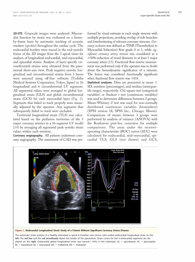
ongitudinal and 6 circumferential LV segments.ll segmental values were averaged to global lon-
itudinal strain (GLS) and global circumferentialtrain (GCS) for each myocardial layer (Fig. 1).egments that failed to track properly were manu-lly adjusted by the operator. Any segments thatubsequently failed to track were excluded.
Territorial longitudinal strain (TLS) was calcu-ated based on the perfusion territories of the 3
ajor coronary arteries in a 16-segment LV model14) by averaging all segmental peak systolic strainalues within each territory.Coronary angiography. All patients underwent coro-
ary angiography. The assessment of CAD was per-
Figure 1. Endocardial Longitudinal Strain Study of a Patient Wi
The automatic strain analysis in a healthy individual in apical 4-chaleft. The red line and the red arrowheads depict the border of theplayed on the right. Endocardial global longitudinal strain was n
BL � basolateral; BS � basoseptal; ML � midlateral; MS � midseptal.ormed by visual estimate in each single stenosis withultiple projections, avoiding overlap of side branches
nd foreshortening of relevant coronary stenoses. Cor-nary occlusion was defined as TIMI (Thrombolysis in
yocardial Infarction) flow grade 0 or 1, while sig-ificant coronary artery stenosis was considered as a50% reduction of vessel diameter in at least 1 major
oronary artery (15). Fractional flow reserve measure-ent was performed only if the operator was in doubt
bout the hemodynamic significance of a stenosis.he lesion was considered functionally significanthen fractional flow reserve was �0.8.
Statistical analyses. Data are presented as mean �D, numbers (percentages), and median (interquar-ile range), respectively. Chi-square test (categoricalariables) or Student t test (continuous variables)as used to determine differences between 2 groups.ann-Whitney U test was used for non-normally
istributed continuous variables (biomarkers)SPSS version 18, SPSS Inc., Chicago, Illinois).omparisons of means between 3 groups wereerformed by analysis of variance (ANOVA) withhe Bonferroni post-hoc correction for multipleomparisons. The areas under the receiver-perating characteristic (ROC) curves (AUC) werealculated for endocardial, mid-myocardial, epi-ardial TLS, GLS (not shown) and GCS,
t Significant Coronary Artery Disease
r view shows color-coded endocardial longitudinal strain on thecardium. Strain curves for the 6 endocardial segments are dis-al (–19%) in this individual. AL � apicolateral; AS � apicoseptal;
thou
mbeepiorm
LMW � low molecular we
J A C C : C A R D I O V A S C U L A R I M A G I N G , V O L . 6 , N O . 5 , 2 0 1 3
M A Y 2 0 1 3 : 5 3 5 – 4 4
Sarvari et al.
Layer-Specific Strain and CAD
538
WMSI, EF, and troponin T, and all variableswere compared by using a nonparametric U test(Analyse-it, Analyse-it Software, Ltd., Leeds,United Kingdom). The value closest to the upperleft corner of the ROC curves determined opti-mal sensitivity and specificity for the ability ofendocardial, mid-myocardial, and epicardialTLS, GLS and GCS, WMSI, EF, and troponin T topredict the presence of significant CAD. Logisticregression analysis was performed for factors thatcould potentially influence myocardial function and todetermine the diagnostic value of endocardial TLS forpredicting significant CAD. Parameters influencingmyocardial contraction, such as age, body mass index,systolic and diastolic blood pressure, plus the categor-
line Characteristics of Patients With and Without
No Significant CAD(n � 28)
Significant CAD(n � 49) p Value
61.4 � 8.9 63.3 � 9.3 0.4
14 (50) 43 (88) �0.001
n 67 � 13 66 � 11 0.73
143 � 17 151 � 24 0.16
78 � 11 82 � 12 0.11
80 � 18 86 � 12 0.15
174 � 9 176 � 9 0.28
26 � 4 28 � 3 0.16
d 15 (54) 33 (67) 0.23
pe 1 1 (4) 0 (0) 0.18
pe 2 3 (11) 11 (22) 0.20
ia 8 (29) 18 (37) 0.47
9 (32) 11 (22) 0.35
6 (21) 11 (22) 0.92
mission 4 (14) 11 (22) 0.4
n admission
28 (100) 49 (100) 1.00
8 (29) 22 (45) 0.16
5 (18) 18 (37) 0.08
0 (0) 1 (2) 0.45
20 (71) 41 (84) 0.20
13 (46) 28 (57) 0.37
5 (18) 6 (12) 0.50
25 (89) 48 (98) 0.10
191 (14–368) 588 (505–990) 0.13
3.1 (2.6–3.5) 11.0 (4.4–27) 0.07
%), or median (interquartile range). *CAD in a first-degree relative prior to age
erting enzyme inhibitor; ARB � angiotensin II receptor blocker; BMI � bodyary artery disease; CCB � calcium channel blocker; CK-MB � creatine kinaseiastolic blood pressure; ECG � electrocardiography; IQR � interquartile range;
ight; SBP � systolic blood pressure.ical variables hypertension and diabetes mellitus, inaddition to endocardial TLS, were included in amultivariate logistic regression analysis. To avoid over-fitting the model, in each analysis only 3 variables wereincluded with endocardial TLS as a constant factor inthe model. All possible combinations with 3 variableswere tested. Due to co-linearity of strain parameters,only endocardial TLS was selected for inclusion in themultivariate analysis, and blood pressure parameterswere included separately in the model. Correlation ofstrain parameters with troponin T was tested by theSpearman correlation coefficient. Reproducibility wasexpressed as intraclass correlation coefficient. p Valueswere 2-tailed; values �0.05 were consideredsignificant.
R E S U L T S
We included 77 patients with suspected NSTE-ACS (Table 1). By coronary angiography, 49 (64%)had significant CAD. Patients with significantCAD were more frequently male compared withpatients without (Table 1). No differences in age,comorbidity, or medication were observed betweenthose with and without significant CAD at admis-sion. Of the patients with significant CAD, 21(28%) had significant coronary stenosis, and 28(36%) had coronary artery occlusion in 1 or morecoronary arteries. Five fractional flow reserve mea-surements were performed in 4 patients. In these 5arteries, hemodynamically significant stenosis wasfound in 4 and no significant stenosis in 1 coronaryartery. Detailed angiographic findings and revascu-larization results are shown in Table 2.Echocardiographic findings. Patients with significantCAD had worse function in all 3 myocardial layersassessed by TLS, GLS, and GCS compared withpatients without (Table 3). However, endocardialTLS (–14.0 � 3.3% vs. –19.2 � 2.2%; p � 0.001),GLS (–15.3 � 2.2% vs. –19.2 � 2.2%; p � 0.001),and GCS (–19.3 � 4.0% vs. –24.3 � 3.4%; p �0.001) were most affected (Fig. 2). In general, therewas a worsening of myocardial function assessed byTLS, GLS, and GCS in all 3 layers in individualswithout significant CAD versus patients with signif-icant coronary artery stenosis and patients with occlu-sion. However, this method could not differentiatebetween patients with significant coronary artery ste-nosis and patients with occlusion (Table 3).
Patients with coronary artery stenosis or occlusionshowed generally lower TLS magnitude than GLS(Table 3); however, the difference between these param-
Table 1. Clinical BaseSignificant CAD
Demographics
Age, yrs
Male
Medical history
Heart rate, beats/mi
SBP, mm Hg
DBP, mm Hg
Weight, kg
Height, cm
BMI, kg/m2
Risk factors
Hypertension, treate
Diabetes mellitus ty
Diabetes mellitus ty
Hypercholesterolem
Smoking
Family history*
Ischemic ECG on ad
Concomitant therapy o
Acetylsalicylic acid
Clopidogrel
LMW heparin
Warfarin
�-Receptor blocker
ACEI/ARB
CCB
Statin
Biomarkers
Troponin T, ng/l
CK-MB, �g/l
Values are mean � SD, n (55 (men) or 65 (women).ACEI � angiotensin-convmass index; CAD � coronmyocardial band; DBP � d
eters generally did not reach statistical significance. The

J A C C : C A R D I O V A S C U L A R I M A G I N G , V O L . 6 , N O . 5 , 2 0 1 3
M A Y 2 0 1 3 : 5 3 5 – 4 4
Sarvari et al.
Layer-Specific Strain and CAD
539
absence of statistically significant differences can probablybe explained by the relatively low sample size.
Endocardial TLS, GLS, and GCS (–15.4 �4.0%, –16.7 � 2.9%, and –21.1 � 4.5%, respec-tively) were significantly greater in magnitude thanwere epicardial TLS, GLS, and GCS (–11.8 �2.9%, –12.7 � 2.0%, and –13.0 � 2.1%, respec-tively; all, p � 0.001) in the total study population(N � 77).
The absolute differences between endocardialand epicardial TLS, GLS, and GCS were lower inpatients with significant CAD (patients with sig-nificant stenosis and/or occlusion; n � 49) (�2.4 �3.6%, �3.4 � 1.3%, and �6.7 � 3.8%, respectively)
Table 2. Angiographic Findings and Revascularization
No CoronaryOcclusion(n � 49)
CoronaryOcclusion(n � 28)
pValue
Angiographic findings
No significant CAD 28 (36) 0 (0) �0.001
1–Vessel disease 13 (27) 7 (25) 0.88
2–Vessel disease 3 (6) 8 (29) 0.007
3–Vessel disease 5 (10) 13 (46) �0.001
Revascularization
PCI 15 (31) 14 (50) 0.09
CABG 5 (10) 12 (43) 0.001
No intervention 29 (59) 2 (7) �0.001
Values are n (%).CABG � coronary artery bypass grafting; CAD � coronary artery disease;PCI � percutaneous coronary intervention.
Table 3. Echocardiographic Data
No Significant Coronary Stenosis(n � 28)
Sig
EF, % 62.0 � 6.0
EDV, ml 111.0 � 26.0
ESV, ml 44.0 � 16.0
WMSI 1.06 � 0.1
TLS, %
Endocardial –19.2 � 2.2
Mid–myocardial –15.9 � 1.5
Epicardial –13.9 � 1.8
GLS, %
Endocardial –19.2 � 2.2
Mid–myocardial –15.9 � 1.5
Epicardial –13.9 � 1.8
GCS, %
Endocardial –24.3 � 3.6
Mid–myocardial –17.5 � 2.8
Epicardial –13.9 � 2.1
Values are mean � SD. *ANOVA (F test). Flags for significance were obtained fro3 groups. †p � 0.05 compared with individuals with no significant coronary stANOVA � analysis of variance; EDV � end–diastolic volume; EF � ejection frac
longitudinal strain; TLS � territorial longitudinal strain; WMSI � wall motion scorethan in those without significant CAD (n � 28;values are presented in Table 4) (p � 0.001). Thisreflects a pronounced decrease in endocardial func-tion in patients with significant CAD. The absolutedifferences between epicardial and endocardial de-formation were generally lower in territorial analysisthan in the global analyses. The difference betweenthese parameters, however, did not reach statisticalsignificance (Table 4). The transmural gradient ofmyocardial strain was less pronounced in individu-als with coronary artery occlusion (n � 28) andsignificant stenosis (n � 21) than in patients with-out significant CAD (n � 28) (Table 4).
Of 1,232 segments, 1,087 (88%) were analyzableby wall motion scoring. Of these segments, 124(11%) had wall motion abnormalities: hypokinesisin 119 and akinesis in 5 segments. In hypokineticand akinetic segments, the absolute differences be-tween endocardial and epicardial longitudinalstrains were lower than in segments with normalfunction (�3.1 � 6.3 vs. �5.2 � 2.3; p � 0.01).
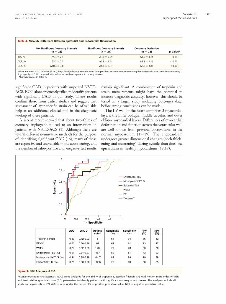
Figures 3 and 4 demonstrate ROC analyses ofthe ability of different strain parameters, WMSI,EF, and troponin T, to identify patients withsignificant CAD. Comparison of ROC curvesshowed that endocardial and mid-myocardial TLSand endocardial GLS were significantly better foridentifying significant CAD than were epicardialTLS and GLS (all, p � 0.05), WMSI (all, p �0.01), and EF (all, p � 0.001). In addition, endo-
cant Coronary Stenosis(n � 21)
Coronary Occlusion(n � 28) p Value*
59.0 � 6.0 59.0 � 7.0 0.09
117.0 � 23.0 124.0 � 21.0 0.43
47.0 � 12.0 51.0 � 13.0 0.46
1.12 � 0.1 1.2 � 0.2†‡ �0.001
–14.6 � 2.3† –13.6 � 3.9† �0.001
–12.6 � 2.6† –12.4 � 2.6† �0.001
–12.0 � 2.5† –11.7 � 2.6† �0.001
–16.1 � 1.9† –14.8 � 2.3† �0.001
–14.0 � 1.6† –12.8 � 2.1† �0.001
–12.3 � 1.1† –11.7 � 2.1† �0.001
–20.2 � 4.2† –18.5 � 3.7† �0.001
–15.5 � 2.5† –14.2 � 2.3† �0.001
–13.4 � 1.9 –11.9 � 2.0†‡ 0.002
ost-hoc pair-wise comparison using the Bonferroni correction when comparingis. ‡p � 0.05 compared with patients with significant coronary stenosis.; ESV � end–systolic volume; GCS � global circumferential strain; GLS � global
nifi
m penostion
index.

edu
J A C C : C A R D I O V A S C U L A R I M A G I N G , V O L . 6 , N O . 5 , 2 0 1 3
M A Y 2 0 1 3 : 5 3 5 – 4 4
Sarvari et al.
Layer-Specific Strain and CAD
540
cardial GCS was better for identifying significantCAD than were epicardial GCS (p � 0.005) andEF (p � 0.01). Troponin T and mid-myocardialGLS were better only than EF (p � 0.05). TLSparameters had greater AUCs than did GLS pa-rameters; however, the differences were not signif-icant. The other apparent differences betweenAUCs also were not significant.
Multivariate regression analyses, including pa-rameters influencing myocardial function, showedthat reduced myocardial function by endocardialTLS (per 1% change) was the only predictor of thepresence of significant CAD (Table 5), indepen-dently of which variables were included in themodel in addition to endocardial TLS.
There was a significant difference in myocardialfunction by mid-myocardial TLS between those with1-vessel occlusion and those with occlusion and3-vessel disease (p � 0.02), confirming a relationshipof function and apparent ischemic burden. There wasa similar significant difference in epicardial TLS (p �0.05). There were, however, no differences in myocar-dial function assessed by GLS or GCS betweenpatients with 1-, 2-, or 3-vessel disease.
All longitudinal and circumferential strain pa-
Figure 2. Endocardial Longitudinal Strain Study of a Patient Wi
The automatic strain analysis in a patient with non–ST-segment elein apical 4-chamber view shows reduced color-coded endocardial sThe red line and the red arrowheads depict the border of the epic�30% to –30%. Yellow indicates preserved strain. Brown indicatesments are displayed on the right. The curves representing the segmto –17% (white arrow). Endocardial global longitudinal strain was r
rameters showed significant correlations with
troponin-T values. Endocardial GLS and TLS andendocardial GCS demonstrated greatest correla-tion, with correlation coefficients of 0.64, 0.58, and0.47, respectively (all, p � 0.01).
Assessment of longitudinal strain could be per-formed in 1,303 (94%) endocardial, 1,149 (83%)mid-myocardial, and 1,142 (82%) epicardial LV seg-ments. The corresponding circumferential analysescould be done in 361 (78%) endocardial, 361 (78%)mid-myocardial, and 343 (74%) epicardial segments.
Intraobserver and interobserver intraclass corre-lations were performed in 10 patients and were 0.96and 0.96, respectively, for endocardial GLS; 0.87and 0.92 for mid-myocardial GLS; 0.94 and 0.93for epicardial GLS; 0.81 and 0.84 for endocardialGCS; 0.84 and 0.81 for mid-myocardial GCS; and0.82 and 0.76 for epicardial GCS measurements.
D I S C U S S I O N
This study demonstrates impaired LV function inall 3 myocardial layers in patients with NSTE-ACSand significant CAD compared with patients with-out significant CAD. We introduce LV layer-specificlongitudinal and circumferential strain assessed by
oronary Artery Occlusion
n acute coronary syndrome with occluded circumflex (CX) arteryvalues in the segments supplied by the CX artery on the left.um. Color-coding from yellow to green indicates strain froms with reduced strain. Strain curves for the 6 endocardial seg-s supplied by the CX artery show reduced strain values of –15%ced in this patient to –15%. Abbreviations as in Figure 1.
th C
vatiotrainardiareaent
2D-STE as a novel method for the identification of
J A C C : C A R D I O V A S C U L A R I M A G I N G , V O L . 6 , N O . 5 , 2 0 1 3
M A Y 2 0 1 3 : 5 3 5 – 4 4
Sarvari et al.
Layer-Specific Strain and CAD
541
significant CAD in patients with suspected NSTE-ACS. ECG alone frequently failed to identify patientswith significant CAD in our study. These resultsconfirm those from earlier studies and suggest thatassessment of layer-specific strain can be of valuablehelp as an additional clinical tool in the diagnosticworkup of these patients.
A recent report showed that about two-thirds ofcoronary angiographies lead to an intervention inpatients with NSTE-ACS (1). Although there areseveral different noninvasive methods for the purposeof identifying significant CAD (16), many of theseare expensive and unavailable in the acute setting, andthe number of false-positive and -negative test results
AUC 95% CI Opt cu
Troponin T (ng/l) 0.83 0.72-0.93
EF (%) 0.63 0.50-0.76 6
WMSI 0.74 0.62-0.85 1
Endocardial TLS (%) 0.91 0.84-0.97 -1
Mid-myocardial TLS (%) 0.91 0.85-0.98 -1
Epicardial TLS (%) 0.79 0.68-0.90 -1
1
0.8
0.6
0.4
0.2
00 0.2 0.4
1 - Specif
Sen
siti
vity
Figure 3. ROC Analyses of TLS
Receiver-operating characteristic (ROC) curve analyses for the abilityand territorial longitudinal strain (TLS) parameters to identify patien
Table 4. Absolute Difference Between Epicardial and Endocardi
No Significant Coronary Stenosis(n � 28)
Signific
TLS, % ∆5.3 � 2.1
GLS, % ∆5.3 � 2.1
GCS, % ∆10.4 � 3.0
Values are mean � SD. *ANOVA (F test). Flags for significance were obtained fro3 groups. †p � 0.01 compared with individuals with no significant coronary stAbbreviations as in Table 3.
study participants (N � 77). AUC � area under the curve; PPV � positi
remain significant. A combination of troponin andstrain measurements might have the potential toincrease diagnostic accuracy; however, this should betested in a larger study including outcomes data,before strong conclusions can be made.
The LV wall of the heart comprises 3 myocardiallayers: the inner oblique, middle circular, and outeroblique myocardial layers. Differences of myocardialdeformation and function across the ventricular wallare well known from previous observations in thenormal myocardium (17–19). The endocardiumundergoes greater dimensional changes (both thick-ening and shortening) during systole than does theepicardium in healthy myocardium (17,18).
l Sensitivity Specificity PPV NPV (%) (%) (%) (%)
64 94 86 82
61 61 73 47
79 73 63 86
89 81 73 93
82 88 79 89
78 69 58 85
0.8 1
Endocardial TLS
Mid-myocardial TLS
Epicardial TLS
WMSI
EF
Troponin T
troponin T, ejection fraction (EF), wall motion score index (WMSI),ith significant coronary artery disease. The analyses include all
eformation
Coronary Stenosisn � 21)
Coronary Occlusion(n � 28) p Value*
.0 � 2.9† ∆1.9 � 4.1† 0.001
.8 � 1.4† ∆3.1 � 1.1† �0.001
.8 � 3.8† ∆6.6 � 3.8† �0.001
ost-hoc pair-wise comparison using the Bonferroni correction when comparingis.
imatoff
8
2
.07
6.4
4.7
2.6
0.6
icity
ofts w
al D
ant(
∆3
∆3
∆6
m penos
ve predictive value; NPV � negative predictive value.

artic
J A C C : C A R D I O V A S C U L A R I M A G I N G , V O L . 6 , N O . 5 , 2 0 1 3
M A Y 2 0 1 3 : 5 3 5 – 4 4
Sarvari et al.
Layer-Specific Strain and CAD
542
In patients with CAD, the inner oblique myo-cardium is most susceptible to ischemic injury (2).Myocardial infarction models as well as reperfusionstudies of myocardial infarction have indicated thatthe endocardial layer is first affected by ischemia (4),causing morphologic (3) and functional alterationspredominant in this layer. With increasing severity,ischemia and necrosis propagate in a transmuralwave front extending from the endocardium to theepicardium (5). Due to the fact that greatest short-ening in a healthy myocardium occurs in the endo-cardial layer and that endocardium is first affectedby ischemia, one will expect that significant CADcauses greatest reduction of function in this partic-ular layer. Reduced endocardial function in patients
AUC 95% CI Opt cu
Troponin T (ng/l) 0.83 0.72-0.93
EF (%) 0.63 0.50-0.76 6
WMSI 0.74 0.62-0.85 1
Endocardial GCS (%) 0.85 0.76-0.95 -2
Mid-myocardial GCS (%) 0.77 0.65-0.88 -1
Epicardial GCS (%) 0.68 0.56-0.81 -1
1
0.8
0.6
0.4
0.2
00 0.2 0.4
1 - Specif
Sen
siti
vity
Figure 4. ROC Analyses of GCS
ROC curve analyses for the ability of troponin T, EF, WMSI, and globsignificant coronary artery disease. The analyses include all study p
Table 5. Endocardial TLS and Parameters Influencing Myocardia
Univariate Logistic Regress
OR 95% CI
Endocardial TLS, % 1.88 1.42–2.49
Age, yrs 1.02 0.97–1.08
BMI, kg/m2 1.10 0.96–1.26
BMI � body mass index; CI � confidence interval; NSTE-ACS � non–ST-segment
strain.with significant CAD can be explained by eithercoronary occlusion and direct myocardial damage orby reversible myocardial dysfunction caused bymyocardial stunning or hibernation (20).
Most imaging techniques focus on the evaluationof global and regional LV function. Traditionally,the entire myocardial wall thickness is considered inthe analysis of myocardial function without takinginto account the differences in the layers of themyocardium. Imaging techniques allowing layer-specific myocardial function analysis are likely toincrease the morphologic and pathophysiologic un-derstanding of myocardial ischemia and may help toimprove characterization of patients with CAD.
l Sensitivity Specificity PPV NPV (%) (%) (%) (%)
64 94 86 82
61 61 73 47
79 73 63 86
85 85 76 91
62 80 64 79
73 65 54 81
0.8
Endocardial GCS
Mid-myocardial GCS
Epicardial GCS
WMSI
EF
Troponin T
1
ircumferential strain (GCS) parameters to identify patients withipants (N � 77). Abbreviations as in Figure 3.
nction in Patients With Suspected NSTE-ACS (N � 77)
Multivariate Logistic Regression
Value OR 95% CI p Value
0.001 2.10 1.47–3.09 �0.001
0.39 1.05 0.97–1.14 0.21
0.16 1.00 0.89–1.12 0.98
tion-acute coronary syndrome(s); OR � odds ratio; TLS � territorial longitudinal
imatoff
8
2
.07
1.7
6.9
2.9
0.6
icity
al c
l Fu
ion
p
�
eleva

cc
st
J A C C : C A R D I O V A S C U L A R I M A G I N G , V O L . 6 , N O . 5 , 2 0 1 3
M A Y 2 0 1 3 : 5 3 5 – 4 4
Sarvari et al.
Layer-Specific Strain and CAD
543
In our study, endocardial longitudinal and circum-ferential strains were better than epicardial strains foridentifying patients with significant CAD. Further-more, our results indicate that all 3 myocardial layerswere affected by both coronary artery occlusion andsignificant coronary artery stenosis, although to differ-ent extents. Reduction of function in all 3 layers couldbe explained by several factors. First, deformations ofthe myocardial layers are not independent. The con-traction of viable myocardium may result in thedeformation of the neighboring nonviable myocar-dium through passive translational or tethering move-ments. On the contrary, nonviable myocardium maynegatively influence contraction in the adjacent viablemyocardium. The deformation of each layer is a sumof an active contraction within the layer and thepassive influence of the adjacent layer. Second, non-occlusive lesions and occlusions may represent differ-ent degrees of transmural ischemia. Coronary arteryocclusions, in the absence of collaterals, may causetransmural ischemia, affecting all 3 myocardial layerswith both longitudinal and circumferential dysfunc-tion. Third, lateral resolution might be a factor limit-ing the delineation of delicate structures withoutapparent boundaries such as the endocardium, mid-myocardium, and epicardium. These problems arefurther exaggerated in the basal LV segments, wherethe density of echo beams is lower.
Both global and territorial strains were assessed inthis study for a number of reasons. One is thevariability of coronary anatomy (21). Myocardial func-tion assigned to a specific vascular territory by Cer-queira et al. (22) may not necessarily reflect the realcoronary distribution. Assessment of global strain isnot exposed to these anatomic variations and has beenshown to detect even minor reductions in myocardialfunction (9). Furthermore, networks formed by mi-rovascular communications between coronary arteriesan give rise to zones of dual arterial perfusion (23),
making strict regional analysis somewhat inaccurate.However, CAD results primarily in segmental wallmotion abnormalities; therefore, TLS was assessed aswell. Conversely, we chose to omit territorial circum-ferential strain results because we had short-axis ac-quisition from only 1 LV slice. The results fromterritorial circumferential strain would therefore con-sist of only 2 LV segments each.
All 3 myocardial layers are affected by significantCAD. The greatest decrease in myocardial func-tion, however, occurs in the endocardial layer be-cause shortening normally is most prominent in thislayer. Therefore, layer-specific strain analyses might
increase diagnostic accuracy in these patients. tStudy limitations. Longitudinal and circumferential,but not radial, strain was assessed in the presentstudy. We chose not to analyze radial strain becauseit has methodological limitations and has beenshown to be inferior to longitudinal and circumfer-ential strain in identifying ischemia and necrosis(24). In addition, longitudinal and circumferentialstrains have been well validated against EF in previousstudies (11,25), are reproducible, and are easily ob-tained with only a minor increase of procedure dura-tion. We did not have 3 short-axis projections avail-able for the assessment of circumferential strain in a16-segment model. In our study, 16 longitudinalsegments were analyzed by strain compared with only6 mid-circumferential segments.
Fractional flow reserve measurements were not per-formed in all patients; therefore, the true hemodynamicrelevance of the stenoses is not known.
It is not known whether coronary artery occlusionsrepresent transmural or nontransmural infarctions be-cause no viability studies have been performed on ourpatients.
All patients were treated according to guidelines onadmission. Obviously, this could have caused dissolutionof thrombi, allowing recovery of the affected myocar-dium. However, one may also speculate that even in caseof dissolution of thrombi, the myocardium might be stillstunned or hibernated within the first 48 h.
Deformation of the entire wall thickness of themyocardium was not assessed because the softwaredid not allow such analysis. Consequently, we couldnot evaluate whether layer-specific strain analysishas additional value compared with traditionalglobal strain analysis in diagnosing CAD.
The gradient of strain across the myocardium is anonlinear phenomenon, and the definition of the layers isarbitrary and is based on simple division into 3 parts.Because the spatial resolution of ultrasound is limited,there will always be a certain degree of overlap.
C O N C L U S I O N S
Assessment of endocardial and mid-myocardial TLSby layer-specific strain echocardiography providedhigher accuracy than did epicardial strain, WMSI, andEF in the identification of patients with NSTE-ACSand significant CAD. Endocardial function was moreaffected in patients with significant CAD compared tothose with epicardial function.
Reprint requests and correspondence: Prof. Thor Edvard-en, Department of Cardiology, Oslo University Hospi-al, Rikshospitalet, N-0027 Oslo, Norway. E-mail:
[email protected].
J A C C : C A R D I O V A S C U L A R I M A G I N G , V O L . 6 , N O . 5 , 2 0 1 3
M A Y 2 0 1 3 : 5 3 5 – 4 4
Sarvari et al.
Layer-Specific Strain and CAD
544
1
1
1
1
1
1
1
1
m
R E F E R E N C E S
1. Damman P, van GN, Wallentin L, etal. Timing of angiography with a rou-tine invasive strategy and long-termoutcomes in non–ST-segment eleva-tion acute coronary syndrome: a col-laborative analysis of individual pa-tient data from the FRISC II (Fragminand Fast Revascularization DuringInstability in Coronary Artery Dis-ease), ICTUS (Invasive Versus Con-servative Treatment in UnstableCoronary Syndromes), and RITA-3(Intervention Versus ConservativeTreatment Strategy in Patients WithUnstable Angina or Non-ST Eleva-tion Myocardial Infarction) Trials. J AmColl Cardiol Intv 2012;5:191–9.
2. Duncker DJ, Traverse JH, IshibashiY, Bache RJ. Effect of NO on trans-mural distribution of blood flow inhypertrophied left ventricle duringexercise. Am J Physiol 1999;276:H1305–H1312.
3. Geer JC, Crago CA, Little WC,Gardner LL, Bishop SP. Subendocar-dial ischemic myocardial lesions asso-ciated with severe coronary atheroscle-rosis. Am J Pathol 1980;98:663–80.
4. Ono S, Waldman LK, Yamashita H,Covell JW, Ross J Jr. Effect of coro-nary artery reperfusion on transmuralmyocardial remodeling in dogs. Cir-culation 1995;91:1143–53.
5. Reimer KA, Jennings RB. The “wave-front phenomenon” of myocardialischemic cell death. II. Transmuralprogression of necrosis within theframework of ischemic bed size (myo-cardium at risk) and collateral flow.Lab Invest 1979;40:633–44.
6. Haugaa KH, Smedsrud MK, Steen T,et al. Mechanical dispersion assessedby myocardial strain in patients aftermyocardial infarction for risk predic-tion of ventricular arrhythmia. J AmColl Cardiol Img 2010;3:247–56.
7. Sarvari SI, Haugaa KH, AnfinsenOG, et al. Right ventricular mechan-ical dispersion is related to malignantarrhythmias: a study of patients witharrhythmogenic right ventricular car-diomyopathy and subclinical rightventricular dysfunction. Eur Heart J2011;32:1089–96.
8. Sjoli B, Orn S, Grenne B, Ihlen H,Edvardsen T, Brunvand H. Diagnos-tic capability and reproducibility of
strain by Doppler and by speckletracking in patients with acute myo-cardial infarction. J Am Coll CardiolImg 2009;2:24–33.
9. Gjesdal O, Hopp E, Vartdal T, et al.Global longitudinal strain measuredby two–dimensional speckle trackingechocardiography is closely related tomyocardial infarct size in chronic isch-aemic heart disease. Clin Sci (Lond)2007;113:287–96.
0. Eek C, Grenne B, Brunvand H, et al.Strain echocardiography predicts acutecoronary occlusion in patients withnon-ST-segment elevation acute cor-onary syndrome. Eur J Echocardiogr2010;11:501–8.
1. Grenne B, Eek C, Sjoli B, et al. Acutecoronary occlusion in non-ST-elevationacute coronary syndrome: outcome andearly identification by strain echocardi-ography. Heart 2010;96:1550–6.
2. Bassand JP, Hamm CW, ArdissinoD, et al. Guidelines for the diagnosisand treatment of non-ST-segment el-evation acute coronary syndromes.Eur Heart J 2007;28:1598–660.
3. Alpert JS, Thygesen K, Jaffe A, WhiteHD. The universal definition of myo-cardial infarction: a consensus docu-ment: ischaemic heart disease. Heart2008;94:1335–41.
4. Lang RM, Bierig M, Devereux RB, etal. Recommendations for chamberquantification: a report from theAmerican Society of Echocardiogra-phy’s Guidelines and Standards Com-mittee and the Chamber Quantifica-tion Writing Group, developed inconjunction with the European Asso-ciation of Echocardiography, a branchof the European Society of Cardiol-ogy. J Am Soc Echocardiogr 2005;18:1440–63.
5. Arnold JR, Karamitsos TD, Bhamra-Ariza P, et al. Myocardial oxygenationin coronary artery disease: insights fromblood oxygen level–dependent mag-netic resonance imaging at 3 Tesla.J Am Coll Cardiol 2012;59:1954–64.
6. Achenbach S, Kramer CM, ZoghbiWA, Dilsizian V. The year in coro-nary artery disease. J Am Coll CardiolImg 2010;3:1065–77.
7. Adamu U, Schmitz F, Becker M,Kelm M, Hoffmann R. Advancedspeckle tracking echocardiography al-lowing a three-myocardial layer-specific analysis of deformation pa-
rameters. Eur J Echocardiogr 2009;10:303–8. m18. Sabbah HN, Marzilli M, Stein PD.The relative role of subendocardiumand subepicardium in left ventricularmechanics. Am J Physiol 1981;240:H920–H926.
19. Leitman M, Lysiansky M, LysyanskyP, et al. Circumferential and longitu-dinal strain in 3 myocardial layers innormal subjects and in patients withregional left ventricular dysfunction.J Am Soc Echocardiogr 2010;23:64–70.
20. Shivalkar B, Flameng W, Szilard M,Pislaru S, Borgers M, Vanhaecke J.Repeated stunning precedes myocar-dial hibernation in progressive multi-ple coronary artery obstruction. J AmColl Cardiol 1999;34:2126–36.
21. Segall GM, Atwood JE, BotvinickEH, Dae MW, Lucas JR. Variabilityof normal coronary anatomy: implica-tions for the interpretation of thallium-SPECT myocardial perfusion images insingle-vessel disease. J Nucl Med 1995;36:944–51.
22. Cerqueira MD, Weissman NJ, Dilsi-zian V, et al. Standardized myocardialsegmentation and nomenclature fortomographic imaging of the heart: astatement for healthcare professionalsfrom the Cardiac Imaging Committeeof the Council on Clinical Cardiologyof the American Heart Association.Circulation 2002;105:539–42.
23. Cicutti N, Rakusan K, Downey HF.Coronary artery occlusion extendsperfusion territory boundaries throughmicrovascular collaterals. Basic ResCardiol 1994;89:427–37.
24. Gjesdal O, Helle-Valle T, Hopp E, etal. Noninvasive separation of large,medium, and small myocardial in-farcts in survivors of reperfused ST-elevation myocardial infarction: acomprehensive tissue Doppler andspeckle-tracking echocardiographystudy. Circ Cardiovasc Imaging 2008;1:189–96.
25. Stanton T, Leano R, Marwick TH.Prediction of all-cause mortality fromglobal longitudinal speckle strain: com-parison with ejection fraction and wallmotion scoring. Circ Cardiovasc Imag-ing 2009;2:356–64.
Key Words: coronary arterydisease y echocardiography y
yocardial function y
yocardial layers y strain.