Embed Size (px)
Citation preview

6/9/2007
1
Late Decelerations
Gary A. Dildy III, MD
Perinatologist
St. Mark’s Hospital
Salt Lake City, UT
Director of MFM, MountainStar Division
Hospital Corporation of America
Nashville, TN
Clinical Professor of OB/GYN
LSU Health Sciences Center
New Orleans, LA
Late
DecelerationsMichael A. Belfort, MD, PhD
Professor of OB/GYN
Division of Maternal-Fetal Medicine
University of Utah School of Medicine
Salt Lake City, UT
Director of Perinatal Research
HCA, Nashville TN
LATE QUOTES
How did it get so late so soon?
Its night before its afternoon.
December is here before its June.
My goodness how the time has flewn.
How did it get so late so soon?
Theodor Geisel
American Writer
1904 - 1991
Outline
• Acid Base Balance
• Definition
• Etiology
• Historical perspective
• RSA and late decelerations
• Management
• Late Decelerations and CP

6/9/2007
2
Lactic acid
Anerobic MetabolismAerobic Metabolism
Energy + CO2 + H2O
Krebs cycle
IN THE PERIPHERAL TISSUES
Glucose
Glycolysis
Accumulates in
excess of
buffering capacity
Metabolic acidosis
Oxidative phosphorylation
AEROBIC METABOLISM
6 O2
GLUCOSE
METABOLISM
6 CO2
6 H2O
36 ATP
HEAT (417 kcal)
Oxidative phosphorylation: Very efficient form of energy
production. Each pyruvic acid is converted into 34 ATP
ANAEROBIC METABOLISM
GLUCOSE METABOLISM
2 LACTIC ACID
2 ATP
HEAT (32 kcal)
Glycolysis: Inefficient source of energy production; 2
ATP for every glucose; produces pyruvic acid
Anaerobic Metabolism
• Occurs without oxygen
–oxydative phosphorylation can’t occur
without oxygen
–glycolysis can occur without oxygen
–cellular death leads to tissue and organ
death
–can occur even after return of perfusion
• ⇒ organ or organism death

6/9/2007
3
InadequateCellularOxygenDelivery
Anaerobic
Metabolism
InadequateEnergy
Production
Metabolic
Failure
Lactic
Acid
Production
Metabolic
AcidosisCELL
DEATH
Ultimate Effects
of Anaerobic
Metabolism Alveolus
Inspired Air - 150 mm Hg
PARTIAL PRESSURE OF OXYGEN -- pO2
pO2 95 mmHg
pO2 105 mmHg
95 mmHg
45 mmHg40-45 mmHg
Spiral artery
35 mmHg25-35 mmHg
25 mmHg15-25 mmHg
40 mmHg
Normal Fetal Arterial pO2
Cordocentesis (N = 35) – 23 mmHg (14 – 32)Nicolaides Am J Obstet Gynecol 1989;161:996-1001
Elective cesarean without labor (N = 665) – 23 mmHg (± 14%)
Cesarean after labor (N = 1,609) – 18 mmHg (± 13%)
Vaginal delivery (N = 14,285) – 24 mmHg (± 15%)Richardson Am J Obstet Gynecol 1998;178:572-9
Umbilical artery 16 mmHg (± 4.6)
Placental artery 16.5 mmHg (± 3.6)Nodwell Obstet Gynecol 2005;105:129-38
5-minute Apgar score ≥ 7 (N = 15,073) – 17 mmHg (2SD 6-30)Helwig Am J Obstet Gynecol 1996;174:1807-12

6/9/2007
4
American Journal of Obstetrics and Gynecology
© Mosby-Year Book Inc. 1997. All Rights Reserved.
Volume 177(6) December 1997 pp 1385-1390
Electronic fetal heart rate monitoring: Research guidelines for interpretation
[Clinical Opinion]
National Institute of Child Health and Human Development Research Planning Workshop.
“The purpose of the National Institutes of Health research planning
workshops is to assess the research status of clinically important areas.
This article reports on a workshop whose meetings were held between
May 1995 and November 1996 in Bethesda, Maryland, and Chicago,
Illinois. Its specific purpose was to develop standardized and
unambiguous definitions for fetal heart rate tracings.”
“Late deceleration of the FHR is a visually
apparent gradual (onset to nadir ≥ to 30 sec) decrease and return to baseline FHR
associated with a uterine contraction.”
AJOG Volume 177(6) December 1997 pp 1385-1390
“Decelerations are tentatively defined as
recurrent if they occur with ≥ 50% of
uterine contractions in any 20-minute
segment.”

6/9/2007
5
NIHCD Terminology
Pattern NICHD Definitions
Acceleration • Abrupt � in FHR above baseline, peak 15
bpm above baseline, acceleration sustained
for 15 sec (10 x 10 in fetus < 32 wks)
Early
Deceleration • Gradual onset-to-nadir (> 30 seconds) with
nadir coinciding with peak of contraction
Late
Deceleration • Gradual onset-to-nadir (> 30 seconds) with
nadir occurring after peak of contraction
Variable
Deceleration • Abrupt onset-to-nadir (< 30 seconds)
Prolonged
Decelerations • Deceleration > 2 minutes but < 10 minutes
NICHD
NICHD
NICHD
NICHD
NIHCD Terminology
Pattern NICHD Definitions
Baseline FHR Approximate mean FHR rounded to 5 bpm
increments. 10 minutes. Minimum 2-minute
segment
• Normal
FHR
110-160 (10 minutes)
• Bradycardia FHR < 110 bpm for 10 minutes
• Tachycardia FHR > 160 bpm for 10 minutes
Variability Fluctuations or variations in baseline FHR
• Absent Amplitude range undetectable
• Minimal > undetectable but < 5 bpm
• Moderate 6-25 bpm
• Marked > 25 bpm
NICHD
NICHD
LATE DECELERATIONS
Historical
• Little known before EFM
• Description of FHR changes with UPI
– Hon’s “Late deceleration”
– Caldeyro-Barcia’s “Type II Dip”
HON EH. Observations on pathologic fetal bradycardia.
Am J Obstet Gynecol 1959;77:1084-99.
FETAL HEART RATE MONITORING
Ed Hon
• HON EH. The electronic evaluation of the fetal heart rate; preliminary report. Am J Obstet Gynecol 1958;75:1215-30.
• HON EH. Observations on pathologic fetal bradycardia. Am J Obstet Gynecol 1959;77:1084-99.
• HON EH. The fetal heart rate patterns preceding death in utero. Am J Obstet Gynecol 1959;78:47-56.
• HON EH, BRADFIELD AH, HESS OW. The electronic evaluation of the fetal heart rate. V. The vagal factor in fetal bradycardia. Am J Obstet Gynecol 1961;82:291-300.
• HON EH. THE CLASSIFICATION OF FETAL HEART RATE. I. A WORKING CLASSIFICATION. Obstet Gynecol 1963;22:137-46.
• HON EH, LEE ST. THE FETAL ELECTROCARDIOGRAM. I. THE ELECTROCARDIOGRAM OF THE DYING FETUS. Am J Obstet Gynecol 1963;87:804-13.
• HON EH, LEE ST. ELECTRONIC EVALUATION OF THE FETAL HEART RATE. VIII. PATTERNS PRECEDING FETAL DEATH, FURTHER OBSERVATIONS. Am J Obstet Gynecol 1963;87:814-26.

6/9/2007
6
LATE DECELERATION
Terminology
• Type– Reflex
• Hypoxia – chemoreceptors – vagal discharge
• Accompanied by normal FHR variability (normal CNS)
– Nonreflex• Hypoxia – chemoreceptors – vagus - myocardial depression
• Abnormal variability
• Fetal CNS & CV decompensation
• Degree– Mild <15
– Moderate
– Severe >45
Parer & Nageotte, in Creasy & Resnik 5th Edition
Recurrent late decelerations suggest
recurrent episodes of hypoxemia
during contractions.
Recurrent late decelerations suggest
recurrent episodes of hypoxemia
during contractions.
LATE DECELERATIONS & HYPOXIA
Myers RE, Mueller-Heubach E, Adamsons K. Predictability of the state
of fetal oxygenation from a quantitative analysis of the components of
late deceleration. Am J Obstet Gynecol 1973;115:1083-94.
Late Decelerations
• Late decelerations with intact variability are mediated by vagal reflex, are unlikely to indicate fetal acidemia and are likely to respond to O2, fluids and position changes.
• Late decelerations with decreased or absent variability are caused by myocardial depression, likely associated with fetal acidemia and do not respond to conservative measures.
Paul RH, Suidan AK, Yeh S, Schifrin BS, Hon EH. Clinical fetal
monitoring. VII. The evaluation and significance of intrapartum
baseline FHR variability. Am J Obstet Gynecol 1975;123:206-10.

6/9/2007
7
LATE DECELERATIONS
Vagus Nerve Mediated I
• Late deceleration FHR pattern produced in fetal sheep by periodic occlusion of the maternal common hypogastric artery for 30-60 s
– Transient fetal hypertension occurred during the occlusions
• Alpha-adrenergic blockade with phentolamine
– Eliminated or markedly reduced the hypertensive response
– FHR deceleration character was greatly altered
• Parasympathetic blockade with atropine
– Decelerations replaced by periodic FHR accelerations during occlusions
– Accelerations eliminated by the beta-adrenergic blocker propranolol
Martin CB Jr, de Haan J, van der Wildt B, Jongsma HW, Dieleman A, Arts TH.
Mechanisms of late decelerations in the fetal heart rate. A study with autonomic
blocking agents in fetal lambs. Eur J Obstet Gynecol Reprod Biol 1979;9:361-73.
LATE DECELERATIONS
Vagus Nerve Mediated II
• Combined parasympathetic, alpha- and beta-adrenergic blockade
– FHR remained constant during occlusions in non-acidemic fetuses
– FHR decelerations persisted after parasympathetic or total autonomic blockade when fetuses significantly hypoxic
– BTBV persisted in the face of severe hypoxia and acidosis
• CONCLUSIONS…Reflex mechanisms are involved in acutely hypoxemic fetus, direct myocardial depression a factor as hypoxic acidosis develops
Martin CB Jr, de Haan J, van der Wildt B, Jongsma HW, Dieleman A, Arts TH.
Mechanisms of late decelerations in the fetal heart rate. A study with autonomic
blocking agents in fetal lambs. Eur J Obstet Gynecol Reprod Biol 1979;9:361-73.
Vagus Nerve + Chemoreceptor
• Studied initially normoxic chronically instrumented sheep
fetuses after sudden occlusion of uterine blood flow (20s):
– Late decelerations occurred only after significant decrease in fetal
O2 consumption, O2 content, and O2 tension occurred
• No significant change in BP
• Vagal blockade led to a late acceleration which was
eliminated by beta-blockade
• Late deceleration in initially normoxic sheep are vagally
mediated and are due to chemoreceptor, rather than,
baroreceptor activity
Parer JT, Krueger TR, Harris JL. Fetal oxygen consumption and mechanisms of
heart rate response during artificially produced late decelerations of fetal heart
rate in sheep. Am J Obstet Gynecol 1980;136:478-82.
LATE DECELS & ACIDOSIS
• Nine chronically instrumented rhesus monkeys.
• After recovery, all the fetuses had shown accelerations and no late decelerations with spontaneous uterine contractions.
• Late decelerations were the first sign of fetal deterioration and occurred with a slight but significant decrease in fetal PaO2 without changes in pH, whereas accelerations in FHR were still present.
• The loss of FHR accelerations was a later phenomenon and was associated with significant reductions in fetal pH and PaO2.
Murata Y, Martin CB Jr, Ikenoue T, Hashimoto T, Taira S, Sagawa T, Sakata H.
Fetal heart rate accelerations and late decelerations during the course of
intrauterine death in chronically catheterized rhesus monkeys. Am J Obstet
Gynecol 1982;144:218-23.

6/9/2007
8
Vagus Nerve + Chemoreceptor
• Studied chronically hypoxic sheep fetuses after transient occlusion of maternal aorta (20s):– Late decelerations occurred
• Significant decrease in fetal BP and UA flow
• Vagal blockade with atropine before occlusion reduced but did not eliminate the late deceleration
• Late deceleration was associated with decreased myocardial O2 consumption:
• Conclusion: Late decelerations are caused by 2 mechanisms – chemoreceptor vagal reflex
– myocardial depression
Harris JL, Krueger TR, Parer JT. Mechanisms of late decelerations of the fetal
heart rate during hypoxia. Am J Obstet Gynecol 1982;144:491-6.
Late Decels: Mechanism
• Studied normoxemic and chronically hypoxic sheep fetuses after transient obstruction of uterine artery of ewe:
– In initially normoxic fetuses a chemoreceptor reflex induced bradycardia and decreased LV output developed after the carotid PaO2 decreased to < 20 mmHg
• This reaction was abolished by atropine (vagally mediated)
• In the chronically hypoxic fetuses there was a late deceleration composed of three stages:
– chemoreceptor induced, vagally mediated bradycardia
– baroreceptor induced bradycardia with slow and late recovery
– non-reflex bradycardia due to myocardial depression
• The time to onset depended, and the depth and duration of the deceleration depended on the initial state of fetal oxygenation
Itskovitz J, Goetzman BW, Rudolph AM. The mechanism of late deceleration
of the heart rate and its relationship to oxygenation in normoxemic and
chronically hypoxemic fetal lambs. Am J Obstet Gynecol 1982;142:66-73.
pO2 < 15-20 mmHg
sensed by carotid and
aortic chemoreceptors
Combined effect � Increased BP
Medullary
vasomotor
center
(Brain, heart, adrenals)
“Central”
Vasodilatation
(Brain, heart, adrenals)
Peripheral
Vasoconstriction
(Gut, kidneys, limbs)
LATE DECELERATION
Chemoreceptors sense
↓ pO2 and trigger reflex
peripheral
vasoconstriction.
Chemoreceptors sense
↓ pO2 and trigger reflex
peripheral
vasoconstriction.
Hypoxemia during contraction
pO2 falls below critical threshold ~ 15 - 20 mm Hg
near peak of contraction.
Increased BP is
sensed by
baroreceptors,
triggering increased
vagal outflow to
reduce HR, reduce
CO and return BP to
normal.
Oxygenated blood is
shunted away from
non-essential vascular
beds toward essential
vascular beds in brain,
heart and adrenals.
Oxygenated blood is
shunted away from
non-essential vascular
beds toward essential
vascular beds in brain,
heart and adrenals.
Blood pressure rises.

6/9/2007
9
HYPOXEMIAHYPOXEMIA
INITIAL FETAL RESPONSE TO HYPOXEMIA IN THE LAMB
90Ball (9)
5Itskovitz (8)
90Ball (7)
35Jensen (6)
20Reid (5)
90Field (4)
420Richardson (3)
60Peeters (2)
4-
44
Cohn (1)
����
����
NS
����
����
����
����
����
����
BrainMinReference
NS
����
����
����
����
����
����
����
Heart
����
����
����
����
����
����
����
����
Adrenal
����
NS
����
NS
����
����
����
����
Kidney
����
����
����
����
����
����
����
����
����
Carcass
1. AJOG 1974;120:817-24
2. AJOG 1979;135:637-46
3. J Dev Physiol 1989;11:37-43
4. J Dev Physiol 1990;14:131-7
5. J Dev Physiol 1991;15:183-8
6. J Dev Physiol 1991;15:309-23
7. AJOG 1994;170:156-61
8. Am J Physiol 1987;252:H100-99. AJOG 1994;171:1549-55
Blood Flow
MAP
����
����
����
����
����
����
NS
����
����
Base Excess and Hypoxia
• Normal acid base balance in the fetus
– Base Excess: -2.3 +/- 0.6 mmol/L
– pH: 7.39 +/- 0.05 mmol/L
– Base excess does not change with gestation
Lazarevic et al. Clin Exp Obset Gynecol 1991;18:81
• BE after SNVD with 5 min Apgar > 7:
– UA: -4 +/- 3 mmol/L
– UV: -3 +/- 3 mmol/L
– 2.5% for the UA was –11 mmol/LHelwig et al. Am J Obset Gynecol 1996;174:1807
Base Excess and Hypoxia
• A normal baby enters labor with a BE of –2
mmol/L and can be expected to decrease by
an additional 3 mmol/L
• Expected normal decrease in BE during:
– First stage is –1 mmol/L per 3 to 6 hours
– Second Stage is –1 mmol/L per 1 hourHagelin and Leyon. Acta Obset Gynecol Scand 1998;77:841
Base Excess and Hypoxia
• When there are repetitive decels for hours buffer base decreases 1mmol/L per 30 minutes
Low et al. Am J Obstet Gynecol 1977;129:857
• When there is intrapartum hypoxia causing asphyxia buffer base decreases 1mmol/L per 6 to 15 minutes
Low et al. Am J Obstet Gynecol 1984;148:533
• When there is terminal bradycardia from uterine rupture buffer base decreases 1mmol/L per 2 to 3 minutes
Leung et al. Am J Obstet Gynecol 1993;169:945

6/9/2007
10
Base Excess and Hypoxia
• With BE –12 to –16 mmol/L about 10% have:
– Moderate to severe encephalopathy
– Respiratory complicationsLow et al. Am J Obset Gynecol 1997;177:1391
• Buffer base: Normal = 46 mmol/L
– < 34 mmol/L = asphyxia (~BE -12)
– Major/minor deficits 20%
– Increased to 80% when buffer base < 22 mmol/L (~BE -24)
Base Excess and Hypoxia
• BE in umbilical artery and vein:
– UV BE represents the situation at the time
initial cord compressionRoss and Gala. Am J Obstet Gynecol 2002;187:1
– A wide (> 6 mmol/L) UA/UV BE difference
suggests short duration asphyxia and a
narrow (< 6 mmol/L) difference suggests
long term asphyxia Low et al. Am J Obstet Gynecol 1995;172:805
Base Excess and Hypoxia
• No change in BE is likely in the immediate neonatal period:
– immediate effective resuscitation with no bicarb
– no sepsis
– no severe hypoxia or hypotensionRoss and Gala. Am J Obstet Gynecol 2002;187:1
• BE is likely to continue to fall at about 1mmol/L per 2 minutes if resuscitation is ineffective
Leung et al. Am J Obstet Gynecol 1993;169:945
Base Excess and Hypoxia
• Most neonates with a BE of –12 mmol/L
do not exhibit encephalopathy
• Even with a BE < -16 mmol/L most
babies will either die or survive normally,
only a small proportion develop CP
• Most babies that do get CP have an
underlying propensity to CPRoss and Gala. Am J Obstet Gynecol 2002;187:1
6/9/2007
11
Late Decelerations
• Experiment on monkey’s brain showed only pH <7.00 for >30 minutes can cause hypoxic damage. Metabolic acidosis pH >7.00 lasting for several hours did not cause neurological injury.
Myers RE 1977 In Gluck Yearbook, inc 37-97
• pH <7.00 (Base deficit >16mmol/L) is a realistic threshold below which brain dysfunction significantly increases. At this level systemic hypotension start to occur.
Low 1993 ; Clinical OB&GYN 36:82
Acidemia and FHR
Analysis of a number of observational studies to determine relationship between FHR patterns and fetal acidemia
1. Moderate variability strongly associated with UA pH > 7.15 and 5 min Apgar > 7
2. Absent or minimal variability with late or variable decels was strongest predictor of acidemia BUT only a 23% association
3. Strong association between the degree of acidemia and the depth of the deceleration
4. Except for sudden profound bradycardia, acidemia with decreasing variability in combination with decels takes about 1 hour to develop
Grade III observation/uncontrolled studies mostly
Parer JT, King T, Flanders S, Fox M, Kilpatrick SJ. Fetal acidemia and
electronic fetal heart rate patterns: is there evidence of an association?
J Matern Fetal Neonatal Med 2006;19:289-94.
Acidemia and FHR
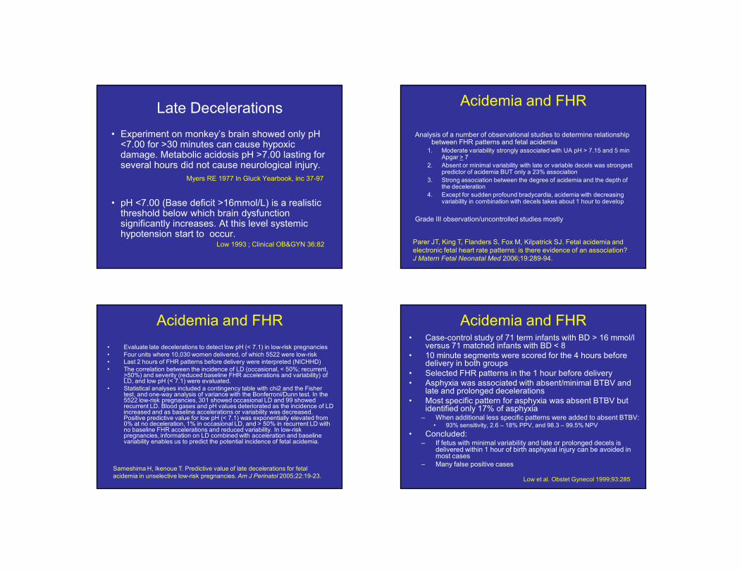
• Evaluate late decelerations to detect low pH (< 7.1) in low-risk pregnancies
• Four units where 10,030 women delivered, of which 5522 were low-risk
• Last 2 hours of FHR patterns before delivery were interpreted (NICHHD)
• The correlation between the incidence of LD (occasional, < 50%; recurrent, >50%) and severity (reduced baseline FHR accelerations and variability) of LD, and low pH (< 7.1) were evaluated.
• Statistical analyses included a contingency table with chi2 and the Fisher test, and one-way analysis of variance with the Bonferroni/Dunn test. In the 5522 low-risk pregnancies, 301 showed occasional LD and 99 showed recurrent LD. Blood gases and pH values deteriorated as the incidence of LD increased and as baseline accelerations or variability was decreased. Positive predictive value for low pH (< 7.1) was exponentially elevated from 0% at no deceleration, 1% in occasional LD, and > 50% in recurrent LD with no baseline FHR accelerations and reduced variability. In low-risk pregnancies, information on LD combined with acceleration and baseline variability enables us to predict the potential incidence of fetal acidemia.
Sameshima H, Ikenoue T. Predictive value of late decelerations for fetal
acidemia in unselective low-risk pregnancies. Am J Perinatol 2005;22:19-23.
Acidemia and FHR• Case-control study of 71 term infants with BD > 16 mmol/l
versus 71 matched infants with BD < 8
• 10 minute segments were scored for the 4 hours before delivery in both groups
• Selected FHR patterns in the 1 hour before delivery
• Asphyxia was associated with absent/minimal BTBV and late and prolonged decelerations
• Most specific pattern for asphyxia was absent BTBV but identified only 17% of asphyxia– When additional less specific patterns were added to absent BTBV:
• 93% sensitivity, 2.6 – 18% PPV, and 98.3 – 99.5% NPV
• Concluded: – If fetus with minimal variability and late or prolonged decels is
delivered within 1 hour of birth asphyxial injury can be avoided in most cases
– Many false positive cases
Low et al. Obstet Gynecol 1999;93:285

6/9/2007
12
Predictive value of FHR variables for fetal asphyxia
• METHODS:
– Asphyxia group of 71 term infants with UA BD >16 mmol/L
– Control group of 71 term infants with UA BD <8 mmol/L
• RESULTS:
– FHR variables associated with fetal asphyxia
• absent and minimal baseline variability
• late and prolonged decelerations
– FHR patterns with absent baseline variability• Were the most specific but identified only 17%
• Sensitivity increased to 93% with addition of less specific patterns
• PPV (18.1% to 2.6%) NPV (98.3% to 99.5%)
• CONCLUSION:
– A narrow 1-hour window of FHR patterns including minimal baseline variability and late or prolonged decelerations will predict fetal asphyxial exposure before decompensation.
– Predictive FHR patterns can be a useful screening test.
– Supplementary tests are required to confirm the diagnosis and to identify the large number of false-positive patterns to avoid unnecessary intervention.
Low JA, Victory R, Derrick EJ. Predictive value of electronic fetal monitoring for
intrapartum fetal asphyxia with metabolic acidosis. Obstet Gynecol 1999 ;93:285-91.
Late Decels: Aortic Compression
• 14% (126/902) laboring patients had late decelerations
• Of these 19% (24/126) only had lates when supine
• Associated with reduced maternal femoral BP and decreased pulse pressure in the big toe
• Conclusion: Supine hypotensive syndrome can cause late decelerations
Abitbol MM. Supine position in labor and associated fetal heart
rate changes. Obstet Gynecol 1985;65:481-6.
Late Decels and Abruption
• Case control study of 69 patients with severe early onset preeclampsia with and without abruptio placentae
• Late decelerations: 58% abruption vs. 32% controls
• Only recognizable warning sign noted for abruption in preeclamptic women
Odendaal HJ, Hall DR, Grove D. Risk factors for and perinatal mortality of
abruptio placentae in patients hospitalised for early onset severe pre-
eclampsia - a case controlled study. J Obstet Gynaecol 2000;20:358-64.
Respiratory Sinus Arrhythmia
• RSA is heart rate variability in synchrony with respiration.
• R-R interval is shortened during inspiration & prolonged during expiration.
• Used as an index of cardiac vagal function.
• Reflects respiratory-circulatory interactions universally among vertebrates.
• Efficiency of pulmonary gas exchange is improved by RSA, suggesting active physiologic role.
• The matched timing of alveolar ventilation and its perfusion with RSA within each respiratory cycle could save energy expenditure by suppressing unnecessary heartbeats during expiration and ineffective ventilation during the ebb of perfusion.
• Evidence of a possible dissociation between RSA and vagal control of the heart rate, suggesting differential controls between the respiratory modulation of cardiac vagal outflow and cardiac vagal tone.
• RSA or heart rate variability in synchrony with respiration is a biological phenomenon, which may have a positive influence on gas exchange at the level of the lung via efficient ventilation/perfusion matching.
Yasuma F, Hayano J. Respiratory sinus arrhythmia: why does the heartbeat
synchronize with respiratory rhythm? Chest 2004;125:683-90.

6/9/2007
13
RSA and Polyvagal Theory
• Model defining the role of the parasympathetic NS and particularly vagus N in mammalian stress reaction
• Phylogenetic shift in neural development of ANS has 3 stages:
– Primitive unmyelinated visceral vagus – immobilization, depressed metabolic activity
– Sympathetic NS – increasing metabolic output, inhibit visceral vagus
– Myelinated vagus – rapidly regulates cardiac output
• 2 primary medullary source nucleii of the vagus (X nerve)
– Nucleus Ambiguus – myelinated (more developed)
– Dorsal Motor Nucleus – unmyelinated (primitive)
• Both nucleii have fibers terminating on the SA node BUT only the NA fibers regulate RSA
Porges SW. Orienting in a defensive world: mammalian modifications of our
evolutionary heritage. A Polyvagal Theory. Psychophysiology 1995;32:301-18.
RSA and Polyvagal Theory• Neurophysiological model based on the Polyvagal Theory to interpret FHR patterns.
• Beat-to-beat heart rate data from 7 fetuses monitored during the first and second stages of labor were analyzed.
• Transitory FHR accelerations and reduced beat-to-beat variability reliably preceded FHR decelerations.
• Data are interpreted within the context of the Polyvagal Theory
– Transitory FHR accelerations and the depression of the respiratory rhythm in the beat-to-beat heart rate pattern reflect a withdrawal of the vagal tonedetermined by myelinated vagal pathways originating in the nucleus ambiguus.
– Withdrawal of vagal tone originating in the NA results in the cardiac pacemaker becoming vulnerable to sympathetic influences and to the more-primitive unmyelinated vagal pathways originating in the dorsal motor nucleus of the vagus, which may contribute to clinically relevant bradycardia.
Reed SF, Ohel G, David R, Porges SW. A neural explanation of fetal heart
rate patterns: A test of the polyvagal theory. Dev Psychobiol 1999;35:108-18.
Late Decels and Fetal Breathing
• 6 patients evaluated between 37 – 42 weeks
• “Late decelerations” in association with fetal breathing seen on US or EFM
• antepartum/intrapartum
• 3 “high risk” (diabetes, IUGR, postdates)
• New onset of “late decels” in previously normal tracings – shallow, increased BTBV– no change after position change and O2
– normal rate and BTBV after deceleration
– normal neonatal outcomes
• Suggested that this represents fetal breathing
Schifrin BS, Artenos J, Lyseight N. Late-onset fetal cardiac
decelerations associated with fetal breathing movements. J Matern
Fetal Neonatal Med 2002;12:253-9.
Late Decels and AV Block
• ECG evaluated in 15 fetuses during decelerations during labor
• 6/15 cases had sinus bradycardia
• 7/15 had complete dissociation of p wave from QRS – complete heart block
• Most decelerations are vagally mediated but some may be due to hypoxia of the Bundle of His with reversible AV node dysfunction
Mohajer et al. Arch Dis Child Fetal Neonatal Ed. 1995;72:F51

6/9/2007
14
Oxygenation• Exclude pulmonary disease (asthma, PE, AFE)
• Supplemental oxygen
• Breathing techniques
Uterine Blood Flow• Exclude uterine rupture, abruption, hyperstimulation
• Discontinue uterine stimulants
• Uterine relaxants as needed
• Anxiety regulation
• Modify pushing technique
Umbilical Blood Flow• Exclude umbilical cord prolapse, vasa previa
• Amnioinfusion as needed
• Manual elevation of fetal vertex
Cardiac Output• Exclude hypotension (supine, epidural)
• Lateral decubitus position
• Hydration
CORRECTIVE MEASURES – FETAL “ABCs”Cerebral Palsy
• Nonprogressive motor deficit of early onset
• One or more limbs with paralysis, spasticity
or problems of motor control
• Type is determined by location of the
lesions(s), nature of timing of its
occurrence, and the ability of the CNS to
compensate
Fetal Heart Rate PatternsCP No CP
(n = 78) (n = 300) Odds Ratio
Tachycardia
>160 bpm 22 85 1.0 (0.6-1/7)
>180 bpm 5 16 1.3 (0.4-3.4)
Bradycardia
<100 bpm 27 75 1.5 (0.9-2.5)
< 80 bpm 13 35 1.5 (0.8-3.0)
Multiple lates 11 12 3.9 (1.7-9.3)
↓ variability 13 21 2.7 (1.1-5.8)
Nelson et al NEJM 1996; 334:613-8
LATE DECELERATIONS
Summary
• Know and use NICHD EFM definitions !
• Lates mediated by multiple mechanisms– Old News - Hypoxemia, hypertension, myocardial depression
– New News - RSA and conduction abnormalities
• By understanding physiology, you will– Know appropriate treatments
– Positively affect some outcomes

6/9/2007
15
LATE QUOTES
“Better never than late.”
George Bernard Shaw
1856-1950
Irish Essayist
A. Hypoxemia
B. Hypoxia
C. Acidemia
D. Acidosis
E. Asphyxia
A. Hypoxemia
B. Hypoxia
C. Acidemia
D. Acidosis
E. Asphyxia
Question: A late deceleration is the result of fetal
Question: What is the normal fetal umbilical arterial
pO2?
A. 85 – 95 mmHg
B. 70 – 80 mmHg
C. 55 – 65 mmHg
D. 35 – 45 mmHg
E. 15 – 25 mmHg
Question: What is the normal fetal umbilical arterial
pO2?
A. 85 – 95 mmHg
B. 70 – 80 mmHg
C. 55 – 65 mmHg
D. 35 – 45 mmHg
E. 15 – 25 mmHg
A late deceleration is a protective reflex that is
triggered when fetal arterial pO2 falls below a
critical threshold (hypoxemia).
A late deceleration is a protective reflex that is
triggered when fetal arterial pO2 falls below a
critical threshold (hypoxemia).
A. Stimulation of aortic chemoreceptors
B. Increased blood flow to the brain
C. Decreased blood flow to the extremities
D. Decreased blood flow to the kidneys
E. Transient hypotension
A. Stimulation of aortic chemoreceptors
B. Increased blood flow to the brain
C. Decreased blood flow to the extremities
D. Decreased blood flow to the kidneys
E. Transient hypotension
Question: Fetal hypoxemia leads to all of
the following, except:
Question: Fetal hypoxemia leads to all of
the following, except:

6/9/2007
16