Embed Size (px)
Citation preview
Beyea et al. Journal of Otolaryngology - Head and Neck Surgery 2013, 42:17http://www.journalotohns.com/content/42/1/17
ORIGINAL RESEARCH ARTICLE Open Access
Laser Doppler Vibrometry measurements ofhuman cadaveric tympanic membrane vibrationJason A Beyea1, Seyed Alireza Rohani2, Hanif M Ladak1,2,3,4 and Sumit K Agrawal1,4,5*
Abstract
Objective: To determine the feasibility of measuring tympanic membrane (TM) vibrations at multiple locations onthe TM to differentiate normal eardrums from those with associated ossicular pathologies.
Design: Cadaveric human temporal bone study.
Setting: Basic science laboratory.
Methods: A mastoidectomy and facial recess approach was performed on four cadaveric temporal bones to obtainaccess to the ossicles without disrupting the TM. Ossicles were palpated to ensure normal mobility and an intactossicular chain. Laser Doppler Vibrometry (LDV) measurements were then taken on all four TMs. LDV measurementswere repeated on each TM following stapes footplate fixation, incudo-stapedial joint dislocation, and malleus headfixation.
Main outcome measures: LDV measurements of TM vibration at the umbo, the lateral process of the malleus, andin each of the four quadrants of the TM.
Results: The best signal-to-noise ratios were found between 2 and 4 kHz, at the umbo, the anterior superiorquadrant, the anterior inferior quadrant, and the posterior inferior quadrant. Since our goal was to assess theossicular chain, we selected the TM locations closest to the ossicular chain (the umbo and lateral process of themalleus) for further analysis. Differences could be seen between normals and the simulated ossicular pathologies,but values were not statistically significant.
Conclusions: LDV measurements are technically challenging and require optimization to obtain consistentmeasurements. This study demonstrates the potential of LDV to differentiate ossicular pathologies behind an intacttympanic membrane. Future studies will further characterize the clinical role of this diagnostic modality.
Keywords: Laser Doppler Vibrometry, Malleus head fixation, Incudo-stapedial joint separation, Otosclerosis,Tympanic membrane vibration, Conductive hearing loss
IntroductionLaser Doppler Vibrometry (LDV) is a non-contacting op-tical technique which can be used to measure tympanicmembrane (TM) vibration and middle ear function. Thishas previously been reported in fresh [1] and embalmed[2] cadaveric human temporal bones, and in live humansubjects [3-5]. Although previous studies have analyzedossicular abnormalities with LDV [3-6], this method is not
* Correspondence: [email protected] of Otolaryngology – Head and Neck Surgery, Schulich Schoolof Medicine and Dentistry, Western University, London, ON, Canada4Department of Electrical and Computer Engineering, Western University,London, ON, CanadaFull list of author information is available at the end of the article
© 2013 Beyea et al.; licensee BioMed Central LCommons Attribution License (http://creativecreproduction in any medium, provided the or
clinically diagnostic at present in patients with an intactTM and a conductive hearing loss (CHL).Diagnosis of the specific cause of a CHL in a patient
with an intact TM is not presently possible with currenttesting modalities. However, current audiologic and im-pedance testing can suggest possible causes. For instance,a type As tympanogram can suggest ossicular fixation,whereas a type Ad tympanogram can suggest ossiculardiscontinuity. Furthermore, the absence of acousticreflexes can imply an abnormality of the ossicles. A patientwith a CHL and intact acoustic reflexes should bescreened for vestibular symptoms and undergo a high-resolution CT scan of the temporal bones to rule out Su-perior Semicircular Canal Dehiscence Syndrome. At
td. This is an Open Access article distributed under the terms of the Creativeommons.org/licenses/by/2.0), which permits unrestricted use, distribution, andiginal work is properly cited.
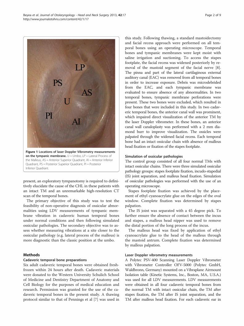
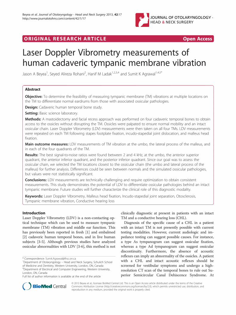
Figure 1 Locations of laser Doppler Vibrometry measurementson the tympanic membrane. U = Umbo, LP = Lateral Process ofthe Malleus, AS = Anterior Superior Quadrant, AI = Anterior InferiorQuadrant, PS = Posterior Superior Quadrant, PI = PosteriorInferior Quadrant.
Beyea et al. Journal of Otolaryngology - Head and Neck Surgery 2013, 42:17 Page 2 of 9http://www.journalotohns.com/content/42/1/17
present, an exploratory tympanotomy is required to defini-tively elucidate the cause of the CHL in these patients withan intact TM and an unremarkable high-resolution CTscan of the temporal bones.The primary objective of this study was to test the
feasibility of non-operative diagnosis of ossicular abnor-malities using LDV measurements of tympanic mem-brane vibration in cadaveric human temporal bonesunder normal conditions and then following simulatedossicular pathologies. The secondary objective was to as-sess whether measuring vibrations at a site closer to theossicular pathology (e.g. lateral process of the malleus) ismore diagnostic than the classic position at the umbo.
MethodsCadaveric temporal bone preparationsSix adult cadaveric temporal bones were obtained fresh-frozen within 24 hours after death. Cadaveric materialswere donated to the Western University Schulich Schoolof Medicine and Dentistry Department of Anatomy andCell Biology for the purposes of medical education andresearch. Permission was granted for the use of the ca-daveric temporal bones in the present study. A thawingprotocol similar to that of Pennings et al [7] was used in
this study. Following thawing, a standard mastoidectomyand facial recess approach were performed on all tem-poral bones using an operating microscope. Temporalbones and tympanic membranes were kept moist withsaline irrigation and suctioning. To access the stapesfootplate, the facial recess was widened posteriorly by re-moval of the mastoid segment of the facial nerve [8].The pinna and part of the lateral cartilaginous externalauditory canal (EAC) was removed from all temporal bonesin order to increase exposure. Debris was microdebridedfrom the EAC, and each tympanic membrane wasevaluated to ensure absence of any abnormalities. In twotemporal bones, tympanic membrane perforations werepresent. These two bones were excluded, which resulted infour bones that were included in this study. In two cadav-eric temporal bones, the anterior canal wall was prominent,which impaired direct visualization of the anterior TM bythe laser Doppler vibrometer. In these bones, an anteriorcanal wall canaloplasty was performed with a 2 mm dia-mond burr to improve visualization. The ossicles werepalpated through the widened facial recess. Each temporalbone had an intact ossicular chain with absence of malleushead fixation or fixation of the stapes footplate.
Simulation of ossicular pathologiesThe control group consisted of all four normal TMs withintact ossicular chains. There were three simulated ossicularpathology groups: stapes footplate fixation, incudo-stapedial(IS) joint separation, and malleus head fixation. Simulationof ossicular pathologies was performed with the use of anoperating microscope.Stapes footplate fixation was achieved by the place-
ment of ethyl cyanoacrylate glue on the edges of the ovalwindow. Complete fixation was determined by stapespalpation.The IS joint was separated with a 45 degree pick. To
further ensure the absence of contact between the incusand stapes, a malleus head nipper was used to removethe distal portion of the long process of the incus.The malleus head was fixed by application of ethyl
cyanoacrylate glue to the head of the malleus throughthe mastoid antrum. Complete fixation was determinedby malleus palpation.
Laser Doppler vibrometry measurementsA Polytec PSV-400 Scanning Laser Doppler Vibrometerwith Vibrometer Controller OFV-5000 (Polytec GmbH,Waldbronn, Germany) mounted on a Vibraplane AirmountIsolation table (Kinetic Systems, Inc., Boston, MA, U.S.A.)was used for all LDV measurements. LDV measurementswere obtained in all four cadaveric temporal bones fromthe normal TM with intact ossicular chain, the TM afterstapes fixation, the TM after IS joint separation, and theTM after malleus head fixation. For each cadaveric ear in
Figure 2 Experimental laser Doppler Vibrometry setup. The laser Doppler vibrometer is mounted on an isolation table, adjacent to thespeaker. The microphone is placed in the external auditory canal. An operating microscope is available for ossicular inspection, palpation,and modifications.
Beyea et al. Journal of Otolaryngology - Head and Neck Surgery 2013, 42:17 Page 3 of 9http://www.journalotohns.com/content/42/1/17
each of the experimental conditions, LDV measurementswere taken from six locations on the TM (Figure 1): theumbo, the lateral process of the malleus, the anterior super-ior quadrant, the anterior inferior quadrant, the posteriorsuperior quadrant, and the posterior inferior quadrant. Foreach TM location, LDV measurements were taken fromnine points arranged in a 3×3 grid, then averaged.The TMs were stimulated with a 75-80 dB stimulus
from 10 to 8000 Hz with a frequency sweep measured ateach increment of 2.5 Hz, delivered through a Harman-Kardon HK-195 speaker (Harman Kardon, Stamford,CT, U.S.A.) placed adjacent to the cadaveric head. Soundpressure was measured with an ER-7C microphone(Etymotic Research, Elk Grove Village, IL, U.S.A.) placedwithin 2 mm of the TM, and a sound pressure of >75 dBwas measured from 200-8000 Hz. LDV measurementswere collected within the frequency range of 10–8000Hz. Measurements were repeated until two comparabletracings were obtained from all TM locations in all ex-perimental groups. Once obtained, the two comparabletracings were averaged to produce one tracing for eachTM location in each cadaveric head in each experimen-tal group. Our experimental setup is shown in Figure 2.
Data analysisAll data were obtained with supplied Polytec software ver-sion 8.5 (Polytec GmbH, Waldbronn, Germany). Data wereexported to Matlab (Mathworks, Natick, Massachusetts, U.S.A.) for further analysis. To meet our signal-to-noise ratio(SNR) criteria, two conditions must have been met: (1) TMdisplacement was 10 dB greater than the background TMdisplacement with stimulus off, or TM velocity was 10 dBgreater than the background TM velocity with stimulus off,and (2) sound pressure was 20 dB SPL (sound pressurelevel) greater than the background sound pressurewith stimulus off. If the data did not meet the SNR, theywere excluded from further analysis. Data were plottednormalized to the EAC sound pressure. Displacement ver-sus sound pressure and velocity versus sound pressureacross a frequency range of 0.1 to 8 kHz were plotted to re-flect prior publications in the literature [3,9]. Raw data ispresented (the data were not smoothed). For the umbo andlateral process of the malleus data, a t-test was performedat α = 0.05 at all frequencies comparing each of the experi-mental groups (stapes footplate fixation, IS joint separation,and malleus head fixation) to the normal group using aBonferonni correction.
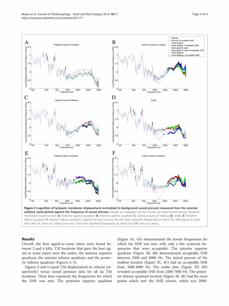
Figure 3 Logarithm of tympanic membrane displacement normalized to background sound pressure measured from the externalauditory canal plotted against the frequency of sound stimulus. Results are averaged over four bones per experimental group. Tympanicmembrane locations were: A. Posterior superior quadrant, B. Anterior superior quadrant, C. Lateral process of malleus, D. Umbo, E. Posteriorinferior quadrant, F. Anterior inferior quadrant. Legend: For each tracing, the thin lines represent frequencies at which the SNR (signal to noiseratio) did not meet our criteria (see text). Thick lines represent frequencies at which the SNR met our criteria.
Beyea et al. Journal of Otolaryngology - Head and Neck Surgery 2013, 42:17 Page 4 of 9http://www.journalotohns.com/content/42/1/17
ResultsOverall, the best signal-to-noise ratios were found be-tween 2 and 4 kHz. TM locations that gave the best sig-nal to noise ratios were the umbo, the anterior superiorquadrant, the anterior inferior quadrant, and the poster-ior inferior quadrant (Figures 3, 4).Figures 3 and 4 report TM displacement or velocity (re-
spectively) versus sound pressure data for all six TMlocations. Thick lines represent the frequencies for whichthe SNR was met. The posterior superior quadrant
(Figure 3A, 4A) demonstrated the fewest frequencies forwhich the SNR was met, with only a few scattered fre-quencies that were acceptable. The anterior superiorquadrant (Figure 3B, 4B) demonstrated acceptable SNRbetween 2500 and 8000 Hz. The lateral process of themalleus location (Figure 3C, 4C) had an acceptable SNRfrom 3000-4000 Hz. The umbo data (Figure 3D, 4D)revealed acceptable SNR from 2500-7000 Hz. The poster-ior inferior quadrant location (Figure 3E, 4E) had the mostpoints which met the SNR criteria, which was 2000-
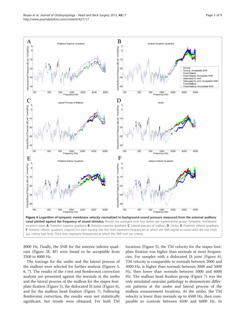
Figure 4 Logarithm of tympanic membrane velocity normalized to background sound pressure measured from the external auditorycanal plotted against the frequency of sound stimulus. Results are averaged over four bones per experimental group. Tympanic membranelocations were: A. Posterior superior quadrant, B. Anterior superior quadrant, C. Lateral process of malleus, D. Umbo, E. Posterior inferior quadrant,F. Anterior inferior quadrant. Legend: For each tracing, the thin lines represent frequencies at which the SNR (signal to noise ratio) did not meetour criteria (see text). Thick lines represent frequencies at which the SNR met our criteria.
Beyea et al. Journal of Otolaryngology - Head and Neck Surgery 2013, 42:17 Page 5 of 9http://www.journalotohns.com/content/42/1/17
8000 Hz. Finally, the SNR for the anterior inferior quad-rant (Figure 3F, 4F) were found to be acceptable from2500 to 8000 Hz.The tracings for the umbo and the lateral process of
the malleus were selected for further analysis (Figures 5,6, 7). The results of the t-test and Bonferonni correctionanalysis are presented against the normals at the umboand the lateral process of the malleus for the stapes foot-plate fixation (Figure 5), the dislocated IS joint (Figure 6),and for the malleus head fixation (Figure 7). FollowingBonferonni correction, the results were not statisticallysignificant, but trends were obtained. For both TM
locations (Figure 5), the TM velocity for the stapes foot-plate fixation was higher than normals at most frequen-cies. For samples with a dislocated IS joint (Figure 6),TM velocity is comparable to normals between 2000 and3000 Hz, is higher than normals between 3000 and 5000Hz, then lower than normals between 5000 and 6000Hz. The malleus head fixation group (Figure 7) was theonly simulated ossicular pathology to demonstrate differ-ent patterns at the umbo and lateral process of themalleus measurement locations. At the umbo, the TMvelocity is lower than normals up to 4500 Hz, then com-parable to controls between 4500 and 6000 Hz. In
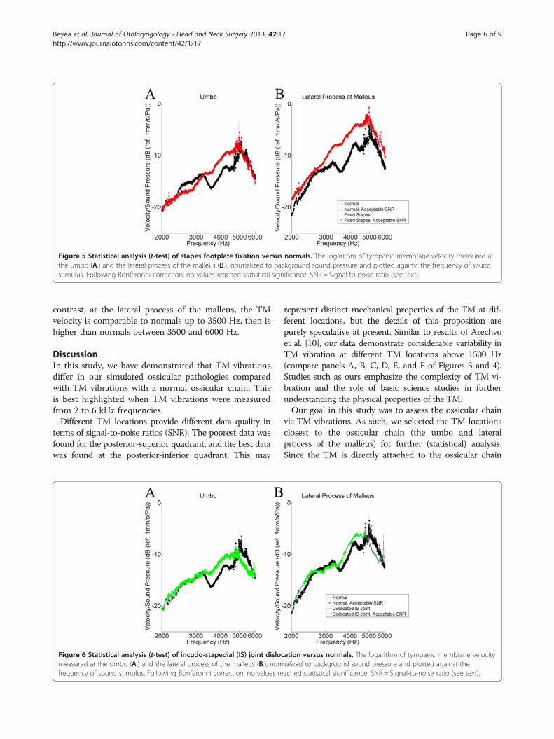
Figure 5 Statistical analysis (t-test) of stapes footplate fixation versus normals. The logarithm of tympanic membrane velocity measured atthe umbo (A.) and the lateral process of the malleus (B.), normalized to background sound pressure and plotted against the frequency of soundstimulus. Following Bonferonni correction, no values reached statistical significance. SNR = Signal-to-noise ratio (see text).
Beyea et al. Journal of Otolaryngology - Head and Neck Surgery 2013, 42:17 Page 6 of 9http://www.journalotohns.com/content/42/1/17
contrast, at the lateral process of the malleus, the TMvelocity is comparable to normals up to 3500 Hz, then ishigher than normals between 3500 and 6000 Hz.
DiscussionIn this study, we have demonstrated that TM vibrationsdiffer in our simulated ossicular pathologies comparedwith TM vibrations with a normal ossicular chain. Thisis best highlighted when TM vibrations were measuredfrom 2 to 6 kHz frequencies.Different TM locations provide different data quality in
terms of signal-to-noise ratios (SNR). The poorest data wasfound for the posterior-superior quadrant, and the best datawas found at the posterior-inferior quadrant. This may
Figure 6 Statistical analysis (t-test) of incudo-stapedial (IS) joint dislocmeasured at the umbo (A.) and the lateral process of the malleus (B.), normfrequency of sound stimulus. Following Bonferonni correction, no values re
represent distinct mechanical properties of the TM at dif-ferent locations, but the details of this proposition arepurely speculative at present. Similar to results of Arechvoet al. [10], our data demonstrate considerable variability inTM vibration at different TM locations above 1500 Hz(compare panels A, B, C, D, E, and F of Figures 3 and 4).Studies such as ours emphasize the complexity of TM vi-bration and the role of basic science studies in furtherunderstanding the physical properties of the TM.Our goal in this study was to assess the ossicular chain
via TM vibrations. As such, we selected the TM locationsclosest to the ossicular chain (the umbo and lateralprocess of the malleus) for further (statistical) analysis.Since the TM is directly attached to the ossicular chain
ation versus normals. The logarithm of tympanic membrane velocityalized to background sound pressure and plotted against theached statistical significance. SNR = Signal-to-noise ratio (see text).
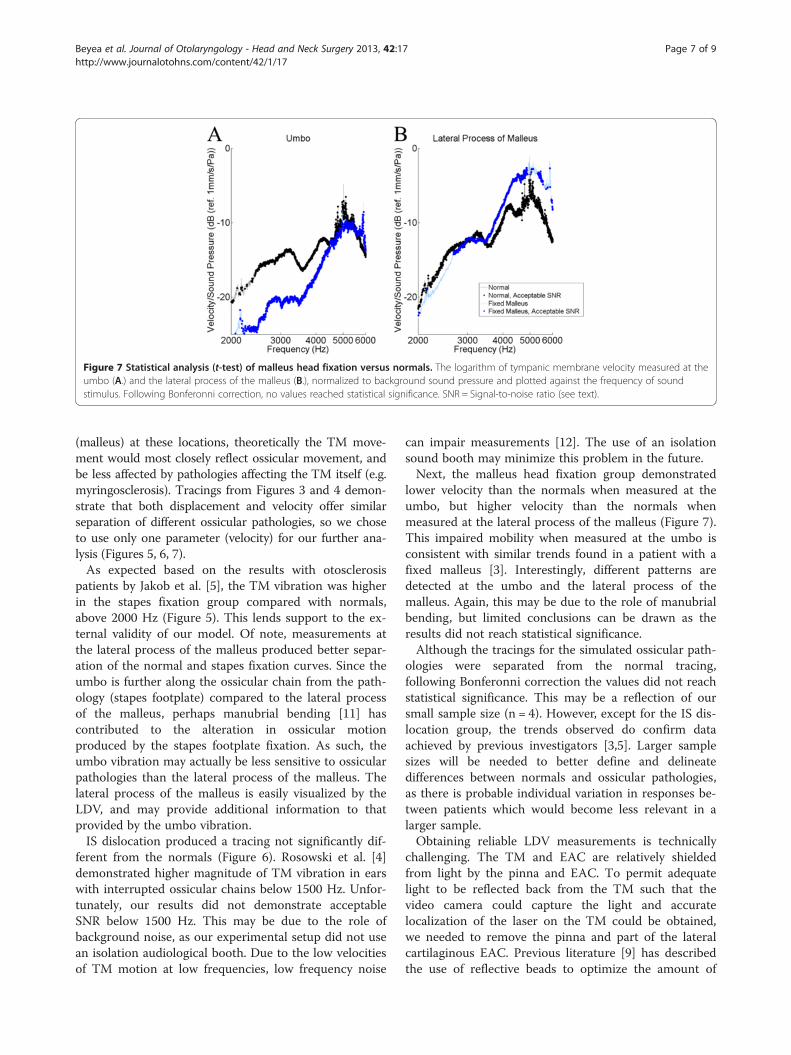
Figure 7 Statistical analysis (t-test) of malleus head fixation versus normals. The logarithm of tympanic membrane velocity measured at theumbo (A.) and the lateral process of the malleus (B.), normalized to background sound pressure and plotted against the frequency of soundstimulus. Following Bonferonni correction, no values reached statistical significance. SNR = Signal-to-noise ratio (see text).
Beyea et al. Journal of Otolaryngology - Head and Neck Surgery 2013, 42:17 Page 7 of 9http://www.journalotohns.com/content/42/1/17
(malleus) at these locations, theoretically the TM move-ment would most closely reflect ossicular movement, andbe less affected by pathologies affecting the TM itself (e.g.myringosclerosis). Tracings from Figures 3 and 4 demon-strate that both displacement and velocity offer similarseparation of different ossicular pathologies, so we choseto use only one parameter (velocity) for our further ana-lysis (Figures 5, 6, 7).As expected based on the results with otosclerosis
patients by Jakob et al. [5], the TM vibration was higherin the stapes fixation group compared with normals,above 2000 Hz (Figure 5). This lends support to the ex-ternal validity of our model. Of note, measurements atthe lateral process of the malleus produced better separ-ation of the normal and stapes fixation curves. Since theumbo is further along the ossicular chain from the path-ology (stapes footplate) compared to the lateral processof the malleus, perhaps manubrial bending [11] hascontributed to the alteration in ossicular motionproduced by the stapes footplate fixation. As such, theumbo vibration may actually be less sensitive to ossicularpathologies than the lateral process of the malleus. Thelateral process of the malleus is easily visualized by theLDV, and may provide additional information to thatprovided by the umbo vibration.IS dislocation produced a tracing not significantly dif-
ferent from the normals (Figure 6). Rosowski et al. [4]demonstrated higher magnitude of TM vibration in earswith interrupted ossicular chains below 1500 Hz. Unfor-tunately, our results did not demonstrate acceptableSNR below 1500 Hz. This may be due to the role ofbackground noise, as our experimental setup did not usean isolation audiological booth. Due to the low velocitiesof TM motion at low frequencies, low frequency noise
can impair measurements [12]. The use of an isolationsound booth may minimize this problem in the future.Next, the malleus head fixation group demonstrated
lower velocity than the normals when measured at theumbo, but higher velocity than the normals whenmeasured at the lateral process of the malleus (Figure 7).This impaired mobility when measured at the umbo isconsistent with similar trends found in a patient with afixed malleus [3]. Interestingly, different patterns aredetected at the umbo and the lateral process of themalleus. Again, this may be due to the role of manubrialbending, but limited conclusions can be drawn as theresults did not reach statistical significance.Although the tracings for the simulated ossicular path-
ologies were separated from the normal tracing,following Bonferonni correction the values did not reachstatistical significance. This may be a reflection of oursmall sample size (n = 4). However, except for the IS dis-location group, the trends observed do confirm dataachieved by previous investigators [3,5]. Larger samplesizes will be needed to better define and delineatedifferences between normals and ossicular pathologies,as there is probable individual variation in responses be-tween patients which would become less relevant in alarger sample.Obtaining reliable LDV measurements is technically
challenging. The TM and EAC are relatively shieldedfrom light by the pinna and EAC. To permit adequatelight to be reflected back from the TM such that thevideo camera could capture the light and accuratelocalization of the laser on the TM could be obtained,we needed to remove the pinna and part of the lateralcartilaginous EAC. Previous literature [9] has describedthe use of reflective beads to optimize the amount of
Beyea et al. Journal of Otolaryngology - Head and Neck Surgery 2013, 42:17 Page 8 of 9http://www.journalotohns.com/content/42/1/17
light reflected. We chose not to use reflective beads inorder to create a TM model that could be transitionedinto a future in vivo protocol, as reflective beads wouldnot be appropriate with live human participants. ThePolytec vibrometer detected that there was sufficient signalwithout reflective beads in order to make measurements;however, there likely would have been a better SNR if re-flective beads were used. Next, the anterior EAC canal wallcan be prominent, and thus impairs visualization and laserDoppler measurements from the anterior TM. For thesetemporal bones, an anterior canal wall canaloplasty wasrequired to provide adequate visualization. Following ourmodifications, we were able to obtain reproducible tracingsfrom all TM locations. This finding, in addition to thecomparable nature of our results to those of previousinvestigators, serves as both internal and external valid-ation of our methodology, and establishes the basis for fu-ture basic science and clinical LDV studies at our centre.We should highlight, however, potential future difficultiesin our transition to live human subjects. Clearly, themodifications which were made to the cadavers would notbe performed in a human study. This will necessitatefurther optimization of lighting and laser positioningto achieve reproducible results. Given our use of the anter-ior canaloplasty in this study, perhaps anterior TMmeasurements may not be possible in all human subjects.Note should be made that although a clinical hearing laservibrometer is commercially available (Polytec GmbH,Waldbronn, Germany) that can be used in vivo, it doesnot provide unobstructed access to the entire TM via theEAC. Overcoming these technical challenges will be para-mount in future experiments in our centre.Previous literature [13] demonstrates that the cadav-
eric temporal bone is a valid model of middle and innerear mechanics as measured by LDV. As LDV is a non-contact technique, the mechanical properties of the TMwere not altered by our measurements. Therefore, themeasured cadaveric TM vibrations in our study mimicthe vibrations which would occur in a normal livehuman ear exposed to the same acoustic stimulus. Ourcadaveric measurements in this study will permit aneducated evaluation of live human TM vibrations in fu-ture studies.This study also describes a useful model for the inves-
tigation of ossicular abnormalities in cadaveric temporalbones, a variant of the models described by Stasche et al.[14]. Fixation of the stapes footplate and the malleushead can be performed with a mastoidectomy, facial re-cess, and removal of the mastoid segment of the facialnerve. Adequacy of fixation is readily confirmed byossicular palpation. Other ossicular manipulations, suchas the IS joint dislocation in the present study, can alsoreadily be performed and confirmed by inspection andpalpation. This model is inexpensive, easy to perform for
otolaryngologists, and uses commonly available materials(ethyl cyanoacrylate glue).The motivation for this study was to evaluate the pos-
sibility of diagnosis of ossicular pathology in patientswith a conductive hearing loss in the presence of an in-tact TM. Often patients undergoing an exploratorytympanotomy do not get a preoperative CT scan unlessthere is concern for cholesteatoma or if the patient hashad prior ear surgery, as a CT scan would expose thesepatients to unnecessary radiation. Furthermore, ossicularabnormalities often cannot be diagnosed by CT. Ifossicular pathologies could be diagnosed preoperativelywith LDV, this would theoretically permit better surgicalplanning, and would limit the role of diagnostic explora-tory tympanotomy. From the current study, LDV cansuggest a diagnosis of ossicular abnormalities, but atpresent exploratory tympanotomy remains the goldstandard for diagnosis.
ConclusionsLaser Doppler Vibrometry measurements are challen-ging. However, once proper TM positioning and lightinghave been obtained, LDV delivers consistent and repro-ducible results which characterize TM vibrationresponses to a sound stimulus. This modality holds greatpromise as a diagnostic tool to characterize the status ofthe ossicles in patients with a conductive hearing lossand an intact TM.
This manuscript was presented at: Canadian Society of Otolaryngology -Head and Neck Surgery 66th Annual Meeting, Fairmont Royal York Hotel,Toronto, Ontario, Canada. May 21, 2012.
Competing interestThe authors have no actual or potential conflict of interest to disclose.
Authors’ contributionsJB, HL, and SA designed the experiments. JB and SR collected and analyzedthe data. JB, SR, HL, and SA wrote the manuscript. All authors discussed theresults and implications and commented on the manuscript at all stages ofpreparation. All authors read and approved the final manuscript.
AcknowledgementsThe authors would like to thank the Western University Department ofOtolaryngology – Head and Neck Surgery Resident Research Program, theNatural Sciences and Engineering Research Council of Canada (NSERC),Medtronic of Canada Ltd., and the Ontario Research Fund (ORF) for financialsupport of this project.
Grants/Financial Support1) Western University Department of Otolaryngology – Head and NeckSurgery Resident Research Program, 2) Natural Sciences and EngineeringResearch Council of Canada (NSERC), 3) Ontario Research Fund, 4) Medtronicof Canada Ltd.
Author details1Department of Otolaryngology – Head and Neck Surgery, Schulich Schoolof Medicine and Dentistry, Western University, London, ON, Canada.2Biomedical Engineering Graduate Program, Western University, London, ON,Canada. 3Department of Medical Biophysics, Western University, London, ON,Canada. 4Department of Electrical and Computer Engineering, WesternUniversity, London, ON, Canada. 5Schulich School of Medicine and Dentistry,
Beyea et al. Journal of Otolaryngology - Head and Neck Surgery 2013, 42:17 Page 9 of 9http://www.journalotohns.com/content/42/1/17
Department of Otolaryngology - Head and Neck Surgery, Western University,339 Windermere Road, P.O. Box 5339, London, ON, Canada.
Received: 20 November 2012 Accepted: 6 January 2013Published: 25 February 2013
References1. Morris DP, Bance M, Van Wijhe RG: Vibration characteristics and function
of atelectatic segments in the tympanic membrane in fresh humancadaveric temporal bones. Clin Otolaryngol Allied Sci 2004, 29:133–137.
2. Stieger C, Candreia C, Kompis M, et al: Laser Doppler VibrometricAssessment of Middle Ear Motion in Thiel-Embalmed Heads.Otol Neurotol 2012, Feb 28.[Epub ahead of print].
3. Rosowski JJ, Mehta RP, Merchant SN: Diagnostic utility of laser-Dopplervibrometry in conductive hearing loss with normal tympanic membrane.Otol Neurotol 2003, 24:165–75.
4. Rosowski JJ, Nakajima HH, Merchant SN: Clinical utility of laser-Dopplervibrometer measurements in live normal and pathologic human ears.Ear Hear 2008, 29:3–19.
5. Jakob A, Bornitz M, Kuhlisch E, et al: New aspects in the clinical diagnosisof otosclerosis using laser Doppler vibrometry. Otol Neurotol 2009,30:1049–57.
6. Feeney MP, Grant IL, Mills DM: Wideband energy reflectancemeasurements of ossicular chain discontinuity and repair in humantemporal bone. Ear Hear 2009, 30:391–400.
7. Pennings RJE, Ho A, Brown J, et al: Analysis of Vibrant SoundbridgePlacement Against the Round Window Membrane in a HumanCadaveric Temporal Bone Model. Otol Neurotol 2010, 31:998–1003.
8. Voss SE, Rosowski JJ, Merchant SN, et al: Acoustic responses of the humanmiddle ear. Hear Res 2000, 150:43–69.
9. Bance M, Morris DP, Van Wijhe R: Effects of ossicular prosthesis mass andsection of the stapes tendon on middle ear transmission. J Otolaryngol2007, 36:113–9.
10. Arechvo I, Lasurashvili N, Bornitz M, et al: Laser Doppler vibrometry of themiddle ear in humans: derivation dependence, variability, and bilateraldifferences. Medicina (Kaunas) 2009, 45:878–86.
11. Funnell WR, Khanna SM, Decraemer WF: On the degree of rigidity of themanubrium in a finite-element model of the cat eardrum. J Acoust SocAm 1992, 91:2082–90.
12. Akache F, Funnell WR, Daniel SJ: An experimental study of tympanicmembrane and manubrium vibrations in rats. Audiol Neurootol 2007,12:49–58.
13. Chien W, Ravicz ME, Rosowski JJ, et al: Measurements of human middle-and inner-ear mechanics with dehiscence of the superior semicircularcanal. Otol Neurotol 2007, 28:250–7.
14. Stasche N, Foth HJ, Hörmann K, et al: Middle ear transmission disorders–tympanic membrane vibration analysis by laser-Doppler-vibrometry.Acta Otolaryngol 1994, 114:59–63.
doi:10.1186/1916-0216-42-17Cite this article as: Beyea et al.: Laser Doppler Vibrometrymeasurements of human cadaveric tympanic membrane vibration.Journal of Otolaryngology - Head and Neck Surgery 2013 42:17.
Submit your next manuscript to BioMed Centraland take full advantage of:
• Convenient online submission
• Thorough peer review
• No space constraints or color figure charges
• Immediate publication on acceptance
• Inclusion in PubMed, CAS, Scopus and Google Scholar
• Research which is freely available for redistribution
Submit your manuscript at www.biomedcentral.com/submit