Embed Size (px)
Citation preview

Hindawi Publishing CorporationCase Reports in Gastrointestinal MedicineVolume 2011, Article ID 107087, 6 pagesdoi:10.1155/2011/107087
Case Report
Laparoscopic Drainage of a Hepatic Echinococcal Cyst:A Case Report
Steven B. Goldin, James J. L. Mateka, Michael J. Schnaus, and Sujat Dahal
Department of Surgery, College of Medicine, University of South Florida, Tampa, FL 33606, USA
Correspondence should be addressed to Steven B. Goldin, [email protected] and James J. L. Mateka, [email protected]
Received 2 June 2011; Accepted 26 June 2011
Academic Editors: G. Nardone and I. Siddique
Copyright © 2011 Steven B. Goldin et al. This is an open access article distributed under the Creative Commons AttributionLicense, which permits unrestricted use, distribution, and reproduction in any medium, provided the original work is properlycited.
The Echinococcus granulosus tapeworm causes hepatic echinococcosis. It is endemic in the Mediterranean region, Middle East,and South America. Human infection is secondary to accidental consumption of ova in feces. Absorption through the bowel walland entrance into the portal circulation leads to liver infection. This case involves a 34 y/o Moroccan male with an echinococcalliver cyst. His chief complaint was RUQ pain. The patient was treated with albendazole and praziquantel. His PMH and PSH wasnoncontributory. Patient was not on any other medications. ROS was otherwise unremarkable. The patient was AF VSS. He wastender to palpation in RUQ. Liver function tests were normal. Echinococcal titers were positive. CT demonstrated a large cysticlesion in the right lobe of the liver measuring 13.5 cm in diameter. The patient underwent successful laparoscopic drainage andexcision of echinococcal cyst. Final pathology demonstrated degenerating parasites (E. granulosus) of echinococcal cyst.
1. Introduction
Hepatic echinococcus is acquired by humans secondary toaccidental consumption of ova in dog feces [1]. It resultsfrom infection by the dog tapeworm. It is endemic insheep and cattle raising areas in Europe, China, Russia, theMediterranean region, Middle East, South America, Aus-tralia, New Zealand, and Southern Africa. It is exceedinglyrare in the United States [2] and mainly occurs in membersof high risk groups such as sheep farmers in the west,some native American Indians in the southwest and Alaska,and in immigrants [3]. Its incidence is estimated to be lessthan 1 case per 1 million population in the continentalUnited States and can range from 1 to 220 cases per100,000 persons in endemic areas. Hepatic echinococcal cystsare described most frequently using the ultrasonographicclassification system of Gharbi et al. [4] (Table 1). Based onthe Gharbi classification system, the World Health Organi-zation (WHO) Informal Working Group on Echinococcosismodified their International Classification System [5] toidentify the functional state of parasites to define treatment(Table 2).
2. Case Report
A 34-year-old Moroccan male presented with a severalmonth history of moderately severe, constant, and progres-sively worsening, dull aching right upper quadrant pain(RUQ). There were no alleviating and precipitating factors.A computed tomography (CT) scan revealed a 13.5 cm cysticlesion in the right lobe of the liver (Gharbi Type I) anddensely calcified masses in the right and left sides of the liver(Gharbi Type V). An ELISA test for echinococcal infectionwas positive. The patient was treated with albendazole andpraziquantel for 3 months with no clinical or radiographicimprovement (Figure 1). Laparoscopic cyst evacuation andpartial cyst excision was undertaken.
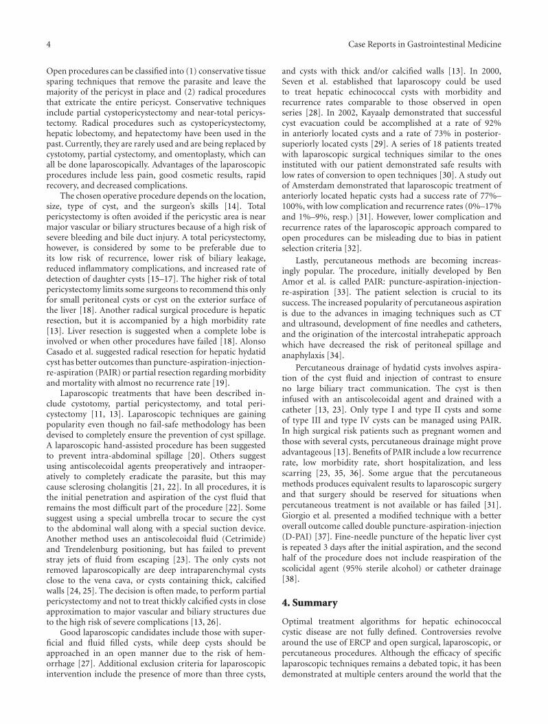
The procedure was done using four 12 mm ports asshown in Figure 2. The cyst was first surrounded withpads soaked in a scolicidal agent (20% hypertonic saline)(Figure 3), to protect the abdominal contents from contam-ination. Approximately, 400 mL was aspirated from the cyst,which was then filled with 1 liter of 20% hypertonic saline(Figure 4). This saline was aspirated, and the cyst refilledagain with hypertonic saline. The saline was allowed to dwell

2 Case Reports in Gastrointestinal Medicine
Figure 1: CT after 3 month course of albendazole and praziquantel.
Table 1: Gharbi classification of hepatic echinococcal cysts.
Type I Pure fluid
Type II Fluid collection, spilt-wall floatingmembrane
Type III Fluid collection with septa, daughtercysts, and honeycomb image
Type IV Heterogeneous echographic pattern
Type V Reflecting thick walls
in the cyst cavity for 30 minutes per cycle to ensure completekilling of organisms. The cyst was then opened and a portionof the cyst wall excised using a harmonic scalpel and endo-GIA stapler with 2.5 mm vascular loads (Figures 5 and 6).A “double bag” technique was used to remove the excised cystwall and debris, which were placed into an Endo CATCH bagand a Lapsac Surgical Tissue Pouch prior to being removedfrom the abdomen. An argon beam coagulator was thenused on the entire remaining cyst wall. A Yankauer suckerwas placed through the midepigastric port site, to aid inirrigation of the abdomen with a Clorpactin solution. A 10French flat Jackson-Pratt drain was placed in the cyst bed.
The patient was discharged on postoperative day threewith the drain, which had collected a small amount of biliousfluid. The drain was removed after seven days. Pathologyshowed degenerating stage cysts without any living forms ofE. granulosus. The infectious disease service recommended28 more days of albendazole. At six-month followup, thepatient remained asymptomatic and disease-free.
3. Discussion
Surgery is the primary treatment for echinococcal disease.However, controversy still surrounds the preoperative med-ical treatment and type of operative procedure. The role ofpreoperative endoscopic retrograde cholangiopancreatogra-phy (ERCP) continues to be debated, with proposed benefitsincluding preoperative definition of cystobiliary relation-ships, treatment of cholangitis and biliary obstruction, andpossible definitive treatment in cases of intrabiliary rupture.In one study, ERCP was reported to be safe method totreat biliary complications of hepatic hydatidosis before and
Trocar placement
1
2
34
Figure 2: Trocar placement.
Figure 3: Cyst surrounded by hypertonic saline-soaked pediatriclaparotomy pads.
Figure 4: Aspiration of cyst contents and instillation of hypertonicsaline.

Case Reports in Gastrointestinal Medicine 3
Table 2: International classification of ultrasound images in cystic echinococcosis for application in clinical and field epidemiologicalsettings.
Tybe of Cyst Status Ultrasound features Remarks
CL Active Signs no pathognomonic, unilocular, and no cyst wallUsually early stage, not fertile, anddifferential diagnosis necessary
CE 1 Active Cyst wall, hydatid sand Usually fertile
CE 2 Active Multivesicular, c st wall, and “rosette-like” Usually fertile
CE 3 Transitional Detachment of laminated membrane, “water-lil sign,”less round-decreased intracystic pressure
Starting to degenerate, may producedaughter cysts
CE 4 Inactive Heterogeneous hypo- or hyperechogenic degenerativecontents, no daughter cysts
Usually no living protoscoleces,differential diagnosis necessary
CE 5 InactiveThick calcified wall, calcification partial to complete,and not pathognomonic but highly suggestive ofdiagnosis
Usually no living protoscoleces
Figure 5: Excision of cyst wall using a harmonic scalpel.
Figure 6: Excision of cyst wall using endo-GIA stapler with 2.5 mmvascular loads.
after surgical management [6]. ERCP, however, may causecomplications and has a false negative rate of 17%–20%in identifying small cystobiliary communications due toelevated cyst pressure, minimal communication, or transientobstruction of the communication by daughter cysts [7].
Therefore, even if no cystobiliary communications are iden-tified preoperatively, cysts must still be carefully inspectedintraoperatively, since small cystobiliary communicationsdevelop in 80%–90% of patients with hepatic echinococcaldisease.
Biliary obstruction only occurs in 5%–10% of patientsdue to biliary communications that are at least 5 mm indiameter. Communications of this size or larger are moresignificant due to their ability to carry hydatid debris anddaughter cysts into the common bile duct causing biliaryobstruction in over 66.6% of cases. In patients without jaun-dice or cholangitis, ERCP findings are normal in 50% anddemonstrate cystobiliary rupture, biloma, or bile duct com-pression in the other 50% [8, 9]. Ozaslan and Bayraktar [7]suggest that preoperative or postoperative ERCP should beused only for complicated cases and that for uncomplicatedcases, routine use of ERCP should not be recommendedexcept for surgery planning. Another recent study suggestedthat cyst diameter independently predicts risk for biliary-cystcommunication in asymptomatic patients. The mean cystsize in patients with biliary leakage was 10.2 cm, compared to6.1 cm in patients without biliary leakage, and they suggestedthat preoperative ERCP should only be used in asymptomaticpatients large cysts [10].
Use of an intraoperative cholangiogram is also controver-sial. Ramachandran and Arora suggest that an intraoperativecholangiogram is unnecessary and increases morbidity [11].They state that inspection of the cyst wall using thelaparoscope will identify all major bile leaks. Ozmen andCoskun [12] also states biliary tract communications can becontrolled by inspection of the cyst cavity and ligation ofruptured bile ducts less than 5 mm in diameter. If bile ductsgreater than 5 mm in diameter are identified, intraoperativecholangiogram is done to assess the common bile duct(CBD) for debris, which requires CBD exploration with T-tube placement if present. 5 mm is used as a cutoff as bileducts smaller than this rarely transmit particulate matterto the CBD, while 65% of bile ducts 5 mm or larger allowpassage of material into the CBD.
All surgical treatments require complete cyst exposure,cyst decompression evacuation and sterilization, ligation ofbile duct communications, and cavity management [13].
4 Case Reports in Gastrointestinal Medicine
Open procedures can be classified into (1) conservative tissuesparing techniques that remove the parasite and leave themajority of the pericyst in place and (2) radical proceduresthat extricate the entire pericyst. Conservative techniquesinclude partial cystopericystectomy and near-total pericys-tectomy. Radical procedures such as cystopericystectomy,hepatic lobectomy, and hepatectomy have been used in thepast. Currently, they are rarely used and are being replaced bycystotomy, partial cystectomy, and omentoplasty, which canall be done laparoscopically. Advantages of the laparoscopicprocedures include less pain, good cosmetic results, rapidrecovery, and decreased complications.
The chosen operative procedure depends on the location,size, type of cyst, and the surgeon’s skills [14]. Totalpericystectomy is often avoided if the pericystic area is nearmajor vascular or biliary structures because of a high risk ofsevere bleeding and bile duct injury. A total pericystectomy,however, is considered by some to be preferable due toits low risk of recurrence, lower risk of biliary leakage,reduced inflammatory complications, and increased rate ofdetection of daughter cysts [15–17]. The higher risk of totalpericystectomy limits some surgeons to recommend this onlyfor small peritoneal cysts or cyst on the exterior surface ofthe liver [18]. Another radical surgical procedure is hepaticresection, but it is accompanied by a high morbidity rate[13]. Liver resection is suggested when a complete lobe isinvolved or when other procedures have failed [18]. AlonsoCasado et al. suggested radical resection for hepatic hydatidcyst has better outcomes than puncture-aspiration-injection-re-aspiration (PAIR) or partial resection regarding morbidityand mortality with almost no recurrence rate [19].
Laparoscopic treatments that have been described in-clude cystotomy, partial pericystectomy, and total peri-cystectomy [11, 13]. Laparoscopic techniques are gainingpopularity even though no fail-safe methodology has beendevised to completely ensure the prevention of cyst spillage.A laparoscopic hand-assisted procedure has been suggestedto prevent intra-abdominal spillage [20]. Others suggestusing antiscolecoidal agents preoperatively and intraoper-atively to completely eradicate the parasite, but this maycause sclerosing cholangitis [21, 22]. In all procedures, it isthe initial penetration and aspiration of the cyst fluid thatremains the most difficult part of the procedure [22]. Somesuggest using a special umbrella trocar to secure the cystto the abdominal wall along with a special suction device.Another method uses an antiscolecoidal fluid (Cetrimide)and Trendelenburg positioning, but has failed to preventstray jets of fluid from escaping [23]. The only cysts notremoved laparoscopically are deep intraparenchymal cystsclose to the vena cava, or cysts containing thick, calcifiedwalls [24, 25]. The decision is often made, to perform partialpericystectomy and not to treat thickly calcified cysts in closeapproximation to major vascular and biliary structures dueto the high risk of severe complications [13, 26].
Good laparoscopic candidates include those with super-ficial and fluid filled cysts, while deep cysts should beapproached in an open manner due to the risk of hem-orrhage [27]. Additional exclusion criteria for laparoscopicintervention include the presence of more than three cysts,
and cysts with thick and/or calcified walls [13]. In 2000,Seven et al. established that laparoscopy could be usedto treat hepatic echinococcal cysts with morbidity andrecurrence rates comparable to those observed in openseries [28]. In 2002, Kayaalp demonstrated that successfulcyst evacuation could be accomplished at a rate of 92%in anteriorly located cysts and a rate of 73% in posterior-superiorly located cysts [29]. A series of 18 patients treatedwith laparoscopic surgical techniques similar to the onesinstituted with our patient demonstrated safe results withlow rates of conversion to open techniques [30]. A study outof Amsterdam demonstrated that laparoscopic treatment ofanteriorly located hepatic cysts had a success rate of 77%–100%, with low complication and recurrence rates (0%–17%and 1%–9%, resp.) [31]. However, lower complication andrecurrence rates of the laparoscopic approach compared toopen procedures can be misleading due to bias in patientselection criteria [32].
Lastly, percutaneous methods are becoming increas-ingly popular. The procedure, initially developed by BenAmor et al. is called PAIR: puncture-aspiration-injection-re-aspiration [33]. The patient selection is crucial to itssuccess. The increased popularity of percutaneous aspirationis due to the advances in imaging techniques such as CTand ultrasound, development of fine needles and catheters,and the origination of the intercostal intrahepatic approachwhich have decreased the risk of peritoneal spillage andanaphylaxis [34].
Percutaneous drainage of hydatid cysts involves aspira-tion of the cyst fluid and injection of contrast to ensureno large biliary tract communication. The cyst is theninfused with an antiscolecoidal agent and drained with acatheter [13, 23]. Only type I and type II cysts and someof type III and type IV cysts can be managed using PAIR.In high surgical risk patients such as pregnant women andthose with several cysts, percutaneous drainage might proveadvantageous [13]. Benefits of PAIR include a low recurrencerate, low morbidity rate, short hospitalization, and lessscarring [23, 35, 36]. Some argue that the percutaneousmethods produces equivalent results to laparoscopic surgeryand that surgery should be reserved for situations whenpercutaneous treatment is not available or has failed [31].Giorgio et al. presented a modified technique with a betteroverall outcome called double puncture-aspiration-injection(D-PAI) [37]. Fine-needle puncture of the hepatic liver cystis repeated 3 days after the initial aspiration, and the secondhalf of the procedure does not include reaspiration of thescolicidal agent (95% sterile alcohol) or catheter drainage[38].
4. Summary
Optimal treatment algorithms for hepatic echinococcalcystic disease are not fully defined. Controversies revolvearound the use of ERCP and open surgical, laparoscopic, orpercutaneous procedures. Although the efficacy of specificlaparoscopic techniques remains a debated topic, it has beendemonstrated at multiple centers around the world that the

Case Reports in Gastrointestinal Medicine 5
principles of open surgical treatment of hepatic echinococcuscan be adhered to by laparoscopic intervention.
References
[1] P. Bouree, “Hydatidosis: dynamics of transmission,” WorldJournal of Surgery, vol. 25, no. 1, pp. 4–9, 2001.
[2] W. Trolan, “R. Echinococcosis,” eMedicine, 2009.
[3] M. Chrieki, “Echinococcosis—an emerging parasite in theimmigrant population,” American Family Physician, vol. 66,no. 5, pp. 817–821, 2002.
[4] H. A. Gharbi, W. Hassine, M. W. Brauner, and K. Dupuch,“Ultrasound examination of the hydatic liver,” Radiology, vol.139, no. 2, pp. 459–463, 1981.
[5] C. N. L. MacPherson, D. A. Vuitton, H. A. Gharbi et al.,“International classification of ultrasound images in cysticechinococcosis for application in clinical and field epidemio-logical settings,” Acta Tropica, vol. 85, no. 2, pp. 253–261, 2003.
[6] K. Goumas, A. Poulou, D. Dandakis et al., “Role of endoscopicintervention in biliary complications of hepatic hydatid cystdisease,” Scandinavian Journal of Gastroenterology, vol. 42, no.9, pp. 1113–1119, 2007.
[7] E. Ozaslan and Y. Bayraktar, “Endoscopic therapy in the man-agement of hepatobiliary hydatid disease,” Journal of ClinicalGastroenterology, vol. 35, no. 2, pp. 160–174, 2002.
[8] T. Ponchon, R. Bory, and A. Chavaillon, “Endoscopic retro-grade cholangiography and sphincterotomy for complicatedhepatic hydatid cyst,” Endoscopy, vol. 19, no. 4, pp. 174–177,1987.
[9] A. Uccheddu, C. Murgia, S. Licheri, C. Dazzi, and M. Cagetti,“Endoscopic retrograde cholangiography in the diagnosis ofthe liver hydatidosis,” Giornale di Chirurgia, vol. 10, no. 1-2,pp. 46–50, 1989.
[10] M. Kilic, O. Yoldas, M. Koc et al., “Can biliary-cyst communi-cation be predicted before surgery for hepatic hydatid disease:does size matter?” American Journal of Surgery, vol. 196, no. 5,pp. 732–735, 2008.
[11] C. S. Ramachandran and V. Arora, “Laparoscopic surgeryin hepatic hydatid cysts: a technical improvement,” SurgicalLaparoscopy, Endoscopy and Percutaneous Techniques, vol. 11,no. 1, pp. 14–18, 2001.
[12] M. M. Ozmen and F. Coskun, “New technique for findingthe ruptured bile duct into the liver cysts: scope in the cavetechnique,” Surgical Laparoscopy, Endoscopy and PercutaneousTechniques, vol. 12, no. 3, pp. 187–189, 2002.
[13] C. Dervenis, S. Delis, C. Avgerinos, J. Madariaga, and M.Milicevic, “Changing concepts in the management of liverhydatid disease,” Journal of Gastrointestinal Surgery, vol. 9, no.6, pp. 869–877, 2005.
[14] W. C. Meyers and R. D. kim, “Echinococcal cyst,” in SabistonTextbook of Surgery: The Biological Basis of Modern SurgicalPractice, C. M0 Townsend, Ed., pp. 1053–1055, WB Saunders,Philadelphia, Pa, USA, 2001.
[15] A. Cirenei and I. Bertoldi, “Evolution of surgery for liverhydatidosis from 1950 to today: analysis of a personalexperience,” World Journal of Surgery, vol. 25, no. 1, pp. 87–92, 2001.
[16] C. Dervenis, S. Delis, C. Avgerinos, J. Madariaga, and M.Milicevic, “Changing concepts in the management of liverhydatid disease,” Journal of Gastrointestinal Surgery, vol. 9, no.6, pp. 869–877, 2005.
[17] J. Prousalidis, E. Tzardinoglou, C. Kosmidis, K. Katsohis, andO. Aletras, “Surgical management of calcified hydatid cysts ofthe liver,” HPB Surgery, vol. 11, no. 4, pp. 253–259, 1999.
[18] M. Safioleas, E. Misiakos, C. Manti, D. Katsikas, G. Skalkeas,and E. Moreno-Gonzalez, “Diagnostic evaluation and surgicalmanagement of hydatid disease of the liver,” World Journal ofSurgery, vol. 18, no. 6, pp. 859–865, 1994.
[19] O. Alonso Casado, E. Moreno Gonzalez, C. Loinaz Segurolaet al., “Results of 22 years of experience in radical surgicaltreatment of hepatic hydatid cysts,” Hepato-Gastroenterology,vol. 48, no. 37, pp. 235–243, 2001.
[20] H. Bensaadi and G. Champault, “Laparoscopic Hand-AssistedSurgery for Hydatid Cysts of the Liver,” Surgical Laparoscopy,Endoscopy and Percutaneous Techniques, vol. 14, no. 2, pp. 91–92, 2004.
[21] A. O. Aktan and R. Yalin, “Preoperative albendazole treatmentfor liver hydatid disease decreases the viability of the cyst,”European Journal of Gastroenterology and Hepatology, vol. 8,no. 9, pp. 877–879, 1996.
[22] A. Saglam, “Laparoscopic treatment of liver hydatid cysts,”Surgical Laparoscopy, Endoscopy and Percutaneous Techniques,vol. 6, no. 1, pp. 16–21, 1996.
[23] A. Bickel, N. Loberant, and J. A. Lujan-Mompean, “Laparo-scopic treatment of a liver hydatid cyst,” British Journal ofSurgery, vol. 81, no. 4, p. 627, 1994.
[24] M. Ertem, C. Uras, T. Karahasanoglu, S. Erguney, and K.Alemdaroglu, “Laparoscopic approach to hepatic hydatiddisease,” Digestive Surgery, vol. 15, no. 4, pp. 333–336, 1998.
[25] J. A. Lujan Mompean, P. Parrilla Paricio, R. Robles Campos,and J. Garcia Ayllon, “Laparoscopic treatment of a liverhydated cyst,” British Journal of Surgery, vol. 80, no. 7, pp. 907–908, 1993.
[26] M. Uravic, D. Stimac, T. Lenac et al., “Diagnosis and treatmentof liver hydatid disease,” Hepato-Gastroenterology, vol. 45, no.24, pp. 2265–2269, 1998.
[27] I. Sayek and M. Cakmakci, “Laparoscopic treatment ofechinococcal cysts of the liver,” Zentralblatt fur Chirurgie, vol.124, no. 12, pp. 1143–1146, 1999.
[28] R. Seven, E. Berber, S. Mercan, L. Eminoglu, and D. Budak,“Laparoscopic treatment of hepatic hydatid cysts,” Surgery,vol. 128, no. 1, pp. 36–40, 2000.
[29] C. Kayaalp, “Evacuation of hydatid liver cysts using laparo-scopic trocar,” World Journal of Surgery, vol. 26, no. 11, pp.1324–1327, 2002.
[30] V. Baskaran and P. K. Patnaik, “Feasibility and safety oflaparoscopic management of hydatid disease of the liver,”Journal of the Society of Laparoendoscopic Surgeons, vol. 8, no.4, pp. 359–363, 2004.
[31] H. G. Schipper and P. A. Kager, “Diagnosis and treatmentof hepatic echinococcosis: an overview,” Scandinavian Journalof Gastroenterology, Supplement, vol. 39, no. 241, pp. 50–55,2004.
[32] K. Acarli, “Controversies in the laparoscopic treatment ofhepatic hydatid disease,” HPB, vol. 6, no. 4, pp. 213–221, 2004.
[33] N. Ben Amor, M. Gargouri, H. A. Gharbi, Y. J. Golvan,K. Ayachi, and H. Kchouck, “Trial therapy of inoperableabdominal hydatid cysts by puncture,” Annales de ParasitologieHumaine et Comparee, vol. 61, no. 6, pp. 689–692, 1986.
[34] S. Men, B. Hekimoglu, C. Yucesoy, I. S. Arda, and I.Baran, “Percutaneous treatment of hepatic hydatid cysts: analternative to surgery,” American Journal of Roentgenology, vol.172, no. 1, pp. 83–89, 1999.

6 Case Reports in Gastrointestinal Medicine
[35] M. S. Khuroo, S. A. Zargar, and R. Mahajan, “Echinococcusgranulosus cysts in the liver: management with percutaneousdrainage,” Radiology, vol. 180, no. 1, pp. 141–145, 1991.
[36] X. Wang, Y. Li, and S. Feng, “Clinical treatment of hepatic andabdominal hydatid cyst by percutaneous puncture, drainageand curettage,” Chinese Journal of Parasitology & ParasiticDiseases, vol. 12, no. 4, pp. 285–287, 1994.
[37] A. Giorgio, L. Tarantino, G. Francica et al., “Unilocularhydatid liver cysts: treatment with US-guided, double percu-taneous aspiration and alcohol injection,” Radiology, vol. 184,no. 3, pp. 705–710, 1992.
[38] A. Giorgio, G. De Stefano, V. Esposito et al., “Long-termresults of percutaneous treatment of hydatid liver cysts: asingle center 17 years experience,” Infection, vol. 36, no. 3, pp.256–261, 2008.

Submit your manuscripts athttp://www.hindawi.com
Stem CellsInternational
Hindawi Publishing Corporationhttp://www.hindawi.com Volume 2014
Hindawi Publishing Corporationhttp://www.hindawi.com Volume 2014
MEDIATORSINFLAMMATION
of
Hindawi Publishing Corporationhttp://www.hindawi.com Volume 2014
Behavioural Neurology
EndocrinologyInternational Journal of
Hindawi Publishing Corporationhttp://www.hindawi.com Volume 2014
Hindawi Publishing Corporationhttp://www.hindawi.com Volume 2014
Disease Markers
Hindawi Publishing Corporationhttp://www.hindawi.com Volume 2014
BioMed Research International
OncologyJournal of
Hindawi Publishing Corporationhttp://www.hindawi.com Volume 2014
Hindawi Publishing Corporationhttp://www.hindawi.com Volume 2014
Oxidative Medicine and Cellular Longevity
Hindawi Publishing Corporationhttp://www.hindawi.com Volume 2014
PPAR Research
The Scientific World JournalHindawi Publishing Corporation http://www.hindawi.com Volume 2014
Immunology ResearchHindawi Publishing Corporationhttp://www.hindawi.com Volume 2014
Journal of
ObesityJournal of
Hindawi Publishing Corporationhttp://www.hindawi.com Volume 2014
Hindawi Publishing Corporationhttp://www.hindawi.com Volume 2014
Computational and Mathematical Methods in Medicine
OphthalmologyJournal of
Hindawi Publishing Corporationhttp://www.hindawi.com Volume 2014
Diabetes ResearchJournal of
Hindawi Publishing Corporationhttp://www.hindawi.com Volume 2014
Hindawi Publishing Corporationhttp://www.hindawi.com Volume 2014
Research and TreatmentAIDS
Hindawi Publishing Corporationhttp://www.hindawi.com Volume 2014
Gastroenterology Research and Practice
Hindawi Publishing Corporationhttp://www.hindawi.com Volume 2014
Parkinson’s Disease
Evidence-Based Complementary and Alternative Medicine
Volume 2014Hindawi Publishing Corporationhttp://www.hindawi.com