Embed Size (px)
Citation preview
Laparoscopic Primary Repair with Omentopexy for Duodenal Ulcer Perforation: A Single Institution Experience of 21 Cases
Chung Hyeun Ma, and Min Gyu Kim
Department of Surgery, Hanyang University Guri Hospital, Hanyang University College of Medicine, Guri, Korea
Purpose: Despite the great advances in laparoscopic techniques, most active general surgeons do not apply laparoscopic surgery in the treatment of duodenal ulcer perforation when facing a real-life emergency. Therefore, our study was designed to evaluate the feasibility of laparoscopic surgery in duodenal ulcer perforation, and provide a step-by-step protocol with tips and recommendations for less experi-enced surgeons. Materials and Methods: Between March, 2011 and May, 2012, 21 patients presenting with duodenal ulcer perforation underwent lap-aroscopic primary repair with omentopexy. There were no contraindications to perform laparoscopic surgery, and the choice of primary repair was decided according to the size of the perforation. The procedure for laparoscopic primary repair with omentopexy consisted of peritoneal lavage, primary suture, and omentopexy using a knot pusher. Results: During the operation, no conversion to open surgery or intra-operative events occurred. The median operation time was 45.0 minutes (20~80 minutes). Median day of commencement of a soft diet was day 6 (4~17 days). After surgery, the median hospital stay was 8.0 days (5~27 days). Postoperative complications occurred in one patient, which included a minor leakage. This complica-tion was resolved by conservative management. Conclusions: Although our study was carried out on a small number of patients at a single institution, we conclude that laparoscopic pri-mary repair can be an effective surgical method in the treatment of duodenal ulcer perforation. We believe that the detailed explanation of our procedure will help beginners to perform laparoscopic primary repair more easily.
Key Words: Duodenal ulcer; Perforation; Laproscopy; Primary repair
Original ArticleJ Gastric Cancer 2012;12(4):237-242 http://dx.doi.org/10.5230/jgc.2012.12.4.237
Correspondence to: Min Gyu Kim
Department of Surgery, Hanyang University Guri Hospital, 153, Gyeongchun-ro, Guri 471-701, KoreaTel: +82-31-560-2294, Fax: +82-31-566-4409E-mail: [email protected] July 25, 2012Revised September 10, 2012Accepted September 11, 2012
Copyrights © 2012 by The Korean Gastric Cancer Association www.jgc-online.org
This is an open-access article distributed under the terms of the Creative Commons Attribution Non-Commercial License (http://creativecommons.org/licenses/by-nc/3.0) which permits unrestricted noncommercial use, distribution, and reproduction in any medium, provided the original work is properly cited.
Introduction
There have been many changes in the surgical techniques used
to treat complicated duodenal ulcer disease.(1-7) Especially in the
case of duodenal ulcer perforation, primary repair is one of the
most common methods used by acting surgeons in emergency
surgery because of the convenience of the surgical technique and
advances in medical treatment. After laparoscopic repair was first
introduced for duodenal ulcer perforation, many surgeons tried to
perform the technique and they reported their surgical outcomes
and experiences.(4,6,8,9)
With regards to surgical outcomes in laparoscopic primary re-
pair for duodenal ulcer perforation, there are still many questions as
to whether the surgeon should perform this type of surgery in an
emergency. Some surgeons have suggested that laparoscopic pri-
mary repair is the best way to improve early surgical outcomes.(10-
15) On the other hand, others believe that there is no significant
difference between laparoscopic primary repair group and open
primary repair group.(16-20)
Experience is a very important factor in improving the surgical
outcome of laparoscopic surgery. Many investigators have reported
that a learning period is necessary to improve the surgical outcomes
Ma CH and Kim MG
238
of this type of surgery in the treatment of stomach cancer.(21-24)
Therefore, only experienced laparoscopic surgeons participated in
this study to resolve this problem. This study aims to evaluate the
feasibility and safety of laparoscopic primary repair for duodenal
ulcer perforation, providing detailed technical information about
the procedure.
Materials and Methods
1. Patients
We enrolled 21 consecutive patients who underwent primary
repair with omentopexy for duodenal ulcer perforation between
March, 2011 and May, 2012 at Hanyang University Guri Hospital.
We retrospectively reviewed the prospectively collected data. In this
study, experienced surgeons were defined as those who had abun-
dant experience in laparoscopic surgery and other major operations
as the operating surgeon. One experienced surgeon had performed
139 laparoscopic gastrectomies to treat gastric cancer by March,
2010.(25)
There were no laparoscopic explorations during the study pe-
riod and there were no contraindications to perform laparoscopic
surgery. Laparoscopic primary repair was carried out regardless of
time from the onset of symptoms, previous laparotomy, old age,
and comorbidity. The choice of primary repair was decided by an
intra-operative assessment of the perforation size. During the same
period, five patients who were diagnosed with giant duodenal ulcer
perforation (>2 cm) underwent laparoscopic distal gastrectomy
with truncal vagotomy. These patients were excluded from the
study.(18)
2. Surgical techniques
Each patient was placed in thereverse Trendelenburg position. A
carbon dioxide pneumoperitoneum was formed from the umbilical
port, and pressure was maintained between 12 and 15 mmHg. Two
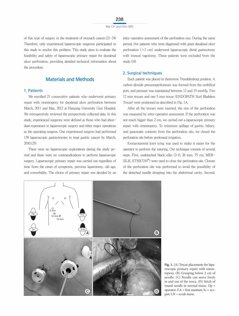
12 mm trocars and one 5 mm trocar (ENDOPATH Xcel Bladeless
Trocar) were positioned as described in Fig. 1A.
After all the trocars were inserted, the size of the perforation
was measured by intra-operative assessment. If the perforation was
not much bigger than 2 cm, we carried out a laparoscopic primary
repair with omentopexy. To minimize spillage of gastric, biliary,
and pancreatic contents from the perforation site, we closed the
perforation site before peritoneal irrigation.
Extracorporeal knot tying was used to make it easier for the
operator to perform the suturing. Our technique consists of several
steps. First, undetached black silks (3-0, 26 mm, 75 cm, MER-
SILK, ETHICON®) were used to close the perforation site. Closure
of the perforation site was performed to avoid the possibility of
the detached needle dropping into the abdominal cavity. Second,
Fig. 1. (A) Trocar placements for laparos copic primary repair with omentopexy. (B) Grasping below 2 cm of needle. (C) Needle can move freely in and out of the troca. (D) Stitch of round needle in normal tissue. Op = operator; F.A = first assistant; Sc = scopist; S.N = scrub nurse.
Laparoscopy for Duodenal Ulcer Perforation
239
the needle was prevented from dropping through the trocar dur-
ing insertion and extraction by grasping the thread 2 cm below the
needle (Fig. 1B). This thread was then moved into the right lower
trocar (Fig. 1C). Third, the needle was held just below the swage to
perform suture repair of full thickness around the perforation site
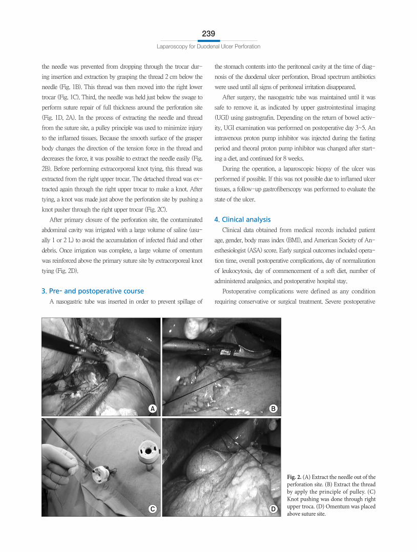
(Fig. 1D, 2A). In the process of extracting the needle and thread
from the suture site, a pulley principle was used to minimize injury
to the inflamed tissues. Because the smooth surface of the grasper
body changes the direction of the tension force in the thread and
decreases the force, it was possible to extract the needle easily (Fig.
2B). Before performing extracorporeal knot tying, this thread was
extracted from the right upper trocar. The detached thread was ex-
tracted again through the right upper trocar to make a knot. After
tying, a knot was made just above the perforation site by pushing a
knot pusher through the right upper trocar (Fig. 2C).
After primary closure of the perforation site, the contaminated
abdominal cavity was irrigated with a large volume of saline (usu-
ally 1 or 2 L) to avoid the accumulation of infected fluid and other
debris. Once irrigation was complete, a large volume of omentum
was reinforced above the primary suture site by extracorporeal knot
tying (Fig. 2D).
3. Pre- and postoperative course
A nasogastric tube was inserted in order to prevent spillage of
the stomach contents into the peritoneal cavity at the time of diag-
nosis of the duodenal ulcer perforation. Broad spectrum antibiotics
were used until all signs of peritoneal irritation disappeared.
After surgery, the nasogastric tube was maintained until it was
safe to remove it, as indicated by upper gastrointestinal imaging
(UGI) using gastrografin. Depending on the return of bowel activ-
ity, UGI examination was performed on postoperative day 3~5. An
intravenous proton pump inhibitor was injected during the fasting
period and theoral proton pump inhibitor was changed after start-
ing a diet, and continued for 8 weeks.
During the operation, a laparoscopic biopsy of the ulcer was
performed if possible. If this was not possible due to inflamed ulcer
tissues, a follow-up gastrofiberscopy was performed to evaluate the
state of the ulcer.
4. Clinical analysis
Clinical data obtained from medical records included patient
age, gender, body mass index (BMI), and American Society of An-
esthesiologist (ASA) score. Early surgical outcomes included opera-
tion time, overall postoperative complications, day of normalization
of leukocytosis, day of commencement of a soft diet, number of
administered analgesics, and postoperative hospital stay.
Postoperative complications were defined as any condition
requiring conservative or surgical treatment. Severe postoperative
Fig. 2. (A) Extract the needle out of the perforation site. (B) Extract the thread by apply the principle of pulley. (C) Knot pushing was done through right upper troca. (D) Omentum was placed above suture site.
Ma CH and Kim MG
240
complications were defined as those that required management by
an endoscopic or interventional procedure or a re-operation (ex-
panded classification, over level 3).(26)
In this study, a liquid diet was started after confirming the safety
of the suture site by performing UGI between postoperative days
3 and 5. The soft diet was started when patients felt comfortable
enough to consume a liquid diet twice, consecutively. Patients were
discharged if they had no problems eating a soft diet, showed an
absence of inflammatory conditions including leukocytosis, un-
stable vital signs and abrupt onset abdominal pain, and if they were
generally comfortable. The final decision regarding discharge was
left up to each patient.
Results
1. Patient characteristics
The clinical characteristics of the patients in this study are pre-
sented in Table 1. Twenty-one patients, 16 men and five women,
presenting with duodenal ulcer perforation were treated with lapa-
roscopic primary repair with omentopexy. The median age was 53
years (19~82 years). Median BMI at the time of operation was 21.7
(14.3~31.2). Ten of the 21 patients showed ASA class 2, 3. Fourteen
patients underwent the operation more than 24 hours after the on-
set of symptoms. Two patients arrived at our institution in a state of
shock.
2. Early surgical outcomes of patients who under-
went primary repair with omentopexy
Details of early surgical outcomes are listed in Table 2. There
was no conversion to open surgery and no postoperative mortality
in any patients. Postoperative complications occurred in one patient
(4.7%). The median operation time was 45.0 minutes. The me-
dian day of commencement of a soft diet was 6.0 days. The mean
number of administered analgesics was 1.3 during the postoperative
period. The duration of the postoperative hospital stay was 8.0 days.
3. Details of postoperative complications
A 58-year-old male patient was operated on due to a duodenal
ulcer perforation. The time period between onset of symptoms
and diagnosis was over 48 hours. Before being transferred to our
hospital, he was hospitalized in a specialized dementia hospital
because of alcohol dementia. Based on operative findings, the size
of the perforation measured about 2 cm. The whole abdominal
cavity had been severely contaminated by infected bowel contents.
Two stitches were used to close the perforation. The operation took
1 hour from skin incision to skin closure. The first postoperative
UGI series showed a contrast leakage from the suture site, although
there were no clinical signs of leakage during the recovery period.
Radiologic leakage disappeared by postoperative day 15. After
confirming closure of the perforation, the patient started a soft diet
Table 1. Clinical characteristics of patients who underwent laparoscopic primary repair (n=21)
Variables Value
Age 53.0 (19.0~82.0)
Sex
Male 16
Female 5
Body mass index (kg/m2) 21.7 (14.3~31.2)
History of treatment for duodenal ulcer
Yes 1
Alcohol consumption (per week, 21%, 360 ml per bottle)
Over 3 bottle 10
Smoking consumption (per day)
Over 20 pieces 13
ASA score
ASA 1 11
ASA 2 8
ASA 3 2
Shock state when arrived at emergency department
No 19
Yes 2
Duration from symptom onset before surgery
Below 24 hours 7
Over 24 hours 14
Values are presented as median (range) or number. ASA = American Society of Anesthesiologist.
Table 2. Early surgical outcomes of laparoscopic primary repair between expert and beginners
Variables Value
Operation time (min) 49.2 (±15.1) 45.0 (20~80)
Overall postoperative complications 1.0 (4.7)
Severe postoperative complications 0.0 (0.0)
Day of commencement of soft diet 6.6 (±3.1) 6.0 (4~17)
Number of administration of analgesics 1.3 (± 2.0) 0.0 (0~7)
Postoperative hospital stay (day) 9.6 (±5.5) 8.0 (5~27)
Values are presented as mean (±standard deviation) or median (range) or number (%).
Laparoscopy for Duodenal Ulcer Perforation
241
on postoperative day 17. He was discharged from the hospital on
postoperative day 18.
Discussion
After laparoscopic surgery was first described as a surgical
method to repair a duodenal ulcer perforation in 1990, several
investigators suggested that this surgery could be a better way
to improve surgical outcomes in patients undergoing emergency
surgery for duodenal ulcer perforation.(1-6,9) By contrast, several
researchers suggested that laparoscopic primary repair did not im-
prove surgical outcomes compared to open primary repair.(7-11)
Despite recent controversies, laparoscopic surgery is likely to
play an increasing role in the future, as surgical techniques evolve
and more surgeons are expected to apply laparoscopic techniques in
the treatment of surgical emergencies such as duodenal ulcer per-
foration. In reality, many surgeons hesitate to perform laparoscopic
emergency surgery due to their inexperience. Therefore, this study
was designed to assess the feasibility of laparoscopic primary repair
for duodenal ulcer perforation and to provide less experienced sur-
geons with detailed information about the practical procedures.
In our study, the analysis of surgical outcomes after laparoscopic
primary repair yielded positive surgical outcomes, similarly to
previous reports.(1-6) Particularly with consideration of the study
population, we believe that our results were satisfactory. We did
not have any contraindications to laparoscopic emergency surgery
in our study. The interval between symptom onset and diagnosis,
the grade of peritoneal contamination, and the ASA score were not
taken into consideration.
In the present study, postoperative complications occurred in
one patient. The leakage at the suture site was diagnosed by routine
postoperative UGI. Before and after the diagnosis of this complica-
tion, there were no clinical signs of leakage. During the operation,
the size of the perforation was measured at 2 cm. We could find
marked wall thickening around the perforation site. In the case of
a large perforation or severely inflamed ulcer tissues, we avoided
creating a tight closure as it can cause substantial disruption. A large
volume of omentum was placed above the suture site as possible.
We believe that the leakage observed radiologically did not fall
under the heading of morbidity. On balance, we are confident that
laparoscopic primary repair is a good method for the surgical treat-
ment of duodenal ulcer perforation.
With respect to surgical outcomes, we believe that there were
several reasons for our findings. First, surgeons experienced in
laparoscopic gastrectomy participated in this study. The operating
surgeon had experience on more than 139 cases of laparoscopic
gastrectomy for stomach cancer before performing this procedure.
We believe that hands-on experience is a key element to success-
ful laparoscopic emergency surgery. Therefore, the accumulation
of experience in this technique is expected to help inexperienced
surgeons in many ways. Second, it is important to find a way to
make this procedure more convenient. For example, we did not
change the operating setup, such as the positioning of the operating
surgeon and patient, during laparoscopic gastrectomy for stomach
cancer. In addition, we did not insist on performing intracorporeal
knottying and suturing, as many surgeons are anxious to perform
this procedure due to their inexperience. Therefore, extracorporeal
knottying and intracorporeal suturing techniques were used suc-
cessfully. Third, we tried to avoid tight suturing at the perforation
site because this may lead to dehiscence of anastomosis by pen-
etration of the thread into inflamed tissues. In our institution, we
inserted and extracted the needle tip into normal tissue outside of
the inflamed tissue range.
We acknowledge that there are some limitations to our study.
We could not compare surgical outcomes between open and
laparoscopic primary repair because open primary repair was
performed by other surgeons. In addition, our sample size was
relatively small.
In conclusion, surgeons should adapt to the changing needs of
future generations. Especially in the case of benign diseases such
as duodenal ulcer perforation, laparoscopic surgery should be per-
formed, as the outcome of such surgery is very important in terms
of quality of life. Easier and more detailed techniques are needed
for less experienced surgeons to perform laparoscopic primary
repair. We hope that our detailed method will help ‘beginners’ to
perform laparoscopic primary repair easily in the case of duodenal
ulcer perforation.
References
1. AbdelSalam WN, Katri KM, Bessa SS, ElKayal elSA. Laparoscopicassisted truncal vagotomy and gastrojejunostomy: trial of simplification. J Laparoendosc Adv Surg Tech A 2009;19:125127.
2. Jani K, Saxena AK, Vaghasia R. Omental plugging for largesized duodenal peptic perforations: a prospective randomized study of 100 patients. South Med J 2006;99:467471.
3. Lal P, Vindal A, Hadke NS. Controlled tube duodenostomy in
Ma CH and Kim MG
242
the management of giant duodenal ulcer perforation: a new technique for a surgically challenging condition. Am J Surg 2009;198:319323.
4. Lam PW, Lam MC, Hui EK, Sun YW, Mok FP. Laparoscopic repair of perforated duodenal ulcers: the"threestitch" Graham patch technique. Surg Endosc 2005;19:16271630.
5. Lee J, Sung K, Lee D, Lee W, Kim W. Singleport laparoscopic repair of a perforated duodenal ulcer: intracorporeal "cross and twine" knotting. Surg Endosc 2011;25:229233.
6. Song KY, Kim TH, Kim SN, Park CH. Laparoscopic repair of perforated duodenal ulcers: the simple "onestitch" suture with omental patch technique. Surg Endosc 2008;22:16321635.
7. Kim MG, Park HK, Park JJ, Lee HG, Nam YS. The applicability of laparoscopic gastrectomy in the surgical treatment of giant duodenal ulcer perforation. Surg Laparosc Endosc Percutan Tech 2012;22:122126.
8. Lunevicius R, Morkevicius M. Comparison of laparoscopic versus open repair for perforated duodenal ulcers. Surg Endosc 2005;19:15651571.
9. Nathanson LK, Easter DW, Cuschieri A. Laparoscopic repair/peritoneal toilet of perforated duodenal ulcer. Surg Endosc 1990;4:232233.
10. Siu WT, Leong HT, Law BK, Chau CH, Li AC, Fung KH, et al. Laparoscopic repair for perforated peptic ulcer: a randomized controlled trial. Ann Surg 2002;235:313319.
11. Lau WY, Leung KL, Kwong KH, Davey IC, Rovertson C, Dawson JJ, et al. A randomized study comparing laparoscopic versus open repair of perforated peptic ulcer using suture or sutureless technique. Ann Surg 1996;224:131138.
12. Seelig MH, Seelig SK, Behr C, Schönleben K. Comparison between open and laparoscopic technique in the management of perforated gastroduodenal ulcers. J Clin Gastroenterol 2003;37:226229.
13. Mehendale VG, Shenoy SN, Joshi AM, Chaudhari NC. Laparoscopic versus open surgical closure of perforated duodenal ulcers: a comparative study. Indian J Gastroenterol 2002;21:222224.
14. Sø JB, Kum CK, Fernandes ML, Goh P. Comparison between laparoscopic and conventional omental patch repair for perforated duodenal ulcer. Surg Endosc 1996;10:10601063.
15. Siu WT, Chau CH, Law BK, Tang CN, Ha PY, Li MK. Routine use of laparoscopic repair for perforated peptic ulcer. Br J Surg 2004;91:481484.
16. Robertson GS, WemyssHolden SA, Maddern GJ. Laparoscopic repair of perforated peptic ulcers. The role of laparoscopy in generalised peritonitis. Ann R Coll Surg Engl 2000;82:610.
17. Bergamaschi R, Mårvik R, Johnsen G, Thoresen JE, Ystgaard B, Myrvold HE. Open vs laparoscopic repair of perforated peptic ulcer. Surg Endosc 1999;13:679682.
18. Michelet I, Agresta F. Perforated peptic ulcer: laparoscopic approach. Eur J Surg 2000;166:405408.
19. Matsuda M, Nishiyama M, Hanai T, Saeki S, Watanabe T. Laparoscopic omental patch repair for perforated peptic ulcer. Ann Surg 1995;221:236240.
20. Naesgaard JM, Edwin B, Reiertsen O, Trondsen E, Faerden AE, Rosseland AR. Laparoscopic and open operation in patients with perforated peptic ulcer. Eur J Surg 1999;165:209214.
21. Jin SH, Kim DY, Kim H, Jeong IH, Kim MW, Cho YK, et al. Multidimensional learning curve in laparoscopyassisted gastrectomy for early gastric cancer. Surg Endosc 2007;21:2833.
22. Kim MC, Jung GJ, Kim HH. Learning curve of laparoscopyassisted distal gastrectomy with systemic lymphadenectomy for early gastric cancer. World J Gastroenterol 2005;11:75087511.
23. Kunisaki C, Makino H, Yamamoto N, Sato T, Oshima T, Nagano Y, et al. Learning curve for laparoscopyassisted distal gastrectomy with regional lymph node dissection for early gastric cancer. Surg Laparosc Endosc Percutan Tech 2008;18:236241.
24. Zhang X, Tanigawa N. Learning curve of laparoscopic surgery for gastric cancer, a laparoscopic distal gastrectomybased analysis. Surg Endosc 2009;23:12591264.
25. Kim MG, Kim KC, Yook JH, Kim BS, Kim TH, Kim BS. A practical way to overcome the learning period of laparoscopic gastrectomy for gastric cancer. Surg Endosc 2011;25:38383844.
26. Strasberg SM, Linehan DC, Hawkins WG. The accordion severity grading system of surgical complications. Ann Surg 2009;250:177186.


![Laparoscopic Repair of Perforated Duodenal Ulcer …...post-operative pain without long incision, faster recovery and shorter hospital stay [5]. However, Laparos- [4] However, Laparos-](https://img.dokumen.tips/doc/110x75/5fc2b84cbdeb7a1bbe119db0/laparoscopic-repair-of-perforated-duodenal-ulcer-post-operative-pain-without.jpg)