Embed Size (px)
Citation preview

RESEARCH ARTICLE2050
Development 139, 2050-2060 (2012) doi:10.1242/dev.078360© 2012. Published by The Company of Biologists Ltd
INTRODUCTIONCells exhibit multiple asymmetries during animal development inresponse to successive cues that create and change cell polarity. Forexample, the one-cell C. elegans embryo develops anterior-posterior polarity cued by the sperm-contributed centrosome, butblastomeres at the four-cell stage exhibit inner-outer polarity cuedby cell contacts (reviewed by Nance and Zallen, 2011). Perhaps themost dramatic changes in cell polarity occur during tissue andorgan morphogenesis, when large groups of mesenchymal cellscoordinately polarize to form epithelial sheets or tubes, an eventtermed mesenchymal to epithelial transition (MET) (reviewed byChaffer et al., 2007).
Studies using cultured cells have long suggested thatextracellular matrix components such as laminin and collagenmight function as polarity cues for developing epithelia (Ekblom,1989). Laminin is a secreted heterotrimeric protein composed of a,b and g subunits. Cells use laminin receptors, such as integrins anddystroglycans, to bind and polymerize laminin at the cell surface.Laminin is an integral basement membrane component and can actas a scaffold for the assembly of other components (reviewed byYurchenco, 2011). Mutations in laminin can cause widespreaddevelopmental abnormalities in mice and humans, and theexpression of certain laminin heterotrimers is causally related to thedevelopment of some aggressive, malignant epithelial cancers inhumans (reviewed by Marinkovich, 2007; Miner and Yurchenco,2004). Evidence that laminin might function as a polarizing cueduring MET has come primarily from work on culturedmammalian kidney cells. After several days in three-dimensionalculture, Madin-Darby canine kidney (MDCK) cells form epithelialcysts that differentiate apical surfaces facing an internal lumen.MDCK cysts can develop inverted polarity when grown without
basement membrane-containing substrate or after expression of adominant-negative Rac1 (O’Brien et al., 2001; Wang et al., 1990).Addition of high levels of exogenous laminin can rescue thisinverted polarity, suggesting that laminin can orient MDCKpolarity (O’Brien et al., 2001). In other studies, antibodies againstlaminin a1 were shown to block epithelial polarization in kidneyorgan culture (Klein et al., 1988). Thus, laminin might function toeither initiate or orient polarity in the cultured cells.
The analysis of laminin function in mice and humans iscomplicated by the presence of at least 16 laminin heterotrimers(Aumailley et al., 2005) and by the severe defects associated withmutations in even single laminin subunits (Miner et al., 1998;Miner and Yurchenco, 2004; Ryan et al., 1999; Smyth et al., 1999).In C. elegans, only two laminin heterotrimers are assembled,composed of either of the two laminin a subunits LAM-3 or EPI-1, and single laminin b and g subunits called LAM-1 and LAM-2,respectively (Huang et al., 2003). lam-3 and epi-1 mutants havecomplex terminal phenotypes, including ruptured tissues, ectopiccell adhesions and abnormally positioned adherens junctions(Huang et al., 2003). Developmental studies have not resolvedwhether these abnormalities result from primary defects in cellpolarity or from more general requirements for basementmembranes in tissue integrity and support.
In the present study, we analyze how embryonic cells that formthe C. elegans pharynx acquire and coordinate their polarity andhow laminin affects these events. The pharynx is an elongatedepithelial tube that contains myoepithelial cells, epithelial supportcells, glands and neurons (Albertson and Thomson, 1976). Thepharynx develops from pharyngeal precursor cells (PPCs) thatcluster together to form a primordium in the interior of the embryo.Through unknown mechanisms, the primordium undergoes a METto transform into a short, cylindrical epithelial cyst; the cystelongates and narrows during subsequent development to form thepharyngeal tube (Leung et al., 1999; Portereiko and Mango, 2001).The cyst represents the architectural foundation for later pharyngealdifferentiation, prefiguring the basic symmetry and patterning ofthe mature pharynx. Although some pharyngeal cells migrateand/or develop elaborate shapes (Mörck et al., 2003; Raharjo et al.,2011; Rasmussen et al., 2008), many of the cells in the mature
1Fred Hutchinson Cancer Research Center, Seattle, WA 98109, USA. 2HowardHughes Medical Institute, Seattle, WA 98019, USA. 3Molecular and Cellular BiologyProgram, University of Washington, Seattle, WA 98195, USA. 4Department ofBiology, University of Washington, Seattle, WA 98195, USA.
*Author for correspondence ([email protected])
Accepted 19 March 2012
SUMMARYThe development of many animal organs involves a mesenchymal to epithelial transition, in which cells develop and coordinatepolarity through largely unknown mechanisms. The C. elegans pharynx, which is an epithelial tube in which cells polarize arounda central lumen, provides a simple system with which to understand the coordination of epithelial polarity. We show that cell fateregulators cause pharyngeal precursor cells to group into a bilaterally symmetric, rectangular array of cells called the doubleplate. The double plate cells polarize with apical localization of the PAR-3 protein complex, then undergo apical constriction toform a cylindrical cyst. We show that laminin, but not other basement membrane components, orients the polarity of the doubleplate cells. Our results provide in vivo evidence that laminin has an early role in cell polarity that can be distinguished from itslater role in basement membrane integrity.
KEY WORDS: Epithelial polarity, Laminin, Mesenchymal to epithelial transition, Morphogenesis, Tubulogenesis
Laminin is required to orient epithelial polarity in the C. elegans pharynxJeffrey P. Rasmussen1,2,3, Sowmya Somashekar Reddy1,2 and James R. Priess1,2,4,*
DEVELO
PMENT

2051RESEARCH ARTICLELaminin cues epithelial polarity
pharynx retain their basic shapes and positions in the cyst. ThePAR-3 complex (PAR-3, PAR-6 and PKC-3) is associated withearly stages of epithelial polarity in many systems (Nance andZallen, 2011) and localizes to the apical surfaces of cyst stage PPCs(Bossinger et al., 2001; Leung et al., 1999; McMahon et al., 2001).PAR-3 function is required for the subsequent apical localizationof several proteins in pharyngeal cells, including components ofadherens junctions (Achilleos et al., 2010). However, the molecularcues that localize the PAR-3 complex to the apical surfaces ofPPCs have not been identified.
We show that development of the pharyngeal cyst is precededby a distinct morphogenetic intermediate that we call the doubleplate. PPC polarization and PAR localization begin in the doubleplate, which transforms into a cyst by apical constriction. We showthat laminin provides a crucial cue that orients apical localizationof the PAR-3 complex in the double plate PPCs. Surprisingly,laminin function does not appear necessary to orient the apicallocalization of the PAR-3 complex in the C. elegans intestine, asecond type of epithelial tube. Thus, different epithelial organsappear to utilize at least partially distinct polarity cues, andpharyngeal development provides a model for laminin-basedsignaling in epithelial polarity.
MATERIALS AND METHODSNematodesC. elegans were cultured as described (Brenner, 1974). Mutant alleles used(details available at http://www.wormbase.org): LG I, lam-3(n2561)(Huang et al., 2003); LG II, unc-52(st549) (Williams and Waterston, 1994);LG III, unc-119(ed3) (Maduro and Pilgrim, 1995), wrm-1(ne1982)(Nakamura et al., 2005), unc-36(e251) emb-9(g23cg46) (Gupta et al.,1997), pat-3(st564) (Williams and Waterston, 1994); LG IV, lam-1(ok3139,ok3221), epi-1(rh199) (Zhu et al., 1999); LG V, fog-2(q71) pha-4(q490) (Mango et al., 1994). Transgenes used: stIs10088 [hlh-1(3.3kb)::HIS-24::mCherry] (Murray et al., 2008), pxIs6 [pha-4::GFP::HIS-11] (Portereiko and Mango, 2001), wgIs37 [PHA-4::TY1::EGFP::3xFLAG] (Zhong et al., 2010), zuIs45 [nmy-2::NMY-2::GFP] (Nance et al., 2003), xnIs3 [par-6::PAR-6::GFP] (Totong et al.,2007), pie-1::mCherry::PAR-6 (Schonegg et al., 2007), urEx131 [lam-1::LAM-1::GFP] (Kao et al., 2006), qyIs43 [PAT-3::GFP; ina-1(genomic)](Hagedorn et al., 2009), ltIs44 [pie-1::mCherry::PH(PLC1d1)] (Kachur etal., 2008), zuEx276, zuIs270 [pha-4::GFP::dMoesin-ABD], zuEx254[lin-12pm8::mCherry::CAAX; lin-12pm8::GFP::HIS-11] and zuEx288 [lam-1(+); SUR-5::GFP]. RNAi against mex-1 and pat-3 was performed usingpreviously described double-stranded (ds) RNA feeding strains (Kamath etal., 2003; Tenlen et al., 2006). Developmental staging of embryos wasperformed as described (Sulston et al., 1983).
Transgene constructionTo create pSSR18 [pha-4::GFP::dMoesin-ABD], the dMoesin-ABDfragment from pJWZ6 (Ziel et al., 2009) was PCR amplified, fused to GFPand cloned into the AvrII and ApaI sites of pJIM20-pha-4 (Murray et al.,2008). zuEx276 was created by injection of pSSR18 at 20 ng/l andpJN254 at 100 ng/l (Nance et al., 2003) into unc-119(ed3) worms, andintegrated by gamma irradiation to create zuIs270. Construction of zuEx254will be described elsewhere.
ImagingEmbryos were mounted for longitudinal optical sectioning as described(Sulston et al., 1983). For transverse sectioning with confocal microscopy,~4 cm2 pieces of 35 micron Nitex mesh (Dynamic Aqua-Supply, Surrey,Canada) were treated with 1% osmium tetroxide (Sigma) overnight andthen washed extensively; this treatment reduces Nitex autofluorescence.Embryos were pipetted in a small volume of liquid onto a piece of theNitex filter on top of a 3% agarose pad. The embryos/filter were coveredwith a coverslip and sealed with petroleum jelly. Time-lapse movies wereacquired with a spinning disk confocal system (Yokogawa CSU-10) on a
Nikon TE-2000 inverted microscope equipped with a Hamamatsu C9100camera, running Volocity 5.3.3 (Improvision, Lexington, MA, USA). 60�and 100� oil-immersion objectives (Nikon) were used for longitudinaloptical sectioning, and a 60� water-immersion objective (Nikon) was usedfor transverse sectioning. Image stacks were analyzed and adjusted forbrightness and contrast using ImageJ (http://rsbweb.nih.gov/ij/) or AdobePhotoshop. Brightness and contrast were adjusted for each time pointseparately. Quantification of midplane enrichment of NMY-2 and PAR-6was performed by analyzing maximum intensity projections of longitudinalviews of embryos co-expressing NMY-2::GFP and mCherry::PAR-6. Theintegrated intensities in two 5�12 m regions (PPC midplane and PPClateral) were corrected for camera noise, and then normalized to the initialtime point. The midplane enrichment at time i was calculated as: (PPCmid; ti/PPClateral; ti)/(PPCmid; t0/PPClateral; t0). Confidence intervalswere calculated using Microsoft Excel.
ImmunofluorescenceFixation and staining of embryos were performed as described (Leung etal., 1999). The following primary antibodies and antisera were used: rabbitanti-GFP (ab6556, Abcam), anti-UNC-52 (Mullen et al., 1999), anti-LET-2 (Graham et al., 1997), rat anti-NMY-1 (Piekny et al., 2003), mouse anti-PAR-3 (Nance et al., 2003), MH25 (Francis and Waterston, 1985),mAbGJ1 (anti-LAM-3), mAbGJ2 (pan-laminin) (Rasmussen et al., 2008),IFA (Pruss et al., 1981) and mAbPL1 (G. Hermann and J.R.P.,unpublished). For quantification of centrosome orientation, image stackswere acquired of PHA-4::GFP-expressing embryos immunostained withGFP and IFA antibodies (a marker of centrosomes) (Leung et al., 1999).
Characterization of lam-1 mutantslam-1 deletion alleles ok3139 and ok3221 were generated by theinternational C. elegans Gene Knockout Consortium. ok3139 wasoutcrossed five times and maintained over the GFP-marked balancernT1[qIs51] or the visible marker unc-5(e53). Alleles ok3139 and ok3221were sequenced and both found to cause frameshifts N-terminal to thecoiled-coil domains required for assembly of the laminin subunits(supplementary material Fig. S2A) (Miner and Yurchenco, 2004); thus,these mutations are predicted to eliminate heterotrimer secretion. Embryoshomozygous for either allele are embryonic lethal, and lam-1(ok3221)embryos show a multi-lumen phenotype similar to that described here forlam-1(ok3139) (data not shown). The ok3139 mutation was fully rescuedby the fosmid WRM0633dC11, which contains the wild-type lam-1 locus.For mosaic analysis, lam-1(ok3139)/nT1[qIs51] adults were injected withWRM0633dC11 at 10 ng/l, pTG96 at 100 ng/l (Yochem et al., 1998)and pRF4 at 50 ng/l (Mello et al., 1991); viable ok3139 homozygoteswere isolated and maintained from this strain.
RESULTSPharyngeal precursor cells form a bilaterallysymmetric double plateThe pharynx forms from pharyngeal precursor cells (PPCs) thatexpress PHA-4, a FoxA transcription factor essential forpharyngeal-specific differentiation (Fig. 1A) (Horner et al., 1998;Kalb et al., 1998; Mango et al., 1994). The PPCs are descendantsof four early embryonic blastomeres (Fig. 1C) that are born on theventral surface of the embryo, but after multiple cycles of celldivision and ingression all PPCs are internalized (Fig. 1A) (Sulstonet al., 1983). At ~400 minutes (timing from the first embryonic celldivision), the PPCs undergo a MET to become a cylindrical cyst ofpolarized cells (Fig. 1A). Under electron microscopy, the cyst cellsappear wedge shaped in transverse section, with narrow apical tipsand broad peripheral surfaces (Leung et al., 1999).
For live imaging of cyst formation, we used a PPC-specificnuclear reporter (pha-4::HIS-GFP) and a ubiquitous plasmamembrane reporter [pie-1::mCherry::PH(PLC1d1), hereaftermemb-mCherry]. Conventional mounting techniques for C. elegansembryos allow longitudinal optical sectioning (Fig. 1A,D). D
EVELO
PMENT

2052
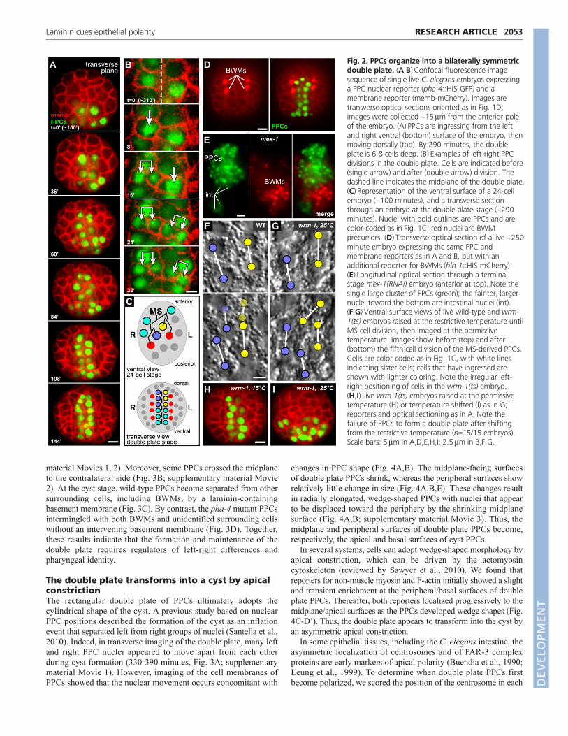
However, this orientation compresses either the left-right or dorsal-ventral axis, and makes the wedge shapes of cyst cells difficult toscore. Thus, we developed a technique to mount live embryos fortransverse optical sectioning (Fig. 1D; see Materials and methods).We found that left and right PPCs at the ventral surface appear tosweep into the embryo interior from 150-260 minutes, converginginto a dorsal-ventral-oriented rectangular array of cells (Fig. 2A).The array extends dorsally within the embryo as internal PPCsdivide and additional PPCs ingress from the ventral surface.Although there are shifts in nuclear positions within the growingarray, inspection of PPC boundaries showed that the array remainsbilaterally symmetric and almost exclusively two cells wide. Thus,we term the array of PPCs the ‘double plate’ stage, and refer to theplane between the left and right halves of the double plate as themidplane (Fig. 2A, see also 2C).
We wanted to determine how bilateral symmetry is maintainedduring growth of the double plate. Previous studies have shownthat most embryonic cell divisions are oriented longitudinally(anterior-posterior) rather than left-right, and that cell positions inthe pharyngeal cyst and at later stages are correlated with thepositions of some early PPCs (Santella et al., 2010; Sulston et al.,1983). Thus, bilateral symmetry of the double plate might derivesimply from the original left-right positions of the PPCs and the
RESEARCH ARTICLE Development 139 (11)
subsequent anterior-posterior divisions of their descendants (Fig.2C). Consistent with previous studies, we found that PPCs on theleft and right sides of the double plate were the respectivedescendants of early left and right PPCs on the ventral surface ofthe embryo (Fig. 2C). In addition, we found that body wall muscles(BWMs) flank the left and right sides of the double plate PPCs, andprecursors of the BWMs flank the left and right sides of the earlyPPCs (Fig. 2C,D). Although most PPC divisions in the double platehad the typical anterior-posterior orientation, we found that at leasttwo PPCs consistently divided left-right (Fig. 2B). The left andright daughters of those PPCs remained on their respective sides ofthe midplane, indicating that the bilateral symmetry of the doubleplate is not maintained solely by an absence of left-right divisions.
We next addressed whether bilateral symmetry of the doubleplate might arise from a lack of PPC motility, thus maintainingearly left-right differences in cell positions. We examinedpharyngeal development in mex-1(RNAi) embryos that mislocalizethe transcription factor SKN-1 (Bowerman et al., 1993). SKN-1 isrequired to specify the fate of an early blastomere called MS, theleft and right daughters of which contribute to the left and rightsides, respectively, of the double plate and flanking BWMs. Inmex-1 mutants, mislocalized SKN-1 can cause five embryonic cellsto adopt MS-like fates and produce ectopic PPCs and BWMs(Mello et al., 1992). If PPCs simply remain in place after birth,mex-1 mutants should form multiple islands of PPCs separated byBWMs. Instead, we found that terminal stage mex-1(RNAi)embryos usually formed a single block of pharyngeal cells withoutintervening BWMs (Fig. 2E). These results suggest that PPCs havean ability to move and aggregate with other PPCs.
We examined wrm-1 mutants to ascertain whether cell fatedifferences between left and right PPCs contributed to the bilateralsymmetry of the double plate. Previous studies showed that somegene expression differences between the left and right daughters ofMS (MSa and MSp) and their respective descendants depend onthe transcription factor POP-1/TCF and proteins that regulate POP-1 activity, such as WRM-1/b-catenin (Hermann et al., 2000;Rocheleau et al., 1999). We cultured temperature-sensitive wrm-1(ne1982) mutant embryos at the restrictive temperature (25°C)until after the left-right MS daughters were born, then downshiftedthe embryos to the permissive temperature (15°C) to follow thedevelopment of the PPCs. We found that MS daughters were bornin their normal left-right positions and that the early descendantsof both cells ingressed at the normal times. However, the borderbetween the early left-right descendants quickly became moreirregular than in wild-type embryos (Fig. 2F,G). At later stages,PPCs in control wrm-1(ne1982) embryos maintained at 15°Cformed an apparently normal double plate (Fig. 2H), whereas PPCsin the temperature-shifted embryos formed an abnormal cluster thatwas more than two cells wide and remained ventral rather thanextending dorsally (Fig. 2I).
Because the transcription factor PHA-4 (FoxA) is thought to berequired for most, if not all, aspects of pharyngeal-specificdifferentiation (reviewed by Mango, 2009), we examined formationof the double plate in pha-4(q490) null mutants. We found that thetiming and patterning of the early PPC divisions in pha-4(q490)embryos appeared similar to wild type (data not shown) and thatall of the PPCs ingressed (Fig. 1B). After ingressing, the mutantPPCs assembled into an array that approximated the shape of thedouble plate and was surrounded by BWM precursors (Fig. 3B, 0�panel; data not shown). However, the width of the array was morevariable than in wild-type embryos, often with three or more cellsdorsally and only one cell ventrally (Fig. 3A,B; supplementary
Fig. 1. Pharyngeal cyst development in wild-type and pha-4mutant C. elegans embryos. (A,B)Longitudinal optical sectionsthrough the centers of wild-type (A) and pha-4(q490) (B) embryos at~420 minutes in development. Embryos express a nuclear reporter(pha-4::HIS-GFP, green) in all pharyngeal precursor cells (PPCs) and inintestinal cells (int). Because the PPC reporter does not encode thePHA-4 protein, it does not rescue the pha-4 mutation and ‘PPCs’ in themutant embryos do not differentiate as pharyngeal cells. Arrows in Aindicate the periphery of the pharyngeal cyst and the red arrowheadindicates the developing apical lumen. (C)Partial cell lineage diagramindicating the origins of early cells described in this study. Body wallmuscles (BWMs) are a non-pharyngeal type of muscle. (D)Opticalsectioning planes used in this study superimposed on a depiction of anembryo midway through development. The internal position of thePPCs is indicated (green). D, dorsal; V, ventral; A, anterior; P, posterior.Scale bar: 5m.
DEVELO
PMENT
material Movies 1, 2). Moreover, some PPCs crossed the midplaneto the contralateral side (Fig. 3B; supplementary material Movie2). At the cyst stage, wild-type PPCs become separated from othersurrounding cells, including BWMs, by a laminin-containingbasement membrane (Fig. 3C). By contrast, the pha-4 mutant PPCsintermingled with both BWMs and unidentified surrounding cellswithout an intervening basement membrane (Fig. 3D). Together,these results indicate that the formation and maintenance of thedouble plate requires regulators of left-right differences andpharyngeal identity.
The double plate transforms into a cyst by apicalconstrictionThe rectangular double plate of PPCs ultimately adopts thecylindrical shape of the cyst. A previous study based on nuclearPPC positions described the formation of the cyst as an inflationevent that separated left from right groups of nuclei (Santella et al.,2010). Indeed, in transverse imaging of the double plate, many leftand right PPC nuclei appeared to move apart from each otherduring cyst formation (330-390 minutes, Fig. 3A; supplementarymaterial Movie 1). However, imaging of the cell membranes ofPPCs showed that the nuclear movement occurs concomitant with
2053RESEARCH ARTICLELaminin cues epithelial polarity
changes in PPC shape (Fig. 4A,B). The midplane-facing surfacesof double plate PPCs shrink, whereas the peripheral surfaces showrelatively little change in size (Fig. 4A,B,E). These changes resultin radially elongated, wedge-shaped PPCs with nuclei that appearto be displaced toward the periphery by the shrinking midplanesurface (Fig. 4A,B; supplementary material Movie 3). Thus, themidplane and peripheral surfaces of double plate PPCs become,respectively, the apical and basal surfaces of cyst PPCs.
In several systems, cells can adopt wedge-shaped morphology byapical constriction, which can be driven by the actomyosincytoskeleton (reviewed by Sawyer et al., 2010). We found thatreporters for non-muscle myosin and F-actin initially showed a slightand transient enrichment at the peripheral/basal surfaces of doubleplate PPCs. Thereafter, both reporters localized progressively to themidplane/apical surfaces as the PPCs developed wedge shapes (Fig.4C-D�). Thus, the double plate appears to transform into the cyst byan asymmetric apical constriction.
In some epithelial tissues, including the C. elegans intestine, theasymmetric localization of centrosomes and of PAR-3 complexproteins are early markers of apical polarity (Buendia et al., 1990;Leung et al., 1999). To determine when double plate PPCs firstbecome polarized, we scored the position of the centrosome in each
Fig. 2. PPCs organize into a bilaterally symmetricdouble plate. (A,B)Confocal fluorescence imagesequence of single live C. elegans embryos expressinga PPC nuclear reporter (pha-4::HIS-GFP) and amembrane reporter (memb-mCherry). Images aretransverse optical sections oriented as in Fig. 1D;images were collected ~15m from the anterior poleof the embryo. (A)PPCs are ingressing from the leftand right ventral (bottom) surface of the embryo, thenmoving dorsally (top). By 290 minutes, the doubleplate is 6-8 cells deep. (B)Examples of left-right PPCdivisions in the double plate. Cells are indicated before(single arrow) and after (double arrow) division. Thedashed line indicates the midplane of the double plate.(C)Representation of the ventral surface of a 24-cellembryo (~100 minutes), and a transverse sectionthrough an embryo at the double plate stage (~290minutes). Nuclei with bold outlines are PPCs and arecolor-coded as in Fig. 1C; red nuclei are BWMprecursors. (D)Transverse optical section of a live ~250minute embryo expressing the same PPC andmembrane reporters as in A and B, but with anadditional reporter for BWMs (hlh-1::HIS-mCherry).(E)Longitudinal optical section through a terminalstage mex-1(RNAi) embryo (anterior at top). Note thesingle large cluster of PPCs (green); the fainter, largernuclei toward the bottom are intestinal nuclei (int).(F,G)Ventral surface views of live wild-type and wrm-1(ts) embryos raised at the restrictive temperature untilMS cell division, then imaged at the permissivetemperature. Images show before (top) and after(bottom) the fifth cell division of the MS-derived PPCs.Cells are color-coded as in Fig. 1C, with white linesindicating sister cells; cells that have ingressed areshown with lighter coloring. Note the irregular left-right positioning of cells in the wrm-1(ts) embryo.(H,I)Live wrm-1(ts) embryos raised at the permissivetemperature (H) or temperature shifted (I) as in G;reporters and optical sectioning as in A. Note thefailure of PPCs to form a double plate after shiftingfrom the restrictive temperature (n15/15 embryos).Scale bars: 5m in A,D,E,H,I; 2.5m in B,F,G.
DEVELO
PMENT

2054
PPC relative to the center of the nucleus, measuring whether thecentrosome was proximal or distal to the midplane. Centrosomesinitially showed an equal proximal-distal distribution, as expectedfrom the predominantly anterior-posterior divisions of the PPCs(Fig. 4F). However, between ~290 and 310 minutes there was ashift in centrosome positions to face the midplane surfaces, beforeany apparent enrichment of non-muscle myosin or F-actin at themidplane (Fig. 4F). Similarly, proteins in the PAR-3 complexbecame enriched at the midplane prior to the non-muscle myosinsNMY-1 and NMY-2 (Fig. 4G, Fig. 5A; data not shown). Thisdifference in timing contrasts with the polarization of early one-celland four-cell embryos, where the asymmetric localization of PAR-6 occurs simultaneously with NMY-2 (Munro et al., 2004).
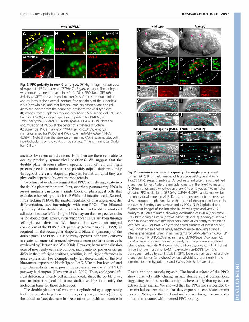
Laminin orients polarity in double plate PPCsIn analyzing fixed, immunostained mex-1(RNAi) embryos, wenoticed that ectopic PPCs at the embryo periphery (superficial PPCs)appeared to organize into cyst-like structures that were surroundedby laminin and that expressed lumen-specific apical markers (Fig.6A; supplementary material Fig. S1). In live recordings, we foundthat the cyst-like structures developed from PPCs that were born, andremained, at the periphery of the mex-1(RNAi) embryos (Fig. 6B).These superficial PPCs lacked direct contact with BWMs or othernon-PPCs, but localized PAR-6 to their interior or apical surfaces andappeared to undergo at least some apical constriction (Fig. 6B;supplementary material Movie 5). These results suggest that PPCpolarization does not require direct cell contact with non-PPCs andmight involve diffusible signals.
RESEARCH ARTICLE Development 139 (11)
Because in vitro studies suggest that the secreted protein lamininfunctions as a polarizing cue for MDCK cells (see Introduction),we immunostained fixed wild-type embryos for PAR-3 and eitherLAM-3/laminin a or PAT-3/b-integrin, a candidate lamininreceptor. We found that all three proteins were present in doubleplate PPCs, and all showed asymmetry at about the same stage;PAR-3 localized to the midplane/apical surfaces and LAM-3 andPAT-3 localized to the peripheral/basal surfaces (Fig. 5D,E).Similarly, live recordings of separate embryos expressing reportersfor PAR-6, laminin or b-integrin showed asymmetric localizationat similar developmental stages (Fig. 5A-C). We next constructeda strain that expressed both mCherry::PAR-6 and LAM-1::GFP andfound that basal enrichment of LAM-1 preceded apical enrichmentof PAR-6 by at least 15 minutes (Fig. 5F; supplementary materialMovie 6).
To determine whether laminin has a role in polarizing the doubleplate PPCs, we examined embryos homozygous for lam-1(ok3139),a predicted null mutation in the sole laminin b subunit(supplementary material Fig. S2; see Materials and methods). lam-1(–) embryos have abnormal pharyngeal basement membranes butlocalize at least some basement membrane components(supplementary material Fig. S3). Although lam-1(–) embryosappear highly abnormal by light microscopy, they appear to produceall of the major differentiated tissue types (Fig. 7A,B), consistentwith previous studies on other mutations in laminin subunits (Huanget al., 2003; Kao et al., 2006). In particular, differentiatedpharyngeal tissue contained internal lines of material resembling thenormal cuticle-lined pharyngeal lumen. Remarkably, however, the
Fig. 3. PHA-4 shapes and maintains thedouble plate. (A)Transverse opticalsections through a live wild-type C.elegans embryo showing the PPC (pha-4::HIS-GFP) transition from double plate tocyst. Images are from supplementarymaterial Movie 1; the dashed linerepresents the midplane. (B)pha-4(q490)embryo showing PPCs inappropriatelycrossing the midplane (arrow) and thefailure to form a cyst. Images are fromsupplementary material Movie 2. (C-D�) Longitudinal optical sectionsthrough the anterior of a fixed wild-typeembryo and a pha-4(q490) mutant embryoimmunostained as follows: PPC nuclei[anti-GFP (pha-4::HIS-GFP)], BWM nuclei(hlh-1::HIS-mCherry), laminin (mAbGJ2)and total nuclei (DAPI). Laminin outlinesthe body in both embryos (bluearrowhead). Laminin localizes to theperiphery of the developing pharynx in thewild-type embryo (yellow arrow) andseparates PPCs from the surroundingBWMs (see inset of bracketed region inC�). Note that laminin is present in anirregular pattern throughout the pha-4mutant PPCs (D�) and does not separatePPCs from BWMs (see inset of bracketedregion in D�). The insets in C and D showmerged images of PPC nuclei (green) andtotal nuclei (blue). Note that PPCs do notintermingle with non-PPCs in wild-typeembryos, but mix in pha-4 mutants. Timeis in minutes. Scale bars: 5m.
DEVELO
PMENT

lam-1(–) pharynx usually contained two or more such lines (Fig.7B; n96/97 embryos); transverse imaging showed that thesupernumerary lines were not simply aberrant radial outgrowthsfrom the single normal lumen, but instead appeared to be separatelumens (Fig. 7C,D). Interestingly, the intestine developed a singlelumen in lam-1(–) mutants (Fig. 7E,F), consistent with ourobservation that proteins in the PAR-3 complex localize to the apicalsurfaces of intestinal cells before laminin is detectable at the basalsurfaces (J. Feldman and J.R.P., unpublished). The multi-lumenpharyngeal defect did not result from a hyperplasia of pharyngealtissue as the mutant embryos contained the wild-type number ofPPCs (88±2, n4 embryos) (Kalb et al., 1998). Null mutants insingle laminin a subunits [lam-3(n2561) or epi-1(rh199)], in thebasement membrane components perlecan [unc-52(st549)] or intype IV collagen [emb-9(g23cg46)] had widespread defects in tissuemorphology, but did not exhibit the multi-lumen defect (Fig. 7G-J).Thus, laminin appears to have a specific role in specifying the singlelumen of the wild-type pharynx.
To determine the ontogeny of the multi-lumen pharyngealdefect, we examined PAR-3 and PAR-6 localization in live andfixed lam-1(–) embryos. PPCs in the lam-1(–) embryos organized
2055RESEARCH ARTICLELaminin cues epithelial polarity
into a double plate that appeared similar in morphology to the wild-type double plate (supplementary material Movie 9). Surprisingly,however, many of the mutant PPCs showed inverted polarity andlocalized PAR-3 and PAR-6 to the peripheral/basal surfaces at thesame time that these proteins normally localize to themidplane/apical surfaces (Fig. 8A-D; supplementary materialMovies 7, 8). With simultaneous imaging of PAR-6 and a generalmembrane reporter, we confirmed that the peripheral PAR-6 waswithin the mutant PPCs rather than in adjacent BWMs (Fig. 8E,F).We considered the possibility that the inverted polarity of the PPCsmight result from inappropriate interactions with neighboring non-PPCs, which normally are separated by the basementmembrane. However, we found that the superficial PPCs in mex-1(RNAi); lam-1(–) embryos showed a similar inverted polarity,with PAR-3 localized primarily to the peripheral, contact-freesurfaces (Fig. 6C).
We found that the multi-lumen pharyngeal defect of terminalstage lam-1(–) embryos results from cell movements thatinternalize the peripheral surfaces of PPCs. Live imaging of lam-1(–) embryos expressing reporters for cell membranes and eitherPAR-6 or non-muscle myosin showed that the peripheral surfaces
Fig. 4. Cyst formation by apical constriction of double plate PPCs. (A-D�)Transverse optical sections of live C. elegans embryos showing thetransition from double plate to cyst. Arrows at the first time point indicate the left and right margins of the double plate. (A)Image sequence fromsupplementary material Movie 3 showing all cell membranes (memb-mCherry). (B)Image sequence showing cell shape changes for a single PPCmarked by zuEx254, which is expressed in only a few PPCs. (C)F-actin expression (pha-4::GFP::dMoesin-ABD) in the PPCs. Note the enrichmentalong the midplane/apical surface. (D)Non-muscle myosin expression (nmy-2::NMY-2::GFP). (D�)High magnification of the periphery of the doubleplate (arrow with bracket in D). Note that non-muscle myosin is initially enriched at the periphery of PPCs, but disappears from the periphery as itconcentrates apically. (E)Average membrane length (m) of midplane and peripheral PPC surfaces. Error bars indicate 95% confidence intervals.(F)The left panel is a longitudinal view (anterior at top) of the double plate PPCs (DAPI , blue) at ~310 minutes showing the positions ofcentrosomes (IFA, red) relative to the midplane (dashed line). Note the number of midplane-facing centrosomes. The right panel quantifiescentrosome positions over time (n>55 centrosomes per time point). (G)Quantification of midplane enrichment of PAR-6 (pie-1::mCherry::PAR-6)and NMY-2 (nmy-2::NMY-2::GFP) in single embryos expressing both reporters (n4 embryos). See also supplementary material Movie 4. Error barsindicate 95% confidence intervals. Scale bars: 5m in A,C,D; 2.5m in B,F; 1m in D�.
DEVELO
PMENT

2056
of several PPCs constricted at about the same time as the midplanesurfaces normally constrict in wild-type PPCs (Fig. 8G-G�;supplementary material Movies 8, 9). Neighboring PPCs spreadacross and enclosed the peripheral constrictions, which underwentlumenal differentiation. We conclude that lam-1(–) mutants havean early and highly penetrant defect in orienting PPC polarity thatis partially masked by subsequent morphogenetic movements.
Laminin can act non-cell-autonomously to orientPPC polarityBecause several C. elegans tissues have been reported to expresslaminin, including the pharynx, intestine and BWMs (Huang et al.,2003; Kao et al., 2006), we investigated which cells must expresslam-1 to properly orient PPC polarity. We constructed ahomozygous lam-1(ok3139) strain that carried anextrachromosomal array (zuEx288) containing a wild-type lam-1(+) gene plus a cell-autonomous marker (SUR-5::GFP); suchtransgenic arrays are lost stochastically during cell divisions,resulting in animals that are mosaic for gene expression. Asexpected, animals that lacked zuEx288 had the multi-lumen defect(n60/61), whereas animals that had zuEx288 in most or all cellshad a normal pharynx with a single lumen (Fig. 7K; n46/47). We
RESEARCH ARTICLE Development 139 (11)
then searched for rare mosaic animals that lacked zuEx288 in allpharyngeal cells but that retained the transgene in other cell groups.We found that the pharynx had a single lumen in 6/8 animals withzuEx288 only in intestinal cells (Fig. 7L) and in 14/17 animals withzuEx288 only in skin (hypodermal) cells and BWMs (Fig. 7M).These results suggest that laminin expressed by non-PPCs diffusesthrough the embryo and accumulates on the available basal surfaceof PPCs where it acts as a polarity cue.
DISCUSSIONMorphogenesis of the pharyngeal cyst proceeds through a distinct,bilaterally symmetric intermediate termed the double plate, wherePPCs first develop apical/basal polarity. The mature C. eleganspharynx has threefold symmetry, but has an underlying bilateralsymmetry evident in the cell lineages of MS descendants thatproduce about half of the pharynx (Albertson and Thomson, 1976;Sulston et al., 1983). For example, symmetrical and identical cellsin the mature pharynx called mc3DL and mc3DR are descendantsof the left and right daughters of MS, respectively, and their fatesappear to be determined by a lineage mechanism rather than cellposition (Priess et al., 1987). These cells do not contact each otherin the mature pharynx and are separated from their nearest common
Fig. 5. Onset of apical and basal polarity in double plate PPCs. (A-C)Transverse optical sections of live C. elegans embryos showing thetransition from double plate to cyst. Arrows indicate the left and right margins of the double plate. Embryos express the following reporters: (A)PAR-6 (pie-1::mCherry::PAR-6), (B) laminin b (lam-1::LAM-1::GFP) and (C) b-integrin (pat-3::PAT-3::GFP). See supplementary material Movie 6 foronset of lam-1::LAM-1::GFP expression. (D,E)Longitudinal optical sections through the center of the double plate (brackets) and intestine (int,arrowheads). The embryos were immunostained for PAR-3 and either laminin a (mAbGJ1) (D) or b-integrin (MH25) (E). The embryos were selectedfor the earliest detectable asymmetry of PAR-3 in the PPCs. Note that at this stage both laminin a and b-integrin are enriched in puncta at theperiphery of the double plate; at later stages, laminin a and b-integrin uniformly coat the basal PPC surfaces (see Fig. 3C�; supplementary materialFig. S4). LAM-3 is not enriched at the periphery of the intestinal cells (arrowheads). (F)Kymograph analysis (top) from supplementary material Movie6 of an embryo co-expressing pie-1::mCherry::PAR-6 and lam-1::LAM-1::GFP showing the width of the double plate and surrounding BWMs.Beneath are shown fluorescence intensity line scans across the kymographs at 35 and 60 minutes. The dashed line indicates the midplane. Scalebars: 5m.
DEVELO
PMENT
ancestor by seven cell divisions. How then are these cells able tooccupy precisely symmetrical positions? We suggest that thedouble plate structure allows specific pairs of left and rightprecursor cells to maintain, and possibly adjust, their proximitythroughout the early stages of pharynx formation, until they arephysically separated by cyst morphogenesis.
Two lines of evidence suggest that PPCs actively aggregate intothe double plate primordium. First, ectopic supernumerary PPCs inmex-1 mutants can form a single block of pharyngeal cells thatexcludes other cell types such as BWMs and intestinal cells. Second,PPCs lacking PHA-4, the master regulator of pharyngeal-specificdifferentiation, can intermingle with non-PPCs. The bilateralsymmetry of the double plate is likely to involve differential celladhesion because left and right PPCs stay on their respective sidesas the double plate grows, even when these PPCs are born throughleft-right cell divisions. We showed that wrm-1, an integralcomponent of the POP-1/TCF pathway (Rocheleau et al., 1999), isrequired for the rectangular shape and bilateral symmetry of thedouble plate. The POP-1/TCF pathway operates in the early embryoto create numerous differences between anterior-posterior sister cells(reviewed by Herman and Wu, 2004). However, because the divisionaxes of most early cells are oblique, many anterior-posterior sistersdiffer in their left-right positions, resulting in left-right differences ingene expression. For example, only left descendants of the MSblastomere express the Notch ligand LAG-2/Delta, but both left andright descendants can express this protein when the POP-1/TCFpathway is disrupted (Hermann et al., 2000). Thus, analogous left-right differences in early cell adhesion could shape the double plate,and an important goal of future studies will be to identify themolecular basis for those differences.
The double plate transforms into a cylindrical cyst, apparentlyby PPCs constricting their midplane, or apical, surfaces (Fig. 9);the apical surfaces decrease in size concomitant with an increase in
2057RESEARCH ARTICLELaminin cues epithelial polarity
F-actin and non-muscle myosin. The basal surfaces of the PPCsshow relatively little change in size during apical constriction,suggesting that these surfaces might adhere to neighboring cells orextracellular matrix. We showed that the PPCs are surrounded bylaminin before constriction, that they express the candidate lamininreceptor PAT-3, and that the basal surface can change size markedlyin laminin mutants with inverted PPC polarity.
Fig. 6. PPC polarity in mex-1 embryos. (A)High-magnification viewof superficial PPCs in a mex-1(RNAi) C. elegans embryo. The embryowas immunostained for laminin a (mAbGJ1), PPCs [anti-GFP (pha-4::PHA-4::GFP)] and a lumenal marker (mAbPL1). Note that lamininaccumulates at the external, contact-free periphery of the superficialPPCs (arrowheads) and that lumenal markers differentiate one celldiameter inward from the periphery, similar to the wild-type cyst.(B)Images from supplementary material Movie 5 of superficial PPCs in alive mex-1(RNAi) embryo expressing reporters for PAR-6 (pie-1::mCherry::PAR-6) and PPC nuclei (pha-4::PHA-4::GFP). Note theaccumulation of PAR-6 at the center of a cyst-like structure.(C)Superficial PPCs in a mex-1(RNAi); lam-1(ok3139) embryoimmunostained for PAR-3 and PPC nuclei [anti-GFP (pha-4::PHA-4::GFP)]. Note that in the absence of laminin, PAR-3 accumulates withinverted polarity on the contact-free surface. Time is in minutes. Scalebar: 2.5m.
Fig. 7. Laminin is required to specify the single pharyngeallumen. (A,B)Brightfield images of late stage wild-type and lam-1(ok3139) C. elegans embryos. Arrowheads indicate the cuticle-linedpharyngeal lumen. Note the multiple lumens in the lam-1(–) mutant.(C,D)Immunostained wild-type and lam-1(–) embryos at 470 minutesshowing PPC nuclei [anti-GFP (pha-4::PHA-4::GFP)] and a marker forthe pharyngeal lumen (mAbPL1). Insets are reconstructed transverseviews through the pharynx. Note that both of the apparent lumens inthe lam-1(–) embryo are surrounded by PPCs. (E,F)Brightfield andfluorescent images of the intestine in live wild-type and lam-1(–)embryos at ~280 minutes, showing localization of PAR-6 (par-6::PAR-6::GFP) to a single lumen (arrow). Although lam-1(–) embryos showedsome mispositioning of intestinal cells, each of 28 embryos examinedlocalized PAR-3 or PAR-6 only to the apical surfaces of intestinal cells.(G-J)Brightfield images of newly hatched larvae showing a singleinternal pharyngeal lumen in null mutants for LAM-3/laminin a (G), EPI-1/laminin a (H), UNC-52/perlecan (I) and EMB-9/type IV collagen (J).n>50 animals examined for each genotype. The pharynx is outlined(blue dashed line). (K-M)Newly hatched homozygous lam-1(–) mutantlarvae that are mosaic for LAM-1 expression [zuEx288: lam-1(+)transgene marked by sur-5::SUR-5::GFP]. Note the formation of a singlepharyngeal lumen (arrowhead) when zuEx288 is present only in theintestine (L) or in hypodermis and BWMs (M). Scale bars: 5m.
DEVELO
PMENT

2058
The coordinated polarization of large groups of cells is ahallmark of epithelial development that allows tubular epithelia toform a single, continuous lumen. Our results show that lamininprovides a crucial cue for the coordinate polarization of epithelialcells in the pharynx, although not for epithelial cells in theintestine. A previous study showed that C. elegans mutants lackingLAM-3, one of two laminin a subunits, develop ruptures in thepharyngeal basement membrane and that some pharyngeal cellsappear to adhere to surrounding tissues through these gaps (Huanget al., 2003). However, all of the mutant pharyngeal cells appearedto connect to a central lumen, as do the wild-type pharyngeal cells.Although pharyngeal cells lacking LAM-1, the sole laminin bsubunit, similarly connect to a lumenal surface, we showed thatthere are multiple lumenal surfaces in the mutant pharynx and thatthese arise from polarity defects in double plate PPCs. The doubleplate PPCs polarize abnormally in lam-1 mutants, with manylocalizing PAR-3 and PAR-6 to their peripheral (normally basal)surfaces. Remarkably, these peripheral surfaces can constrict, likethe normal apical surface, and become repositioned into the interior
RESEARCH ARTICLE Development 139 (11)
of the developing pharynx by the neighboring PPCs. These polaritydefects occur at least an hour before perlecan and type IV collagenlocalize to the wild-type pharyngeal basement membrane (Grahamet al., 1997; Mullen et al., 1999) and were not observed in mutantsfor either basement membrane component. Thus, we propose thatthe defects represent a specific requirement for laminin in PPCpolarization, rather than general functions of the basementmembrane in tissue integrity.
Several embryonic cell types, including PPCs, normally expresslaminin. Our analysis of laminin mosaics shows that lamininsupplied by non-PPCs is sufficient to orient PPC polarity.Consistent with previous observations on laminin and type IVcollagen, this result suggests that basement membrane componentscan diffuse through the embryo (Graham et al., 1997; Huang et al.,2003). We presume that PPC adhesion, as evidenced by theaggregation of supernumerary PPCs in mex-1(RNAi) embryos,normally restricts laminin to the peripheral surface of the doubleplate. PPCs do not appear to maintain normal adhesion in pha-4mutants, and we showed that laminin distributes in an irregular
Fig. 8. Laminin cues PPC polarity. (A,B)Longitudinal optical sections through the double plate PPCs of immunostained wild-type and lam-1(ok3139) C. elegans embryos. Anterior is top. Images show PPC nuclei [anti-GFP (pha-4::PHA-4::GFP)], total nuclei (DAPI) and PAR-3 as maximumintensity projections through the double plate; arrows indicate the midplane. Note that PAR-3 localizes to midplane surfaces in the wild-type doubleplate, but predominantly to peripheral surfaces in the lam-1(–) embryo [n>50 for wild type; n18/18 for lam-1(–)]. (C,D)Optical sections as in A andB of live embryos expressing a PAR-6 reporter (par-6::PAR-6::GFP). Note that PAR-6 localization begins, and persists, at the peripheral surfaces ofthe double plate in the lam-1(–) embryos (n16/16). See supplementary material Movies 7 and 8 for cell membranes of PPCs. (E,F)High-magnification images of part of a longitudinal optical section through the double plate of live wild-type (E) and lam-1(–) (F) embryos showing PAR-6(par-6::PAR-6::GFP) and plasma membranes (memb-mCherry). Line scans through the double plate are shown beneath each image. Note thatectopic PAR-6 is localized within the PPCs in the lam-1(–) embryo. (G-G�) Images from supplementary material Movie 9 showing transverse opticalsections of a live lam-1(–) embryo expressing reporters for non-muscle myosin (nmy-2::NMY-2::GFP) and plasma membranes (memb-mCherry).Images in G� are high-magnification views of PPCs at the periphery of the double plate (arrowhead and bracketed region in G and G�). Thediagrams show tracings of individual color-coded PPCs within the double plate (light gray region), taken from movie frames at left. Time is inminutes. Scale bars: 2.5m in A-F,G�; 5m in G.
DEVELO
PMENT

pattern between the mutant PPCs. We have no evidence thatlaminin receptors are localized to the peripheral surfaces of doubleplate PPCs prior to laminin enrichment. Indeed, we found that PAT-3 localizes to the basal surface at about the same time as laminin,and that laminin function is required for PAT-3 localization(supplementary material Fig. S4). We do not yet know whetherPAT-3 is required to orient the midplane/apical localization of thePAR-3 complex: embryos homozygous for a null pat-3 mutationexpressed PAT-3, suggesting that pat-3 mRNA is contributed fromthe heterozygous mother, and exposing the mothers to pat-3dsRNA resulted in early embryonic arrest that precluded analysisof the pharynx (data not shown).
In Drosophila, similar to C. elegans, some laminin subunits areencoded by single genes; however, the respective Drosophilamutants do not exhibit defects in coordinating epithelial polarity(Urbano et al., 2009). Thus, laminin could function as a polaritysignal in only certain epithelia, such as the pharynx, or mightfunction redundantly with other signals such as other basementmembrane proteins. Alternatively, later morphogenetic movementscould obscure early polarity defects, as we demonstrated in the C.elegans pharynx. A third possibility is that some epithelia recognizeinappropriate features of their environment as polarity cues whenlaminin is absent. By analogy, when the sperm-suppliedcentrosome is not available to polarize the one-cell C. elegansembryo, the meiotic spindle at the opposite pole of the egg appearsto cue an inverted polarity (Wallenfang and Seydoux, 2000).Similarly, Drosophila wing epithelial cells polarize in the absenceof planar cell polarity components, but fail to coordinate theirpolarity at the tissue level (Wong and Adler, 1993). Basementmembrane defects in laminin mutants might allow some PPCs tomake inappropriate contacts with surrounding tissues. However, wedo not believe that those tissues induce the inversion of anotherwise normal PPC polarity because superficial PPCs in mex-1(RNAi); lam-1(–) embryos that do not contact any non-PPCs
2059RESEARCH ARTICLELaminin cues epithelial polarity
nevertheless develop inverted polarity. Thus, the present studyprovides in vivo evidence that laminin acts as a polarizing cue forsome, although not all, epithelia. We conclude that the C. eleganspharynx should provide a useful genetic model for dissectinglaminin-based signaling prior to the general requirements forlaminin in basement membrane assembly and tissue integrity.
AcknowledgementsWe thank James Kramer, Susan Mango, Donald Moerman, John Murray,Jeremy Nance, Alisa Piekny, William Wadsworth and the DevelopmentalStudies Hybridoma Bank for providing antisera/strains and members of theJ.R.P. lab for valuable discussions. Some strains used in this study wereobtained from the Caenorhabditis Genetics Center, which is supported by theNIH National Center for Research Resources.
FundingThis work was supported by a Developmental Biology Predoctoral TrainingGrant from the National Institute of Child Health and Human Development[T32HD007183 to J.P.R.]; the NIH [R01GM098583 to J.R.P.]; and HowardHughes Medical Institute (to J.R.P.). Deposited in PMC for release after 6months.
Competing interests statementThe authors declare no competing financial interests.
Supplementary materialSupplementary material available online athttp://dev.biologists.org/lookup/suppl/doi:10.1242/dev.078360/-/DC1
ReferencesAchilleos, A., Wehman, A. M. and Nance, J. (2010). PAR-3 mediates the initial
clustering and apical localization of junction and polarity proteins during C.elegans intestinal epithelial cell polarization. Development 137, 1833-1842.
Albertson, D. G. and Thomson, J. N. (1976). The pharynx of Caenorhabditiselegans. Philos. Trans. R. Soc. Lond. B Biol. Sci. 275, 299-325.
Aumailley, M., Bruckner-Tuderman, L., Carter, W. G., Deutzmann, R., Edgar,D., Ekblom, P., Engel, J., Engvall, E., Hohenester, E., Jones, J. C. et al.(2005). A simplified laminin nomenclature. Matrix Biol. 24, 326-332.
Bossinger, O., Klebes, A., Segbert, C., Theres, C. and Knust, E. (2001). Zonulaadherens formation in Caenorhabditis elegans requires dlg-1, the homologue ofthe Drosophila gene discs large. Dev. Biol. 230, 29-42.
Bowerman, B., Draper, B. W., Mello, C. C. and Priess, J. R. (1993). Thematernal gene skn-1 encodes a protein that is distributed unequally in early C.elegans embryos. Cell 74, 443-452.
Brenner, S. (1974). The genetics of Caenorhabditis elegans. Genetics 77, 71-94.Buendia, B., Bré, M. H., Griffiths, G. and Karsenti, E. (1990). Cytoskeletal
control of centrioles movement during the establishment of polarity in Madin-Darby canine kidney cells. J. Cell Biol. 110, 1123-1135.
Chaffer, C. L., Thompson, E. W. and Williams, E. D. (2007). Mesenchymal toepithelial transition in development and disease. Cells Tissues Organs 185, 7-19.
Ekblom, P. (1989). Developmentally regulated conversion of mesenchyme toepithelium. FASEB J. 3, 2141-2150.
Francis, G. R. and Waterston, R. H. (1985). Muscle organization inCaenorhabditis elegans: localization of proteins implicated in thin filamentattachment and I-band organization. J. Cell Biol. 101, 1532-1549.
Graham, P. L., Johnson, J. J., Wang, S., Sibley, M. H., Gupta, M. C. andKramer, J. M. (1997). Type IV collagen is detectable in most, but not all,basement membranes of Caenorhabditis elegans and assembles on tissues thatdo not express it. J. Cell Biol. 137, 1171-1183.
Gupta, M. C., Graham, P. L. and Kramer, J. M. (1997). Characterization ofalpha1(IV) collagen mutations in Caenorhabditis elegans and the effects ofalpha1 and alpha2(IV) mutations on type IV collagen distribution. J. Cell Biol.137, 1185-1196.
Hagedorn, E. J., Yashiro, H., Ziel, J. W., Ihara, S., Wang, Z. and Sherwood, D.R. (2009). Integrin acts upstream of netrin signaling to regulate formation of theanchor cell’s invasive membrane in C. elegans. Dev. Cell 17, 187-198.
Herman, M. A. and Wu, M. (2004). Noncanonical Wnt signaling pathways in C.elegans converge on POP-1/TCF and control cell polarity. Front. Biosci. 9, 1530-1539.
Hermann, G. J., Leung, B. and Priess, J. R. (2000). Left-right asymmetry in C.elegans intestine organogenesis involves a LIN-12/Notch signaling pathway.Development 127, 3429-3440.
Horner, M. A., Quintin, S., Domeier, M. E., Kimble, J., Labouesse, M. andMango, S. E. (1998). pha-4, an HNF-3 homolog, specifies pharyngeal organidentity in Caenorhabditis elegans. Genes Dev. 12, 1947-1952.
Huang, C. C., Hall, D. H., Hedgecock, E. M., Kao, G., Karantza, V., Vogel, B.E., Hutter, H., Chisholm, A. D., Yurchenco, P. D. and Wadsworth, W. G.
Fig. 9. Model of cyst morphogenesis in C. elegans. Summary of theformation of the pharyngeal cyst (transverse views). Left (yellow) andright (light blue) PPCs ingress from the ventral (bottom) surface of theembryo and aggregate to form the bilaterally symmetric double plate.Laminin accumulates on the exposed peripheral surface of the doubleplate in wild-type embryos, but is absent in lam-1(–) embryos. Lamininat the peripheral surface precedes, and is required for, localization ofproteins in the PAR-3 complex to the opposite, midplane surface. Inlam-1 mutants, PAR proteins accumulate ectopically on the peripheralsurface. Cell surface constriction occurs where PAR proteins localize,leading to midplane constriction in wild-type embryos and theformation of a single-lumen cyst. In lam-1 mutants, peripheralconstriction and shifts in PPC position internalize the PAR-containingperipheral surfaces, leading to a multi-lumen cyst.
DEVELO
PMENT

2060 RESEARCH ARTICLE Development 139 (11)
(2003). Laminin alpha subunits and their role in C. elegans development.Development 130, 3343-3358.
Kachur, T. M., Audhya, A. and Pilgrim, D. B. (2008). UNC-45 is required forNMY-2 contractile function in early embryonic polarity establishment andgermline cellularization in C. elegans. Dev. Biol. 314, 287-299.
Kalb, J. M., Lau, K. K., Goszczynski, B., Fukushige, T., Moons, D., Okkema, P.G. and McGhee, J. D. (1998). pha-4 is Ce-fkh-1, a fork head/HNF-3alpha,beta,gamma homolog that functions in organogenesis of the C. eleganspharynx. Development 125, 2171-2180.
Kamath, R. S., Fraser, A. G., Dong, Y., Poulin, G., Durbin, R., Gotta, M.,Kanapin, A., Le Bot, N., Moreno, S., Sohrmann, M. et al. (2003). Systematicfunctional analysis of the Caenorhabditis elegans genome using RNAi. Nature421, 231-237.
Kao, G., Huang, C. C., Hedgecock, E. M., Hall, D. H. and Wadsworth, W. G.(2006). The role of the laminin beta subunit in laminin heterotrimer assemblyand basement membrane function and development in C. elegans. Dev. Biol.290, 211-219.
Klein, G., Langegger, M., Timpl, R. and Ekblom, P. (1988). Role of laminin Achain in the development of epithelial cell polarity. Cell 55, 331-341.
Leung, B., Hermann, G. J. and Priess, J. R. (1999). Organogenesis of theCaenorhabditis elegans intestine. Dev. Biol. 216, 114-134.
Maduro, M. and Pilgrim, D. (1995). Identification and cloning of unc-119, agene expressed in the Caenorhabditis elegans nervous system. Genetics 141,977-988.
Mango, S. (2009). The molecular basis of organ formation: insights from the C.elegans foregut. Annu. Rev. Cell Dev. Biol. 25, 597-628.
Mango, S. E., Lambie, E. J. and Kimble, J. (1994). The pha-4 gene is required togenerate the pharyngeal primordium of Caenorhabditis elegans. Development120, 3019-3031.
Marinkovich, M. P. (2007). Tumour microenvironment: laminin 332 in squamous-cell carcinoma. Nat. Rev. Cancer 7, 370-380.
McMahon, L., Legouis, R., Vonesch, J. L. and Labouesse, M. (2001). Assemblyof C. elegans apical junctions involves positioning and compaction by LET-413and protein aggregation by the MAGUK protein DLG-1. J. Cell Sci. 114, 2265-2277.
Mello, C. C., Kramer, J. M., Stinchcomb, D. and Ambros, V. (1991). Efficientgene transfer in C.elegans: extrachromosomal maintenance and integration oftransforming sequences. EMBO J. 10, 3959-3970.
Mello, C. C., Draper, B. W., Krause, M., Weintraub, H. and Priess, J. R. (1992).The pie-1 and mex-1 genes and maternal control of blastomere identity in earlyC. elegans embryos. Cell 70, 163-176.
Miner, J. H. and Yurchenco, P. D. (2004). Laminin functions in tissuemorphogenesis. Annu. Rev. Cell Dev. Biol. 20, 255-284.
Miner, J. H., Cunningham, J. and Sanes, J. R. (1998). Roles for laminin inembryogenesis: exencephaly, syndactyly, and placentopathy in mice lacking thelaminin alpha5 chain. J. Cell Biol. 143, 1713-1723.
Mörck, C., Axäng, C. and Pilon, M. (2003). A genetic analysis of axon guidancein the C elegans pharynx. Dev. Biol. 260, 158-175.
Mullen, G., Rogalski, T., Bush, J., Gorji, P. and Moerman, D. (1999). Complexpatterns of alternative splicing mediate the spatial and temporal distribution ofperlecan/UNC-52 in Caenorhabditis elegans. Mol. Biol. Cell 10, 3205-3221.
Munro, E., Nance, J. and Priess, J. R. (2004). Cortical flows powered byasymmetrical contraction transport PAR proteins to establish and maintainanterior-posterior polarity in the early C. elegans embryo. Dev. Cell 7, 413-424.
Murray, J. I., Bao, Z., Boyle, T. J., Boeck, M. E., Mericle, B. L., Nicholas, T. J.,Zhao, Z., Sandel, M. J. and Waterston, R. H. (2008). Automated analysis ofembryonic gene expression with cellular resolution in C. elegans. Nat. Methods5, 703-709.
Nakamura, K., Kim, S., Ishidate, T., Bei, Y., Pang, K., Shirayama, M.,Trzepacz, C., Brownell, D. R. and Mello, C. C. (2005). Wnt signaling drivesWRM-1/beta-catenin asymmetries in early C. elegans embryos. Genes Dev. 19,1749-1754.
Nance, J. and Zallen, J. A. (2011). Elaborating polarity: PAR proteins and thecytoskeleton. Development 138, 799-809.
Nance, J., Munro, E. M. and Priess, J. R. (2003). C. elegans PAR-3 and PAR-6 arerequired for apicobasal asymmetries associated with cell adhesion andgastrulation. Development 130, 5339-5350.
O’Brien, L. E., Jou, T. S., Pollack, A. L., Zhang, Q., Hansen, S. H., Yurchenco, P.and Mostov, K. E. (2001). Rac1 orientates epithelial apical polarity througheffects on basolateral laminin assembly. Nat. Cell Biol. 3, 831-838.
Piekny, A. J., Johnson, J. L., Cham, G. D. and Mains, P. E. (2003). TheCaenorhabditis elegans nonmuscle myosin genes nmy-1 and nmy-2 function asredundant components of the let-502/Rho-binding kinase and mel-11/myosinphosphatase pathway during embryonic morphogenesis. Development 130,5695-5704.
Portereiko, M. F. and Mango, S. E. (2001). Early morphogenesis of theCaenorhabditis elegans pharynx. Dev. Biol. 233, 482-494.
Priess, J. R., Schnabel, H. and Schnabel, R. (1987). The glp-1 locus and cellularinteractions in early C. elegans embryos. Cell 51, 601-611.
Pruss, R. M., Mirsky, R., Raff, M. C., Thorpe, R., Dowding, A. J. andAnderton, B. H. (1981). All classes of intermediate filaments share a commonantigenic determinant defined by a monoclonal antibody. Cell 27, 419-428.
Raharjo, W. H., Ghai, V., Dineen, A., Bastiani, M. and Gaudet, J. (2011). Cellarchitecture: surrounding muscle cells shape gland cell morphology in theCaenorhabditis elegans pharynx. Genetics 189, 885-897.
Rasmussen, J., English, K., Tenlen, J. and Priess, J. (2008). Notch signaling andmorphogenesis of single-cell tubes in the C. elegans digestive tract. Dev. Cell 14,559-569.
Rocheleau, C. E., Yasuda, J., Shin, T. H., Lin, R., Sawa, H., Okano, H., Priess, J.R., Davis, R. J. and Mello, C. C. (1999). WRM-1 activates the LIT-1 proteinkinase to transduce anterior/posterior polarity signals in C. elegans. Cell 97, 717-726.
Ryan, M. C., Lee, K., Miyashita, Y. and Carter, W. G. (1999). Targeteddisruption of the LAMA3 gene in mice reveals abnormalities in survival and latestage differentiation of epithelial cells. J. Cell Biol. 145, 1309-1323.
Santella, A., Du, Z., Nowotschin, S., Hadjantonakis, A. K. and Bao, Z. (2010).A hybrid blob-slice model for accurate and efficient detection of fluorescencelabeled nuclei in 3D. BMC Bioinformatics 11, 580.
Sawyer, J. M., Harrell, J. R., Shemer, G., Sullivan-Brown, J., Roh-Johnson, M.and Goldstein, B. (2010). Apical constriction: a cell shape change that candrive morphogenesis. Dev. Biol. 341, 5-19.
Schonegg, S., Constantinescu, A. T., Hoege, C. and Hyman, A. A. (2007). TheRho GTPase-activating proteins RGA-3 and RGA-4 are required to set the initialsize of PAR domains in Caenorhabditis elegans one-cell embryos. Proc. Natl.Acad. Sci. USA 104, 14976-14981.
Smyth, N., Vatansever, H. S., Murray, P., Meyer, M., Frie, C., Paulsson, M.and Edgar, D. (1999). Absence of basement membranes after targeting theLAMC1 gene results in embryonic lethality due to failure of endodermdifferentiation. J. Cell Biol. 144, 151-160.
Sulston, J. E., Schierenberg, E., White, J. G. and Thomson, J. N. (1983). Theembryonic cell lineage of the nematode Caenorhabditis elegans. Dev. Biol. 100,64-119.
Tenlen, J. R., Schisa, J. A., Diede, S. J. and Page, B. D. (2006). Reduced dosageof pos-1 suppresses Mex mutants and reveals complex interactions amongCCCH zinc-finger proteins during Caenorhabditis elegans embryogenesis.Genetics 174, 1933-1945.
Totong, R., Achilleos, A. and Nance, J. (2007). PAR-6 is required for junctionformation but not apicobasal polarization in C. elegans embryonic epithelialcells. Development 134, 1259-1268.
Urbano, J. M., Torgler, C. N., Molnar, C., Tepass, U., López-Varea, A., Brown,N. H., de Celis, J. F. and Martín-Bermudo, M. D. (2009). Drosophila lamininsact as key regulators of basement membrane assembly and morphogenesis.Development 136, 4165-4176.
Wallenfang, M. R. and Seydoux, G. (2000). Polarization of the anterior-posterioraxis of C. elegans is a microtubule-directed process. Nature 408, 89-92.
Wang, A. Z., Ojakian, G. K. and Nelson, W. J. (1990). Steps in themorphogenesis of a polarized epithelium. I. Uncoupling the roles of cell-cell andcell-substratum contact in establishing plasma membrane polarity in multicellularepithelial (MDCK) cysts. J. Cell Sci. 95, 137-151.
Williams, B. D. and Waterston, R. H. (1994). Genes critical for muscledevelopment and function in Caenorhabditis elegans identified through lethalmutations. J. Cell Biol. 124, 475-490.
Wong, L. L. and Adler, P. N. (1993). Tissue polarity genes of Drosophila regulatethe subcellular location for prehair initiation in pupal wing cells. J. Cell Biol. 123,209-221.
Yochem, J., Gu, T. and Han, M. (1998). A new marker for mosaic analysis inCaenorhabditis elegans indicates a fusion between hyp6 and hyp7, two majorcomponents of the hypodermis. Genetics 149, 1323-1334.
Yurchenco, P. D. (2011). Basement membranes: cell scaffoldings and signalingplatforms. Cold Spring Harb. Perspect. Biol. 3, a004911.
Zhong, M., Niu, W., Lu, Z. J., Sarov, M., Murray, J. I., Janette, J., Raha, D.,Sheaffer, K. L., Lam, H. Y., Preston, E. et al. (2010). Genome-wideidentification of binding sites defines distinct functions for Caenorhabditiselegans PHA-4/FOXA in development and environmental response. PLoS Genet.6, e1000848.
Zhu, X., Joh, K., Hedgecock, E. M. and Hori, K. (1999). Identification of epi-1locus as a laminin alpha chain gene in the nematode Caenorhabditis elegansand characterization of epi-1 mutant alleles. DNA Seq. 10, 207-217.
Ziel, J., Hagedorn, E., Audhya, A. and Sherwood, D. (2009). UNC-6 (netrin)orients the invasive membrane of the anchor cell in C. elegans. Nat. Cell Biol.11, 183-189.
DEVELO
PMENT

![Epithelial Polarity, Villin Expression, and Enterocytic ......[CANCER RESEARCH 48, 1936-1942, April 1, 1988] Epithelial Polarity, Villin Expression, and Enterocytic Differentiation](https://img.dokumen.tips/doc/110x75/5f0bffd27e708231d433428e/epithelial-polarity-villin-expression-and-enterocytic-cancer-research.jpg)


![Laminin-332 and Integrins: Signaling Platform for …Laminin-332 and Integrins 31 laminin globular (LG) subdomains (LG1-5) [2]. The latter is the major interaction sites for cell surface](https://img.dokumen.tips/doc/110x75/5f712e9e3f945d798f112220/laminin-332-and-integrins-signaling-platform-for-laminin-332-and-integrins-31-laminin.jpg)


