Embed Size (px)
Citation preview

CASE REPORT Open Access
Lameness in fattening pigs – Mycoplasmahyosynoviae, osteochondropathy andreduced dietary phosphorus level as threeinfluencing factors: a case reportB. Wegner1, J. Tenhündfeld2, J. Vogels3, M. Beumer3, J. Kamphues4, F. Hansmann5, H. Rieger4,E. grosse Beilage3 and I. Hennig-Pauka3*
Abstract
Background: Multiple diagnostic procedures, their results and interpretation in a case with severe lameness infattening pigs are described. It is shown that selected diagnostic steps lead to identification of various risk factorsfor disease development in the affected herd. One focus of this case report is the prioritization of diagnostic stepsto verify the impact of the different conditions, which finally led to the clinical disorder. Assessing a sufficientdietary phosphorus (P) supply and its impact on disease development proved most difficult. The diagnosticapproach based on estimated calculation of phosphorus intake is presented in detail.
Case presentation: On a farrow-to-finishing farm, lameness occurred in pigs with 30–70 kg body weight. Necropsyof three diseased pigs revealed claw lesions and alterations at the knee and elbow joints. Histologic findings werecharacteristic of osteochondrosis. All pigs were positively tested for Mycoplasma hyosynoviae in affected joints. Pvalues in blood did not indicate a P deficiency, while bone ashing in one of three animals resulted in a levelindicating an insufficient mineral supply. Analysis of diet composition revealed a low phosphorus content in twodiets, which might have led to a marginal P supply in individuals with high average daily gains with respect todevelopment of bone mass and connective tissue prior to presentation of affected animals. Finally, the impact ofdietary factors for disease development could not be evidenced in all submitted animals in this case.
Conclusions: Mycoplasma (M.) hyosynoviae was identified to be an important etiologic factor for disease. Other,non-infectious factors, such as osteochondrosis and claw lesions might have favored development of lameness. Inaddition, a relevant marginal P supply for pigs was found in a limited time period in a phase of intense growing,but the potential interaction with infection by M. hyosynoviae is unknown.The presented case of severe lameness in fattening pigs revealed that three different influences presumably act inpathogenesis. Focusing only on one factor and ignoring others might be misleading regarding subsequentdecision-making for prevention and therapy. Finally, clinical symptoms disappeared after some changes in dietcomposition and anti-inflammatory treatment of individual animals.
Keywords: Locomotor disorder, Mineral supply, Mycoplasma hyosynoviae, Nutrition, Swine
© The Author(s). 2020 Open Access This article is licensed under a Creative Commons Attribution 4.0 International License,which permits use, sharing, adaptation, distribution and reproduction in any medium or format, as long as you giveappropriate credit to the original author(s) and the source, provide a link to the Creative Commons licence, and indicate ifchanges were made. The images or other third party material in this article are included in the article's Creative Commonslicence, unless indicated otherwise in a credit line to the material. If material is not included in the article's Creative Commonslicence and your intended use is not permitted by statutory regulation or exceeds the permitted use, you will need to obtainpermission directly from the copyright holder. To view a copy of this licence, visit http://creativecommons.org/licenses/by/4.0/.The Creative Commons Public Domain Dedication waiver (http://creativecommons.org/publicdomain/zero/1.0/) applies to thedata made available in this article, unless otherwise stated in a credit line to the data.
* Correspondence: [email protected] Station for Epidemiology, University of Veterinary Medicine Hannover,Foundation, Hannover, GermanyFull list of author information is available at the end of the article
Wegner et al. Porcine Health Management (2020) 6:41 https://doi.org/10.1186/s40813-020-00184-w

BackgroundMycoplasma (M.) hyosynoviae is a commensal of theupper respiratory tract, especially in the tonsils [1], thatmay lead to clinical disease mainly in older pigs (> 10weeks) [2]. The detection rate has increased in the lastyears in pigs with arthritis in Denmark with approxi-mately 20% already in 2001 [3], and in the USA with ap-proximately 8% in 2003, 23% in 2010 [2] and 26% in2015 [4]. Affected pigs show avoidance of standing up oran impaired ability to stand within approximately 24 hpost infection [5, 6]. The clinical symptoms include sud-den lameness affecting one or more limbs, with the ani-mals balancing their weight from one leg to the other[6] up to a dog sitting position [7]. Young adults oftenexperience two-three lameness episodes over a 4-6-weekperiod [8]. M. hyosynoviae is probably present in manyherds worldwide, high morbidity rates occurring in fat-tening pigs between 50 and 60 kg; gilts and breedingboars could be affected as well [1]. It is also known thatjoint infections with M. hyosynoviae can be clinicallyasymptomatic, since the pathogen has also been diag-nosed in synovial fluid of non-lame pigs [3]. For this rea-son, additional trigger factors for disease developmentshould be taken into account during the diagnosticprocedure.A common degenerative disorder in skeletally imma-
ture pigs is osteochondrosis, which is initiated by failureof the blood supply in the growth plate cartilage and is-chemic chondronecrosis resulting also in bone lesions[9–11]. The etiology of osteochondrosis is mostly multi-factorial, with several factors triggering disease develop-ment as heritable and anatomical traits and physicaltrauma, while rapid growth and dietary factors were alsodiscussed [12]. Specific anatomical traits such as un-favorable joint shapes, carcass length and weight of hamswere correlated with osteochondrotic lesions, especiallyif combined with biomechanical stressors [13, 14].Other important diseases in growing pigs are meta-
bolic systemic osteopathies due to mineral imbalances.In recent years, the German Fertilizer Ordinance (FO)has been made stricter, with the aim of reducing the en-vironmental nitrogen and phosphorus load. Accordingto national and international legislation, several restric-tions in manuring came into force to reduce emissionsfrom intensive animal production [15–17]. A high po-tential measure for emission reduction is an N- and P-reduced feeding concept strictly adapted to the animal’srequirements [18]. Reference standard concentrationsfor nutrients in animal feedstuff are commonly used forcalculating the necessary amount of nutrients in a dietto fulfil animal needs. Usually it is not taken into ac-count that dietary nutrient levels vary depending on dif-ferent growth and weather conditions. For this reason,the routinely performed conception of a diet, which is
based on reference standard concentrations, bears therisk of a marginal nutrient supply. A specific problem isthe correct quantification of digestible P, which cannotbe defined by chemical analysis of the diet, but by feed-ing experiments only. The digestible P levels vary be-tween but also within different crops, especially due tophytate as well as phytase levels [19].Phosphorus, in particular, is frequently reduced in the
diet of pigs to lower P-content in manure and to savecosts for this expensive component. General weight-group dependent estimates for digestible P in diets of3.3 g/kg (20–28 kg body weight (bw)) and 3.1 g/kg (20–40 kg bw) are oriented according to growth demands[20]. Digestible P is primarily used for growth and is inparallel accumulated in bones with increasing concentra-tions until a plateau is reached and excess P is excretedalso in urine [21]. In general, requirements for digestibleP are higher for bone mineralization than for maximiz-ing gains and are defined to be approximately 4.3–5.5 g/kg [21–23] for pigs with 20–40 kg bw, which were alsodependent on the Ca: digestible P ratio, which should beapproximately 2.5:1 [22].Reduced dietary P levels in the weaner-grower period
can lead to impaired bone mineralization [24]. It has tobe underlined that bone mineralization in the earlygrowth phase of mammals is fundamental for maximumbone mineralization in later life, and is therefore crucialfor prevention of locomotor disorders [25]. P utilizationis not only dependent on the form of the P sources used(inorganic vs. organic), but also on the activity of phy-tases present in the diet (endogenous or added) and theCa:P ratio [26]. Both elements are tightly regulated inparallel to maintaining homeostasis with bone as a targetbut also as endocrine tissue for the regulating hormonesand modulating factors [27].A minimum Ca:P ratio of 1:1 is required, while the
recommendation for pigs with a 50–80 kg bw is 1.25:1[20]. Dietary Ca concentrations for maximalmineralization were highly variable between differentstudies (6.5–10 g/kg) [22]. A Ca deficiency can lead to aweakening of the growth plate located between meta-and epiphysis at the ends of long bones, resulting in de-velopment of osteoid as an unmineralized organic bonematrix [28, 29]. Other nutritional factors, such as vita-min D supplementation, influence the Ca-P homeostasis[30]. Adequate supply of minerals for bone developmentmight also be dependent on the general herd health be-cause metabolic changes during infection and inflamma-tion, immune deficiency or disease-related stress canalter feed uptake and protein accretion [31]. In addition,age, gender, economic and several biological factors in-fluence bone development [32].In the present case report, it became apparent that the
veterinary practitioner and the involved diagnostic
Wegner et al. Porcine Health Management (2020) 6:41 Page 2 of 11

institute faced a disease picture, which represented thefinal outcome of the exposure to etiologic causes datingback to earlier periods of life-time (Fig. 1). It was shownthat three different etiologic factors were interlockedduring pathogenesis of lameness in fattening pigs. Thereport describes a large panel of diagnostic measures. Fi-nally, the most straightforward diagnostic approach cov-ering the different facets of “lameness” is discussed as alesson for clinical practice learnt from this case.
Case historyOn a farrow-to-finishing farm with 450 productive sowsand 4900 fattening pigs in North Rhine-Westphalia,Germany, lameness occurred in fattening pigs of bothsexes with a body weight of 40–70 kg. Male pigs werenot castrated on this farm. The genetic background ofsows was DanBred, while the piglets were crossbreds ofDanBred x PIC 408. The farm produced in a three-weekbatch system with seven batches in sizes of approxi-mately 64 productive sows and 2.35 litters per sow peryear. After a lactation of 4 weeks, piglets were raised ingroups of 42 animals per pen up to a weight of 28 kg inthe nursery unit on plastic slatted flooring until week 11of age. The totally slatted flooring in the fattening stablecomplied with the German Animal Welfare LivestockFarming Regulation with 18 mm gaps and 80 mm wideconcrete elements. The slatted plastic flooring in thenursery was assessed as soft ground, while the slattedconcrete flooring in the fattening stable was assessed ashard. In total, the nursery had approximately 3000 placesand the fattening stable approximately 4900 places with35 pigs per pen. All-in-all-out (AIAO) was performed inall production units. Average daily weight gain was 820 gin fatteners within an average fattening period of 110
days. The average body weight of pigs at slaughter wasapproximately 116–118 kg.The final diet in the trough was a combination of fer-
mented ingredients (cereals and rapeseed meal with0.14% Ca and 0.4% P) and a supplementary feed (Table 1:AZ-3, VM28, AM40, MM65) in varying proportions fordifferent age groups. The mixture of ingredients thatwere used in the controlled fermentation (13 h at 37–39 °C) and the supplementary feed were delivered by aconventional feed company. Fermentation resulted in afinal pH of 3.8 using starting cultures of Lactobacillusplantarum and Pediococcus acidilactici (Proferm HCL-FL®, Agravis Raiffaisen AG, Münster, Germany). The fer-mented part had been automatically added to the final li-quid diet immediately before feeding starting at day 15after weaning. The share of fermented ingredients withinthe final diet increased from 5% up to 65% in the finalfattening period. The final diet contained thermostableand pH-tolerant phytase-6 (4a24 DuPont Axtra®-Phy-thermostabil, DuPont Nutrition & Biosciences,Copenhagen, Denmark). The drinking water was farmown ground water. Samples of the different final liquiddiets were analysed by the LUFA/Chamber of Agricul-ture North Rhine-Westphalia, Germany for nutrientcontents (Table 1). Diets were analyzed by standard pro-cedures following the routine methods of the Associ-ation of German Agricultural Investigation and ResearchInstitutions (Verband Deutscher LandwirtschaftlicherUntersuchungs- und Forschungsanstalten). Crude pro-tein was determined on the basis of nitrogen quantifica-tion following the method of Kjeldahl. Nitrogen wasmeasured in a high temperature elemental analyzer(Vario Max®, Elementar, Hanau, Germany). The calciumcontent was determined by atomic absorption spectrom-etry, phosphorus colorimetrically [33, 34]. For the
Fig. 1 Schematic timeline indicating common challenges for veterinary diagnostic activities in case of later involvement and faced with a herdhealth problem with a case history of unknown duration
Wegner et al. Porcine Health Management (2020) 6:41 Page 3 of 11

analysis of crude fiber the fat- and ash-free samples werecooked with acid (1.25% sulphuric acid) and alkaline(1.25% sodium hydroxide) solutions in a crude fibre de-termination device (Fibertec™ 2010 Hot Extractor, Fa.Foss GmbH, Hamburg) followed by drying, ashing andweighing [33].As shown in the feed declaration in Table 1, compared
to official recommendations shown in Table 2, the total
phosphorus content was reduced in the diet and did notmeet the officially recommended values.Recurring diseases on farm were arthritis and meningi-
tis caused by Streptococcus (S.) suis, which was treatedby amoxicillin application via the liquid diet in cases ofdisease outbreaks. Intermittent cases of fever and re-spiratory distress occurred in fatteners due to the influ-enza virus. At the time of this case report, neither S.-suis- nor influenza-related disease cases were observed.Routinely, all sows were vaccinated against four influ-enza virus subtypes at day 80 of gestation (RespiporcFLU3 and Respiporc FLUPAN H1N1, CEVA Tierge-sundheit GmbH, Düsseldorf, Germany) and againstparvovirus and swine erysipelas at day 14 after farrowing(Parvoruvac, CEVA Tiergesundheit, Düsseldorf,Germany). Routinely, all piglets were vaccinated withcommercial products against the Porcine Reproductiveand Respiratory Syndrome virus (Unistrain® PRRS,Hipra, Amer, Spain), Porcine Circovirus 2 (Ingelvac Cir-coflex®, Boehringer Ingelheim, Vetmedica GmbH, Ingel-heim, Germany) and M. hyopneumoniae (Hyogen®,CEVA Tiergesundheit GmbH, Düsseldorf, Germany) at
Table 1 Feeding techniques and diets’ composition (in all groups: liquid diet offered ad libitum, probe system)
Diet (AZ-3) forgrowing pigs(88% DM)
Diet 1 (VM28) forfattening pigs(88% DM)
Diet 2 (AM40) forfattening pigs(88% DM)
Diet 3 (MM65) forfattening pigs(88% DM)
Week 9–11 12–14 15–17 18–20
Body weight (kg) 15–28 28–40 41–65 66–80
Pigs, n per group 42 35
Pigs, n per valve 84 70
Length of trough (m) 2.5 3.8
Dry matter (DM) of the final liquid diet (%)a 25.60 21.80 24.40 23.80
Analyzed nutrients (88% DM):
Crude protein (%) 18.10 18.20 16.80 17.40
Crude fiber (%) 3.00 2.90 3.50 3.80
Ca (%) 0.92 0.81 0.87 0.79
P (%) 0.46 0.46 0.46 0.49
Ca:P ratio 2:1 1.8:1 1.9:1 1.6:1
Labeled nutrient content in supplementary feed (88% DM):
Energy (MJ ME) 13.20 13.20 13.20 13.10
Crude protein (%) 18.50 16.14 15.25 14.00
Lysine (%) 1.50 1.15 1.08 0.98
Fat (%) 5.00 3.25 3.00 3.00
Fiber (%) 3.50 4.10 4.25 4.50
Ca (%) 1.00 0.70 0.67 0.65
P (%) 0.45 0.40 0.40 0.37
6-Phytase (FTU/kg) 1125 750 750 750
Analyzed values of diet composition and feeding techniques in the different age groups are shown. P-content is highlighted in bold as it is assessed to becritically low compared with the officially recommended values [20] regarding total dietary phosphorus in Table 2aShare of fermented ingredients varied between 28 and 50%
Table 2 Official recommendations for feed intake, energy,protein and Ca and P contents according to the weight rangeof pigs [20]
Body weight range (kg) 11–25 25–50 50–75 75–100
Daily feed intake (g, 88% DM) 953 1582 2229 2636
Energy (MJ ME/kg, 88% DM) 14.0 13.8 13.8 13.8
Protein (g/kg, 88% DM) 189 157 138 121
Calcium (g/kg, 88% DM) 7.0 6.6 5.9 5.2
Phosphorus (g/kg, 88% DM)
-total 6.0 5.6 5.2 4.7
-digestible 3.3 3.1 2.7 2.4
Wegner et al. Porcine Health Management (2020) 6:41 Page 4 of 11
an age of 24 days, i.e., prior to weaning. At an age of 50and 70 days, pigs were vaccinated against Actinobacilluspleuropneumoniae (Coglapix®, CEVA TiergesundheitGmbH, Düsseldorf, Germany).Clinical examination of pigs in the pens was per-
formed daily by the farmer by inspecting groups of pigsin pens and counting those pigs with suspected lamenessaccording to behavior during resting, standing up, lyingdown and walking. Diseased pigs were color-marked onthe back for later inspection, treatment and follow-upassessment on the following days. After assessing thenumber of affected pigs, selected individual pigs wereexamined by visual examination of the joints by the vet-erinarian at its weekly visits. Main clinical symptomswere high-grade lameness with a stiff walk in approxi-mately 10–35% of the fattening pigs at an age of 80–140days (30–70 kg bw). Limbs were free of swellings and noobvious signs of arthritis were observed. All diseased an-imals and the individual treatments were recorded bythe farmer. Treatment data were digitalized within theofficial antibiotic database of the obligatory German sur-veillance system. For treating those pigs with lameness,amoxicillin trihydrate (Hostamox LA 150mg/mL, MSDTiergesundheit Deutschland GmbH, Unterschleißheim,Germany, 15 mg/kg bw) was injected two-four times at24 h intervals. In addition, 0.1 mg dexamethasone per kgbw (Rapidexon Albrecht, Dechra Veterinary ProductsDeutschland GmbH, Aulendorf, Germany) was injectedtwice with an interval of 1 day. Approximately 90% ofthe treated pigs recovered within 1 week after treatment.The oral treatment with 10 mg tiamulin fumarate (Dena-gard 45%, Elanco Deutschland GmbH, Bad Homburg,Germany) per kg bw via the liquid diet was notsuccessful.
Case presentationIn February 2019, three untreated pigs (32–45 kg bw)from the affected age-groups showing severe tripping inthe hind legs were selected by the veterinarian for fur-ther diagnostics. The attending veterinarian performedX-rays of limbs under deep anesthesia after intramuscu-larly administering 2mg azaperone/kg bw (Stresnil®,Elanco Deutschland GmbH, Bad Homburg, Germany)and 20 mg ketamine/kg bw (Ursotamin®, SerumwerkBernburg, Bernburg, Germany). Only those joints withhigh probability of osteochondrosis based on observedmovement behavior of the animals (tripping in hindlimbs) were x-rayed. Joints selected for x-raying are de-scribed in Table 3. Pigs 1 and 3 were reluctant to movethe forelimbs before standing up, so that the shoulderand elbow joints were x-rayed as well. Digital x-rayexamination of the joints was performed in a radio-logical unit (Vetsystem 30, IBM Inc., Armonk, NY, USA)with automatic exposure with 30 kW. Detailed pre-setting values are recorded in Table 3. Anesthetized ani-mals were placed in lateral or sternal recumbency de-pending on the x-ray path. Slight irregularities werefound in the epiphyseal transition zone at the knees ofpig 2 and pig 3 (Fig. 2) as well as the elbow joints in pig1. No other joints showed pathologic findings in x-raypictures. Subsequently, pigs were transported to theField Station for Epidemiology of the University of Vet-erinary Medicine Hannover in Bakum, Germany, wherea further clinical examination was performed prior toeuthanasia. Examination of the limbs following good vet-erinary practice, starting with inspection from behind,from the front and the side during standing and walkingof the animal, was performed prior to euthanasia. Thecharacter (during support or hanging of the limb) and
Table 3 Macroscopical findings regarding joints and claws of the three pigs
Pig 1(female, 45 kg)
Pig 2(male, 32 kg)
Pig 3(female, 39 kg)
Joints Elbow and tarsal joint:Slightly increased amount of synovia,turbid synovia, redness of synovialis.
Carpal joint:Subcutaneous edema of the joint.Knee joint:Slight increase in synovia, turbid synovia.
Carpal joint:Slightly increased amount of synovia, turbidsynovia, redness of synovialis.Tarsal joint:Subcutaneous edema of the joint.
Claws Both claws of both forelimbs:Medial dew claws: 2 × 1 cm2 lesion atthe lateral wall.All dew claws: skin of the coronary bandnot intact.Left claw of hindlimb:Lateral claw and dew claw each: 1 × 1 cm2
lesion at lateral wall.All claws:Superficial erosive heel lesions(horn detachment at the heel sole horn).
Lateral claw of left forelimb:Upper sole layer lost, lower layers appeardark and rough.All claws:Superficial horn detachments in the cranialparts of the heel sole horn.
Lateral claws of both forelimbs:Wall horn fissures in the caudal part with slightwall horn detachment from corium in an areaof 0.5 × 1 cm2.
X-raypictures
Hips (66 kV, 30 mAs),knees (50 kV, 20 mAs),shoulders/elbows (60 kV, 25 mAs),tarsal joints (50 kV, 10 mAs).
Hips (60 kV, 25 mAs),knees (50 kV, 10 mAs).
Hips (65 kV, 25 mAs),knees (50 kV, 15 mAs)left shoulders/elbows (60 kV, 20 mAs),tarsal joints (50 kV, 10 mAs).
Wegner et al. Porcine Health Management (2020) 6:41 Page 5 of 11

severity grade of lameness, as well as the affected ana-tomical side was assessed by inspection, followed by pal-pation of the joints. Lameness was characterized by stiffwalking and a quick change in weight-strain between thehind limbs during standing (tripping). All animals werereluctant to stand up. No obvious joint swellings wereobserved. Blood samples were taken from the V. jugu-laris of these pigs prior to euthanasia. Blood was centri-fuged after a clotting time of approximately 30 min at2000 g for 10 minutes. Pre-analytical treatment of bloodsamples is critical for P analysis because hemolysis leadsto an artificial increase in P concentrations. For this rea-son, hemoglobin was measured in the serum samples asa quality control. Macroscopic necropsy findings aresummarized in Table 3. Macroscopic cartilage lesionsare shown in Fig. 3 and claw lesions in Fig. 4. Culturalmicrobiological testing of articular swabs showed nopathogens. Real-time PCR was positive for M. hyosyno-viae (cycle threshold (ct) value 29–32) in all threeanimals.Histologic examinations of the stifle joints were per-
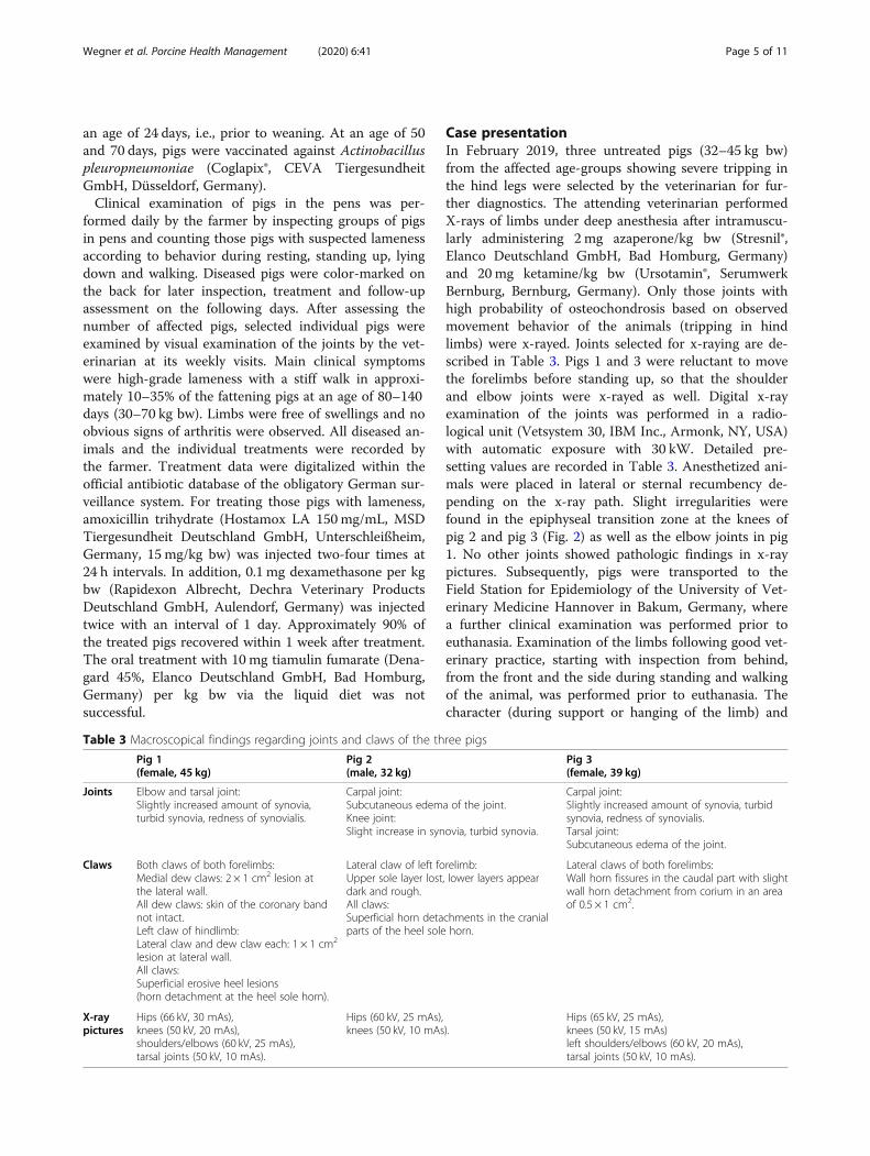
formed at the Department of Pathology of the Universityof Veterinary Medicine Hannover, Germany. Inflamma-tory as well as degenerative lesions were found (Fig. 5,Table 4).The femur bones of all three pigs were measured after
thorough removal of adjacent tissue. The distal third ofthe femur of each pig was ashed at the Institute of Ani-mal Nutrition. Serum samples were sent to the Clinic
for Swine, Small Ruminants and Forensic Medicine ofthe University of Veterinary Medicine Hannover to de-termine the concentration of calcium and phosphorus(Table 5).In addition, individual serum samples of the pigs were
sent to GD (Gezondheidsdienst voor Dieren, Deventer,the Netherlands) to determine bone markers osteocal-cine and C-telopeptid (CTx) reflecting bone metabolism.Serum samples were pooled according to the routinediagnostic procedures. Osteocalcine is indicative of boneformation, while CTx marks bone resorption. Whileosteocalcine-concentration was reduced (20.0 μg/L
Fig. 2 X-ray findings of left knee of pig 3. An osseous tulip-shapedbulge at the distal epiphyseal cartilage of the femur (arrow) as wellas incongruities at the articular surface of the tibia (arrowhead) arevisible. The x-ray picture was produced with pre-settings 30 kV, 50kV, 15 mAS
Fig. 3 Macroscopic lesions in the knee of pig 3. Irregularities at thearticular cartilage of the tibia are visible (arrow)
Fig. 4 Macroscopic lesions at claws of pig 1. Volar surface of clawsof the hind feet with erosive heel lesions and lesions of the coronaryband at the dew claws. In this pig, erosive heel lesions were foundin all claws
Wegner et al. Porcine Health Management (2020) 6:41 Page 6 of 11

[reference value: > 50 μg/L]), CTx was within the refer-ence range (0.17 μg/L [reference value: < 0.2 μg/L]). As aconsequence, the osteocalcine:CTx ratio (osteocalcine:CTx = 117.6 [reference range: > 150]) was reduced.
Interpretation of findings and measuresDiagnostic findings indicated a multifactorial diseasepathogenesis.Clinical findings were typical for both, arthritis caused
by M. hyosynoviae and osteochondropathia. All pigswere infected by M. hyosynoviae. Histologic findingsreflected degenerative cartilage and bone alterationscharacteristic of osteochondrosis (OC). Blood concentra-tions of calcium and phosphorus gave no evidence ofmineral deficiency in the three tested animals.
In a previous experimental study performed at the In-stitute for Animal Nutrition, University of VeterinaryMedicine Hannover, which was involved in this case re-port, serum P values varied between 2.5–2.8 mmol/L inpigs (bw ~ 55 kg) at generous P supply (including inor-ganic P sources and phytase) but dropped to values closeto 1.58 mmol/L at renounced inorganic P and phytase(after 3 weeks of different dietary treatment) [36].Homeostatic mechanisms control P and Ca concentra-tions in the blood so that serum concentrations are notconsidered a reliable indicator of insufficient mineralsupply [38]. In general, P serum concentration dropsonly during severe P deficiency.The youngest pig 2 showed a reduced mineral content
in the femoral bone sample (Table 5). This finding in
Fig. 5 Histologic findings in stifle joints of pig 2. a Histopathology revealed a pannus formation at the articular cartilage of the femur withdemasking of collagen fibers (asterisk). b Within the physis, multifocal cartilage cones (O) were detected. c Additional findings in the physisincluded multifocal chondrocyte degeneration (arrowhead) as well as eosinophilic streaks (arrow). d The synovial membrane revealed severefibrinopurulent (asterisk) inflammation. Hematoxylin and eosin staining, bars = 50 μm (a, c) and 200 μm (b, d)
Table 4 Histologic findings in stifle joints
Pig 1(female, 45 kg)
Pig 2(male, 32 kg)
Pig 3(female, 39 kg)
Femur, articular epiphyseal cartilage Multifocal pannus formation withdemasking of collagen fibers.
Femur, physis Single cartilage cones. Multifocal cartilage cones, multifocalchondrocyte degeneration andeosinophilic streaks.
Multifocal cartilage cones,multifocal chondrocytedegeneration, mild medullaryfibrosis.
Stifle joint, synovial membrane Mild to moderate, multifocalto coalescent, lympho-plasmahistiocytic synovialitis.
Moderate, fibrinosuppurative, partlylympho-plasma-histiocytic synovialitis.
Mild to moderate, multifocal,lympho-plasma-histiocyticsynovialitis.
Wegner et al. Porcine Health Management (2020) 6:41 Page 7 of 11

combination with bone marker values suggested a cata-bolic status of bone metabolism (bone resorption) andtherefore an inadequate mineralization of bones in clin-ically affected individuals.Clinical findings of a progressive, shifting lameness,
which affects one or more limbs, reluctance to move orto stand up, changing posture of the hind legs were typ-ical of the disease. Moreover, claw lesions were found,which could additionally be a trigger factor for diseasedue to disturbed body weight balancing, or which couldbe the result of putting increased weight on the clawsdue to specific postures.
Case outcomeIn April 2019, in the three different diets used at theearly stage of fattening, dietary Ca content was adjustedto 0.7% and total P to 0.48%. The proportion of fermen-ted ingredients in the final liquid diet was restricted to50%. In parallel, feeding technique was checked andfeeding valves were tested and controlled for adequatefunction on a regular basis.Between February and May 2019, individual pigs af-
fected by lameness were treated with amoxicillin trihy-drate and dexamethasone two-four times at 24 hintervals as previously described. Within these months,the incidence rate decreased to less than 5%.
Discussion and conclusionsAll findings in fatteners with impaired mobility on thisfarm led to the assumption that M. hyosynoviae as an
infectious factor was involved in disease pathogenesis incombination with additional factors. A slight increase insynovia volume was found in two of the pigs and in twopigs a subcutaneous edema of the joints was observed. Amild to moderate lymphoplasma-histiocytic inflamma-tion as well as a fibrino-suppurative synovialitis were de-tected by histology. Infectious arthritis caused by M.hyosynoviae often results in decreased profitability forthe farmers due to higher medication costs and time-consuming measures that have to be taken, such as seg-regating diseased pigs in recovery pens. This infectiousagent also further impairs skeletal health in fatteners,which is an important welfare issue [2]. In this case,antibiotic treatment with amoxicillin in combinationwith an anti-inflammatory substance was successful inindividual pigs, although mycoplasma species are intrin-sically resistant to β-lactam antibiotics [39]. In contrast,MIC values for tiamulin are low for M. hyosynoviae(≤0.25 μg/mL) [39], but treatment with this substancewas not successful on the farm in the case report. Withhigh probability, the anti-inflammatory parenteral treat-ment, which was not performed in combination with thein-feed-treatment with a pleuromutilin, was responsiblefor the improvement. If this was the case, infection withM. hyosynoviae might not be the primary cause of theclinical signs, but instead pain either caused by inflam-mation or by the degenerative joint alterations.Due to the generally differing outcome of M.-hyosyno-
viae-infection, identifying further influencing factors inaffected swine is of high importance. On the farm in
Table 5 Results of blood analyses and femoral ashing of the three pigs
Reference values Pig 1(female, 45 kg)
Pig 2(male, 32 kg)
Pig 3(female, 39 kg)
Blood analyses AP [U/I] 0 - 300a 257 175 186
Ca [mmol/L] 2.5–3.1a 2.86 2.53 2.56
P [mmol/L] 2.8–4.3a 2.96 2.69 2.79
hemoglobin [g/L] – 0.27 0.4 0.35
Bone ashing Bone length (mm) – 171 142 158
Bone diameter (mm) – 22 19 22
Total fat-free weight of piece of femur used for analysis (g) – 27.98 20.80 22.99
Total ash content of piece of femur used for analysis (g) – 13.60 9.11 10.92
DM [g/kg] – 473 379 422
Ash [g/kg ffr DM]d 493 ± 20.4b
495-524c486 438 ↓ 475
Ca [g/kg ffr DM]d 154 ± 8.7b
179-196c180 159 177
P [g/kg ffr DM]d 79.3 ± 3.4b
107-118c83 75.1 ↓ 83.5
aReference values for blood in crossbreed grower pigs [35]bReference values for bone ashing results in this age-group had been previously established for the distal femur epiphysis at the institute [36]cIn addition, study findings for the third metacarpal bone of finishing pigs fed different phosphorus sources are recorded [37]. Ranges include results from feedinggroups with calculated Ca of 0.78% and P of 0.56% in nursery and Ca of 0.65% and P of 0.43% in fattening diets [37]dGram per kilogram fat-free dry matter, AP alkaline phosphatase
Wegner et al. Porcine Health Management (2020) 6:41 Page 8 of 11

question, claw lesions and osteochondropathy were iden-tified as additional factors. During nursery, pigs werekept on plastic flooring, which is characterized by a dif-ferent hardness compared to concrete flooring in the fat-tening stable. These differences between both materialsin hardness but also surface roughness are risk factorsfor the development of claw lesions as a consequence ofmismatching in horn quality and underground [40]. Pig-lets experience a sudden change in underground aftermoving to the fattening unit without an adaptationperiod. The observed claw lesions were indicative ofabrasive injuries (sole erosions), but can also be the con-sequence of extended resting periods due to painfulnesswhile moving (skin lesions at the coronary band region).In general, additional factors can support the develop-ment of claw lesions, e.g., genetically determined asym-metries in inner and outer claws and abnormal toeangles, but also nutrient deficiency, e.g., biotin [41, 42],which were not suspected in the present case. Claw le-sions can be the consequence of degenerative joint dis-eases but also a risk factor for the development thereofsuch as OC [43]. Any kind of cartilage pre-damagemight contribute to the adhesion of M. hyosynoviae [44].A negative impact of P deficiency - suspected here atleast in one animal - on the function of the connectivetissue has been shown in other species [45]. Hypotheticallow intake of P would include low P concentrations inthe blood as acute P deficiency, which was only observedin one pig (Table 5). The determined bone markershinted at a stimulated bone resorption maybe as a con-sequence of marginal P supply of individuals with higherdaily weight gain [46, 47].Nevertheless, the hypothesis of an additional nutri-
tional impact on disease development could only be sup-ported by bone composition in one pig in this case. Thecomparably low bone mineral content in combinationwith the bone marker result might suggest that bone for-mation with respect to mineral accretion was reduced.Bone ashing in pigs is a further diagnostic approach toverify the suspicion of impaired mineralization.Standardization of the method has been improved in re-cent years so that preliminary reference values could beelaborated for the femur [36]. Bone ash diagnostic wasfound to be appropriate for diagnostic evaluation ofmarginal supply with minerals lasting at least 3 weeks[36]. In growth periods with insufficient Ca and P sup-ply, at first, the total bone mass is reduced, while boneformation and composition might be maintained. Alower ratio of the diameter of the long bones to bodymass can be the consequence [36]. The pressure on theend of bones covered by cartilage depends on body massand the area of contact within the joint. It could be thatalso in young pigs – as observed in growing dogs- thedietary P supply affects the strength of muscles that keep
the bones in the right position [45]. With an insufficientP intake and impaired muscle tonus, there is a predis-posing effect for alterations of the cartilage especially inpigs with high growth rates.Analysis of diet composition and feeding anamnesis
are fundamental in the diagnostic procedure. The au-thors assume that a marginal mineral supply during aspecific juvenile phase of life with high growth ratesmight be predisposing factors of disease development inlater life. The three pigs examined in this study wereslightly heavier (approximately 3 kg) than the averagepigs in the respective age-group, so that high dailyweight gain can be assumed. In individuals with highgrowth rates (~ 900 g average daily weight gain), the up-take of digestible P might have been insufficient at theend of nursery/beginning of fattening, especially whenthe phytase content in the final diet is reduced by a rela-tively high proportion of phytase-free fermented ingredi-ents. To diagnose a marginal mineral supply in criticalgrowth phases, not only the demand of the pigs with re-spect to feed intake and growth rate, but also for bonemineralization should be considered.A ratio of digestible Ca to digestible P for adequate
mineralization of bones and optimal growth perform-ance was found to be approximately 1.23:1 in cases whendigestible P met the requirements [48]. Excess Ca incombination with P concentrations below recommendedrequirements led to decreased growth rates [48]. Bothrequirements were fulfilled in this case report with low Pconcentrations and a relatively wide Ca:P ratio in thefinal diet. Estimates for Ca requirements of Ca in grow-ing pigs range from 6.3–4.2 g/kg DM in pigs with 20–80kg bw [19, 20]. Different batches of compound feed varyin P and Ca concentrations, which can lead to deficien-cies in short periods depending on batch size. Labeleddiet compositions are only based on an analysis in thefirst charge. In general, digestibility of dietary P is mark-edly improved by fermenting the liquid diet before offer-ing it to the animals and by adding phytase to thecompound feed [49–51]. Both strategies were applied onthis farm.In the three examined pigs in this study, histologic find-
ings were only indicative of osteochondropathy and notmineral or vitamin D deficiency, which would be charac-terized by distinct morphologic entities as e.g. failure ofnewly formed osteoid to mineralize [52, 53]. As metabolicbone diseases are usually reversible, any typical histologicchanges might had been present at an earlier point in timeand would most likely have been superimposed by the de-generative processes at the time of examination. Somestudies support the multifactorial pathogenesis of osteo-chondropathy but there is lack of evidence for an inter-action with mineral deficiency [54, 55]. Underexperimental conditions, hypophosphataemia was found
Wegner et al. Porcine Health Management (2020) 6:41 Page 9 of 11

to cause focal cartilage lesions, which were different fromcartilage degeneration typical of osteochondrosis [56].At the beginning of being involved in this clinical case,
the primary hypothesis was that the development of thedisease was a multifactorial process starting with an im-paired bone mineralization and triggering the develop-ment of osteochondropathy of the joints. Pigs with highgrowth rates develop high pressure on cartilage surfacesof the long bones. In the case that muscular forces areweakened due to a marginal P supply, deviations in limbaxes with the consequence of improper biomechanicalstress on joint cartilage surfaces, aseptic inflammationand subsequent pain during movement might occur.Whether cartilage alterations as shown in these pigs arepredisposing factors for colonization and infection withM. hyosynoviae remains hypothetical. Some authors haveconsidered that OC could be a predisposing factor forjoints to be infected with M. hyosynoviae [44]. However,these assumptions were refuted by authors who did notfind M. hyosynoviae any more frequently in individualsat the slaughterhouse with OC than in individuals with-out OC [3].So far, the potential triad involved in disease develop-
ment can be covered by the most straightforward diag-nostic steps reported in veterinary practice; namely, i)comparison of recommended dietary P levels with thoseanalyzed in the investigated case, during specific growthphases with expected high growth rates based on avail-able farm-specific production and feed data, ii) bacterio-logical diagnostic of articular samples and iii) histologicexamination of articular tissue. The new blood markersfor diagnosis of mineral deficiency should be validated inthe context of more practical cases, because they mightbe a promising new diagnostic tool for assessing skeletalhealth. Bone marker determination should be recom-mended especially in pigs with high daily weight gainsshowing stiff walking, tripping and reluctancy to standup, in order to assess the relation between bone catabol-ism and bone mineralization. In addition, we recom-mend in cases of sudden increased lameness to look atrecent findings without neglecting the period before theepisodes, including the previous dietary supply with P,especially of individuals with higher daily weight gains.Finally, the question concerning the degree of impact
of either P deficiency or osteochondropathy as well asclaw lesions on the disease and especially on diseasepathogenesis in M. hyosynoviae infection cannot be an-swered in this case. Thus, experimental studies understandardized conditions, also taking various geneticbackgrounds into account, would be necessary.
AcknowledgmentsWe wish to thank the farmer for his support in providing anamnestic dataand initiating all diagnostic steps.
Authors’ contributionsIH, EG and JT contributed to the conception and design of the present casereport. BW conducted the literature study and wrote the first draft of themanuscript. JT, JV and MB performed the clinical examination, and JV andMB performed the necropsy. JK, FH and HR gave helpful professional adviceand helped with the processing regarding nutrition and pathology. Allauthors contributed to the development and the revisions of the manuscriptand approved the final version.
FundingOpen Access funding enabled and organized by Projekt DEAL.
Availability of data and materialsData sharing is not applicable to this article as no datasets were generatedor analyzed during the current study.
Ethics approval and consent to participateThe present case report does not include experimental data, and all furtherinvestigations were performed as routine diagnostics during the clinicaloutbreak. Therefore, seeking approval from the animal ethics committee wasunnecessary.
Consent for publicationNot applicable.
Competing interestsThe authors declare that they have no competing interests.
Author details1Veterinary Practice Duemmerland, Steinfeld, Oldenburg, Germany. 2Vetland®Dr. Tenhündfeld & Kollegen, Vreden, Germany. 3Field Station forEpidemiology, University of Veterinary Medicine Hannover, Foundation,Hannover, Germany. 4Institute for Animal Nutrition, University of VeterinaryMedicine Hannover, Foundation, Hannover, Germany. 5Department ofPathology, University of Veterinary Medicine Hannover, Foundation,Hannover, Germany.
Received: 15 June 2020 Accepted: 7 December 2020
References1. Kobisch M, Friis N. Swine mycoplasmoses. Rev Sci Tech. 1996;15(4):1569–
614.2. Gomes Neto J, Gauger P, Strait E, Boyes N, Madson D, Schwartz K.
Mycoplasma-associated arthritis: critical points for diagnosis. J Swine HealthProd. 2012;20(2):82–6.
3. Nielsen E, Nielsen N, Friis N. Mycoplasma hyosynoviae arthritis in grower-finisher pigs. J Vet Med A. 2001;48(8):475–86.
4. Clavijo MJ, Anderson A, Gei-ger J, Lyons W. Can we eliminate mycoplasmahyosynoviae and mycoplasma hyorhinis? National Hog Farmer online.http://www.nationalhogfarmer.com/animal-health/can-we-eliminate-mycoplasma-hyosynoviae-and-mycoplasma-hyorhinis. Accessed 15 Sept2020.
5. Ross R, Duncan J. Mycoplasma hyosynoviae arthritis of swine. J Am Vet MedAssoc. 1970;157(11):1515.
6. Ross RF. Pathogenicity of swine mycoplasmas. Ann N Y Acad Sci. 1973;225(1):347–68.
7. grosse Beilage E, Wendt M. Diagnostik und Gesundheitsmanagement imSchweinebestand. 1st ed. Stuttgart: UTB; 2013.
8. White M. Joint disease in the pig. In Pract. 1994;16(1):37–40.9. Olstad K, Ekman S, Carlson CS. An update on the pathogenesis of
osteochondrosis. Vet Pathol. 2015;52(5):785–802.10. Olstad K, Shea KG, Cannamela PC, Polousky JD, Ekman S, Ytrehus B, Carlson
CS. Juvenile osteochondritis dissecans of the knee is a result of failure of theblood supply to growth cartilage and osteochondrosis. Osteoarthr Cartil.2018;26:1691–8.
11. Olstad K, Wormstrand B, Kongsro J, Grindflek E. Osteochondrosis in thedistal femoral physis of pigs starts with vascular failure. Vet Pathol. 2019;56(5):732–42.
12. Ytrehus B, Carlson CS, Ekman S. Etiology and pathogenesis ofosteochondrosis. Vet Pathol. 2007;44(4):429–48.
Wegner et al. Porcine Health Management (2020) 6:41 Page 10 of 11

13. Grøndalen T. Leg weakness in pigs. II. Litter differences in leg weakness,skeletal lesions, joint shape and exterior conformation. Acta Vet Scand.1974;15:574–86.
14. van der Wal PG, van der Valk PC, Goedegebuure SA, van Essen G.Osteochondrosis in six breeds of slaughter pigs. II. Data concerning carcasscharacteristics in relation to osteochondrosis. Vet Q. 1980;2:42–7.
15. Dodds WK, Smith VH. Nitrogen, phosphorus, and eutrophication in streams.Inland Waters. 2016;6(2):155–64.
16. Ferket P, Van Heugten E, Van Kempen T, Angel R. Nutritional strategies toreduce environmental emissions from nonruminants. J Anim Sci. 2002;80(E.Suppl. 2):E168–82.
17. Directive 2001/81/EC of the European Parliament and of the Council of 23October 2001 on national emission ceilings for certain atmosphericpollutants.
18. Andretta I, Pomar C, Rivest J, Pomar J, Radünz J. Precision feeding cansignificantly reduce lysine intake and nitrogen excretion withoutcompromising the performance of growing pigs. Animal. 2016;10(7):1137–47.
19. Suttle NF. Mineral nutrition of livestock. 4th ed. UK: CABI Oxfordshire; 2010.20. NRC – National Research Counsil. Nutrient requirements of swine. 11th rev
ed. Washington, D.C: Natl. Acad. Press; 2012. Stein HH, Merriman.21. Gutierrez NA, Serão NVL, Elsbernd AJ, Hansen SL, Walk CL, Bedford MR,
Patience JF. Quantitative relationships between standardized total tractdigestible phosphorus and total calcium intakes and their retention andexcretion in growing pigs fed corn–soybean meal diets. J Anim Sci. 2015;93:2174–82.
22. Adeola O, Azain MJ, Carter SD, Crenshaw TD, Estienne MJ, Kerr BJ,Lindemann MD, Maxwell CV, Miller PS, Shannon MC, van Heugten E. Acooperative study on the standardized total-tract digestible phosphorusrequirement of twenty-kilogram pigs. J Anim Sci. 2015;93(12):5743–53.
23. Crenshaw TD. Calcium, phosphorus, vitamin D, and vitamin K in swinenutrition. In: Lewis AJ, Southern LL, editors. Swine nutrition. Boca Raton: CRCPress; 2001. p. 187–212.
24. Varley P, Sweeney T, Ryan M, O'doherty J. The effect of phosphorus restrictionduring the weaner-grower phase on compensatory growth, serum osteocalcinand bone mineralization in gilts. Livest Sci. 2011;135(2–3):282–8.
25. Heaney R, Abrams S, Dawson-Hughes B, Looker A, Looker A, Marcus R,Matkovic V, Weaver C. Peak bone mass. Osteoporos Int. 2000;11(12):985–1009.
26. Simons P, Versteegh HA, Jongbloed AW, Kemme P, Slump P, Bos K, WoltersM, Beudeker R, Verschoor G. Improvement of phosphorus availability bymicrobial phytase in broilers and pigs. Br J Nutr. 1990;64(2):525–40.
27. Zofkova I. Bone tissue as a systematic endocrine regulator. Physiol Res. 2001;64:439–55.
28. González-Vega JC, Stein HH. Calcium digestibility and metabolism in pigs.Asian-Australas J Anim Sci. 2014;27(1):1–9.
29. Doyle ME, Jan de Beur SM. The skeleton. Endocrine regulator of phosphatehomeostasis. Curr Osteoporos Rep. 2008;6(4):134–41.
30. McCue M, Reichert JL, Crenshaw TD. Impact of dietary vitamin D3supplements in nursery diets on subsequent growth and bone responses ofpigs during an immune challenge. J Anim Sci. 2019;97(12):4895–903.
31. Williams NH, Stahly TS, Zimmerman DR. Effect of chronic immune systemactivation on the rate, efficiency, and composition of growth and lysineneeds of pigs fed from 6 to 27 kg. J Anim Sci. 1997;75(9):2463–71.
32. Crenshaw TD, Peo ER, Lewis AJ Jr, Moser BD, Olson D. Influence of age, sexand calcium and phosphorus levels on the mechanical properties of variousbones in swine. J Anim Sci. 1981;52(6):1319–29.
33. Naumann C, Bassler R. Methoden der landwirtschaftlichen Forschungs- undUntersuchungsanstalt, Biochemische Untersuchung von Futtermitteln,Methodenbuch 3 (einschließlich der achten Ergänzungen) VDLUFA,Darmstadt, Germany. 2012.
34. El-Wahab AA, Visscher C, Teitge F, Steinhagen D. Choice preference of dietswith different protein levels depending on water temperature in Nile tilapia.J World Aquacult Soc. 2020;51:512–26.
35. Klem TB, Bleken E, Morberg H, Thoresen SI, Framstad T. Hematologic andbiochemical reference intervals for Norwegian crossbreed grower pigs. VetClin Pathol. 2010;39:221–6.
36. Rieger H. Untersuchungen zum Einfluss einer unterschiedlichenPhosphorversorgung auf die Entwicklung und Mineralisation verschiedenerKnochen wachsender Schweine. Thesis. University of Veterinary MedicineHannover. 2017.
37. Teixeira AO, Corassa A, Moreira LM, Nogueira ET, Lopes JB, Rocha CM,Ferreira VPA. Bone characteristics of pigs fed different sources ofphosphorus. Rev Col Cienc Pec. 2016;29:245–54.
38. O'Doherty JV, Gahan DA, O'Shea C, Callan JJ, Pierce KM. Effects of phytaseand 25-hydroxyvitamin D3 inclusions on the performance, mineral balanceand bone parameters of grower-finisher pigs fed low-phosphorus diets.Animal. 2010;4(10):1634–40.
39. Gautier-Bouchardon AV. Antimicrobial resistance in mycoplasma spp.Antimicrobial resistance in bacteria from livestock and companion animals.Review Microbiol Spectr. 2018;6(4):425–46.
40. Jørgensen B. Influence of floor type and stocking density on leg weakness,osteochondrosis and claw disorders in slaughter pigs. Anim Sci. 2003;77:439–49.
41. Olsson A, Svendsen J, Botermans J, Bergsten C. An experimental model forstudying claw lesions in growing female pigs. Livest Sci. 2016;184:58–63.
42. Webb NG, Penny RH, Johnston AM. Effect of a dietary supplement of biotinon pig hoof horn strength and hardness. Vet Rec. 1984;114(8):185–9.
43. Jensen TB, Kristensen AR, Toft N, Baadsgaard NP, Østergaard S, Houe H. Anobject-oriented Bayesian network modeling the causes of leg disorders infinisher herds. Prev Vet Med. 2009;89(3):237–48.
44. Lawrisuk L, Rothschild M, Ross R, Christian L. Relationship betweenmycoplasma hyosynoviae infection and front limb weakness in Durocswine. Am J Vet Res. 1987;48(9):1395–7.
45. Kiefer-Hecker B, Kienzle E, Dobenecker B. Effects of low phosphorus supplyon the availability of calcium and phosphorus, and musculoskeletaldevelopment of growing dogs of two different breeds. J Anim PhysiolAnim Nutr. 2018;102(3):789–98.
46. Carter S, Cromwell G, Combs T, Colombo G, Fanti P. The determination ofserum concentrations of osteocalcin in growing pigs and its relationship toend-measures of bone mineralization. J Anim Sci. 1996;74(11):2719–29.
47. Peel N, Eastell R. Measurement of bone mass and turnover. Baillieres ClinRheumatol. 1993;7(3):479–98.
48. Lagos LV, Walk CL, Murphy MR, Stein HH. Effects of dietary digestiblecalcium on growth performance and bone ash concentration in 50 to 85 kggrowing pigs fed diets with different concentrations of digestiblephosphorus. Anim Feed Sci Technol. 2019;247:262–72.
49. Pagano AR, Yasuda K, Roneker KR, Crenshaw TD, Lei XG. SupplementalEscherichia coli phytase and strontium enhance bone strength of youngpigs fed a phosphorus-adequate diet. J Nutr. 2007;137(7):1795–801.
50. Veum T, Bollinger D, Buff C, Bedford M. A genetically engineered Escherichiacoli phytase improves nutrient utilization, growth performance, and bonestrength of young swine fed diets deficient in available phosphorus. J AnimSci. 2006;84(5):1147–58.
51. Bunte S. Die Fermentation von Flüssigfutter als Fütterungskonzept imSchweinebestand: Potentiale, aber auch Risiken aus Sicht der Tierernährungund Tiermedizin. Thesis. University of Veterinary Medicine Hannover. 2018.
52. Scholz-Ahrens KE, Glüer CC, Bronner F, Delling G, Açil Y, Hahne HJ,Hassenpflug J, Timm W, Schrezenmeir J. Modulation of vitamin D status anddietary calcium affects bone mineral density and mineral metabolism ingöttingen minipigs. ISRN Rheumatol. 2013;2013:460512.
53. Madsen DM, Ensley SM, Gauger PC, Schwartz KJ, Stevenson GW, Cooper VL,Janke BH, Burrough ER, Goff JP, Horst RL. Rickets: case series and diagnosticreview of hypovitaminosis D in swine. J Vet Diagn Investig. 2012;24(6):1137–44.
54. Fabà L, Gasa J, Tokach M, Varella E, Solà-Oriol D. Effects of supplementingorganic microminerals and methionine with or without limiting growthduring the rearing phase of replacement gilts on lameness, growth, andbody composition1. Transl Anim Sci. 2019;3:717–30.
55. Hartnett P, Boyle L, Younge B, O'Driscoll K. The effect of group compositionand mineral supplementation during rearing on measures of cartilagecondition and bone mineral density in replacement gilts. Animals. 2019;9(9):637.
56. Reiland S, Håglin L, Sjöberg H-E. Experimental hypophosphataemia ingrowing pigs: effects on endochondral ossification in comparison toosteochondrosis. J Comp Pathol. 1991;105(3):247–54.
Publisher’s NoteSpringer Nature remains neutral with regard to jurisdictional claims inpublished maps and institutional affiliations.
Wegner et al. Porcine Health Management (2020) 6:41 Page 11 of 11