Embed Size (px)
Citation preview

OPEN
ORIGINAL ARTICLE
Lactobacillus rhamnosus GG-supplemented formulaexpands butyrate-producing bacterial strains in foodallergic infants
Roberto Berni Canani1,8, Naseer Sangwan2,8, Andrew T Stefka3,8, Rita Nocerino1,Lorella Paparo1, Rosita Aitoro1, Antonio Calignano4, Aly A Khan5, Jack A Gilbert2,6,7and Cathryn R Nagler31Department of Translational Medical Science, Section of Pediatrics, European Laboratory for theInvestigation of Food-Induced Diseases, University of Naples, Federico II, Naples, Italy; 2Institute forGenomics and Systems Biology, Department of Biosciences, Argonne National Laboratory, Argonne, IL, USA;3Committee on Immunology and Department of Pathology, University of Chicago, Chicago, IL, USA;4Department of Pharmacy, University of Naples Federico II, Naples, Italy; 5Toyota Technological Institute atChicago, Chicago, IL, USA; 6Department of Surgery, University of Chicago, Chicago, IL, USA and 7Departmentof Ecology and Evolution, University of Chicago, Chicago, IL, USA
Dietary intervention with extensively hydrolyzed casein formula supplemented with Lactobacillusrhamnosus GG (EHCF+LGG) accelerates tolerance acquisition in infants with cow’s milk allergy(CMA). We examined whether this effect is attributable, at least in part, to an influence on the gutmicrobiota. Fecal samples from healthy controls (n= 20) and from CMA infants (n= 19) before andafter treatment with EHCF with (n= 12) and without (n= 7) supplementation with LGG were comparedby 16S rRNA-based operational taxonomic unit clustering and oligotyping. Differential featureselection and generalized linear model fitting revealed that the CMA infants have a diverse gutmicrobial community structure dominated by Lachnospiraceae (20.5±9.7%) and Ruminococcaceae(16.2±9.1%). Blautia, Roseburia and Coprococcus were significantly enriched following treatmentwith EHCF and LGG, but only one genus, Oscillospira, was significantly different between infants thatbecame tolerant and those that remained allergic. However, most tolerant infants showed asignificant increase in fecal butyrate levels, and those taxa that were significantly enriched in thesesamples, Blautia and Roseburia, exhibited specific strain-level demarcations between tolerant andallergic infants. Our data suggest that EHCF+LGG promotes tolerance in infants with CMA, in part, byinfluencing the strain-level bacterial community structure of the infant gut.The ISME Journal (2016) 10, 742–750; doi:10.1038/ismej.2015.151; published online 22 September 2015
Introduction
The prevalence of allergic responses to food has beenexperiencing an unprecedented increase in devel-oped societies, rising by as much as 20% in a recent10-year period (Branum and Lukacs, 2009; Osborneet al., 2011; Wang and Sampson, 2011; Prescott et al.,2013). Genetic variation alone cannot account for adramatic increase in disease prevalence over such ashort time frame. Emerging evidence suggests thattwenty-first century environmental interventions,including widespread antibiotic use, consumption
of a high-fat/low fiber diet, elimination of previouslycommon enteropathogens (including Helicobacterpylori and helminthic parasites), reduced exposureto infectious disease, Caesarean birth, and formulafeeding, may have perturbed the mutually beneficialinteractions established over millions of years of co-evolution with the bacteria that comprise ourcommensal microbiota (Cho and Blaser, 2012). Thisdysbiosis can predispose genetically susceptibleindividuals to allergic disease (reviewed in ref.Feehley et al., 2012). Cow’s milk allergy (CMA) isone of the most common food allergies of infancyand early childhood with an estimated prevalence of2–3% worldwide (Sicherer, 2011). We have demon-strated that dietary management with a formulacontaining an extensively hydrolyzed form of thecow’s milk protein casein (EHCF), supplementedwith the probiotic Lactobacillus rhamnosus GG(LGG), results in a higher rate of tolerance acquisi-tion in infants with CMA than in those treated with
Correspondence: JA Gilbert, Department of Surgery, University ofChicago, 5842 South Maryland Avenue, Chicago, IL, 60637, USA.E-mail: [email protected] CR Nagler, Department of Pathology, University of Chicago, 924East 57th Street R120, Chicago, IL 60637.E-mail: [email protected] authors contributed equally to this work.Received 16 April 2015; revised 4 July 2015; accepted 8 July 2015;published online 22 September 2015
The ISME Journal (2016) 10, 742–750© 2016 International Society for Microbial Ecology All rights reserved 1751-7362/16
www.nature.com/ismej

EHCF without supplementation or with other non-casein-based formulas (Berni Canani et al., 2012,2013). However, the mechanistic basis for this effectis not known. We hypothesized that it is attributable,in part, to an influence of this dietary intervention onthe composition of the gut microbiota. To test thishypothesis, we performed 16S ribosomal RNA(rRNA)-based amplicon sequencing and oligotypinganalysis on stool samples collected from healthyinfants and from CMA infants before and aftertreatment with EHCF with or without supplementa-tion with LGG.
Materials and methodsPatient enrollment and sample collectionInfants invited to participate in the study werereferred to a tertiary pediatric allergy center(Pediatric Food Allergy Unit at the Department ofTranslational Medical Science of the University ofNaples ‘Federico II’) for a full diagnostic work-up forsuspected CMA. All patients were still receivingcow’s milk protein (mainly from formula feeding) atthe time of enrollment and first stool sampling.The inclusion criteria were infants aged 1–12 monthswith a recent strong suspicion of IgE-mediated CMAbut still receiving cow’s milk protein. Diagnosis ofIgE-mediated CMA was based on clinical history, theresults of a double blind placebo-controlled oral foodchallenge, and the level of serum-specific anti-cow’smilk protein IgE (Berni Canani et al., 2011). Patientsadministered pre- or probiotic products and/orantibiotics in the previous 4 weeks, and patientswith a history of cow’s milk-induced anaphylaxis,eosinophilic disorders of the gastrointestinal tract,food protein-induced enterocolitic syndrome, con-comitant chronic systemic diseases, congenital car-diac defects, active tuberculosis, autoimmunediseases, immunodeficiency, chronic inflammatorybowel diseases, celiac disease, cystic fibrosis, meta-bolic diseases, lactose intolerance, malignancy,chronic pulmonary diseases or malformations ofthe gastrointestinal tract were excluded. Fecalsamples were collected at baseline before diettherapy from patients with a confirmed diagnosisof IgE-mediated CMA according to standardizedcriteria (Berni Canani et al., 2011). Following theinitial visit, patients were treated by dietary manage-ment with a commercially available extensivelyhydrolyzed casein formula (EHCF, Nutramigen,Mead Johnson, Rome, Italy) either with or withoutsupplementation with LGG (at 4.5 × 10⁷–8.5 × 10⁷
colony-forming units per gram of powder (BerniCanani et al, 2012). A second fecal sample wasobtained after 6 months. Samples obtained fromhealthy (non-allergic) infants who visited the studyclinic as part of a vaccination program served ascontrols. These subjects were not at risk for atopicdisorders and their clinical history was negative forany allergic condition. The study was conducted in
accordance with the Declaration of Helsinki andapproved by the Ethics Committee of the Universityof Naples ‘Federico II’.
Fecal DNA isolation and 16S rDNA sequencingFeces were collected and frozen at −20 °C immedi-ately after excretion. To isolate DNA, 100–300mg offecal material was bead beaten before extraction withthe QIAamp DNA stool mini kit (Qiagen, Hilden,Germany). 16S V4-region amplicon libraries wereproduced using previously described primers andsequenced using the Illumina MiSeq platform (150bp×2) at Argonne National Laboratory’s BiosciencesSequencing Core facility (Caporaso et al., 2011).Bacterial load was determined by quantitativePCR using a standard curve derived from a plasmidcontaining a single copy of the 16S rRNA encodinggene (Stefka et al., 2014). Sequence data have beendeposited in MG-RAST (http://metagenomics.anl.gov)under accession numbers 4571868.3–4571924.3 andproject number 10023.
Bioinformatics analysisPaired end reads were quality trimmed and pro-cessed for operational taxonomic unit (OTU) cluster-ing using UPARSE pipeline (Edgar, 2013), set at0.97% identity cutoff. Taxonomic status wasassigned to the high-quality (o1% incorrect bases)candidate OTUs using the ‘parallel_assign_taxono-my_rdp.py’ script of QIIME software (Caporaso et al.,2010). Multiple sequence alignment and phyloge-netic reconstruction were performed using PyNastand FastTree (Caporaso et al., 2010). Phyloseqpackage (McMurdie and Holmes, 2013) was usedfor the detailed downstream analysis on a rarefiedabundance matrix. This matrix was processed toremove OTUs containing less than five reads toreduce the PCR and sequencing based bias; then, theOTU table was rarified to the minimum numbers ofreads present in the smallest library (3746 reads).We used the oligotyping pipeline (Eren et al., 2013)to identify the sub-OTU level differences in the topfive most differentially abundant genera, that is,Roseburia, Blautia, Coprococcus, Faecalibacteriumand Bifidobacterium, as predicted by Metagenome-Seq (Paulson et al., 2013).
Statistical analysisWe used MetagenomeSeq (Paulson et al., 2013)software to determine the differentially abundantOTUs, families, and genera, present across allgroups. We also used nonparametric Kruskal–WallisH-test (post hoc Tukey Kramer tests, Bonferronimultiple test correction) for multi-group compari-sons. Two-group and two-sample comparisons wereperformed using Welch’s t-test and Fisher’s exactt-test, respectively (two-sided with Bonferroni cor-rection). We compiled a de-identified metadata table
LGG expands butyrate-producing bacteriaR Berni Canani et al
743
The ISME Journal

containing all of the clinical and demographic datafor each infant in this study (Supplementary Table1). We then used a generalized linear regressionmodel (GLM) to examine the contribution of sevenmeasurable features from the patient demographicdata ('mode of birth', 'age at introduction of solidfoods', 'age at initial sampling', 'sex', 'body weight','duration of exclusive breastfeeding’ and ‘healthstatus’ (that is, healthy or CMA)) on the bacterialabundance of the differentially abundant bacterialfamilies and strains as predicted by oligotyping. AGLM model was constructed and validated usingrms and ResourceSelection (Lele, 2009) packages,respectively. We modeled bacterial abundancesusing a binomial distribution with a logit linkfunction. To examine whether fecal butyrate levelscorrelated with bacterial diversity (Shannon diver-sity index) and evenness (Pielou’s evenness index)and oligotype abundance patterns across multiplegroups (that is, healthy, CMA, EHCF and EHCF-LGG), we calculated the Spearman correlation usingthe cor.test function implemented in R (http://www.r-project.org/).
Determination of fecal butyrate concentrationFrozen feces weighing 1 g were diluted with saline,vortexed and centrifuged at 13 000 r.p.m. for 10minin 2-ml tubes. The supernatants were filtered(0.45 μm) and used as the fecal extracts, which werestored at − 20 °C until analysis. To determine fecalbutyrate concentration, frozen fecal extracts wereacidified with 20 μl 85% phosphoric acid and 0.5mlethyl acetate, mixed, centrifuged at 14 000 r.p.m. for1 h and extracted in duplicate. A quantity of thepooled extract containing the acidified butyrate wastransferred into a 2-ml glass vial and loaded onto anAgilent Technologies (Santa Clara, CA, USA) 7890gas chromatograph (GC) system with automaticloader/injector. The GC column was an AgilentJ&W DB-FFAP (Agilent Technologies) with thelength 30m, internal diameter 0.25mm and film
thickness 0.25 μm. The GC was programmed toachieve the following run parameters: initial tem-perature 90 °C, hold 0.5min, ramp 20 °Cmin− 1, finaltemperature 190 °C, total run time 8.0min, gas flow7.7mlmin− 1 split less to maintain 3.26 p.s.i. columnhead pressure, septum purge 2.0mlmin− 1. Detectionwas achieved using a flame ionization detector.Peaks were identified using a mixed externalstandard and quantified by peak height/internalstandard ratio.
Results
The gut microbiota of cow’s milk allergic infantsexhibits significantly increased diversity and alteredcompositionA fecal sample was obtained before diet therapy from19 patients with IgE-mediated CMA. During the samestudy period, fecal samples were also obtained from20 age, sex and body weight-matched healthy infantsenrolled in a vaccination program. All study subjectswere breastfed for o1 month after birth and werestill receiving a formula containing cow’s milkproteins at the time of enrollment and first fecalsampling. A second fecal sample was obtained fromeach of the CMA infants after 6 months of treatmentwith EHCF with or without supplementation withLGG. The demographic and clinical characteristicsof the study population are summarized in Table 1.A metadata table containing all of the demographicand clinical information for each patient in thisstudy is provided in Supplementary Table 1. Tocompare the fecal microbiota of healthy and CMAinfants, we generated 1.7 million 16S rRNA V4amplicon sequences, which following quality con-trol, clustered into 592 OTUs (97% nucleotideidentity). Ordination and classification independentanalyses of overall bacterial community structuredemonstrated that allergic infants were significantlymore diverse than age-matched healthy controls(Shannon’s index, healthy=1.7±0.8 vs CMA=2.6±0.4;Figure 1a), and also significantly more even (Pielou’s
Table 1 Demographic and clinical characteristics of the study population
Healthy subjects Patients with IgE-mediated CMA
Treated with EHCF Treated with EHCF+LGG
N 20 7 12Male, n (%) 11 (55.0) 4 (57.1) 9 (75.0)Age, months ( ± s.d.) 4.2 (1.1) 4 (0.8) 4.3 (1.3)Weight, g ( ± s.d.) 6937.5 (793.2) 5607.1 (480.8) 6366.7 (1074.1)Vaginal delivery, n (%) 15 (75) 4 (57.1) 7 (58.3)Duration of exclusive breastfeeding, days ( ± s.d.) 14.4 (4.5) 14.8 (5.4) 15.2 (3.1)Age of solid food introduction, months ( ± s.d.) 4 (0.2) 4.1 (0.4) 4.1 (0.3)
Symptoms at the CMA onsetVomiting, n (%) — 4 (57.1) 8 (66.7)Urticaria/angioedema, n (%) — 3 (42.9) 4 (33.3)Cough/wheezing, n (%) — 1 (14.3) 3 (25.0)
Abbreviations: CMA, cow’s milk allergy; EHCF, extensively hydrolyzed casein formula; LGG, Lactobacillus rhamnosus GG.
LGG expands butyrate-producing bacteriaR Berni Canani et al
744
The ISME Journal

evenness; healthy = 0.52 ± 0.2 vs CMA=0.6 ±0.3;Figure 1b). Bacterial 16S rRNA abundance wassimilar in all samples (Figure 1c).
Taxonomic assignment revealed marked differ-ences between healthy and allergic infants. CMAinfants had a significant reduction in Bifidobacter-iaceae, Streptococcaceae, Enterobacteriaceae andEnterococcaceae, and were significantly enrichedfor Ruminococcaceae (16%) and Lachnospiraceae(20.5%; Figure 1d). The CMA infant gut microbiotacomprised 73% Bacteroidetes and Firmicutes taxa,which are also known to dominate in the adult gut(The Human Microbiome Project Consortium, 2012).Genus-level analysis revealed a significant enrich-ment in CMA infant samples of Ruminococcus andFaecalibacterium, and a significant reduction inBifidobacterium and Escherichia (Welch’s t-test)
compared with healthy samples (SupplementaryTable 2).
To examine whether these significant differencescould be explained by demographic variables, weapplied a GLM for seven features (SupplementaryTable 1) fitted against the relative abundance of thesignificantly different taxa. Health status (that is,healthy or CMA) was the single largest significantcontributor to the differential abundance of Lach-nospiraceae (P=5.74e− 05; Figure 1e), Ruminococ-caceae (P=0.00144; Supplementary Figure S1A),Enterobacteriaceae (P=0.0003; SupplementaryFigure S1B) and Streptococcaceae (P=0.00198;Supplementary Figure S1C). It was also the secondlargest contributor to Bifidobacteriaceae (P=0.0034,Supplementary Figure S1D). Mode of birth andgender did not significantly contribute to these
Figure 1 Significantly diverse bacterial community dynamics across cow’s milk allergy and its treatment. (a) Shannon diversity,(b) Pielou’s evenness and (c) bacterial load in fecal samples from each healthy (n=20) or age-matched pre-treatment cow’s milk allergic(CMA, n=18–19) patients at diagnosis. (d) Family level differential abundance across healthy, CMA pre-treatment and treated groups, ascomputed by MetagenomeSeq. Families depicted are those determined to be differentially abundant. (e) Generalized linear model fitting ofpatient demographic information across relative abundance of family Lachnospiraceae. Parallel x axis represents the relative contributionvalue of every factor, as predicted by the GLM model. *Po0.05, **Po0.0001, by two-sided t-test or by GLM model.
LGG expands butyrate-producing bacteriaR Berni Canani et al
745
The ISME Journal
differences. However, body weight and age at initialsampling were significantly associated with theabundance of Enterobacteriaceae (Po0.0034,Supplementary Figure S1B) and Bifidobacteriaceae(Po0.00633, Supplementary Figure S1D), respec-tively. We validated the GLM model by fitting thesefeatures against genus-level abundances for Faecali-bacterium, (Po0.0001, Supplementary Figure S2A),Ruminococcus (Po0.0029, Supplementary FigureS2B) Escherichia (Po0.0071, SupplementaryFigure S2C) and Bifidobacterium (Po0.0031,Supplementary Figure S2D). These observationssuggested that CMA, more than any other measureddemographic variable, was the most important factoraffecting the significantly different components ofthe gut microbiome.
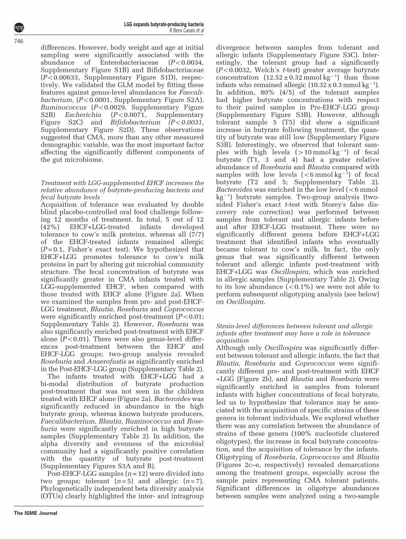
Treatment with LGG-supplemented EHCF increases therelative abundance of butyrate-producing bacteria andfecal butyrate levelsAcquisition of tolerance was evaluated by doubleblind placebo-controlled oral food challenge follow-ing 12 months of treatment. In total, 5 out of 12(42%) EHCF+LGG-treated infants developedtolerance to cow’s milk proteins, whereas all (7/7)of the EHCF-treated infants remained allergic(P=0.1, Fisher’s exact test). We hypothesized thatEHCF+LGG promotes tolerance to cow’s milkproteins in part by altering gut microbial communitystructure. The fecal concentration of butyrate wassignificantly greater in CMA infants treated withLGG-supplemented EHCF, when compared withthose treated with EHCF alone (Figure 2a). Whenwe examined the samples from pre- and post-EHCF-LGG treatment, Blautia, Roseburia and Coprococcuswere significantly enriched post-treatment (Po0.01;Supplementary Table 2). However, Roseburia wasalso significantly enriched post-treatment with EHCFalone (Po0.01). There were also genus-level differ-ences post-treatment between the EHCF andEHCF-LGG groups; two-group analysis revealedRoseburia and Anaerofustis as significantly enrichedin the Post-EHCF-LGG group (Supplementary Table 2).
The infants treated with EHCF+LGG had abi-modal distribution of butyrate productionpost-treatment that was not seen in the childrentreated with EHCF alone (Figure 2a). Bacteroideswassignificantly reduced in abundance in the highbutyrate group, whereas known butyrate producers,Faecalibacterium, Blautia, Ruminococcus and Rose-buria were significantly enriched in high butyratesamples (Supplementary Table 2). In addition, thealpha diversity and evenness of the microbialcommunity had a significantly positive correlationwith the quantity of butyrate post-treatment(Supplementary Figures S3A and B).
Post-EHCF-LGG samples (n=12) were divided intotwo groups; tolerant (n=5) and allergic (n=7).Phylogenetically independent beta diversity analysis(OTUs) clearly highlighted the inter- and intragroup
divergence between samples from tolerant andallergic infants (Supplementary Figure S3C). Inter-estingly, the tolerant group had a significantly(Po0.0032, Welch’s t-test) greater average butyrateconcentration (12.52 ±0.32mmol kg− 1) than thoseinfants who remained allergic (10.32±0.3mmol kg−1).In addition, 80% (4/5) of the tolerant sampleshad higher butyrate concentrations with respectto their paired samples in Pre-EHCF-LGG group(Supplementary Figure S3B). However, althoughtolerant sample 5 (T5) did show a significantincrease in butyrate following treatment, the quan-tity of butyrate was still low (Supplementary FigureS3B). Interestingly, we observed that tolerant sam-ples with high levels (410mmol kg− 1) of fecalbutyrate (T1, 3 and 4) had a greater relativeabundance of Roseburia and Blautia compared withsamples with low levels (o6mmol kg− 1) of fecalbutyrate (T2 and 5; Supplementary Table 2).Bacteroideswas enriched in the low level (o6mmolkg− 1) butyrate samples. Two-group analysis (two-sided Fisher’s exact t-test with Storey's false dis-covery rate correction) was performed betweensamples from tolerant and allergic infants beforeand after EHCF-LGG treatment. There were nosignificantly different genera before EHCF+LGGtreatment that identified infants who eventuallybecame tolerant to cow’s milk. In fact, the onlygenus that was significantly different betweentolerant and allergic infants post-treatment withEHCF+LGG was Oscillospira, which was enrichedin allergic samples (Supplementary Table 2). Owingto its low abundance (o0.1%) we were not able toperform subsequent oligotyping analysis (see below)on Oscillospira.
Strain-level differences between tolerant and allergicinfants after treatment may have a role in toleranceacquisitionAlthough only Oscillospira was significantly differ-ent between tolerant and allergic infants, the fact thatBlautia, Roseburia and Coprococcus were signifi-cantly different pre- and post-treatment with EHCF+LGG (Figure 2b), and Blautia and Roseburia weresignificantly enriched in samples from tolerantinfants with higher concentrations of fecal butyrate,led us to hypothesize that tolerance may be asso-ciated with the acquisition of specific strains of thesegenera in tolerant individuals. We explored whetherthere was any correlation between the abundance ofstrains of these genera (100% nucleotide clusteredoligotypes), the increase in fecal butyrate concentra-tion, and the acquisition of tolerance by the infants.Oligotyping of Roseburia, Coprococcus and Blautia(Figures 2c–e, respectively) revealed demarcationsamong the treatment groups, especially across thesample pairs representing CMA tolerant patients.Significant differences in oligotype abundancesbetween samples were analyzed using a two-sample
LGG expands butyrate-producing bacteriaR Berni Canani et al
746
The ISME Journal
test (Fisher’s exact t-test with two-sided withBonferroni multiple test correction).
The four tolerant patients for whom oligotypes ofthese genera could be detected are marked ‘T’ inFigure 2 (T1–4). Roseburia OTU 26 disassociatedinto 13 oligotypes that each presented differentpatterns (Figure 2c), similar to Coprococcus OTU40, which disassociated into four oligotypes(Figure 2d). However, Blautia OTU 31, whichdisassociated into seven oligotypes, was generallyat very low relative abundance, except in samplesfrom T3, where it had significantly lower relativeabundance post-treatment (Figure 2e).
The strain-level differential abundance patterns ofRoseburia and Coprococcus between the tolerantand allergic groups, and between high and lowbutyrate-producing subgroups of post-EHCF+LGGsamples were assessed. In addition, we also analyzedhow strain-level patterns varied across tolerantsamples before and after EHCF+LGG treatment.Strikingly, the total strain profile of Roseburia(R2 = 0.90) and Coprococcus (R2 = 0.94) was verysimilar in both the tolerant and allergic groups(Po0.001, Welch's t-test). However, Roseburiaoligotype 2 and Coprococcus oligotype 1 weresignificantly enriched in the tolerant group(Supplementary Table 2). The high and lowbutyrate-producing groups revealed lower levels of
community (strain level) overlap, when comparedwith the tolerant- allergic analysis (RoseburiaR2 = 0.64 and Coprococcus R2 = 0.63). The relativeabundance of Roseburia oligotype 2 and Coprococ-cus oligotype 1 were still significantly enriched inthe high butyrate-producing group (SupplementaryTable 2). Interestingly, two-group analysis of tolerantsamples before and after treatment with EHCF+LGGrevealed a significant enrichment in Roseburiaoligotype 2 and Coprococcus oligotype 1 post-treatment (Supplementary Table 2).
Fecal butyrate concentrations were positively cor-related with the abundance of Roseburia oligotype 2(R2 = 0.5, Po0.00061) and Coprococcus oligotype 1(R2 = 0.36, Po0.18). Our data suggest that LGGtreatment enhances acquisition of tolerance to cow’smilk, in part, by changing the strain-level communitystructure of taxa with the potential to producebutyrate. However, this strain-level correlation analy-sis would be best validated with wider sampling sizeincluding longitudinal time series events.
Discussion
The microbiota of CMA infants was significantlymore diverse than that of healthy controls. Bacterialfamilies characteristic of the healthy infant gut
Figure 2 Microbial community dynamics of fecal samples from cow’s milk allergic infants (CMA) before and after treatment. (a) Butyrate (n-butyric acid) concentration in fecal samples from healthy patients (n=20), or from CMA patients before (CMA, n=19) and after treatment (post-EHCF, n=7; post-EHCF+LGG, n=12). (b) Differential features (genera) selection analysis across healthy, CMA pre-treatment and treated groups(EHCF and EHCF+LGG). Abundance matrix was processed using Kruskal–Wallis H-test (post hoc tests=Tukey Kramer, multiple testcorrection=Bonferroni) with hierarchical clustering of both rows (genera; y axis; clusters are represented by color bars) and columns (samples; xaxis; clustering is performed with ‘average’ linkage, using Bray–Curtis’ distance for genera and ‘correlation’ for samples). The heatmap keyshows percent relative abundance. Oligotyping analysis reveals strain-level differential selection in (c) Roseburia, (d) Coprococcus and (d)Blautia enriched across CMA and EHCF+LGG samples. Samples from EHCF+LGG-treated infants determined to be tolerant after double blindplacebo-controlled oral food challenge analysis are labeled as ‘T’. **Po 0.05, ***P o 0.001, by Kruskal–Wallis H-test.
LGG expands butyrate-producing bacteriaR Berni Canani et al
747
The ISME Journal
(notably, Enterobactericeae and Bifidobacteriaceae)were significantly less abundant in the CMA gut, andwere replaced by an increase in Lachnospiraceaeand Ruminococcaceae, representing an emergence ofFirmicutes (particularly, Clostridiales). Blautia,Roseburia and Coprococcus were significantlyenriched following treatment with EHCF and LGG,but only one genus, Oscillospira, was significantlydifferent between infants that became tolerant andthose that remained allergic. However, most tolerantinfants showed a significant increase in fecalbutyrate levels, and those taxa that were significantlyenriched in these samples, Blautia and Roseburia,exhibited specific strain-level demarcations betweentolerant and allergic infants.
Whether or not differences in the composition ofthe microbiota (particularly abundance of Bifidobac-teriaceae) precede the development of atopy, assuggested by other reports (Bjorksten et al., 2001;Kalliomaki et al., 2001a; Penders et al., 2013) is notaddressed in the current study, as the first fecalsample was collected after the onset of CMA signsand symptoms. However, as we have recentlyreviewed, increasing evidence supports a role forthe microbiota in sensitization to food allergens,where the use of antibiotics, anti-bacterial agents anddisruptions in fecal-associated community structurecorrelate with an elevated risk of disease (BerniCanani et al., 2015).
In the current report, the study cohort was selectedbased upon a direct examination of fecal samplesobtained from CMA infants at diagnosis. The use of alocal Italian population with limited racial andethnic diversity and similar environmental influ-ences (for example, diet) is likely to have minimizedinterindividual variation in our study population.Using an unbiased nonparametric statisticalapproach, we demonstrated that allergic status wasthe most significant correlative factor for the compo-sition of the gut microbiota in CMA infants. Severalstudies have suggested that providing combinedantenatal and postnatal supplementation with theprobiotic LGG to infants at risk for atopic diseasesprotects against subsequent allergic sensitization(Kalliomaki et al., 2001b; Kalliomaki et al., 2003;Huurre et al., 2008). LGG may contribute to acquisi-tion of tolerance to food allergens through themodulation of cytokines that influence gut perme-ability, thereby limiting the immune system’s expo-sure to dietary allergens (Pohjavuori et al., 2004;Ghadimi et al., 2008; Mileti et al., 2009; Donato et al.,2010). Treatment with a LGG-supplemented formulahas previously been associated with alterations inthe composition of the gut microbiota (Cox et al.,2010). Although in the present study it is notpossible to determine the mechanism by whichLGG treatment influences microbial communitycomposition and structure in these samples, otherwork has begun to suggest potential ways by whichprobiotics structure the host–gut ecosystem to affectmicrobial ecology. For example, introduction of
Bacteroides fragilis into the gut environment of amouse model of autism influenced the microbialcommunity structure by producing a biofilm thatwas potentially associated with the intestinal wall(Hsiao et al., 2013). This interaction is likely to haveshaped the microbial community by altering the hostimmune response, changing the metabolic interac-tion space in the gut, and altering the physicalenvironment.
Although further analysis will be required toelucidate the mechanisms for their selection theobserved increase in the relative abundance ofspecific strains of Roseburia and Coprococcus inCMA infants successfully treated with EHCF-LGG isnonetheless intriguing. These genera belong to theClostridiales, which comprise a large, heterogeneousbacterial order (Nava and Stappenbeck, 2011;Nagano et al., 2012). Short chain fatty acid (SCFA)production, particularly that of butyrate, is enrichedwithin Clostridium cluster XIVa, (Sokol et al., 2008;Louis and Flint, 2009; Sokol et al., 2009; Miquelet al., 2013; Van den Abbeele et al., 2013). Butyrate isthe preferred energy source for colonocytes and isoften considered a sensor of intestinal health (Miquelet al., 2013). Both bacterial abundance and SCFAproduction are sensitive to dietary manipulation(Duncan et al., 2007; Louis and Flint, 2009; DeFilippo et al., 2010). Indigenous clostridial strainsfrom clusters IV, XIVa and XVIII isolated from bothmice (Atarashi et al., 2011) and humans (Atarashiet al., 2013) are among the most potent inducers ofFoxp3+ regulatory T cells in the colonic laminapropria. Bacteria-produced SCFAs critically regulateboth the proportions and functional capabilities ofcolonic regulatory T cells (Arpaia et al., 2013; Smithet al., 2013) and this phenomenon has beenspecifically linked to butyrate production byspore-forming Clostridiales (Furusawa et al., 2013).We have recently described a novel mechanism bywhich Clostridia regulate innate lymphoid cellfunction to alter epithelial permeability and reduceallergen uptake into the systemic circulation (Stefkaet al., 2014). Preliminary data from our laboratorylinks butyrate, but not other SCFAs, to regulation ofepithelial barrier function (Feehley et al., personalcommunication). It will be of interest to examinewhether the expansion of specific clostridial strainsin infants treated with EHCF+LGG acceleratesacquisition of tolerance by fortifying epithelialbarrier function.
Treatment of CMA infants with extensively hydro-lyzed casein formula containing LGG resulted in theenrichment of specific strains of bacteria that areassociated with butyrate production (Ferrario et al.,2014). The strain-level associations were not con-served, however, between patients who becametolerant, which suggests that the extraordinarydegree of interpersonal strain-level bacterial diver-sity observed in human populations (for exampleRaveh-Sadka et al., 2015) may result in manydifferent ‘tolerance-associated’ microbial profiles.
LGG expands butyrate-producing bacteriaR Berni Canani et al
748
The ISME Journal
Our findings will inform the development of effec-tive strategies to prevent or treat food allergy basedon modulation of the intestinal microbiota.
Conflict of Interest
The authors declare no conflict of interest.
AcknowledgementsThis study was supported by funding from NIAIDAI106302, Food Allergy Research and Education and theUniversity of Chicago (CRN), U. Chicago Digestive DiseasesResearch Core Center, DK42086 (CRN), Chicago Biomedi-cal Consortium IGSB/CBC Fellows Program (AAK) and agrant from the Italian Ministry of Health PE-2011-02348447 (to RBC). This work was also supported in partby the US Department of Energy under Contract DE-AC02-06CH11357 (NS and JAG). We thank D Antonopoulos andS Owens for expertly running our samples on the IlluminaMiSeq at the IGSB-NGS Core Facility at Argonne. We aregrateful to T Patton and S Guandalini for their assistance ininitiating this study.
ReferencesArpaia N, Campbell C, Fan X, Dikiy S, van der Veeken J,
deRoos P et al. (2013). Metabolites produced bycommensal bacteria promote peripheral regulatory Tcell generation. Nature 504: 451–455.
Atarashi K, Tanoue T, Shima T, Imaoka A, Kuwahara T,Momose Y et al. (2011). Induction of colonic regulatoryT Cells by indigenous Clostridium species. Science331: 337–341.
Atarashi K, Tanoue T, Oshima K, Suda W, Nagano Y,Nishikawa H et al. (2013). Treg induction by arationally selected mixture of Clostridia strains fromthe human microbiota. Nature 500: 232–236.
Berni Canani R, Di Costanzo M, Troncone R. (2011). Theoptimal diagnostic workup for children with suspectedfood allergy. Nutrition 27: 983–987.
Berni Canani R, Nocerino R, Terrin G, Coruzzo A, Cosenza L,Leone L et al. (2012). Effect of Lactobacillus GG ontolerance acquisition in infants with cow's milkallergy: a randomized trial. J Allergy Clin Immunol129: 580–582.
Berni Canani R, Nocerino R, Terrin G, Frediani T, LucarelliS, Cosenza L et al. (2013). Formula selection formanagment of children with cow milk allergy influ-ences the rate of acquisition of tolerance: a prospectivemulticenter study. J Pediatr 163: 771–777.
Berni Canani R, Gilbert JA, Nagler CR. (2015). The role ofthe commensal microbiota in the regulation of toler-ance to dietary allergens. Curr Opin Allergy ClinImmunol 15: 243–249.
Bjorksten B, Sepp E, Julge K, Voor T, Mikelsaar M. (2001).Allergy development and the intestinal microfloraduring the first year of life. J Allergy Clin Immunol108: 516–520.
Branum AM, Lukacs SL. (2009). Food allergy amongchildren in the United States. Pediatrics 124:1549–1555.
Caporaso JG, Kuczynski J, Stombaugh J, Bittinger K,Bushman FD, Costello EK et al. (2010). QIIME allowsanalysis of high-throughput community sequencing data.Nat Methods 7: 335–336.
Caporaso JG, Lauber CL, Walters WA, Berg-Lyons D,Lozupone CA, Turnbaugh PJ et al. (2011). Globalpatterns of 16S rRNA diversity at a depth of millions ofsequences per sample. Proc Natl Acad Sci USA108(Suppl 1): 4516–4522.
Cho I, Blaser MJ. (2012). The human microbiome: at theinterface of health and disease. Nat Rev Genet 13:260–270.
Cox MJ, Huang YJ, Fujimura KE, Liu JT, McKean M,Boushey HA et al. (2010). Lactobacillus caseiabundance is associated with profound shifts in theinfant gut microbiome. PLoS One 5: e8745.
De Filippo C, Cavalieri D, Di Paola M, Ramazzotti M,Poullet JB, Massart S et al. (2010). Impact of diet inshaping gut microbiota revealed by a comparativestudy in children from Europe and rural Africa. ProcNatl Acad Sci USA 107: 14691–14696.
Donato KA, Gareau MG, Wang YJ, Sherman PM. (2010).Lactobacillus rhamnosus GG attenuates interferon andtumour necrosis factor-alpha-induced barrier dysfunc-tion and pro-inflammatory signalling. Microbiology156: 3288–3297.
Duncan SH, Belenguer A, Holtrop G, Johnstone AM, FlintHJ, Lobley GE. (2007). Reduced dietary intake ofcarbohydrates by obese subjects results in decreasedconcentrations of butyrate and butyrate-producingbacteria in feces. Appl Environ Microbiol 73:1073–1078.
Edgar RC. (2013). UPARSE: highly accurate OTUsequences from microbial amplicon reads. Nat Meth-ods 10: 996–998.
Eren AM, Maignien L, Sul WJ, Murphy LG, Grim SL,Morrison HG et al. (2013). Oligotyping: Differentiatingbetween closely related microbial taxa using 16S rRNAgene data. Methods Ecol Evol 4: 12.
Feehley T, Stefka AT, Cao S, Nagler CR. (2012). Microbialregulation of allergic responses to food. Semin Immu-nopathol 34: 671–688.
Ferrario C, Taverniti V, Milani C, Fiore W, Laureati M, DeNoni I et al. (2014). Modulation of fecal Clostridialesbacteria and butyrate by probiotic intervention withLactobacillus paracasei DG varies amonghealthy adults. J Nutr 144: 1787–1796.
Furusawa Y, Obata Y, Fukuda S, Endo TA, Nakato G,Takahashi D et al. (2013). Commensal microbe-derivedbutyrate induces differentiation of colonic regulatoryT cells. Nature 504: 446–450.
Ghadimi D, Folster-Holst R, de Vrese M,Winkler P, Heller KJ,Schrezenmeir J. (2008). Effects of probiotic bacteria andtheir genomic DNA on TH1/TH2-cytokine production byperipheral blood mononuclear cells (PBMCs) of healthyand allergic subjects. Immunobiology 213: 677–692.
Hsiao EY, McBride SW, Hsien S, Sharon G, Hyde ER,McCue T et al. (2013). Microbiota modulate behavioraland physiological abnormalities associated with neu-rodevelopmental disorders. Cell 155: 1451–1463.
Huurre A, Laitinen K, Rautava S, Korkeamaki M, Isolauri E.(2008). Impact of maternal atopy and probiotic supple-mentation during pregnancy on infant sensitization: adouble-blind placebo-controlled study. Clin Exp Allergy38: 1342–1348.
Kalliomaki M, Kirjavainen P, Eerola E, Kero P, Salminen S,Isolauri E. (2001a). Distinct patterns of neonatal gut
LGG expands butyrate-producing bacteriaR Berni Canani et al
749
The ISME Journal
microflora in infants in whom atopy was and was notdeveloping. J Allergy Clin Immunol 107: 129–134.
Kalliomaki M, Salminen S, Poussa T, Arvilommi H,Isolauri E. (2003). Probiotics and prevention of atopicdisease: 4-year follow-up of a randomised placebo-controlled trial. Lancet 361: 1869–1871.
Kalliomaki N, Salminen S, Arvilommi H, Kero P,Koskinen P, Isolauri E. (2001b). Probiotics in primaryprevention of atopic disease: a randomised placebo-controlled trial. The Lancet 357: 1076–1079.
Lele SR. (2009). A new method for estimation of resourceselection probability function. J Wildl Manage 73:122–127.
Louis P, Flint HJ. (2009). Diversity, metabolism andmicrobial ecology of butyrate-producing bacteria fromthe human large intestine. FEMS Microbiol Lett 294: 1–8.
McMurdie PJ, Holmes S. (2013). phyloseq: an R package forreproducible interactive analysis and graphics ofmicrobiome census data. PLoS One 8: e61217.
Mileti E, Matteoli G, Iliev ID, Rescigno M. (2009).Comparison of the immunomodulatory properties ofthree probiotic strains of Lactobacilli using complexculture systems: prediction for in vivo efficacy. PLoSOne 4: e7056.
Miquel S, Martin R, Rossi O, Bermudez-Humaran LG,Chatel JM, Sokol H et al. (2013). Faecalibacteriumprausnitzii and human intestinal health. Curr OpinMicrobiol 16: 255–261.
Nagano Y, Itoh K, Honda K. (2012). The induction of Tregcells by gut-indigenous Clostridium. Curr Opin Immu-nol 24: 392–397.
Nava GM, Stappenbeck TS. (2011). Diversity of the auto-chthonous colonic microbiota. Gut Microbes 2: 99–104.
Osborne NJ, Koplin JJ, Martin PE, Gurrin LC, Lowe AJ,Matheson MC et al. (2011). Prevalence of challenge-proven IgE-mediated food allergy using population-basedsampling and predetermined challenge criteria in infants.J Allergy Clin Immunol 127: 668–676 e661-662.
Paulson JN, Stine OC, Bravo HC, Pop M. (2013). Differ-ential abundance analysis for microbial marker-genesurveys. Nat Methods 10: 1200–1202.
Penders J, Gerhold K, Stobberingh EE, Thijs C, Zimmer-mann K, Lau S et al. (2013). Establishment of theintestinal microbiota and its role for atopic dermatitis inearly childhood. J Allergy Clin Immunol 132: 601–607.
Pohjavuori E, Viljanen M, Korpela R, Kuitunen M,Tiittanen M, Vaarala O et al. (2004). Lactobacillus GGeffect in increasing IFN-gamma production in infantswith cow's milk allergy. J Allergy Clin Immunol 114:131–136.
Prescott SL, Pawankar R, Allen KJ, Campbell DE, Sinn JK,Fiocchi A et al. (2013). A global survey of changingpatterns of food allergy burden in children. WorldAllergy Organ J 6: 21.
Raveh-Sadka T., Thomas B. C., Singh A, Firek B, Brooks B,Castelle CJ et al. (2015). Gut bacteria are rarely sharedby co-hospitalized premature infants, regardless ofnecrotizing enterocolitis development. Elife 4: e05477.
Sicherer SH. (2011). Epidemiology of food allergy. J AllergyClin Immunol 127: 594–602.
Smith PM, Howitt MR, Panikov N, Michaud M, Gallini CA,Bohlooly YM et al. (2013). The microbial metabolites,short-chain fatty acids, regulate colonic Treg cellhomeostasis. Science 341: 569–573.
Sokol H, Pigneur B, Watterlot L, Lakhdari O, Bermudez-Humaran LG, Gratadoux JJ et al. (2008). Faecalibacter-ium prausnitzii is an anti-inflammatory commensalbacterium identified by gut microbiota analysis ofCrohn disease patients. Proc Natl Acad Sci USA 105:16731–16736.
Sokol H, Seksik P, Furet JP, Firmesse O, Nion-Larmurier I,Beaugerie L et al. (2009). Low counts of Faecalibacter-ium prausnitzii in colitis microbiota. Inflamm BowelDis 15: 1183–1189.
Stefka AT, Feehley T, Tripathi P, Qiu J, McCoy K,Mazmanian SK et al. (2014). Commensal bacteriaprotect against food allergen sensitization. Proc NatlAcad Sci USA 111: 13145–13150.
The Human Microbiome Project Consortium (2012).Structure, function and diversity of the healthy humanmicrobiome. Nature 486: 207–214.
Van den Abbeele P, Belzer C, Goossens M, Kleerebezem M,De Vos WM, Thas O et al. (2013). Butyrate-producingClostridium cluster XIVa species specifically colonizemucins in an in vitro gut model. ISME J 7: 949–961.
Wang J, Sampson HA. (2011). Food allergy. J Clin Invest121: 827–835.
This work is licensed under a CreativeCommons Attribution 3.0 Unported
License. The images or other third party material inthis article are included in the article’s CreativeCommons license, unless indicated otherwise in thecredit line; if the material is not included under theCreative Commons license, users will need to obtainpermission from the license holder to reproduce thematerial. To view a copy of this license, visit http://creativecommons.org/licenses/by/4.0/
Supplementary Information accompanies this paper on The ISME Journal website (http://www.nature.com/ismej)
LGG expands butyrate-producing bacteriaR Berni Canani et al
750
The ISME Journal