Embed Size (px)
Citation preview

The
Journ
al o
f Exp
erim
enta
l M
edic
ine
BRIEF DEFINITIVE REPORT
JEM © The Rockefeller University Press $15.00
Vol. 204, No. 3, March 19, 2007 475–480 www.jem.org/cgi/doi/10.1084/jem.20062465
475
FOXP3 was discovered through the genetic analysis of patients with the hereditary mono-genic disorder immune dysregulation, poly-endocrinopathy, enteropathy, X-linked syndrome. Concurrently, disruption of the murine homo-logue Foxp3 was found to be the causal muta-tion underlying the scurfy phenotype. The lesions observed in mouse and man included lym-phoadenopathy and splenomegaly, exfoliative dermatitis and eczema, autoimmune thrombo-cytopenia, type 1 diabetes, and autoimmune thyroiditis (1–4). Despite the identifi cation of the genetic basis of the disorder in humans and mice, its immunological mechanism has long been the subject of a great deal of controversy. Although several studies suggested that the autoimmune lesions in mice are mediated by T cells (5–9), early BM transplantation experi-ments showed that the transfer of mutant he-matopoietic stem cells into lethally irradiated wild-type recipients by itself does not result in pathology (2). In agreement with these results, transplantation of mutant Foxp3sf thymi into
nude or SCID recipients resulted in the charac-teristic disease (10). Collectively, these studies implicated impaired diff erentiation of T cells in Foxp3-defi cient thymic stroma as a cause of pathology. The evidence in favor of the latter possibility remained inconclusive, however, be-cause the aforementioned inability of Foxp3 mutant BM-derived cells to induce disease in Foxp3sf ® Foxp3wt recipients can be explained by surviving radio-resistant wild-type host T cells capable of providing protection. Indeed, the presence of as little as 3–5% of wild-type BM is suffi cient to rescue the disease in mixed BM chimeras (11). Furthermore, the transfer of disease via thymic transplant into a lym-phopenic host can be explained by the pres-ence of pathogenic thymocytes within the transplanted thymus.
More recent studies have provided a radi-cally diff erent explanation for of the pathol-ogy associated with Foxp3 defi ciency. Foxp3 was suggested to be a “master regulator” of CD4+CD25+ T regulatory (T reg) cell diff er-entiation because these cells fail to diff erentiate in the absence of Foxp3, and forced expression of Foxp3 facilitates diff erentiation of periph-eral non–T reg cells into T reg cells (12–14).
Lack of Foxp3 function and expression in the thymic epithelium
Adrian Liston,1 Andrew G. Farr,1,2 Zhibin Chen,4 Christophe Benoist,4 Diane Mathis,4 Nancy R. Manley,5 and Alexander Y. Rudensky1,3
1Department of Immunology, 2Department of Biological Structure, and 3Howard Hughes Medical Institute,
University of Washington School of Medicine, Seattle, WA 981954Department of Medicine, Joslin Diabetes Center, Brigham and Women’s Hospital, Harvard Medical School, Boston, MA 022155Department of Genetics, University of Georgia, Athens, GA 30602
Foxp3 is essential for the commitment of differentiating thymocytes to the regulatory
CD4+ T (T reg) cell lineage. In humans and mice with a genetic Foxp3 defi ciency, absence
of this critical T reg cell population was suggested to be responsible for the severe auto-
immune lesions. Recently, it has been proposed that in addition to T reg cells, Foxp3 is also
expressed in thymic epithelial cells where it is involved in regulation of early thymocyte
differentiation and is required to prevent autoimmunity. Here, we used genetic tools to
demonstrate that the thymic epithelium does not express Foxp3. Furthermore, we formally
showed that genetic abatement of Foxp3 in the hematopoietic compartment, i.e. in T cells,
is both necessary and suffi cient to induce the autoimmune lesions associated with Foxp3
loss. In contrast, deletion of a conditional Foxp3 allele in thymic epithelial cells did not
result in detectable changes in thymocyte differentiation or pathology. Therefore, in mice
the only known role for Foxp3 remains promotion of T reg cell differentiation within the
T cell lineage, whereas there is no role for Foxp3 in thymic epithelial cells.
CORRESPONDENCE
Alexander Y. Rudensky:
Z. Chen’s present address is Dept. of Microbiology and
Immunology, University of Miami, Miami, FL 33136.
The online version of this article contains supplemental material.
on May 24, 2007
ww
w.jem
.orgD
ownloaded from
http://www.jem.org/cgi/content/full/jem.20062465/DC1Supplemental Material can be found at:

476 RESTRICTION OF FOXP3 TO T CELLS | Liston et al.
Because transfection of Foxp3 endows the transfected cells expressing high levels of Foxp3 with suppressive properties (13, 14), and in mixed BM chimeras generated upon trans-ferring of Foxp3− and Foxp3+ BM into Rag2−/− recipients T reg cells diff erentiate only from the Foxp3+ BM, a T cell–intrinsic role for Foxp3-mediated regulation of tolerance was proposed. This view was formally established by the observa-tion of indistinguishable disease in mice with the germline- and CD4-Cre–mediated T cell lineage–restricted ablation of a conditional Foxp3Flox allele (15).
However, a recent study by Chang et al. (16, 17) revived the old argument by raising an interesting possibility that Foxp3 is expressed in the thymus not only in diff erentiating T reg cells but also in the thymic epithelium. It was further proposed that Foxp3 expressed in the thymic epithelium plays an essential role in the regulation of double negative (DN) thymocyte maturation and that its dysregulation in the absence of Foxp3 results in fatal autoimmunity. In agreement with this idea, Foxp3wt Rag−/− recipients reconstituted with Foxp3− BM did not manifest fatal autoimmunity (16).
To resolve this continuing controversy, we have used a ge-netic approach to revisit potential Foxp3 expression and its role in the thymic epithelium. We found no evidence for Foxp3 expression or function in the thymic epithelium in suppressing the autoimmune symptoms associated with Foxp3 defi ciency and in guiding early thymocyte diff erentiation. Our results demonstrate that Foxp3 has a solely T cell–intrinsic function required to maintain tolerance and prevent autoimmunity.
RESULTS AND DISCUSSION
No detectable Foxp3 protein expression
in the thymic epithelium
Recently, apparent expression of Foxp3 in the majority of thymic epithelial cells was observed in experiments using fl ow cytometry while a subset of thymic cortical epithelial cells was found positive for Foxp3 by immunofl uorescence (16). Because epithelial cells are notorious for a high degree of nonspecifi c antibody binding in fl ow cytometric assays and are tightly associated with thymocytes in situ, potentially leading to false positive results in immunofl uorescence assays, we sought to reexamine Foxp3 expression in thymic epithe-
lial cells and thymocytes by fl ow cytometric analysis of ge-netically marked Foxp3-expressing cells using knock-in mice harboring a Foxp3gfp reporter allele.
Previously, through examination of thymic tissue sections using immunofl uorescence, we found Foxp3GFP protein pre-dominantly expressed in the medullary region in the thymus with rare Foxp3+ cells in the cortex (15). The cortical ex-pression of GFP was previously ascribed to expression by the few CD4+CD8+ double positive (DP) thymocytes that are GFP+ by fl ow cytometry (15). To determine if this expression was due instead to epithelial cell expression of the Foxp3GFP allele, we performed fl ow cytometric analysis of purifi ed CD45− thymic stroma from Foxp3GFP mice and found no expression of GFP (Fig. 1 A). For in situ analysis of epithelial cells, we crossed the Foxp3GFP allele into the Rag2−/− mouse line. Consistent with our previous studies, no expression of Foxp3 was observed in thymocytes isolated from Rag2−/− Foxp3GFP mice (not depicted). This fi nding allowed us to examine Foxp3 expression in thymic stromal cells using immunofl uorescence in the absence of “contaminating” Foxp3-expressing thymocytes by examining GFP expression using anti-GFP antibody (not depicted) and polyclonal affi nity- purifi ed rabbit antibody specifi c for Foxp3 (Fig. 1 B). We found no sign of Foxp3 expression in Rag-defi cient stroma above background fl uorescence observed for Foxp3− mice, whereas Foxp3 expression was readily detectable in control Rag-suffi cient mice (Fig. 1 B). These data demonstrate that the thymic epithelium did not express detectable levels of Foxp3 protein within the sensitivity limit of these assays.
Disruption of Foxp3 in thymocytes is necessary
and suffi cient to cause autoimmune syndromes
Although the aforementioned studies failed to detect Foxp3 protein in thymic epithelial cells, it can be argued that our detection of Foxp3 protein is not suffi ciently sensitive. Thus, the possibility remained that low level of Foxp3 expression in thymic epithelial cells at a certain stage of T cell diff erentia-tion is required for prevention of autoimmunity. We ad-dressed this possibility by ablation of a conditional Foxp3Flox allele using Cre recombinase expressed exclusively in thymic epithelial cells. Because the Foxn1 gene is the highly specialized
Figure 1. No expression of Foxp3 in the thymic epithelium.
(A) Expression of GFP in CD45− thymic stroma from wild-type (shaded)
and Foxp3GFP mice (line). Data is representative of four independent
experiments. (B) Immunohistochemical analysis of Foxp3 expression in the
thymus of Foxp3GFP, Foxp3GFPRag2−/−, and Foxp3−Rag−/− mice. Thymic
sections were stained with the affi nity-purifi ed rabbit anti-Foxp3 anti-
body. Bar, 100 μm.
on May 24, 2007
ww
w.jem
.orgD
ownloaded from
JEM VOL. 204, March 19, 2007 477
BRIEF DEFINITIVE REPORT
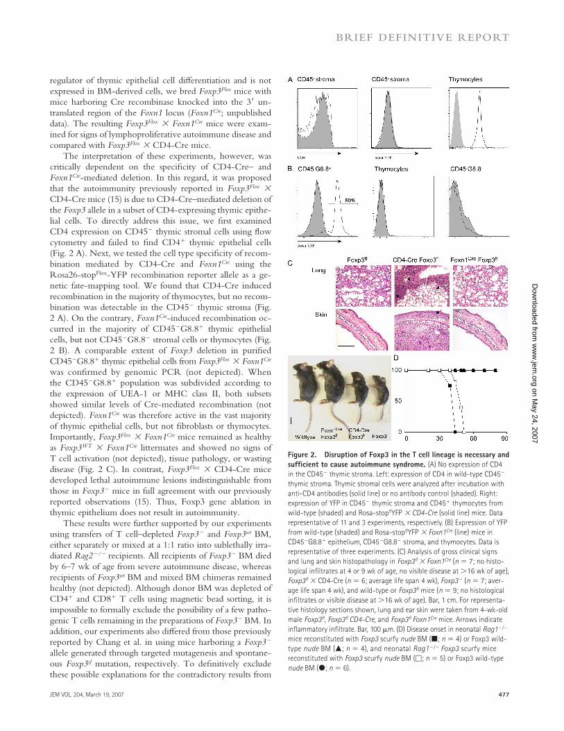
regulator of thymic epithelial cell diff erentiation and is not expressed in BM-derived cells, we bred Foxp3Flox mice with mice harboring Cre recombinase knocked into the 3′ un-translated region of the Foxn1 locus (Foxn1Cre; unpublished data). The resulting Foxp3Flox × Foxn1Cre mice were exam-ined for signs of lymphoproliferative autoimmune disease and compared with Foxp3Flox × CD4-Cre mice.
The interpretation of these experiments, however, was critically dependent on the specifi city of CD4-Cre– and Foxn1Cre-mediated deletion. In this regard, it was proposed that the autoimmunity previously reported in Foxp3Flox × CD4-Cre mice (15) is due to CD4-Cre–mediated deletion of the Foxp3 allele in a subset of CD4-expressing thymic epithe-lial cells. To directly address this issue, we fi rst examined CD4 expression on CD45− thymic stromal cells using fl ow cytometry and failed to fi nd CD4+ thymic epithelial cells (Fig. 2 A). Next, we tested the cell type specifi city of recom-bination mediated by CD4-Cre and Foxn1Cre using the Rosa26-stopFlox-YFP recombination reporter allele as a ge-netic fate-mapping tool. We found that CD4-Cre induced recombination in the majority of thymocytes, but no recom-bination was detectable in the CD45− thymic stroma (Fig. 2 A). On the contrary, Foxn1Cre-induced recombination oc-curred in the majority of CD45−G8.8+ thymic epithelial cells, but not CD45−G8.8− stromal cells or thymocytes (Fig. 2 B). A comparable extent of Foxp3 deletion in purifi ed CD45−G8.8+ thymic epithelial cells from Foxp3Flox × Foxn1Cre was confi rmed by genomic PCR (not depicted). When the CD45−G8.8+ population was subdivided according to the expression of UEA-1 or MHC class II, both subsets showed similar levels of Cre-mediated recombination (not depicted). Foxn1Cre was therefore active in the vast majority of thymic epithelial cells, but not fi broblasts or thymocytes. Importantly, Foxp3Flox × Foxn1Cre mice remained as healthy as Foxp3WT × Foxn1Cre littermates and showed no signs of T cell activation (not depicted), tissue pathology, or wasting disease (Fig. 2 C). In contrast, Foxp3Flox × CD4-Cre mice developed lethal autoimmune lesions indistinguishable from those in Foxp3− mice in full agreement with our previously reported observations (15). Thus, Foxp3 gene ablation in thymic epithelium does not result in autoimmunity.
These results were further supported by our experiments using transfers of T cell–depleted Foxp3− and Foxp3wt BM, either separately or mixed at a 1:1 ratio into sublethally irra-diated Rag2−/− recipients. All recipients of Foxp3− BM died by 6–7 wk of age from severe autoimmune disease, whereas recipients of Foxp3wt BM and mixed BM chimeras remained healthy (not depicted). Although donor BM was depleted of CD4+ and CD8+ T cells using magnetic bead sorting, it is impossible to formally exclude the possibility of a few patho-genic T cells remaining in the preparations of Foxp3− BM. In addition, our experiments also diff ered from those previously reported by Chang et al. in using mice harboring a Foxp3− allele generated through targeted mutagenesis and spontane-ous Foxp3sf mutation, respectively. To defi nitively exclude these possible explanations for the contradictory results from
Figure 2. Disruption of Foxp3 in the T cell lineage is necessary and
suffi cient to cause autoimmune syndrome. (A) No expression of CD4
in the CD45− thymic stroma. Left: expression of CD4 in wild-type CD45−
thymic stroma. Thymic stromal cells were analyzed after incubation with
anti-CD4 antibodies (solid line) or no antibody control (shaded). Right:
expression of YFP in CD45− thymic stroma and CD45+ thymocytes from
wild-type (shaded) and Rosa-stopfl YFP × CD4-Cre (solid line) mice. Data
representative of 11 and 3 experiments, respectively. (B) Expression of YFP
from wild-type (shaded) and Rosa-stopfl YFP × Foxn1Cre (line) mice in
CD45−G8.8+ epithelium, CD45−G8.8− stroma, and thymocytes. Data is
representative of three experiments. (C) Analysis of gross clinical signs
and lung and skin histopathology in Foxp3fl × Foxn1Cre (n = 7; no histo-
logical infi ltrates at 4 or 9 wk of age, no visible disease at >16 wk of age),
Foxp3fl × CD4-Cre (n = 6; average life span 4 wk), Foxp3− (n = 7; aver-
age life span 4 wk), and wild-type or Foxp3fl mice (n = 9; no histological
infi ltrates or visible disease at >16 wk of age). Bar, 1 cm. For representa-
tive histology sections shown, lung and ear skin were taken from 4-wk-old
male Foxp3fl , Foxp3fl CD4-Cre, and Foxp3fl Foxn1Cre mice. Arrows indicate
infl ammatory infi ltrate. Bar, 100 μm. (D) Disease onset in neonatal Rag1−/−
mice reconstituted with Foxp3 scurfy nude BM (■; n = 4) or Foxp3 wild-
type nude BM (▲; n = 4), and neonatal Rag1−/− Foxp3 scurfy mice
reconstituted with Foxp3 scurfy nude BM (□; n = 5) or Foxp3 wild-type
nude BM (●; n = 6).
on May 24, 2007
ww
w.jem
.orgD
ownloaded from

478 RESTRICTION OF FOXP3 TO T CELLS | Liston et al.
the two sets of BM transfer studies, we crossed the Foxp3sf allele to the Foxn1-defi cient nude mice, which are character-ized by an early block in thymic epithelium diff erentiation and thus lack mature T cells. As previously reported, Foxp3sf nude mice were not aff ected by the autoimmunity (2). Thus, we were able to transfer BM isolated from these disease-free mice into Rag1−/− recipients without the risk of contamina-tion with mature pathogenic T cells. In these experiments, Foxp3sf nude BM reconstitution of neonatal Rag1−/− mice and Foxp3sfRag1−/− mice (serving as a positive control) re-sulted in identical lethal lymphoproliferative disease (Fig. 2 D). In contrast, control Foxp3wt nude BM transfers into either neonatal Rag1−/− or Foxp3sfRag1−/− did not result in disease (Fig. 2 D). The fi ndings above demonstrate that genetic abla-tion of Foxp3 in the thymic epithelium is neither necessary nor suffi cient for the development of autoimmune disease.
Distorted thymopoiesis associated with Foxp3 defi ciency is
secondary to lymphoproliferative autoimmune syndrome
Lack of autoimmunity in Foxp3wt nude ® Foxp3sf Rag1−/− BM chimeras and in Foxp3fl ox Foxn1Cre mice did not exclude the previously proposed role for expression of Foxp3 in the thymic epithelium in normal thymopoiesis (16). Major thymic aberra-tions reported for Foxp3 mutant mice include a decrease in thymic cellularity and in the proportion of CD4+CD8+ DP thymocytes (16). To reexamine this possibility, we analyzed thymopoiesis in Foxp3−, Foxp3fl ox CD4-Cre, and Foxp3fl ox Foxn1Cre mice and found that both Foxp3− and the Foxp3fl CD4-Cre mice showed reduced total thymic cellularity and a decrease in the percentage of CD4+CD8+ DP thymocytes, whereas Foxp3fl ox Foxn1Cre mice were identical to their wild-type littermates (Fig. 3, A and B). These results demonstrate that deletion of the Foxp3 gene in thymic epithelial cells does not result in detectable changes in thymic T cell maturation, and that an apparent decrease in DP thymocyte subset size and in total thymocyte numbers is due to loss of Foxp3 in thymo-cytes rather than in the thymic epithelium and is most likely secondary to the severe autoimmunity and cytokine storm common to the Foxp3sf, Foxp3−, and Foxp3fl CD4-Cre mice.
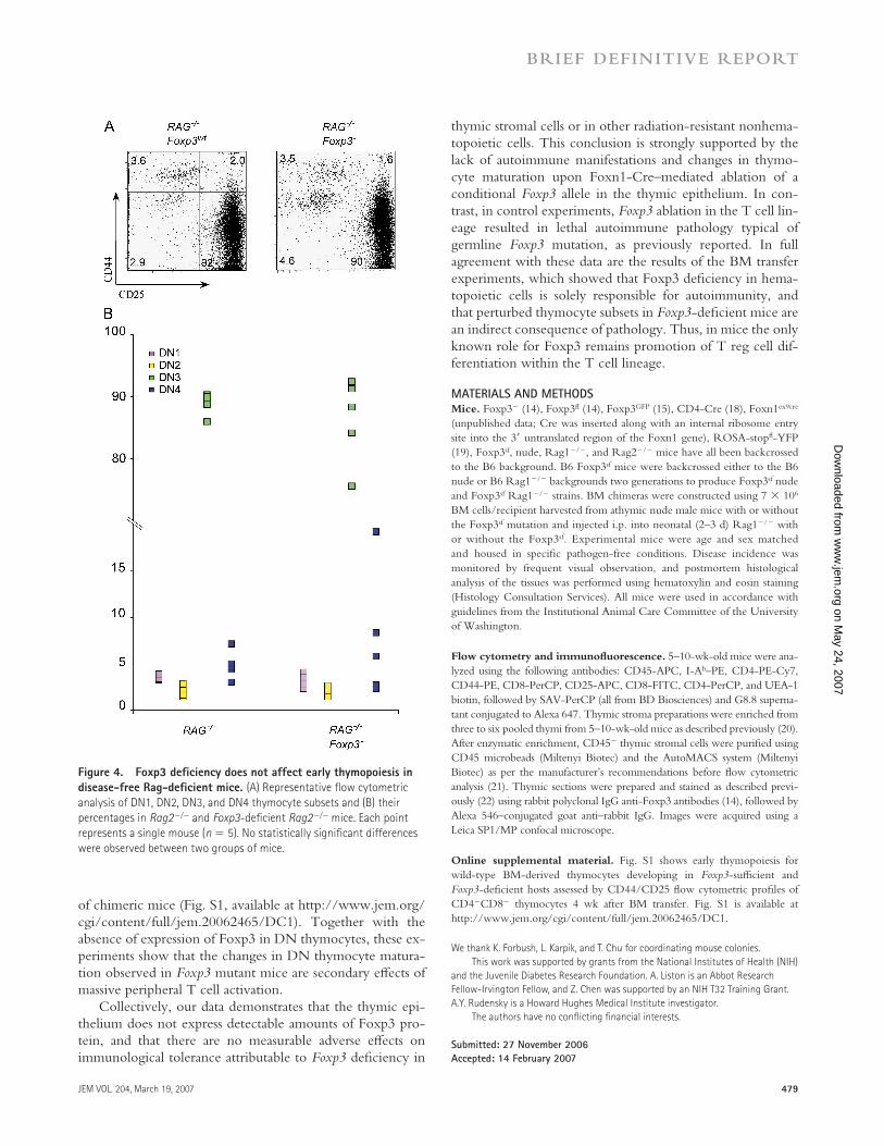
In addition to the aforementioned changes in thymocyte subsets, an expansion of DN1 (CD44+CD25−) cells was re-ported for both Foxp3sf and Foxp3sf Rag−/− mice (16). To ex-amine this phenomenon, we fi rst analyzed the composition of DN thymocyte subsets in Foxp3−, Foxp3fl CD4-Cre, and Foxp3fl Foxn1Cre mice. The relative size of the DN1-4 thy-mocyte subsets was not altered in Foxp3fl Foxn1Cre as com-pared with control mice; however, we found relative increases in the DN1 subset in Foxp3− and Foxp3fl CD4-Cre mice (Fig. 3, C and D). To determine whether this is a cell-intrin-sic defect in developing thymocytes, we analyzed DN thy-mocyte subsets in disease-free Foxp3−Rag2−/− and control Foxp3wtRag2−/− littermates. No diff erences in the relative sizes of DN1, DN2, and DN3 subsets were detected in these mice (Fig. 4, A and B). To exclude the possibility that very few thymic epithelial cells escaping Cre-mediated deletion in Foxp3fl ox × Foxn1Cre mice are capable of supporting normal
thymocyte development, we generated an additional set of BM chimeras by transferring wild-type BM into Foxp3−Rag2−/− and Foxp3wtRag2−/− recipients. The matura-tion of wild-type thymocytes was not diff erent in the two sets
Figure 3. Distorted thymopoiesis is dependent on Foxp3 loss in the
T cell lineage. Thymopoiesis was assessed by examining total thymus
cellularity and by fl ow cytometric analysis of thymocyte subsets. (A) Rep-
resentative CD4 and CD8 thymocyte profi les for wild-type, Foxp3-defi -
cient, Foxp3fl CD4-Cre, and Foxp3fl Foxn1Cre mice. Average thymus
cellularity (mean ± SD) is shown below each graph. (B) Percentages of
thymocytes in CD4−CD8− DN, CD4+CD8+ DP, CD4+ single positive, and
CD8+ single positive subsets. Each data point represents a single mouse.
(C) Representative fl ow cytometric profi les of DN thymocyte subsets and
(D) percentages of DN1, DN2, DN3, and DN4 subsets in wild-type, Foxp3-
defi cient, Foxp3fl CD4-Cre, and Foxp3fl Foxn1Cre mice.
on May 24, 2007
ww
w.jem
.orgD
ownloaded from
JEM VOL. 204, March 19, 2007 479
BRIEF DEFINITIVE REPORT
of chimeric mice (Fig. S1, available at http://www.jem.org/cgi/content/full/jem.20062465/DC1). Together with the absence of expression of Foxp3 in DN thymocytes, these ex-periments show that the changes in DN thymocyte matura-tion observed in Foxp3 mutant mice are secondary eff ects of massive peripheral T cell activation.
Collectively, our data demonstrates that the thymic epi-thelium does not express detectable amounts of Foxp3 pro-tein, and that there are no measurable adverse eff ects on immunological tolerance attributable to Foxp3 defi ciency in
thymic stromal cells or in other radiation-resistant nonhema-topoietic cells. This conclusion is strongly supported by the lack of autoimmune manifestations and changes in thymo-cyte maturation upon Foxn1-Cre–mediated ablation of a conditional Foxp3 allele in the thymic epithelium. In con-trast, in control experiments, Foxp3 ablation in the T cell lin-eage resulted in lethal autoimmune pathology typical of germline Foxp3 mutation, as previously reported. In full agreement with these data are the results of the BM transfer experiments, which showed that Foxp3 defi ciency in hema-topoietic cells is solely responsible for autoimmunity, and that perturbed thymocyte subsets in Foxp3-defi cient mice are an indirect consequence of pathology. Thus, in mice the only known role for Foxp3 remains promotion of T reg cell dif-ferentiation within the T cell lineage.
MATERIALS AND METHODSMice. Foxp3− (14), Foxp3fl (14), Foxp3GFP (15), CD4-Cre (18), Foxn1ex9cre
(unpublished data; Cre was inserted along with an internal ribosome entry
site into the 3′ untranslated region of the Foxn1 gene), ROSA-stopfl -YFP
(19), Foxp3sf, nude, Rag1−/−, and Rag2−/− mice have all been backcrossed
to the B6 background. B6 Foxp3sf mice were backcrossed either to the B6
nude or B6 Rag1−/− backgrounds two generations to produce Foxp3sf nude
and Foxp3sf Rag1−/− strains. BM chimeras were constructed using 7 × 10⁶
BM cells/recipient harvested from athymic nude male mice with or without
the Foxp3sf mutation and injected i.p. into neonatal (2–3 d) Rag1−/− with
or without the Foxp3sf. Experimental mice were age and sex matched
and housed in specifi c pathogen-free conditions. Disease incidence was
monitored by frequent visual observation, and postmortem histological
analysis of the tissues was performed using hematoxylin and eosin staining
(Histology Consultation Services). All mice were used in accordance with
guidelines from the Institutional Animal Care Committee of the University
of Washington.
Flow cytometry and immunofl uorescence. 5–10-wk-old mice were ana-
lyzed using the following antibodies: CD45-APC, I-Ab–PE, CD4-PE-Cy7,
CD44-PE, CD8-PerCP, CD25-APC, CD8-FITC, CD4-PerCP, and UEA-1
biotin, followed by SAV-PerCP (all from BD Biosciences) and G8.8 superna-
tant conjugated to Alexa 647. Thymic stroma preparations were enriched from
three to six pooled thymi from 5–10-wk-old mice as described previously (20).
After enzymatic enrichment, CD45− thymic stromal cells were purifi ed using
CD45 microbeads (Miltenyi Biotec) and the AutoMACS system (Miltenyi
Biotec) as per the manufacturer’s recommendations before fl ow cytometric
analysis (21). Thymic sections were prepared and stained as described previ-
ously (22) using rabbit polyclonal IgG anti-Foxp3 antibodies (14), followed by
Alexa 546–conjugated goat anti–rabbit IgG. Images were acquired using a
Leica SP1/MP confocal microscope.
Online supplemental material. Fig. S1 shows early thymopoiesis for
wild-type BM-derived thymocytes developing in Foxp3-suffi cient and
Foxp3-defi cient hosts assessed by CD44/CD25 fl ow cytometric profi les of
CD4−CD8− thymocytes 4 wk after BM transfer. Fig. S1 is available at
http://www.jem.org/cgi/content/full/jem.20062465/DC1.
We thank K. Forbush, L. Karpik, and T. Chu for coordinating mouse colonies.
This work was supported by grants from the National Institutes of Health (NIH)
and the Juvenile Diabetes Research Foundation. A. Liston is an Abbot Research
Fellow-Irvington Fellow, and Z. Chen was supported by an NIH T32 Training Grant.
A.Y. Rudensky is a Howard Hughes Medical Institute investigator.
The authors have no confl icting fi nancial interests.
Submitted: 27 November 2006
Accepted: 14 February 2007
Figure 4. Foxp3 defi ciency does not affect early thymopoiesis in
disease-free Rag-defi cient mice. (A) Representative fl ow cytometric
analysis of DN1, DN2, DN3, and DN4 thymocyte subsets and (B) their
percentages in Rag2−/− and Foxp3-defi cient Rag2−/− mice. Each point
represents a single mouse (n = 5). No statistically signifi cant differences
were observed between two groups of mice.
on May 24, 2007
ww
w.jem
.orgD
ownloaded from

480 RESTRICTION OF FOXP3 TO T CELLS | Liston et al.
R E F E R E N C E S 1. Wildin, R.S., S. Smyk-Pearson, and A.H. Filipovich. 2002. Clinical and
molecular features of the immunodysregulation, polyendocrinopathy, enteropathy, X linked (IPEX) syndrome. J. Med. Genet. 39:537–545.
2. Godfrey, V.L., J.E. Wilkinson, E.M. Rinchik, and L.B. Russell. 1991. Fatal lymphoreticular disease in the scurfy (sf) mouse requires T cells that mature in a sf thymic environment: potential model for thymic education. Proc. Natl. Acad. Sci. USA. 88:5528–5532.
3. Bennett, C.L., and H.D. Ochs. 2001. IPEX is a unique X-linked syndrome characterized by immune dysfunction, polyendocrinopathy, enteropathy, and a variety of autoimmune phenomena. Curr. Opin. Pediatr. 13:533–538.
4. Ferguson, P.J., S.H. Blanton, F.T. Saulsbury, M.J. McDuffi e, V. Lemahieu, J.M. Gastier, U. Francke, S.M. Borowitz, J.L. Sutphen, and T.E. Kelly. 2000. Manifestations and linkage analysis in X-linked autoimmunity-immunodefi ciency syndrome. Am. J. Med. Genet. 90:390–397.
5. Godfrey, V.L., J.E. Wilkinson, and L.B. Russell. 1991. X-linked lym-phoreticular disease in the scurfy (sf) mutant mouse. Am. J. Pathol. 138:1379–1387.
6. Chatila, T.A., F. Blaeser, N. Ho, H.M. Lederman, C. Voulgaropoulos, C. Helms, and A.M. Bowcock. 2000. JM2, encoding a fork head- related protein, is mutated in X-linked autoimmunity-allergic disregula-tion syndrome. J. Clin. Invest. 106:R75–R81.
7. Clark, L.B., M.W. Appleby, M.E. Brunkow, J.E. Wilkinson, S.F. Ziegler, and F. Ramsdell. 1999. Cellular and molecular characterization of the scurfy mouse mutant. J. Immunol. 162:2546–2554.
8. Blair, P.J., S.J. Bultman, J.C. Haas, B.T. Rouse, J.E. Wilkinson, and V.L. Godfrey. 1994. CD4+CD8− T cells are the eff ector cells in dis-ease pathogenesis in the scurfy (sf) mouse. J. Immunol. 153:3764–3774.
9. Zahorsky-Reeves, J.L., and J.E. Wilkinson. 2001. The murine mutation scurfy (sf) results in an antigen-dependent lymphoproliferative disease with altered T cell sensitivity. Eur. J. Immunol. 31:196–204.
10. Godfrey, V.L., B.T. Rouse, and J.E. Wilkinson. 1994. Transplantation of T cell-mediated, lymphoreticular disease from the scurfy (sf) mouse. Am. J. Pathol. 145:281–286.
11. Smyk-Pearson, S.K., A.C. Bakke, P.K. Held, and R.S. Wildin. 2003. Rescue of the autoimmune scurfy mouse by partial bone marrow trans-
plantation or by injection with T-enriched splenocytes. Clin. Exp. Immunol. 133:193–199.
12. Khattri, R., T. Cox, S.A. Yasayko, and F. Ramsdell. 2003. An essential role for Scurfin in CD4+CD25+ T regulatory cells. Nat. Immunol. 4:337–342.
13. Hori, S., T. Nomura, and S. Sakaguchi. 2003. Control of regula-tory T cell development by the transcription factor Foxp3. Science. 299:1057–1061.
14. Fontenot, J.D., M.A. Gavin, and A.Y. Rudensky. 2003. Foxp3 pro-grams the development and function of CD4+CD25+ regulatory T cells. Nat. Immunol. 4:330–336.
15. Fontenot, J.D., J.P. Rasmussen, L.M. Williams, J.L. Dooley, A.G. Farr, and A.Y. Rudensky. 2005. Regulatory T cell lineage specifi cation by the forkhead transcription factor foxp3. Immunity. 22:329–341.
16. Chang, X., J.X. Gao, Q. Jiang, J. Wen, N. Seifers, L. Su, V.L. Godfrey, T. Zuo, P. Zheng, and Y. Liu. 2005. The Scurfy mutation of FoxP3 in the thymus stroma leads to defective thymopoiesis. J. Exp. Med. 202:1141–1151.
17. Chang, X., P. Zheng, and Y. Liu. 2006. FoxP3: a genetic link between immunodefi ciency and autoimmune diseases. Autoimmun. Rev. 5:399–402.
18. Wolfer, A., T. Bakker, A. Wilson, M. Nicolas, V. Ioannidis, D.R.
Littman, P.P. Lee, C.B. Wilson, W. Held, H.R. MacDonald, and F. Radtke. 2001. Inactivation of Notch 1 in immature thymocytes does not perturb CD4 or CD8T cell development. Nat. Immunol. 2:235–241.
19. Srinivas, S., T. Watanabe, C.S. Lin, C.M. William, Y. Tanabe, T.M. Jessell, and F. Costantini. 2001. Cre reporter strains produced by targeted insertion of EYFP and ECFP into the ROSA26 locus. BMC Dev. Biol. 1:4.
20. Dooley, J., M. Erickson, and A.G. Farr. 2005. An organized medullary epithelial structure in the normal thymus expresses molecules of respi-ratory epithelium and resembles the epithelial thymic rudiment of nude mice. J. Immunol. 175:4331–4337.
21. Gray, D.H., A.P. Chidgey, and R.L. Boyd. 2002. Analysis of thymic stromal cell populations using fl ow cytometry. J. Immunol. Methods. 260:15–28.
22. Lehar, S.M., J. Dooley, A.G. Farr, and M.J. Bevan. 2005. Notch ligands Delta 1 and Jagged1 transmit distinct signals to T-cell precursors. Blood. 105:1440–1447.
on May 24, 2007
ww
w.jem
.orgD
ownloaded from