Embed Size (px)
Citation preview

Laboratory Review for Long Term Care

Why is lab interpretation so difficult in the elderly?
• physiologic changes associated with aging can alter ‘normal values’
• high prevalence of chronic conditions• changes in nutrition and fluid consumption• lifestyle changes• pharmacologic regimes• gender, body mass, diet, stress• collection site, collection time, tourniquet
application, specimen transportation
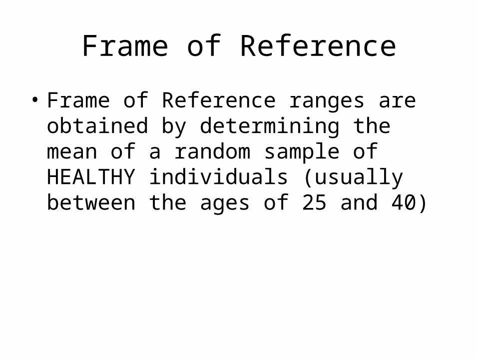
Frame of Reference
• Frame of Reference ranges are obtained by determining the mean of a random sample of HEALTHY individuals (usually between the ages of 25 and 40)

Changes in lab values can be classified into 3 groups:
• those that change with aging
• those that do not change with aging
• those for which it is unclear whether aging, disease, or both influence the

Too many numbers to know!?
A wise wound care nurse once said:“Look at the WHOLE patient and not just the HOLE”
This applies to laboratory interpretation as wellMust consider a total assessment rather than simply relying
on laboratory diagnostic testing
“Can’t see the branches through all the leaves!”Don’t get too focused on the actual laboratory values – you
do need to be aware of normals, but think about the processes that are responsible for the values
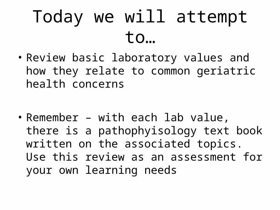
Today we will attempt to…
• Review basic laboratory values and how they relate to common geriatric health concerns
• Remember – with each lab value, there is a pathophyisology text book written on the associated topics. Use this review as an assessment for your own learning needs

Red Blood Cells and Anemias

Hemoglobin(HGB) Part of a complete blood
count (CBC)• Red blood cells (where Hgb is found) live for
approximately 100 days• In a person with Sickle Cell Anemia, red cells
only live for approximately 40 days• Changes in erythrocytes (red blood cell)
synthesis caused by changes in iron and vitamin B12 absorption
• Impaired erythrocyte production, blood loss, increased erythrocyte destruction or a combination, will lower haemoglobin levels
Hgb
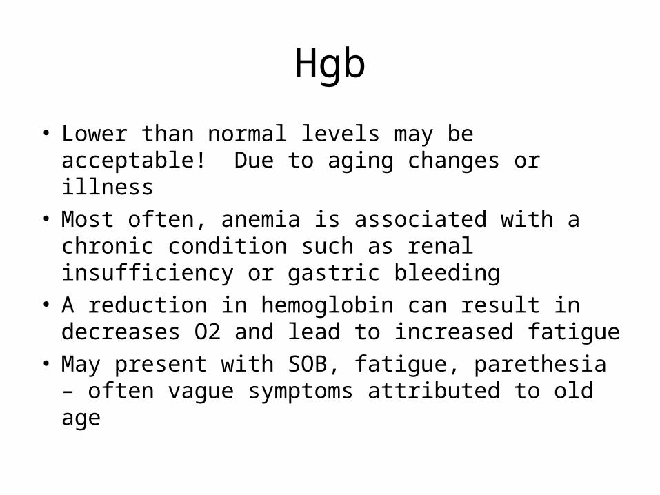
• Lower than normal levels may be acceptable! Due to aging changes or illness
• Most often, anemia is associated with a chronic condition such as renal insufficiency or gastric bleeding
• A reduction in hemoglobin can result in decreases O2 and lead to increased fatigue
• May present with SOB, fatigue, parethesia – often vague symptoms attributed to old age

Hgb
Decreased: anemias, cirrhosis of liver, leukemias, Hodgkin’s disease, cancer (intestine, rectum, liver or bone), kidney disease
Increased: Dehydration, COPD, CHF, polycythemia

Anemia and the Elderly
• According to the Canadian Journal of CME, up to 44% of the geriatric population has some form of anemia
• Decreased serum iron in many older adults, resulting in iron deficiency anemia
• Theory: normal age related decrease in hydrochloric acid (HCl) in the stomach affects iron absorption in stomach….HCLI is important in facilitating iron absorption in intestines
• Medications that decrease HCl secretion!!!• Decrease in iron storage and iron deficiency anemia,
commonly caused by inadequate dietary intake of iron or loss of iron through chronic or acute blood loss

Serum Iron – repeated info??
Decrease: iron deficiency, inflammatory bowel disease, grastric surgery
Increase: Hemolytic, pernicious and folic acid anemias, liver damage, lead toxicity,

Interpreting Anemia with MCV(Mean Corpuscular Volume)
MCV: Microcytic (MCV low)• Iron deficiency anemia• Anemia of chronic disease
Macrocytic (MCV high)- Deficiency of vitamin B12, folic acid- Pernicious anemia – lack of ability to absorb vitamin B12 from food- Hypothyroidism- Alcoholism
Normocytic (MCV normal)- Acute blood loss- Anemia of chronic disease- Aplastic anemia- Hemolytic anemia

B12
• B12 stored in the liver for 5-7 years – 2000 to 5000 mcg
• Approx 1mcg per day is used for making RBCs• Keeps the myelin in the CNS and PNS healthy • Involved in making serotonin – our happy
hormone• Takes about 5-7 years of no B12 to deplete
stores
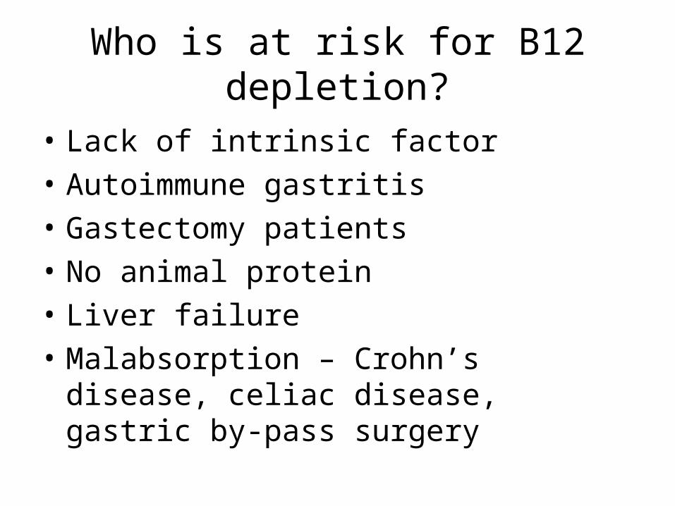
Who is at risk for B12 depletion?
• Lack of intrinsic factor
• Autoimmune gastritis
• Gastectomy patients
• No animal protein
• Liver failure
• Malabsorption – Crohn’s disease, celiac disease, gastric by-pass surgery

B12 Deficiency
• A leading cause of nutritional dementia
• One of the top causes of peripheral neuropathy
• Contributes to depression
• Commonly seen in liver disease, hypothyroidism

Folic Acidaka Vitamin B9
• Used for synthesizing DNA, repairing DNA• Aiding in rapid cell division and growth• Many folic acid fortified foods, therefore, difficult
in north America to be deficient – but you have to eat it!!
• Folate deficiency symptoms include:• Diarrhea, SOB, peripheral neuropathy, mental
confusion, cognitive decline, depression, sore or swollen tongue, peptic or mouth ulcers, headaches, cardiac palpitations, irritability, behavioural disorders

Drugs that can block folic acid synthesis…
• TMP/SFX (Bactrim, Septra)
• Reheumatrex (Methotrexate)
• Phenytoin (Dilantin)

White Blood CellsWBC
A component of a complete Blood Count

White Blood CellsWBC
• Immunity gradually declines after age 30-40; may also result from disease, infection or sepsis, or medications, analgesics, steroids
older persons with infection or sepsis do not always mount the same WBC response (i.e. no fever).
If someone is older and confused, but has a WBC is still in the “normal range,” look closely at the absolute neutrophil levels;
if you see a rise in this, they may have an occult infection despite having a “normal” WBC.

White Blood Cells (WBC)
Increased: acute infections, tissue necrosis, alcoholism, lupus, rheumatoid arthritis, hemolytic anemia, parasitic diseases, stress
Decreased: specific disease (myeloma, collagen disorders), infection or sepsis (pneumonia, UTI), medication (analgesic, phenothiazides, steroids), stress, alcoholism, rheumatoid arthrtis

Breaking down the WBC
• Neutrophils – acute inflammation, bacteria, acute necrosis
• Lymphocytes – first responder to viruses, cells of the immune system (T cells, B cells)
• Monocytes – macrophages in tissues, cells of chronic inflammation
• Eosinophils – cells that respond to parasites and allergies
• Basophils – contains histamine

Neutrophils
• Phagocytic functions – they love to eat!
• Cell of acute inflammation
• First responder to bacterial invasion (strep, staf, E. Coli, H. flu, menigococcus, Pseudomaonas, C. diff)
• Loves acute necrotic tissue (gangrene, MI, appendicitis) Remember – loves to eat!
• Fastest dividing cell in an adult

Drugs and Neutropenia
• Cimetidine (Tegament), ranitidine (Zantac)• Carbamazepine (Tegretol); phenytoin• Captopril (Capoten), enalipril (Vasotec),
amiodarone, quindine• Zidovudine (Retrovir)• Clonapine (Clozaril)• Antibiotics including metronidozole (Flagyl),
gentamiacin, clindamycin, imipenem, tetracylines
• Azothiaprine (Imuran)

Prednisone and Neutrophils
• Inhibits migration and degranulation – halts the antinflammatory process
• Prednisone increases blood sugar by stimulating glycogenolysis in liver and hyperglycemia inhibits funciton of neutrophils
• Fever increases migration of neutorphils

Coagulation

Coagulation
• The process by which blood forms clots• Damage to blood vessel epithlial lining;
exposure of blood to protiens (tissue factors) initiates changes to platelets and fibrinogen (clotting factor)
• Platelets immediately form plug at site of injury• Then fibrin strands (thought clotting cascade) to
strengthen platelet plug• What conditions increase risk of clotting?

Aging and Clotting
• Amount of fibrinogen increases by 1% per year after age 30

Platelets
• Aging usually causes decline in bone marrow function, may contribute to lower platelet counts and decreased platelet function
• BUT platelet adhesiveness increases with age, with no change in numbers
• Therefore, ability to regenerate platelets may be inadequate, leading to inadequate clotting…
• hidden blood loss? Occult blood in stools, emesis

Platelets
• Decrease: anemia, liver disease, kidney disease, idiopathic thrombocytopenia purpura (ITP), cancer, leukemia
• Increase: pulmonary embolism, tuberculosis, polycthemia, trauma, post-splenectomy, metastatic carcinoma

Coagulation Profile
• Platlet norms:
• Hemostasis: platelet count above 100 000
• 50 000 to 100 000 may show increased bruising
• Less than 50 000 need monitoring
• Hemorrhage under 10 000
• INR protocols for residents on Coumadin

What time do most Myocardial Infarctions happen?
• Liver produces clotting factors, cholesterol, glucose, inflammatory mediators overnight then disperses them to the body in the morning
• Inflammatory mediators are highest in the am – triggers plaque rupture
• Platelets are stickiest in the early am due to highest blood sugar
• Platelet plug forms, triggers clotting cascade• Takes 2 hrs to form MI• Therefore MI at 0900• ASA inhibits platelet aggregation

What time will a Pulmonary Embolism happen?
DVT (clot) formation from a few hrs to a few weeks
Attached to the deep veins of the legs and pelvis
Breaks off in the early am and travels to lungs
PE at 0730

Medications and Platelets
• Gingko – increases blood flow to lower limbs
• Glucosamine – affects blood suger
• Ginseng – NA and H2O retiner
• Grapeseed extract –
• Garlic –
• Heparin/Plavix – decreases platelet counts

Kidney Function

Albumin
• Produced in the liver
• Helps keep water inside the blood vessels to prevent dehydration
• Albumin levels decrease each decade over age of 60 with marked decrease over 90yr

Albumin
• Decreased: malnutrition, liver failure, renal disorders, prolong immobilization
• Increased: dehydration, severe vomiting, diarrhea

Total Protein
• Changes in protein may reflect decreased liver functioning, or inadequate nutritional intake
• High: dehydration, vomiting
• Low: decreased intake/absorption, edema, malnutrition, low protein diet, severe liver disease, chronic renal failure

Creatinine
• What is creatinine? A break-down product of creatine phosphate in muscle and is filtered out of the body by the kidneys
• Age related decrease in functioning renal tissue is 30-45%
• Which leads to a decrease in the glomerular filtration rate (GFR)
• Which leads to a decline in creatinine clearance

• Increase: renal failure, shock, acute MI, CHF, diabetic neuropathy
We have a serum creatinine, so why calculate a creatinine clearance?

• A simple creatinine level can overestimate renal function…
• Reduction in lean body mass, decreased dietary protein intake and/or decreased hepatic function may lead to a decrease in the end products of metabolism, and hence, less creatinine production…in a blood test, the creatinine level may appear in ‘normal range’ due to these above mentioned changes in the elderly body
• Therefore, serum creatinine values remain within normal limits despite diminished renal clearence

Creatinine clearance
• A measure of how effectively kidneys are filtering creatinine out of body
• Decrease: renal impairment, hyperthyroidism, thiazide use
• Increase: hypothyroidism, renal vascular hypertension
• Formula for creatinine clearence

Changes in renal function can also be linked to:
• Chronic urinary tract infections, benign prostatic hypertrophy, prostatic tumors, diabetic neuropathy
• One of the early signs of renal failure is mild anemia

Thyroid
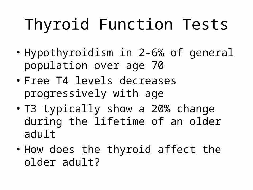
Thyroid Function Tests
• Hypothyroidism in 2-6% of general population over age 70
• Free T4 levels decreases progressively with age
• T3 typically show a 20% change during the lifetime of an older adult
• How does the thyroid affect the older adult?

Thyroid and geriatrics
• Thyroid regulates metabolism, promotes skeletal growth and brain development, stimulates the heart and regulates energy production
• Hypothyroidsm can be masked by clinical features that share symptoms with aging including: general slowing of mental and physical function, tendency of low body temperatures, cold intolerance, weight gain, constipation, hardening of the arteries, elevation of cholesterol, elevation of blood pressure and anemia
• Hyperthyroidism associated with irregular heart rhythms, congestive heart failure, nervousness, sweating, weight loss, muscle weakens

TSH T4 T3 Interpretation
High Normal Normal Mild (subclinical hypothyroidism
High Low Low or Normal Hypothyrodism
Low Normal Normal Mild (subclinical) hyperthyroidism
Low High or Normal High or Normal Hyperthyroidism
Low Low or Normal Low or Normal Nonthyroidal illness; rare pituitary hypothyroidism

TSH
• Decrease: excessive thyroid hormone replacement, Graves’ disease, primary hyperthyroidism
• Increase: primary hypothyroidism, thyroid hormone resistance

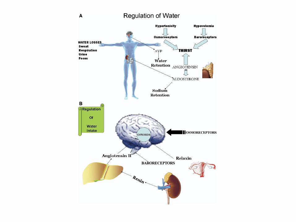
Clues about Dehydration

• Water is 55-65% of body mass• 2/3 of water is intracellular (lean tissue)• 1/3 extracellular – of that, 25% intravascualr (8%
total body water)• With aging, decline in total body water, in both
extra and intracellular fluid volume• Up to 30% more fat than lean muscle• The decrease in total body water, alterations in
water regulation leads to increased vulnerability• In response to heat/exercise, older adults loos
more intracellular fluid and less intersitial fluid

2 kinds of total body water fluid loss:
• Dehydration: loss of body water mainly from intracellular compartments
• Volume depletion: loss of extracellular fluid

• Sodium and Water depletion: diuretics, adrenal insufficiency, renal salt wastage, vomiting and/or diaarrhea, excessive sweating, burns
• Water Depletion: fever, central diabetes insipidus, nephrogenic diabetes insipidus, essential hypernatremia, osmotic diuresis

Electrolytes
• Overall, electrolytes values remain within standard reference values
• Electrically charged minerals found in body tissues and blood in the form of dissolved salts
• Help move nutrients into and wastes out of body’s cells, maintain a healthy water balance and stabilize the body’s pH level
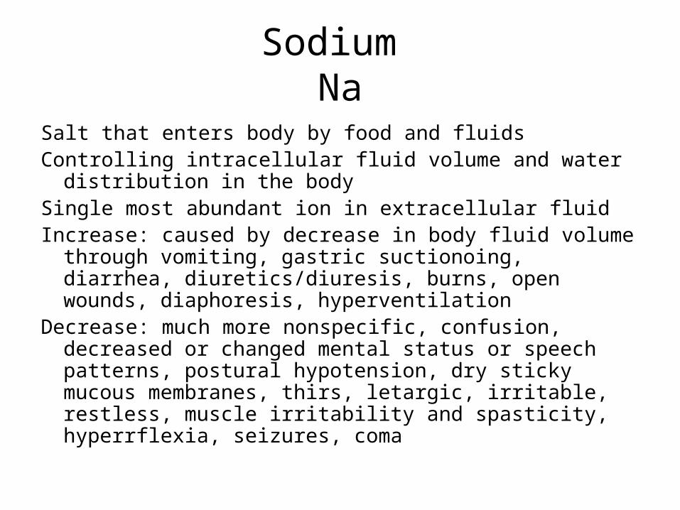
Sodium Na
Salt that enters body by food and fluidsControlling intracellular fluid volume and water distribution
in the bodySingle most abundant ion in extracellular fluidIncrease: caused by decrease in body fluid volume through
vomiting, gastric suctionoing, diarrhea, diuretics/diuresis, burns, open wounds, diaphoresis, hyperventilation
Decrease: much more nonspecific, confusion, decreased or changed mental status or speech patterns, postural hypotension, dry sticky mucous membranes, thirs, letargic, irritable, restless, muscle irritability and spasticity, hyperrflexia, seizures, coma

PotassiumK
• Gained through dietary intake and lost by excretion – if either is altered, hyperkalemia or hypokalemia can rapidly occur (a very narrow index)
• Conducts electricity in the body, crucial for heart function, key role in skeletal and smooth muscle contraction (which inturn aides in normal digestive and muscular function)

K
Decrease: vomiting, diarrhea, dehydration, malnutrition, stress, diabetic acidosis
Increase: acute renal failure, acidosis, crushing injury, Addison’s disease

Hematocrit (part of a CBC)
• An indicator of fluid and/or nutritional status• Increase: cirrosis of liver, protein malnutrition,
peptic ulcer, chronic renal failure, rheumatoid arthritis, anemia, leukemia, Hodgkin’s disease, multiple myeloma
• Decrease: dehydration, sever diarrhea, diabetic acidosis, emphysema, transient cerebral ischemia may indicate fluid overload, dietary deficiencies
Albumin
• Remember – albumin helps keep water inside blood vessels
• High albumin almost always synomonous with dehydration
Urinalysis

Urinalysis Appearance
Colour
Odor
pH
Protein
Specific Gravity
Leukocyte esterase, Nitrates, Ketones, Glucose
Crystals
WBC
WBC casts
RBC
RBC casts
Clear
Light straw to dark amber
No odor/aromatic
4.5-8
2-8 mg/dl
1.005-1.030
Negative
0
3-4
Occasional hyaline
Less than 2
0
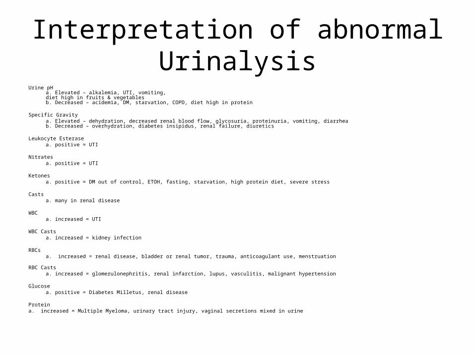
Interpretation of abnormal Urinalysis
Urine pH a. Elevated – alkalemia, UTI, vomiting,diet high in fruits & vegetablesb. Decreased – acidemia, DM, starvation, COPD, diet high in protein
Specific Gravity a. Elevated – dehydration, decreased renal blood flow, glycosuria, proteinuria, vomiting, diarrheab. Decreased – overhydration, diabetes insipidus, renal failure, diuretics
Leukocyte Esterasea. positive = UTI
Nitratesa. positive = UTI
Ketones a. positive = DM out of control, ETOH, fasting, starvation, high protein diet, severe stress
Castsa. many in renal disease
WBCa. increased = UTI
WBC Castsa. increased = kidney infection
RBCsa. increased = renal disease, bladder or renal tumor, trauma, anticoagulant use, menstruation
RBC Castsa. increased = glomerulonephritis, renal infarction, lupus, vasculitis, malignant hypertension
Glucosea. positive = Diabetes Milletus, renal disease
Proteina. increased = Multiple Myeloma, urinary tract injury, vaginal secretions mixed in urine

Urinalysis for Kidney Disease
• Nephritis 1-2+ protein
• Nephrosis 3-4+ protein

You have the lab results – now what?
• Timeliness depends on results and clinical situation
• Report other key clinical data with results
• Documentation of communication

The End

Bones and Red Cell Production
Nutritional Status greatly affects the body’s ability to produce new red cells
• Vitamin A – bone remodeling• Vitamin B12 – combines with folic acid, iron and vitamin C to
improve production of red cells• Vitamin C – promotes formation of collagen (structural synthesis of
bone production)• Vitamin K – supports bone remodeling• Vitamin E – antioxidant that preserves cellular consttuents• Iron – essential part of blood cell production• Magnesium – for calcium metabolism in bone• Zinc – tissue renewal and skeletal development• Copper – form the connective structures of bones and enhances
effectivness of vitamin D• Hematocrit is another good indicator of nutritional status….

Erythrocyte Sedimentation RateESR
• Can be ordered with a CBC
• measures rate at which red cells settle in 1 hr
• Increased rate could be presence of inflammation (causes alteration in blood proteins, making RBCs heavier and causing them to settle faster)

ESR
• Decreased: CHF, degenerative arthritis, angina
• Increased: hepatitis, cirrhosis of liver, rheumatoid arthrtis, rheumatic fever, acute MI, cancer (stomach, colon, breast, liver, kidney), Hodgkin’s disease, multiple myeloma, bacterial endocareditis, gout