Embed Size (px)
Citation preview

WLHS/A&P/Oppelt Name _________________
LAB – Dissection of Sheep’s Pluck
Purpose: Today we will be exploring the structures of the
respiratory system. We will also be examining how our lungs move as
we breathe. Lastly, we will be describing how the structure of the
lungs relate to how we breath.
Procedure:
1. When handling the sheep’s pluck, make sure to wear disposable
gloves. Aprons and goggles are also available for additional protection.
2. Examine your specimen. Describe the color, shape, size, and feel of
the specimen.
3. What organs/body parts are included in your specimen?
4. Identify the trachea. Is the trachea stiff or flexible? What allows the trachea to stay open
and bend? Why is this important?
5. Try to inflate the lungs and observe how they respond. Do not breathe directly into the trachea
to do this! Insert a length of rubber tubing into the trachea and attach it to a bellows or foot
pump to inflate the lungs and then watch them deflate. It is best not to breathe through the tube
into the lungs yourself because of the elastic recoil of the lung tissue: you might inhale a blast of
rather unpleasant air!
a. What happened to the lungs when air was forced into them?
b. Do you think there is a limit to how much air can be pumped into the lungs? Explain your
answer
5. Start of cut down the trachea with your dissecting scissors. Does the trachea stay as one tube
or does it split as it enters the lung?

6. If the larynx is still attached to your lungs, try forcing air through the larynx while you squeeze
it tight. As air moves past the skin and cords in the larynx it may make a noise.
Discuss how similar this is to the noise the animal makes in life.
7. Cut a small piece of spongy lung tissue to examine more closely.
a. Are the lungs hollow bags or spongy? _____________________________
b. What does the lung tissue look like where you cut into it? _______________________
c. Drop the piece of lung into a beaker of water. Does the piece sink or float? ______
d. What does the answer to part c indicate? ___________________________________
8. Now let’s examine the heart as review. Use a scalpel to remove a portion of this layer and
expose the myocardium beneath.
- Note the abundance of fat along the paths of various blood vessels. This adipose tissue
occurs in the loose connective tissue that underlies the visceral pericardium.
- Identify the following: Right Atrium, Right Ventricle, Pulmonary Artery, Left
Atrium, Left Ventricle, and the Aorta
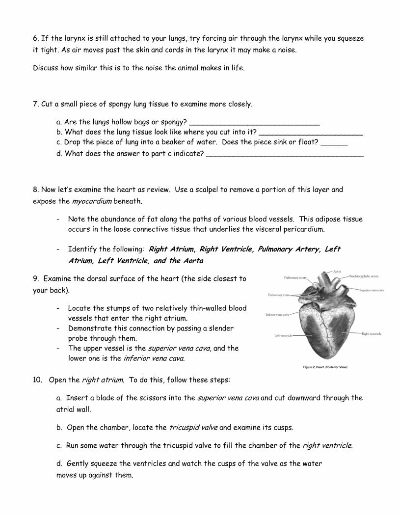
9. Examine the dorsal surface of the heart (the side closest to
your back).
- Locate the stumps of two relatively thin-walled blood
vessels that enter the right atrium.
- Demonstrate this connection by passing a slender
probe through them.
- The upper vessel is the superior vena cava, and the lower one is the inferior vena cava.
10. Open the right atrium. To do this, follow these steps:
a. Insert a blade of the scissors into the superior vena cava and cut downward through the
atrial wall.
b. Open the chamber, locate the tricuspid valve and examine its cusps.
c. Run some water through the tricuspid valve to fill the chamber of the right ventricle.
d. Gently squeeze the ventricles and watch the cusps of the valve as the water
moves up against them.

11. Open the right ventricle as follows:
a. Continue cutting downward through the tricuspid valve and the right ventricular wall until
you reach the apex of the heart.
b. Find the opening to the pulmonary trunk and use the scissors to cut upward through the
wall of the right ventricle. Follow the pulmonary trunk until you have exposed the pulmonary
valve.
c. Examine the valve and its cusps.
12. Open the left side of the heart. To do this, follow these steps:
a. Insert the blade of the scissors through the wall of the left atrium and cut downward to
the apex of the heart.
b. Open the left atrium and locate the four openings of the pulmonary veins. Pass a slender
probe through each opening and locate the stump of its vessel.
c. Examine the bicuspid valve (mitral valve) and its cusps.
d. Also examine the left ventricle and compare the thickness of its wall with that of the
right ventricle.
13. Locate the aorta, which leads away from the left ventricle, and proceed as follows:
a. Compare the thickness of the aortic wall with that of the pulmonary trunk.
b. Use scissors to cut along the length of the aorta to expose the aortic valve at its base.
c. Examine the cusps of the valve and locate the openings of the coronary arteries just
distal to them.
Questions
1. How many chambers are found in the mammalian heart? List these chambers.
2. Which chambers are the pumping chambers of the heart?
3. Which chambers are the receiving chambers of the heart?

4. How do the walls of the atria compare with the walls of the ventricles and why are they
different?
5. What is the purpose of heart valves?
6. Vessels that carry blood away from the heart are called __________, while __________ carry
blood toward the heart.
7. Which artery is the largest and why?
8. How are the heart and lungs’ function(s) related?

WLHS/A&P/Oppelt Name _________________
LAB – Dissection of Sheep’s Pluck
Purpose: Today we will be exploring the structures of the
respiratory system. We will also be examining how our lungs move as
we breathe. Lastly, we will be describing how the structure of the
lungs relate to how we breath.
Procedure:
1. When handling the sheep’s pluck, make sure to wear disposable
gloves. Aprons and goggles are also available for additional protection.
2. Examine your specimen. Describe the color, shape, size, and feel of
the specimen.
3. What organs/body parts are included in your specimen?
4. Identify the trachea. Is the trachea stiff or flexible? What allows the trachea to stay open
and bend? Why is this important?
5. Try to inflate the lungs and observe how they respond. Do not breathe directly into the trachea
to do this! Insert a length of rubber tubing into the trachea and attach it to a bellows or foot
pump to inflate the lungs and then watch them deflate. It is best not to breathe through the tube
into the lungs yourself because of the elastic recoil of the lung tissue: you might inhale a blast of
rather unpleasant air!
a. What happened to the lungs when air was forced into them?
b. Do you think there is a limit to how much air can be pumped into the lungs? Explain your
answer
5. Start of cut down the trachea with your dissecting scissors. Does the trachea stay as one tube
or does it split as it enters the lung?

6. If the larynx is still attached to your lungs, try forcing air through the larynx while you squeeze
it tight. As air moves past the skin and cords in the larynx it may make a noise.
Discuss how similar this is to the noise the animal makes in life.
7. Cut a small piece of spongy lung tissue to examine more closely.
a. Are the lungs hollow bags or spongy? _____________________________
b. What does the lung tissue look like where you cut into it? _______________________
c. Drop the piece of lung into a beaker of water. Does the piece sink or float? ______
d. What does the answer to part c indicate? ___________________________________
8. Now let’s examine the heart as review. Use a scalpel to remove a portion of this layer and
expose the myocardium beneath.
- Note the abundance of fat along the paths of various blood vessels. This adipose tissue
occurs in the loose connective tissue that underlies the visceral pericardium.
- Identify the following: Right Atrium, Right Ventricle, Pulmonary Artery, Left
Atrium, Left Ventricle, and the Aorta
9. Examine the dorsal surface of the heart (the side closest to
your back).
- Locate the stumps of two relatively thin-walled blood
vessels that enter the right atrium.
- Demonstrate this connection by passing a slender
probe through them.
- The upper vessel is the superior vena cava, and the lower one is the inferior vena cava.
10. Open the right atrium. To do this, follow these steps:
a. Insert a blade of the scissors into the superior vena cava and cut downward through the
atrial wall.
b. Open the chamber, locate the tricuspid valve and examine its cusps.
c. Run some water through the tricuspid valve to fill the chamber of the right ventricle.
d. Gently squeeze the ventricles and watch the cusps of the valve as the water
moves up against them.

11. Open the right ventricle as follows:
a. Continue cutting downward through the tricuspid valve and the right ventricular wall until
you reach the apex of the heart.
b. Find the opening to the pulmonary trunk and use the scissors to cut upward through the
wall of the right ventricle. Follow the pulmonary trunk until you have exposed the pulmonary
valve.
c. Examine the valve and its cusps.
12. Open the left side of the heart. To do this, follow these steps:
a. Insert the blade of the scissors through the wall of the left atrium and cut downward to
the apex of the heart.
b. Open the left atrium and locate the four openings of the pulmonary veins. Pass a slender
probe through each opening and locate the stump of its vessel.
c. Examine the bicuspid valve (mitral valve) and its cusps.
d. Also examine the left ventricle and compare the thickness of its wall with that of the
right ventricle.
13. Locate the aorta, which leads away from the left ventricle, and proceed as follows:
a. Compare the thickness of the aortic wall with that of the pulmonary trunk.
b. Use scissors to cut along the length of the aorta to expose the aortic valve at its base.
c. Examine the cusps of the valve and locate the openings of the coronary arteries just
distal to them.
Questions
1. How many chambers are found in the mammalian heart? List these chambers.
2. Which chambers are the pumping chambers of the heart?
3. Which chambers are the receiving chambers of the heart?

4. How do the walls of the atria compare with the walls of the ventricles and why are they
different?
5. What is the purpose of heart valves?
6. Vessels that carry blood away from the heart are called __________, while __________ carry
blood toward the heart.
7. Which artery is the largest and why?
8. How are the heart and lungs’ function(s) related?

WLHS/A&P/Oppelt Name _________________
LAB – Dissection of Sheep’s Pluck
Purpose: Today we will be exploring the structures of the
respiratory system. We will also be examining how our lungs move as
we breathe. Lastly, we will be describing how the structure of the
lungs relate to how we breath.
Procedure:
1. When handling the sheep’s pluck, make sure to wear disposable
gloves. Aprons and goggles are also available for additional protection.
2. Examine your specimen. Describe the color, shape, size, and feel of
the specimen.
3. What organs/body parts are included in your specimen?
4. Identify the trachea. Is the trachea stiff or flexible? What allows the trachea to stay open
and bend? Why is this important?
5. Try to inflate the lungs and observe how they respond. Do not breathe directly into the trachea
to do this! Insert a length of rubber tubing into the trachea and attach it to a bellows or foot
pump to inflate the lungs and then watch them deflate. It is best not to breathe through the tube
into the lungs yourself because of the elastic recoil of the lung tissue: you might inhale a blast of
rather unpleasant air!
a. What happened to the lungs when air was forced into them?
b. Do you think there is a limit to how much air can be pumped into the lungs? Explain your
answer
5. Start of cut down the trachea with your dissecting scissors. Does the trachea stay as one tube
or does it split as it enters the lung?

6. If the larynx is still attached to your lungs, try forcing air through the larynx while you squeeze
it tight. As air moves past the skin and cords in the larynx it may make a noise.
Discuss how similar this is to the noise the animal makes in life.
7. Cut a small piece of spongy lung tissue to examine more closely.
a. Are the lungs hollow bags or spongy? _____________________________
b. What does the lung tissue look like where you cut into it? _______________________
c. Drop the piece of lung into a beaker of water. Does the piece sink or float? ______
d. What does the answer to part c indicate? ___________________________________
8. Now let’s examine the heart as review. Use a scalpel to remove a portion of this layer and
expose the myocardium beneath.
- Note the abundance of fat along the paths of various blood vessels. This adipose tissue
occurs in the loose connective tissue that underlies the visceral pericardium.
- Identify the following: Right Atrium, Right Ventricle, Pulmonary Artery, Left
Atrium, Left Ventricle, and the Aorta
9. Examine the dorsal surface of the heart (the side closest to
your back).
- Locate the stumps of two relatively thin-walled blood
vessels that enter the right atrium.
- Demonstrate this connection by passing a slender
probe through them.
- The upper vessel is the superior vena cava, and the lower one is the inferior vena cava.
10. Open the right atrium. To do this, follow these steps:
a. Insert a blade of the scissors into the superior vena cava and cut downward through the
atrial wall.
b. Open the chamber, locate the tricuspid valve and examine its cusps.
c. Run some water through the tricuspid valve to fill the chamber of the right ventricle.
d. Gently squeeze the ventricles and watch the cusps of the valve as the water
moves up against them.

11. Open the right ventricle as follows:
a. Continue cutting downward through the tricuspid valve and the right ventricular wall until
you reach the apex of the heart.
b. Find the opening to the pulmonary trunk and use the scissors to cut upward through the
wall of the right ventricle. Follow the pulmonary trunk until you have exposed the pulmonary
valve.
c. Examine the valve and its cusps.
12. Open the left side of the heart. To do this, follow these steps:
a. Insert the blade of the scissors through the wall of the left atrium and cut downward to
the apex of the heart.
b. Open the left atrium and locate the four openings of the pulmonary veins. Pass a slender
probe through each opening and locate the stump of its vessel.
c. Examine the bicuspid valve (mitral valve) and its cusps.
d. Also examine the left ventricle and compare the thickness of its wall with that of the
right ventricle.
13. Locate the aorta, which leads away from the left ventricle, and proceed as follows:
a. Compare the thickness of the aortic wall with that of the pulmonary trunk.
b. Use scissors to cut along the length of the aorta to expose the aortic valve at its base.
c. Examine the cusps of the valve and locate the openings of the coronary arteries just
distal to them.
Questions
1. How many chambers are found in the mammalian heart? List these chambers.
2. Which chambers are the pumping chambers of the heart?
3. Which chambers are the receiving chambers of the heart?
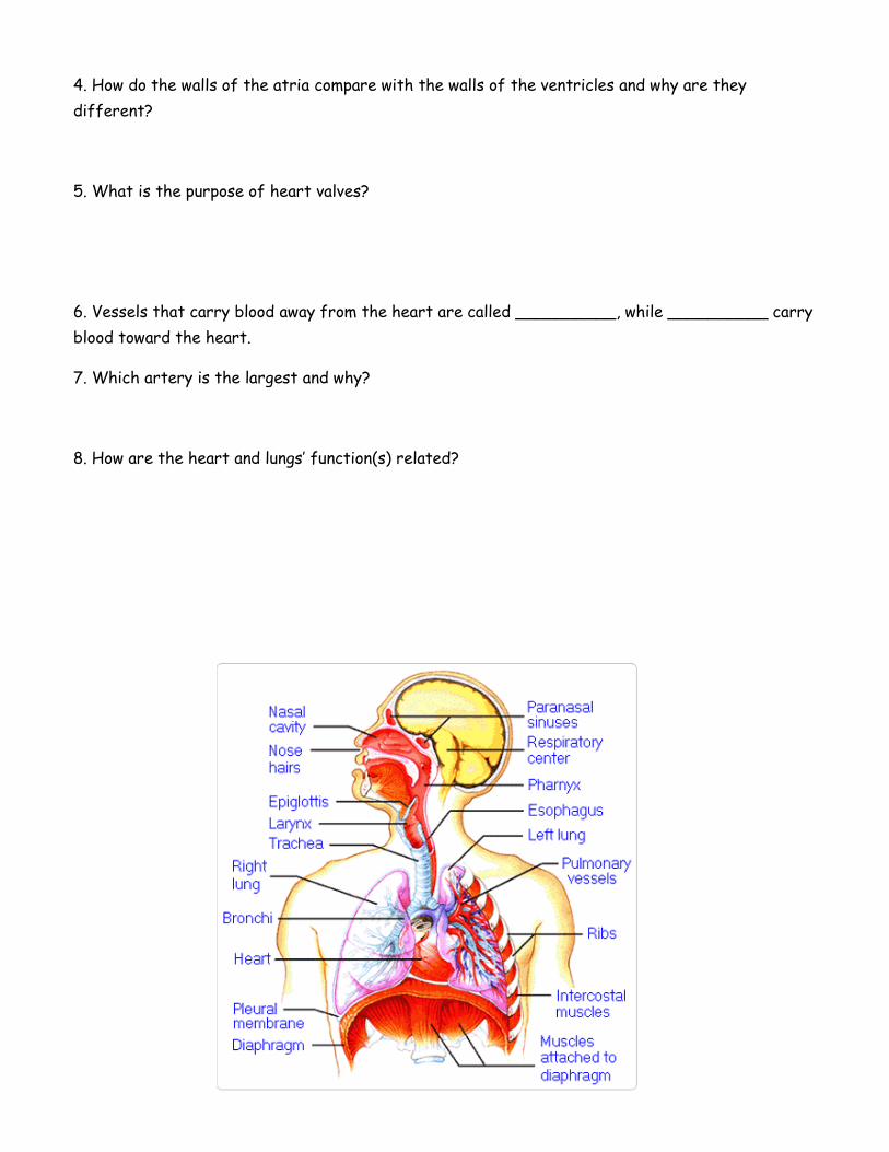
4. How do the walls of the atria compare with the walls of the ventricles and why are they
different?
5. What is the purpose of heart valves?
6. Vessels that carry blood away from the heart are called __________, while __________ carry
blood toward the heart.
7. Which artery is the largest and why?
8. How are the heart and lungs’ function(s) related?

WLHS/A&P/Oppelt Name _________________
LAB – Dissection of Sheep’s Pluck
Purpose: Today we will be exploring the structures of the
respiratory system. We will also be examining how our lungs move as
we breathe. Lastly, we will be describing how the structure of the
lungs relate to how we breath.
Procedure:
1. When handling the sheep’s pluck, make sure to wear disposable
gloves. Aprons and goggles are also available for additional protection.
2. Examine your specimen. Describe the color, shape, size, and feel of
the specimen.
3. What organs/body parts are included in your specimen?
4. Identify the trachea. Is the trachea stiff or flexible? What allows the trachea to stay open
and bend? Why is this important?
5. Try to inflate the lungs and observe how they respond. Do not breathe directly into the trachea
to do this! Insert a length of rubber tubing into the trachea and attach it to a bellows or foot
pump to inflate the lungs and then watch them deflate. It is best not to breathe through the tube
into the lungs yourself because of the elastic recoil of the lung tissue: you might inhale a blast of
rather unpleasant air!
a. What happened to the lungs when air was forced into them?
b. Do you think there is a limit to how much air can be pumped into the lungs? Explain your
answer
5. Start of cut down the trachea with your dissecting scissors. Does the trachea stay as one tube
or does it split as it enters the lung?

6. If the larynx is still attached to your lungs, try forcing air through the larynx while you squeeze
it tight. As air moves past the skin and cords in the larynx it may make a noise.
Discuss how similar this is to the noise the animal makes in life.
7. Cut a small piece of spongy lung tissue to examine more closely.
a. Are the lungs hollow bags or spongy? _____________________________
b. What does the lung tissue look like where you cut into it? _______________________
c. Drop the piece of lung into a beaker of water. Does the piece sink or float? ______
d. What does the answer to part c indicate? ___________________________________
8. Now let’s examine the heart as review. Use a scalpel to remove a portion of this layer and
expose the myocardium beneath.
- Note the abundance of fat along the paths of various blood vessels. This adipose tissue
occurs in the loose connective tissue that underlies the visceral pericardium.
- Identify the following: Right Atrium, Right Ventricle, Pulmonary Artery, Left
Atrium, Left Ventricle, and the Aorta
9. Examine the dorsal surface of the heart (the side closest to
your back).
- Locate the stumps of two relatively thin-walled blood
vessels that enter the right atrium.
- Demonstrate this connection by passing a slender
probe through them.
- The upper vessel is the superior vena cava, and the lower one is the inferior vena cava.
10. Open the right atrium. To do this, follow these steps:
a. Insert a blade of the scissors into the superior vena cava and cut downward through the
atrial wall.
b. Open the chamber, locate the tricuspid valve and examine its cusps.
c. Run some water through the tricuspid valve to fill the chamber of the right ventricle.
d. Gently squeeze the ventricles and watch the cusps of the valve as the water
moves up against them.

11. Open the right ventricle as follows:
a. Continue cutting downward through the tricuspid valve and the right ventricular wall until
you reach the apex of the heart.
b. Find the opening to the pulmonary trunk and use the scissors to cut upward through the
wall of the right ventricle. Follow the pulmonary trunk until you have exposed the pulmonary
valve.
c. Examine the valve and its cusps.
12. Open the left side of the heart. To do this, follow these steps:
a. Insert the blade of the scissors through the wall of the left atrium and cut downward to
the apex of the heart.
b. Open the left atrium and locate the four openings of the pulmonary veins. Pass a slender
probe through each opening and locate the stump of its vessel.
c. Examine the bicuspid valve (mitral valve) and its cusps.
d. Also examine the left ventricle and compare the thickness of its wall with that of the
right ventricle.
13. Locate the aorta, which leads away from the left ventricle, and proceed as follows:
a. Compare the thickness of the aortic wall with that of the pulmonary trunk.
b. Use scissors to cut along the length of the aorta to expose the aortic valve at its base.
c. Examine the cusps of the valve and locate the openings of the coronary arteries just
distal to them.
Questions
1. How many chambers are found in the mammalian heart? List these chambers.
2. Which chambers are the pumping chambers of the heart?
3. Which chambers are the receiving chambers of the heart?

4. How do the walls of the atria compare with the walls of the ventricles and why are they
different?
5. What is the purpose of heart valves?
6. Vessels that carry blood away from the heart are called __________, while __________ carry
blood toward the heart.
7. Which artery is the largest and why?
8. How are the heart and lungs’ function(s) related?
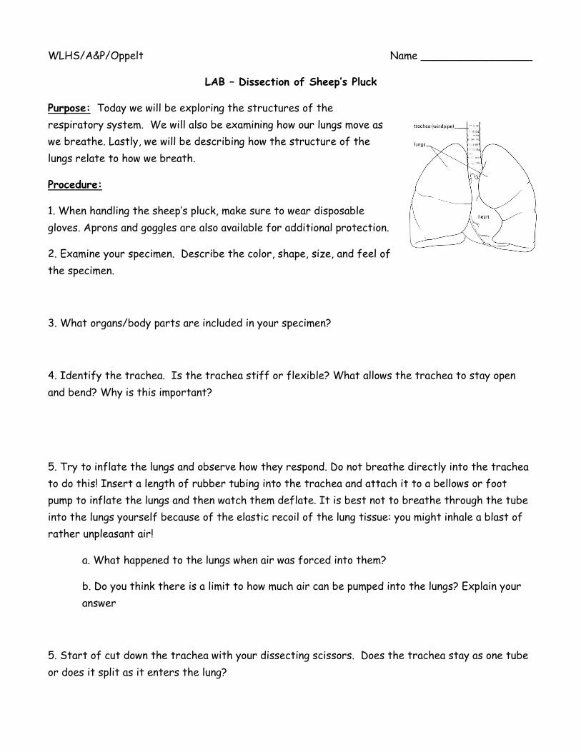
WLHS/A&P/Oppelt Name _________________
LAB – Dissection of Sheep’s Pluck
Purpose: Today we will be exploring the structures of the
respiratory system. We will also be examining how our lungs move as
we breathe. Lastly, we will be describing how the structure of the
lungs relate to how we breath.
Procedure:
1. When handling the sheep’s pluck, make sure to wear disposable
gloves. Aprons and goggles are also available for additional protection.
2. Examine your specimen. Describe the color, shape, size, and feel of
the specimen.
3. What organs/body parts are included in your specimen?
4. Identify the trachea. Is the trachea stiff or flexible? What allows the trachea to stay open
and bend? Why is this important?
5. Try to inflate the lungs and observe how they respond. Do not breathe directly into the trachea
to do this! Insert a length of rubber tubing into the trachea and attach it to a bellows or foot
pump to inflate the lungs and then watch them deflate. It is best not to breathe through the tube
into the lungs yourself because of the elastic recoil of the lung tissue: you might inhale a blast of
rather unpleasant air!
a. What happened to the lungs when air was forced into them?
b. Do you think there is a limit to how much air can be pumped into the lungs? Explain your
answer
5. Start of cut down the trachea with your dissecting scissors. Does the trachea stay as one tube
or does it split as it enters the lung?

6. If the larynx is still attached to your lungs, try forcing air through the larynx while you squeeze
it tight. As air moves past the skin and cords in the larynx it may make a noise.
Discuss how similar this is to the noise the animal makes in life.
7. Cut a small piece of spongy lung tissue to examine more closely.
a. Are the lungs hollow bags or spongy? _____________________________
b. What does the lung tissue look like where you cut into it? _______________________
c. Drop the piece of lung into a beaker of water. Does the piece sink or float? ______
d. What does the answer to part c indicate? ___________________________________
8. Now let’s examine the heart as review. Use a scalpel to remove a portion of this layer and
expose the myocardium beneath.
- Note the abundance of fat along the paths of various blood vessels. This adipose tissue
occurs in the loose connective tissue that underlies the visceral pericardium.
- Identify the following: Right Atrium, Right Ventricle, Pulmonary Artery, Left
Atrium, Left Ventricle, and the Aorta
9. Examine the dorsal surface of the heart (the side closest to
your back).
- Locate the stumps of two relatively thin-walled blood
vessels that enter the right atrium.
- Demonstrate this connection by passing a slender
probe through them.
- The upper vessel is the superior vena cava, and the lower one is the inferior vena cava.
10. Open the right atrium. To do this, follow these steps:
a. Insert a blade of the scissors into the superior vena cava and cut downward through the
atrial wall.
b. Open the chamber, locate the tricuspid valve and examine its cusps.
c. Run some water through the tricuspid valve to fill the chamber of the right ventricle.
d. Gently squeeze the ventricles and watch the cusps of the valve as the water
moves up against them.

11. Open the right ventricle as follows:
a. Continue cutting downward through the tricuspid valve and the right ventricular wall until
you reach the apex of the heart.
b. Find the opening to the pulmonary trunk and use the scissors to cut upward through the
wall of the right ventricle. Follow the pulmonary trunk until you have exposed the pulmonary
valve.
c. Examine the valve and its cusps.
12. Open the left side of the heart. To do this, follow these steps:
a. Insert the blade of the scissors through the wall of the left atrium and cut downward to
the apex of the heart.
b. Open the left atrium and locate the four openings of the pulmonary veins. Pass a slender
probe through each opening and locate the stump of its vessel.
c. Examine the bicuspid valve (mitral valve) and its cusps.
d. Also examine the left ventricle and compare the thickness of its wall with that of the
right ventricle.
13. Locate the aorta, which leads away from the left ventricle, and proceed as follows:
a. Compare the thickness of the aortic wall with that of the pulmonary trunk.
b. Use scissors to cut along the length of the aorta to expose the aortic valve at its base.
c. Examine the cusps of the valve and locate the openings of the coronary arteries just
distal to them.
Questions
1. How many chambers are found in the mammalian heart? List these chambers.
2. Which chambers are the pumping chambers of the heart?
3. Which chambers are the receiving chambers of the heart?

4. How do the walls of the atria compare with the walls of the ventricles and why are they
different?
5. What is the purpose of heart valves?
6. Vessels that carry blood away from the heart are called __________, while __________ carry
blood toward the heart.
7. Which artery is the largest and why?
8. How are the heart and lungs’ function(s) related?