Embed Size (px)
Citation preview

544 BIOCHEMISTRY: IZAWA, ALLFREY, AND MIRSKY PROC. N. A. S.
t Predoctoral fellow trainee in Molecular Genetics (USPH 2G-319).1 Yankofsky, S. A., and S. Spiegelman, these PROCEEDINGS, 48, 1069 (1962).2 Yankofsky, S. A., and S. Spiegelman, these PROCEEDINGS, 48, 1466 (1962).3Spahr, P. F., and A. Tissi~res, J. Mol. Biol., 1, 237 (1959).4Yankofsky, S. A., and S. Spiegelman, unpublished observations (1962).5 Kurland, C. G., J. Mol. Biol., 2, 83 (1960).6 Hall, B. D., and S. Spiegelman, these PROCEEDINGS, 47, 137 (1961).7 Mangalo, R., and J. T. Wachsman, J. Bacteriol., 83, 27 (1962).8 Mangalo, R., and J. T. Wachsman, J Bacteriol., 83, 35 (1962).9 Hayashi, M., and S. Spiegelman, these PROCEEDINGS, 47, 1564 (1961).10 Mandell, J. D., and A. D. Hershey, Analyt. Biochem., 1, 66 (1960)."1 Britten, R. J., and R. B. Roberts, Science, 131, 32 (1960).12 Marmur, J., J. Mol. Biol., 3, 208 (1961).13 Doty, P., B. McGill, and S. A. Rice, these PROCEEDINGS, 44, 432 (1958).14 Kano-Sueoka, T., and S. Spiegelman, these PROCEEDINGS, 48, 1942 (1962).15 Spiegelman, S., in Cellular Regulatory Mechanisms, Cold Spring Harbor Symposia on Quantita-
tive Biology, 26, 75 (1961).16 Woese, C. R., Nature, 189, 918 (1961).17 Giacomoni, D., and S. Spiegelman, Science, 138, 1328 (1962).18 Goodman, H. M., and A. Rich, these PROCEEDINGS, 48, 2101 (1962).19 Spiegelman, S., A. I. Aronson, and P. C. Fitz-James, J. Bacteriol., 75, 102 (1958).
THE RELATIONSHIP BETWEEN RNA SYNTHESIS AND LOOPSTRUCTURE IN LAMPBRUSH CHROMOSOMES*
BY MITSUO IZAWA, V. G. ALLFREY, AND A. E. MIRSKY
THE ROCKEFELLER INSTITUTE
Communicated January 31, 1963
The experiments to be described are concerned with the relationship betweenstructure and activity in the chromosomes of higher organisms, selecting, in par-ticular, the giant "lampbrush" chromosomes of amphibian o6cytes. Ribonucleicacid synthesis in these chromosomes has already been shown by Gall and Callan'to occur on DNA-containing "loops" which project laterally from many sitesalong the chromosome axis. In the present paper, this chromosomal RNA syn-thesis is shown to be DNA-dependent, as judged by its sensitivity to ActinomycinD, and chromosomal structure is found to be intimately related to the capacityto synthesize RNA, as evidenced by changes in "loop" morphology induced by dif-ferent actinomycins and by other agents, such as histones, which can also inhibitnuclear RNA synthesis.2' I
Materials and Methods.-The "lampbrush" chromosomes which appear and increase in size inthe course of meiosis seem especially well-suited for studies of chromosome function. At thediplotene stage, they remain enlarged and active for long periods while the o6cyte continues itsmaturation and growth. Lampbrush chromosomes of the newt, Triturus viridescens, were selectedfor study because of their great size and characteristic loop morphology, which facilitate observa-tion and autoradiography, and because the intense synthetic activity of the nucleus at this stage inobcyte development allows a measure of chromosome function based on the incorporation of radio-active amino acids into protein and of isotopically labeled purine and pyrimidine compounds intoRNA.For full accounts of the structure of lampbrush chromosomes, the reader is referred to E. B.
Dow
nloa
ded
by g
uest
on
Nov
embe
r 26
, 202
1
VOL. 49, 1963 BIOCHEMISTRY: IZAWA, ALLFREY, AND MIRSKY 545
Wilson's text4 and to more recent reviews,5-7 especially to the superb memoir of Callan and Lloyd.8The essentials of structure relevant to these experiments can be summarized briefly as follows:at this stage in meiosis, each chromosome consists of two homologues of enormous length (400-700 ,12), joined at numerous bridges or chiasmata. A typical view is shown in Figure 1A. Eachhomologue has hundreds of loops which extend laterally, in pairs, from dense areas (centromeres)on the main chromosomal axis (Fig. 1B). These structural features are readily observable in theordinary light microscope using freshly dissected,unfixed specimens (as shown by the pioneeringwork of Duryee5), but observations are much im-proved by the use of an inverted microscope withphase-optics (introduced by Gall9) and by employ-ing the suspension media developed by Callan.8 8The morphology of the loops at different sites
differs sufficiently to allow their use for chromo- isome identification.8 Each loop has a definitepolarity, being thin where it leaves the mainchromosomal axis and thick where it rejoins thecentromere. Both the loops and the centromerescontain DNA, as indicated by their destructionupon exposure to deoxyribonuclease. 10 The loops E,,also contain RNA." 12 An important aspect ofthe structure is the inverse relationship betweenthe length of the loops and the size of the cen-tromeres from which they arise8; this is consist- E)ent with the view that the loops represent an un-coiling of the tightly wound chromatid at thecentromere.That loop formation is a sign of chromosomal
activity is indicated by loop regression after theobcyte reaches its full size, and by autoradio-graphic evidence that the majority of the loopsrapidly incorporate H3 uridine into ribonucleicacid. ' The experiments now to be described indi-cate that the synthesis of RNA on the chromo-someis mediatedby aDNA "primer," presumablythrough the activity of an RNA-polymerase simi-lar to those described in other cell types byWeiss1, Hurwitz et al.,'4 Stevens,'" and Rho andBonner.'86
O5cytes of the newt, Triturus viridescens, rang-ing in diameter from 0.9 to 1.2 mm were selectedas corresponding to the "lampbrush" stage ofchromosome development.9 The chromosomeswere isolated by microdissection as recommendedby Gall9' and by Callan and Lloyd.8 The nu- FIG. L-Lampbrush chromosomes of Trcleoplasm was dispersed in Callan's solution C8 turus viridescens, unfixed: (A) low powerand the chromosomes were photographed under view showing homologues joined at chias-a Zeiss ("plankton") microscope with inverted mata, (B) higher magnification showing theoptics.1- 8. 9 loops projecting laterally from the main chro-
The effects of the actinomycins on chromosome mosomal axistructure were determined using both intact o6cytes and isolated chromosomes. ActinomycinD and N-fl-aminoethyl-Actinomycin C3 were the generous gift of Dr. Edward Reich of the Rocke-feller Institute. In whole egg experiments, these reagents were dissolved in 0.1 Ml NaCl-0.01M "tris" (HCl) at pH 7.0 and the odcytes exposed for periods ranging from 2 to 12 hr at 200-22°C. After incubation the chromosomes were isolated and photographed. In testing the effectsof the actinomycins on free chromosomes, the isolation was first carried out as described above,
Dow
nloa
ded
by g
uest
on
Nov
embe
r 26
, 202
1

546 BIOCHEMISTRY: IZAWA, ALLFREY, AND MIRSKY PROC. N. A. S.
and the chromosomes were then immersed in a small volume (0.25 ml) of actinomycin solution ina cylindrical well in a microscope slide. After periods ranging from 2 to 90 min, the chromosomeswere photographed.
Similar techniques were applied in studying the effects of the histones and of polylysine onchromosome structure. The arginine-rich and lysine-rich histones were prepared from calfthymus by the method of Johns and Butler,'7 with the exception that we began with isolatednuclei rather than whole tissue.
In studies of RNA synthesis, H' uridine (0.9 c/mmol), HI adenosine (2 c/mmol) and HIcytidine (1.5 c/mmol) (Schwartz) were used as markers, and the incorporation was allowed toproceed in isolated o6cytes for 1.5, 3, and 6 hr at 20'-220C. The chromosomes were subsequentlyisolated for autoradiography. When the actinomycins were tested, the eggs were preincubated for5 hr at 20°-22° in 0.1 M NaCl-0.01 M "tris" at pH 7.0, containing 10 jg Actinomycin D (orN-3-aminoethyl Actinomycin C3) per ml. After preincubation, the eggs were transferred to anActinomycin-free medium containing 500,uc/ml H3 uridine. Incubaiion was continued for 6 hr.Controls were preincubated in the saline-buffer without added actinomycin.
Localization of the isotope in RNA was tested using ribonuclease and deoxyribonuclease. Afterisolation of the radioactive chromosomes, some were immersed in 0.1 M NaCl-0.01 M "tris,"pH 7.6, containing 0.4 mg/ml pancreatic ribonuclease (Worthington) and incubated for 2 hr at 37°.Other chromosomes were tnmated with deoxyribonuclease under the same conditions, or incubatedin buffer alone. Following treatment, the chromosomes were prepared for autoradiography.
In studies of DNA synthesis, H' thymidine (6 c/mmol) was injected into whole animals. 300lMc, dissolved in 0.2 ml 0.1 M NaCl-0.01 M "tris," pH 7.0, was injected into the coelomic cavity.Five to fifteen days later, oocytes were collected and the chromosomes prepared for autoradiog-raphy as described below.
Autoradiography of the isolated chromosomes following tritium-labeling experiments wascarried out as recommended by Gall and Callan.'
Effect of Actinomycin D on Chromosomal RNA Synthesis.-In previous experi-ments on RNA synthesis in lampbrush chromosomes,' the isotopic precursor wasinjected into the whole animal. We have studied the process in isolated oocytes,incubating them in vitro in the presence of tritiated uridine, cytidine, or adenosine,and then measuring the uptake in the isolated chromosomes by autoradiography.
These RNA precursors are readily incorporated in the in vitro system; increasingamounts of tritium are found in the chromosomes at 1.5, 3, and 6 hr incubation.Both the loops and the main chromosomal axis are labeled and grains are also ob-served over the nucleoli. In some chromosomal preparations, labeling is veryintense along the main chromosomal axis, as shown in Figure 2. Virtually all ofthe isotope is removed from the loops and the axis by digestion with ribonuclease.DNase has no effect.When Actinomycin D (Fig. 4A) is added to the suspension medium, the oocytes
lose the ability subsequently to incorporate tritiated uridine into RNA. Pre-exposure to Actinomycin D (at a concentration of 10 ,jg/ml) for 5 hr at 200 com-pletely blocks RNA synthesis. No grains are seen over the chromosomes, nor isany labeling evident in the nucleoli.
Since Actinomycin D blocks RNA synthesis by selectively combining with the"primer" DNAs needed for the RNA polymerase reaction,'8-0 it can be concludedthat virtually all RNA synthesis in the lampbrush chromosome is DNA-dependent.The failure to observe grains over the nucleoli as well indicates that nucleolar RNAsynthesis is also DNA-dependent. These findings are in accord with earlier experi-ments on isolated thymus nuclei21 and on mammalian cells in tissue culture.18
Effects of Different Actinomycins on Lampbrush Chromosome Structure.-Theeffects of Actinomycin D on chromosome structure were investigated using both in-
Dow
nloa
ded
by g
uest
on
Nov
embe
r 26
, 202
1
VOL. 49, 1963 BIOCHEMISTRY: IZAWA, ALLFREY, AND MIRSKY 547
FIG. 2.-Autoradio-I-./ ,,,graph of lampbrushV chromosomes containing
tritium-labeled RNA.The chromosomes were
i.Hi isolated from oocytes pre-W viously incubated in vitrowith H3 uridine.
E } o-47 &7 * H
tact odcytes and isolated chromosomes. Free odcytes were exposed to 10 jig Actino-mycin D per ml for periods ranging from 2 to 12 hr (an exposure sufficient to blockRNA synthesis in the intact cell). The effects on chromosome morphology arestriking; the characteristic loops disappear, although the maid chromosomal axisremains intact. Presumably, the disappearance of the loops indicates their re-traction into the chromosome.The same effects can be seen in isolated chromosomes. With these, disappearance
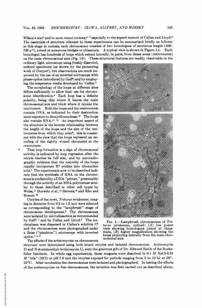
of the loops on exposure to Actinomycin D is very rapid; within 10 min the loopsare gone from chromosomes immersed in Callan's Medium C8 containing 2 ,jg/mlof the antibiotic. Compare the Actinomycin D-treated chromosomes in Figure3A with the control chromosomes in Figure 1B.Did this change in chromosomal morphology result simply from the combination
between DNA and Actinomycin D, or did it follow as a result of the inhibition ofRNA synthesis?Experiments with a modified actinomycin support the latter interpretation.
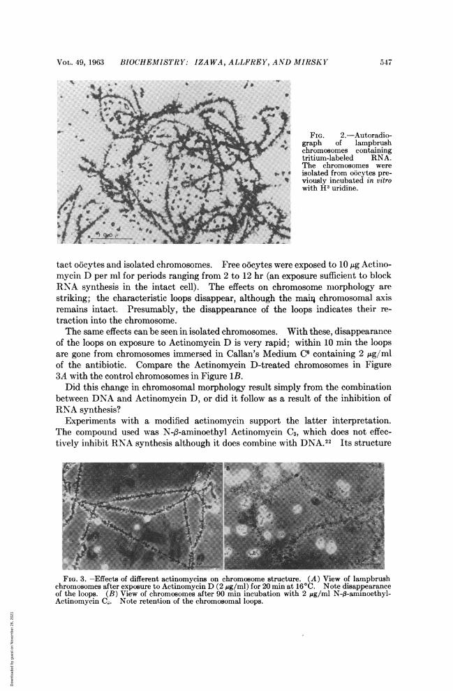
The compound used was N-f-aminoethyl Actinomycin C3, which does not effec-tively inhibit RNA synthesis although it does combine with DNA.22 Its structure
FIG. 3 -Effects of different actinomycins on chromosome structure. (A) View of lampbrushchromosomes after exposure to Actinomycin D(2 jsg/ml) for 20minat16 C. Note disappearanceof the ioops. (B) View of chromosomes after 90 mi incubation with 2 g/ml N-1_-aminoethyl_Actinomycin C. Note retention of the chromosomal loops.
Dow
nloa
ded
by g
uest
on
Nov
embe
r 26
, 202
1
548 BIOCHEMISTRY: IZAWA, ALLFREY, AND MIRSKY PROC. N. A. S
KL-N-METHYL-VAUNE L-N-METHYL-VALINE- L-N-METHYL-VALINE L-N-METHYLA-INE
SARCOSIVE SARCINE SARCOSINK SARCONE0 L-PROLINE L-PROLINE 0 0 L-PROLINE L-PROLINE 0
LD-VALINE D-MALINE -SOLEUCINE a0I-ISOLEUCINEIII
L-THREONINE L-THREONINE L- THREONINE L-THREONINE
CO CO CO CO
(A) (B)C,_NH
a~. tN H, ()NCaNH
CH3 C>3 CH3
Actinomycin D (C,) N-A-aminoethyl-Actinomycin C,
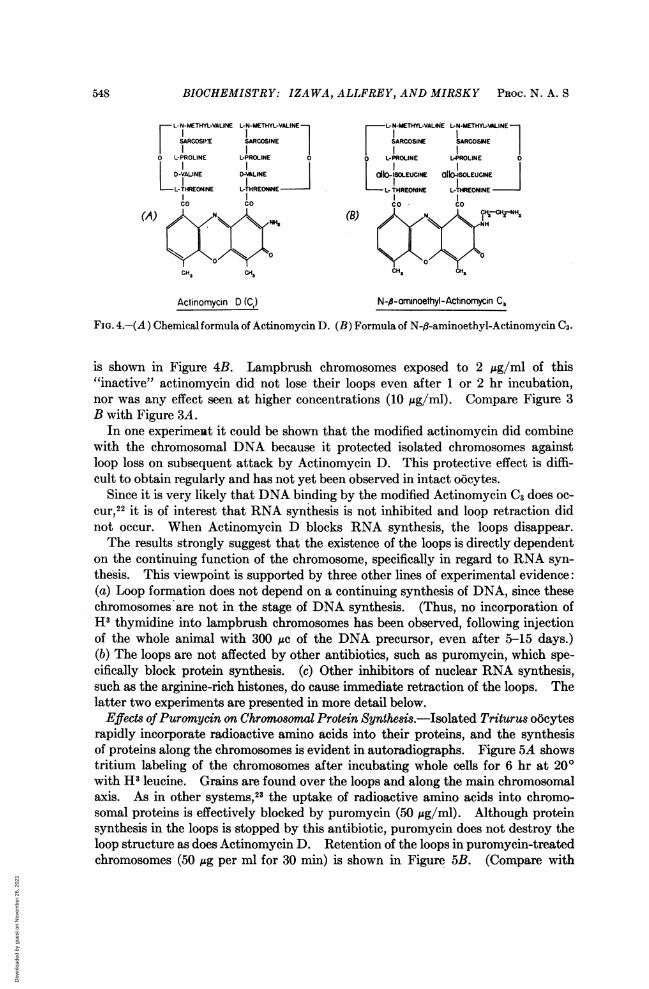
FIG. 4.-(A) Chemical formula of Actinomycin D. (B) Formula of N-0-aminoethyl-Actinomycin C3.
is shown in Figure 4B. Lampbrush chromosomes exposed to 2 /Ag/ml of this"inactive" actinomycin did not lose their loops even after 1 or 2 hr incubation,nor was any effect seen at higher concentrations (10 ;Lg/ml). Compare Figure 3B with Figure 3A.
In one experiment it could be shown that the modified actinomycin did combinewith the chromosomal DNA because it protected isolated chromosomes againstloop loss on subsequent attack by Actinomycin D. This protective effect is diffi-cult to obtain regularly and has not yet been observed in intact oocytes.
Since it is very likely that DNA binding by the modified Actinomycin C3 does oc-cur,22 it is of interest that RNA synthesis is not inhibited and loop retraction didnot occur. When Actinomycin D blocks RNA synthesis, the loops disappear.The results strongly suggest that the existence of the loops is directly dependent
on the continuing function of the chromosome, specifically in regard to RNA syn-thesis. This viewpoint is supported by three other lines of experimental evidence:(a) Loop formation does not depend on a continuing synthesis of DNA, since thesechromosomes are not in the stage of DNA synthesis. (Thus, no incorporation ofH3 thymidine into lampbrush chromosomes has been observed, following injectionof the whole animal with 300 ,uc of the DNA precursor, even after 5-15 days.)(b) The loops are not affected by other antibiotics, such as puromycin, which spe-cifically block protein synthesis. (c) Other inhibitors of nuclear RNA synthesis,such as the arginine-rich histones, do cause immediate retraction of the loops. Thelatter two experiments are presented in more detail below.
Effects of Puromycin on Chromosomal Protein Synthesis.-Isolated Triturus o6cytesrapidly incorporate radioactive amino acids into their proteins, and the synthesisof proteins along the chromosomes is evident in autoradiographs. Figure 5A showstritium labeling of the chromosomes after incubating whole cells for 6 hr at 200with H3 leucine. Grains are found over the loops and along the main chromosomalaxis. As in other systems,23 the uptake of radioactive amino acids into chromo-somal proteins is effectively blocked by puromycin (50 Mg/ml). Although proteinsynthesis in the loops is stopped by this antibiotic, puromycin does not destroy theloop structure as does Actinomycin D. Retention of the loops in puromycin-treatedchromosomes (50 M4g per ml for 30 min) is shown in Figure 5B. (Compare with
Dow
nloa
ded
by g
uest
on
Nov
embe
r 26
, 202
1
VOL. 49, 1963 BIOCHEMISTRY: IZAWA, ALLFREY, AND MIRSKY 549
A~~~ ~ ~ ~ ~ ~ ~ ~ ~
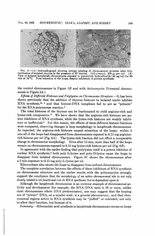
FIG. 5.-(A) Autoradiograph showing intense labeling of chromosomal proteins after 6-hrincubation of isolated oocytes in the presence of H' leucine. (2.8 c/mmol; 500 Mc per ml). (B)View of isolated lampbrush chromosomes exposed to puromycin hydrochloride (50 Mg/mi) for 30-min at 160C. Note retention of the loops, despite inhibition of protein synthesis.
the control chromosomes in Figure lB and with Actinomycin D-treated chromo-somes in Figure 3A.)
Effects of Different Histones and Polylysine on Chromosome Structure.-Jt has beenshown previously that the addition of thymus histones to isolated nuclei inhibitsRNA synthesis,24. 2 and that histone-DNA complexes fail to act as "primers"~for the RNA-polymerase reaction.3The total histones of the thymus can be fractionated to yield arginine-rich and
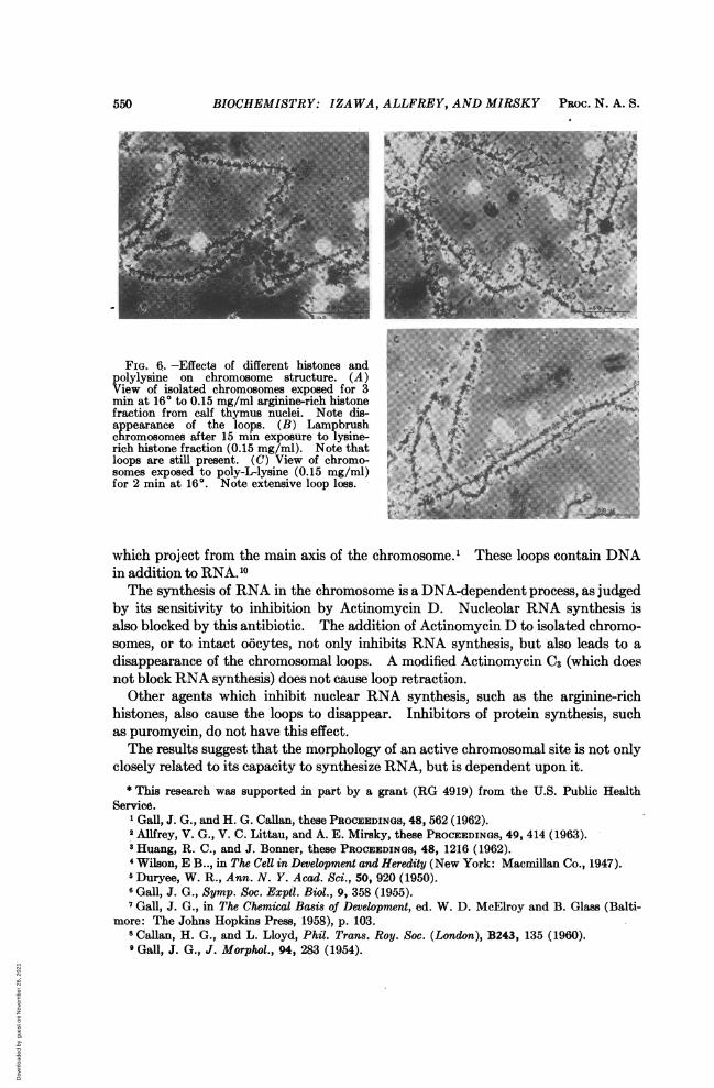
lysine-rich components.'7 We have shown that the arginine-rich histones are po-tent inhibitors of RNA synthesis, while the lysine-rich histones are weakly inhibi-tory or ineffectual.2 For this reason, the effects of these different histone fractionswere compared, observing changes in loop morphology in lampbrush chromosomes.As expected, the arginine-rich histones caused retraction of the loops; within 3mm all of the loops had disappeared from chromosomes exposed to 0.15 mg arginine-rich histone per ml (Fig. GA). The lysine-rich fraction did not effect a comparablechange in chromosome morphology. Even after 15 mm, more than half of the loopsremain on chromosomes exposed to 0.15 mg lysine-rich histone per ml (Fig. GB).In agreement with the earlier finding that polylysine itself is a potent inhibitor of
nuclear RNA synthesis,2 both poly-Llysine and poly-D-lysine cause the loops todisappear from isolated chromosomes. Figure GC shows the chromosomes aftera 2 mth exposure to 0.15 mg poly-L-lysine per ml.
Ribonuclease also causes the loops to disappear from unfixed chromosomes.The complete correlation between the effects of these inhibitors of RNA synthesis
on chromosome structure and the earlier results witthethe actinomycins stronglywsupport the conclusion that the morphology of an active chromosomal site is not onlyclosely related to its functional role in RNA synthesis, but is dependentupoiit.Although the lampbrush chromosome is an extreme example of chromosomal ac-
tivity and development (for example, the RNA/DNA ratio is 10 or more, unlikemost chromosomes where DNA predominates), one may suggest that the loopingout of "primer" DNA, on a smaller scale, is a general phenomenon, and that chro-mosomal regions active in RNA synthesis may be "puffed" or extended, not onlyto allow their function, but because of it.Summary.- Ribonucleic acid synthesis in lampbrush chromosomes occurs on loops
Dow
nloa
ded
by g
uest
on
Nov
embe
r 26
, 202
1
550 BIOCHEMISTRY: IZAWA, ALLFREY, AND MIRSKY PROC. N. A. S.
4~~~~~~ s
i........
FIG. 6.-Effects of different histones andpolylysine on chromosome structure. (A) .0View of isolated chromosomes exposed for 3min at 160 to 0.15 mg/mi arginine-rich histonefraction from calf thymus nuclei. Note disappearance of the loops. (B) Lampbrush :4chromosomes after 15 min exposure to lysine- E i.rich histone fraction (0.15 mg/ml). Note thatloops are still present. (C) View of chromo- ..........
somes exposed to poly-L-lysine (0.15 mg/ml)for 2 min at 160. Note extensive loop loss.
which project from the main axis of the chromosome.' These loops contain DNAin addition to RNA.10The synthesis of RNA in the chromosome is a DNA-dependent process, as judged
by its sensitivity to inhibition by Actinomycin D. Nucleolar RNA synthesis isalso blocked by this antibiotic. The addition of Actinomycin D to isolated chromo-somes, or to intact oicytes, not only inhibits RNA synthesis, but also leads to adisappearance of the chromosomal loops. A modified Actinomycin C3 (which doesnot block RNA synthesis) does not cause loop retraction.
Other agents which inhibit nuclear RNA synthesis, such as the arginine-richhistones, also cause the loops to disappear. Inhibitors of protein synthesis, suchas puromycin, do not have this effect.The results suggest that the morphology of an active chromosomal site is not only
closely related to its capacity to synthesize RNA, but is dependent upon it.* This research was supported in part by a grant (RG 4919) from the U.S. Public Health
Service.1 Gall, J. G., and H. G. Callan, these PROCEEDINGS, 48, 562 (1962).2 AMfrey, V. G., V. C. Littau, and A. E. Mirsky, these PROCEEDINGS, 49, 414 (1963).3Huang, R. C., and J. Bonner, these PROCEEDINGS, 48, 1216 (1962).4 Wilson, E B.., in The Cell in Development and Heredity (New York: Macmillan Co., 1947).5 Duryee, W. R., Ann. N. Y. Acad. Sci., 50, 920 (1950).6 Gall, J. G., Symp. Soc. Exptil. Biol., 9, 358 (1955).7 Gall, J. G., in The Chemical Basis of Development, ed. W. D. McElroy and B. Glass (Balti-
more: The Johns Hopkins Press, 1958), p. 103.8Callan, H. G., and L. Lloyd, Phil. Trans. Roy. Soc. (London), B243, 135 (1960).Gall, J. G., J. Morphol., 94, 283 (1954).
Dow
nloa
ded
by g
uest
on
Nov
embe
r 26
, 202
1
VOL. 49, 1963 GENETICS: T. NAGATA 551
10 Callan, H. G., and H. C. MacGregor, Nature, 181, 1479 (1958).Dodson, E. O., Univ. Calif. Publ. Zool., 53, 281 (1948).
12 Gall, J. G., in Mutation, Brookhaven Symposia in Biology, vol. 8, p. 17 (1956).13 Weiss, S. A., these PROCEEDINGS, 46, 1020 (1960).14 Hurwitz, J., A. Bresler, and R. Diringer, Biochem. and Biophys. Res. Comm., 3, 15 (1960).15 Stevens, A., Biochem. and Biophys. Res. Comm., 3, 92 (1960).16 Rho, J. M., and J. Bonner, these PROCEEDINGS, 47, 1611 (1961).17 Johns, E. W., and J. A. V. Butler, Biochem. J., 82, 15 (1962).18 Reich, E., R. M. Franklin, A. J. Shatkin, and E. L. Tatum, Science, 134, 556 (1961).19 Kirk, J. M., Biochim. et Biophys. Acta, 42, 167 (1960).20Hurwitz, J., J. J. Furth, M. Malamy, and M. Alexander, these PROCEEDINGS, 48, 1222
(1962).21 Allfrey, V. G., and A. E. Mirsky, these PROCEEDINGS, 48, 1590 (1962).22 Reich, E., I. H. Goldberg, and M. Rabinowitz, Nature, 196, 743 (1962).23 Yarmolinsky, M. D., and G. de la Haba, these PROCEEDINGS, 45, 1721 (1959).24 Allfrey, V. G., in The Molecular Basis of Neoplasia (Austin: University of Texas Press,
1962), p. 58.
THE MOLECULAR SYNCHRONY AND SEQUENTIAL REPLICATION OFDNA IN ESCHERICHIA COLI*
BY TOSHio NAGATAt
DEPARTMENT OF ZOOLOGY, COLUMBIA UNIVERSITY
Communicated by E. L. Tatum, February 18, 1963
Cytological and genetic observations on a number of organisms have contributedto formulating the concept of a sequential replication of the chromosome startingfrom one or more fixed points.1 2 Especially in bacteria, as Maal0e3 pointed out,this possibility found support in a mass of data strongly indicating a definite reg-ularity in the synthesis of DNA.4 5 Advantage of the bacterial system was takenin the present work which employed two strains of Escherichia coli K-12 Hfr, aswell as one of F-, and analyses were made of the problem of molecular synchronyand the sequential replication of the chromosomes. A system has been establishedfor determining the course of change in the number of prophage X per bacteriumduring a replication cycle of DNA in populations of lysogenic bacteria under con-ditions of synchronous growth. A substantial amount of evidence indicates thatthe prophage X is indeed a DNA structure, occupies a definite site on the K-12linkage map, replicates in harmony with the host chromosome, and can be induced byvarious agents.6-8 The existence of X as an episome allows us to regard it as a geneon the bacterial chromosome and at the same time provides us with a means ofenumerating its intracellular number at any given time. Thus, the kinetics of thismeasure should directly reflect the pattern of the replication of the host chromosomeitself.
Bacterial Strains.-All the bacteria used are derivatives of Escherichia coli K-12 (Table 1).Superinfecting phase: Xvlh, a weak virulent and host range mutant of X,9 was kindly provided
by Dr. R. K. Appleyard. In contrast to the wild type (v1 +) which forms turbid plaques, themutant vl gives rise to clear plaques. High titer stocks of this phage were prepared by the methodof confluent lysis. l0
Media: (a) M-9 is a minimal salt solution composed of 7.0 g anhydrous Na2HPO4, 3.0 g KHr
Dow
nloa
ded
by g
uest
on
Nov
embe
r 26
, 202
1