Embed Size (px)
DESCRIPTION
Â
Citation preview

Vol 75 | No 2 2013C a N a d i a N J o u r N a l o f o p t o m e t r y | r e V u e C a N a d i e N N e d ’ o p t o m é t r i e 41
CLINICAL STUDY
BY ELISE G. KRAMER OD & LANGIS MICHAUD, OD, MSc, FAAO& VANESSA BACHIR, OD
Introduction
Möbius or Möbius-like syndrome is a disease within a group
of syndromes known as “terminal transverse defects with orofacial malformations”.1 The condition was originally recognized by Von Graefe in 1880 and later, more extensively described by Moebius in 1888.2 It is characterized by a unilateral or bilat-eral, nonprogressive congenital facial palsy with impairments of ocular
abduction. It is also associated with other cranial nerve palsies, orofacial dysmorphism (congenital malforma-tions of the face and/or mouth), and limb or axial malformations.2 Limb anomalies (hypoplasia) can range from mere syndactyly of the digits (webbed fingers or toes) to the absence of a limb. The craniofacial deformities vary even more and can include: micrognathism (undersized jaw), tongue malformations, facial and oral clefts, oligodontism (miss-ing teeth) and cranial nerve palsies.1 The presence of cranial nerve palsies is required to make the diagnosis of Möbius syndrome.1 Patients may be
Mini-scleral Lenses in the Treatment of Neurotrophic Keratopathy Secondary to Möbius or Möbius-like Syndrome
Elise G. Kramer, OD
Langis Michaud, OD, MSc, FAAO (Dipl) Associate Professor, École d’optométrie- Université de Montréal
Vanessa Bachir, OD
Objective: This case report aims to explore the use of large diameter rigid gas permeable lenses (LDRGP) for the treatment of ocular and visual complications in a young patient presenting with neurotrophic corneas.
Methods: After a comprehensive eye exam and specific testing for contact lens fitting, LDRGP lenses were fitted with success and dispensed. RESULTS. Prescribed contact lenses helped the patient achieve optimal visual correction (6/6) as well as ocular protection for the cornea.
Conclusion: Neurotrophic keratopathy is a challenging condition due to its impact on ocular health and vision. LDRGP offer a unique way of addressing many issues raised in this case report such as corneal healing and visual restoration. This lens modality may be considered for any other case involving abnormal corneal tissue healing and reduced visual acuity.
Key words: large diameter rigid gas permeable lenses (LDRGP), Möbius syndrome, neurotrophic keratopathy
Objectif : Cette étude de cas vise à explorer l'utilisation de lentilles de grand diamètre rigides perméables aux gaz (LDRGP) afin de traiter les complications oculaires et visuelles d’une jeune patiente présentant un problème de kératite neurotrophique.
Méthodes : Après un examen de la vue complet et des tests spécialisés pour l’adaptation en lentilles cornéennes, des lentilles LDRGP ont été adaptées avec succès et livrées. RÉSULTATS. Les lentilles prescrites ont aidé la patiente à atteindre une correction visuelle optimale (6/6) et ont protégé la surface oculaire.
Conclusion : La kératite neurotrophique est une condition difficile à gérer a cause de son impact sur la santé oculaire et la vision. La correction avec des lentilles LDRGP offre une solution unique aux nombreux défis soulevés dans ce rapport tels que la cicatrisation de la cornée et la restauration visuelle. Ce type de lentille peut être envisagé afin de solutionner les cas de guérison anormale de la cornée avec baisse d’acuité visuelle.
Mots clés : kératite neurotrophique, lentilles de grand diamètre rigides perméables aux gaz (LDRGP), Syndrome de Möbius
ABSTRACT

C a N a d i a N J o u r N a l o f o p t o m e t r y | r e V u e C a N a d i e N N e d ’ o p t o m é t r i eVol 75 | No 2 201342
affected by bilateral and incomplete facial nerve palsy, impeding them socially, given their inability to con-vey reactions of joy or sorrow.3 Table I below summarizes clinical features that have been reported in cases of Möbius Synrome.
Möbius syndrome has been re-ported to usually occur sporadically but some heterogeneous inheritance patterns have been observed in several affected families.2 The exact pathogenesis remains unclear and controversial. Fetal exposure to teratogens, disturbance in rhomb-encephalic development or acquired ischemic events occurring shortly after the fifth week in utero have all been proposed.1,2 The insult is thought to lead to a chain of events involving a temporary interrup-tion in the fetal blood supply to the lower brainstem. This then causes one or more focal areas of dam-age.2 The facial, abducens, lacrimal and salivary nuclei are all located in the involved brainstem. They also
tend to develop simultaneously in this phase of embryogenesis. This theory is supported by many studies that have found hypoplasia of the lower brainstem with flattening of the floor of the 4th ventricle in ap-proximately 30% of cases of Möbius syndrome. They’ve also found gliosis and calcifactions in the same area.2 The general concensus is that Möbius syndrome is a complex and multifactorial developmental disor-der of the lower part of the brain-stem. Möbius patients are often categorized according to the severity of their abnormalities. Defining the syndrome is confusing at times given the highly heterogeneous presentation.1 Despite its wide spectrum of abnormalities, most patients with Möbius syndrome will present with orofacial findings including micrognatia, cleft palate, tongue anomalies, ear malforma-tions and bifid uvula (split uvula).5
Ocular findings related to Möbius include severe limitations in
abduction, with marked decrease of horizontal saccades, similar to a gaze paresis, most likely due to a supranuclear lesion.3 Consequently, reading problems can occur. Other findings include epicanthal folds, and more rarely hypertelorism, coloboma, heterochromia, hetero-chromic cyclitis, nystagmus and limbal dermoids.1 A significant loss of corneal sensitivity can occur as a result of the trigeminal nerve palsy (V). This nerve, which provides sen-sory innervation to the cornea, also allows constant renewal of epithelial cells. Surface squamous cells are constantly sloughing off every day; this process of continual renewal of cells is imperative for ocular surface integrity. In neurotrophic keratopathy, the cornea is deprived of sensation and does not produce and renew cells, but the normal loss of cells still occurs. In addition, the corneal reflex is attenuated, result-ing in reduced protection of the ocular surface through a lack of blinking. Patients with neurotrophic keratopathy have worse signs than symptoms and are therefore at risk for corneal ulceration, resulting in severe and irreversible vision loss.6 If the facial nerve is also affected, lagophthalmos, an incomplete closure of the eyelids while blink-ing, may result.2,4 This case report explores how optometrists can help restore vision and protect the ocular surface in these patients using LDRGP.
The concept of neutralizing the refractive error induced by the cornea with an enclosed reservoir of liquid was first introduced in 1508 by Leonardo da Vinci.7 Scleral
Table I: Clinical Features Reported in Cases with Möbius Syndrome4
System Features
Neurological Mental retardation, CNS abnormalities, Hypotonia, Epilepsy
Craniofacial Cranium shape defect, Bitemporal narrowing, Epicanthic folds, Hypertelorism, Ptosis, Strabismus, Microphthalmia, Duane anomaly, Lacrimal duct defects, Flat nasal bridge, Teeth anomalies, Highly arched palate, Bifid uvula/cleft palate, Small tongue, Micrognathia, External ear defects, Low set ears, Short neck
Cranial nerves Palsies: III, IV, V, VI, VII, VIII, IX, X, XII
Trunk Poland anomaly, Congenital heart defect, Vertebral abnormalities, Kyphoscoliosis, Aplasia abdominal muscles, Underdeveloped genitalia
Limbs Brachydactyly, Clinodactyly, Camptodactyly, Syndactyly, Ectrodactyly, Low set thumbs, Adducted thumbs, Supernumerary thumb, Flexion deformities of wrist, Hip defects/luxation, Hypoplasia of lower legs, Arthrogryposis, Clubfoot, Pes planus
Skin Hemangiomas

Vol 75 | No 2 2013C a N a d i a N J o u r N a l o f o p t o m e t r y | r e V u e C a N a d i e N N e d ’ o p t o m é t r i e 43
lenses were first described in medi-cal literature in the late 19th century. Adolf Fick proposed his use of glass blown shells in 1888. Eugene Kalt then used these vesicles to improve the vision of a patient with keratoconus. August Mueller later described his attempts to correct his own myopia with these glass lenses. Although these lenses did improve vision, they were not widely used due to challenges with manufac-turing and wearing.7 In the 1940’s, polymethylmethacylate (PMMA), a new lens material was developed by workers such as Feinbloom, Obrig and Gyoffry.8 The lenses were molded based on an impres-sion of the cornea, which facilitated manufacture. However, the poor reproducibility and permeability of these lenses limited their distribu-tion. In the mid-1900’s, corneal contact lenses were introduced.7 They were also originally made of PMMA but were smaller than scleral lenses, which made oxygen and tear exchange as well as fitting easier. With the later development of rigid gas permeable (RGP) materials, as first described by Ezekiel in 1983, oxygen was readily able to penetrate through the lenses themselves and further reduced complications related to contact lens wear.7 These, in addition to soft lenses, stopped the further development of LDRGP fitting.7,8
A few years ago, only few spe-cialized practitioners were fitting LDRGP lenses. Since then, there has been a slow but steady increase in the demand for these lenses as a solution for more challenging cases.8 LDRGP designs have become more
and more popular and are available in several options: a corneo-scleral lens (12.5 mm to 15 mm), supported partly by the cornea and partly by the sclera; a mini-scleral lens (15 mm to 18 mm) vaulting the cornea, supported by the fluid layer and the conjunctiva; or a larger scleral lens (18 mm to 25 mm) with the same fitting philosophy as the mini-scleral lens but with different parameters.8
This case describes the use of mini-scleral lens technology in restoring vision and protecting the ocular surface in a patient suffering from Möbius syndrome.
BackgroundIn the fall of 2010, an 8-year-old Caucasian female was referred for a contact lens evaluation by an oph-thalmologist for the treatment of neurotrophic keratopathy. She had been diagnosed with encephalopa-thy, likely of prenatal origin, resulting in a forme frustre of Möbius-like syndrome. Systemic manifestations of her condition included epilepsy, recurrent episodes of rigidity (especially when tired), agitated sleep, decreased pain sensa-tion resulting in frequent injuries, complete deafness of the right ear, absent gag reflex, nasal congestion and trouble walking due to lower limb deformities. Her ocular history was remarkable for a trigeminal nerve (V) palsy, leading to loss of corneal sensitivity, as well as abdu-cens nerve (VI) palsy. She also presented with lagophthalmia secondary to facial nerve (VII) malfunction. Combined, these
anomalies triggered the develop-ment of neurotrophic corneas, worse in the right eye than the left. Consequently, she required ocular lubricants several times a day to preserve both the ocular surface and her vision. She was seen in the con-tact lens clinic in order to be fitted in RGP contact lenses to maintain constant lubrication of her cornea. The idea was to protect the ocular surface from eventual abrasions as well as improve her vision.
Clinical findingsInitial clinical findings are summa-rized in Table II.
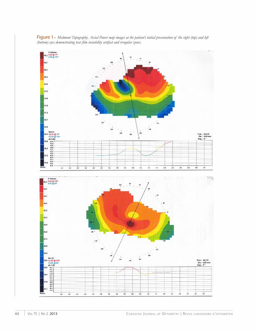
Corneal topography was mea-sured using a Medmont E-300. An axial power map displaying the paraxial power of the surface in diopters with respect to the kerato-scope axis was selected (Figure1). The color scale on the left repre-sents the range of powers that can be found on the cornea, with dark red being the highest and dark blue being the lowest.10 The E values at the top left, formerly what Med-mont called Shape Factor, indicate the elliptical shape index for the Steep (in red) and Flat (in blue) axes of the cornea. The Sim-K values at the bottom left indicate the values for the Steep (in red) and Flat (in blue) axes of the cornea.9 The patient’s topography showed many irregular zones of the corneal surfaces in both eyes, but mostly in the right eye (Figure1). There were large and rapid changes in power and shape. The interruptions in the image rep-resent the device’s inability to cap-ture that part of the corneal surface
C a N a d i a N J o u r N a l o f o p t o m e t r y | r e V u e C a N a d i e N N e d ’ o p t o m é t r i eVol 75 | No 2 201344
Figure 1– Medmont Topography. Axial Power map images at the patient’s initial presentation of the right (top) and left (bottom)eyesdemonstratingtearfilminstabilityartifactandirregularzones.
Vol 75 | No 2 2013C a N a d i a N J o u r N a l o f o p t o m e t r y | r e V u e C a N a d i e N N e d ’ o p t o m é t r i e 45
due to tear film or other artifacts.9 These surface issues contributed to the decrease in best-corrected visual acuity. Figure1 shows Axial Power map images at the patient’s initial presentation.
The patient was diagnosed with moderate hyperopia and astigma-tism, requiring optical correction, and bilateral neurotrophic kera-topathy with scarring and irregular corneal surfaces. Spectacles were prescribed for full-time wear (for distance, near and intermediate activities) to provide an immediate visual correction and to serve as a back-up for contact lenses.
Treatment plan After the glasses were prescribed, contact lenses were strongly recom-mended as the primary treatment for neurotrophic keratopathy. In this condition, the ocular surface must be protected to minimize the risk of erosion; contact lenses help maintain constant lubrication of the corneal surface, which allows for its restoration. A soft bandage does not provide a good outcome for visual correction on a highly irregular cornea. Small diameter RGP lenses can provide a better alternative to improve visual acuity but do not protect the ocular surface. In fact, these lenses can increase mechanical stress on an already altered cornea in this case. One of the ways to
resolve this issue could be to con-sider a piggy-back system, which implies fitting a high oxygen perme-ability soft lens carrier on top of which a high permeability RGP lens is fitted. In that way, the soft carrier aims to protect the cornea while the RGP restores visual acuity. Another solution includes the implementa-tion of hybrid lenses. These consist of a gas permeable rigid center sur-rounded by a silicone hydrogel soft skirt. In fitting this lens, the skirt is designed to lift the rigid center off the corneal surface so that it never has to interact with it. However, there have been some reported cases of warpage with these lenses.10 In addition, most, if not all of the hybrid lenses do not offer enough oxygen permeability to maintain ocular health in the presence of a compromised cornea.10
LDRGP can also be considered. They are fitted in a way to vault the cornea. They maintain a constant reservoir of fluid between the lens and the cornea to ensure that it remains lubricated. Moreover, this fluid layer also compensates for surface irregularities, leading to improved visual acuity. This mo-dality can provide the comfort of a soft lens with the optical quality of a gas permeable lens. In that way, LDRGP designs currently available are considered the best option to provide health benefits and increased comfort compared to smaller corneal RGP and, in this case, soft lenses. In the case of neu-rotrophic keratopathy, in order to determine which type of LDRGP to use, any touch on the cornea should be avoided. Corneo-scleral
Table II: Clinical Findings on Initial Presentation to the Contact Lens Clinic
Clinical Findings OD OS
Unaided Visual Acuities 6/12-2 6/9-1
Cycloplegic Refraction +4.75 −1.25 × 130 with VA: 6/7.5
+3.25 −1.50 × 175 with VA: 6/7.5
Anterior Segment Evaluation
Cornea:l Mid-peripheral scarringl Moderate diffuse
punctate epithelial erosions (positive staining)
TBUT:l >10s (normal)Lacrymal lake:l Moderately reduced at
inferior margins
Cornea:l Mild to moderate central haze
without scarringl Moderate diffuse punctate
epithelial erosions (positive staining)
TBUT:l >10s (normal)Lacrymal lake:l Moderately reduced at inferior
margins
Dilated Fundus examination
Optic nerve:lUnremarkableMacula:lUnremarkableVessels:lUnremarkablePeriphery:lNo breaks, holes or tears 360
Optic nerve:lUnremarkableMacula:lUnremarkableVessels:lUnremarkablePeriphery:lNo breaks, holes or tears 360

C a N a d i a N J o u r N a l o f o p t o m e t r y | r e V u e C a N a d i e N N e d ’ o p t o m é t r i eVol 75 | No 2 201346
lenses are contraindicated because a small portion of the cornea sup-ports most of the weight of the lens. This may result in a stress to the tissue that could aggravate the corneal epithelial defect and/or generate scarring. The mini-scleral lenses represent an improved option, where cornea-lens touch is absent with a limited amount of fluid layer. In addition, they are smaller than scleral lenses and are therefore easier to handle and less intimidating for young patients to insert into their eyes.8
Lens trials The patient was fitted successfully with a relatively new LDRGP on the market (One Fit P&A, Tyro 97 Blanchard Laboratories, Sherbrooke, Quebec.). The apical clearance and peripheral edge of this mini-scleral lens are designed to correct regular ametropia (high refractive errors, astigmatism, dryness related to con-tact lens wear (P&A profile) and are very successful in correcting irregu-lar corneas (KC profile). The fitting process is simple and easy to learn. They are proven as easy to wear and as comfortable, once properly fitted, as a soft lens.11 Based on the fitting guide, the initial base curve is selected 0.3 mm steeper than flat K, to provide a central clearance of 150 µm after 30 minutes of wear. This can be directly assessed at the slit lamp, using the known or esti-mated corneal thickness (555 µm on average), by comparing the width of the space between the lens and the cornea (the green fluorescein layer) to the slit width of the cornea.12
This can be efficiently re-evaluated at subsequent follow-up visits. The lens diameter should exceed the cornea by at least 1 mm in every quadrant (Figure2). The lens should offer no resistance on push-up, compression (blanching of the con-junctival vessels) or impingement (pinching of the conjunctival tissue resulting in staining).8 The lens is inserted into the eye once it has been filled with fluid (non preserved saline solution or artificial tears). With LDRGP wear, the need for topical ocular lubricants during the day can be substantially decreased because the fluid inside the lens constantly surrounds and lubricates the cornea. It was recommended that the patient wear the contact lenses the majority of the time with the option of spectacle wear when the lenses were removed. The final prescription was made with the following parameters: +4.25 OD
and +3.75 OS with base curves of 7.80 mm and diameters of 14.0 mm OU. The lenses were made of Tyro 97, a fluoro-silicone material with a Dk of 97 and a wetting angle of less than 10 degrees. This was the only contact lens, among all that were attempted, that satisfied both the physiological requirements of the ocular surface and the visual needs of the patient. In theory, the lifespan of these lenses is two years. An example of a One Fit lens in a pediatric patient can be appreciated above. (Figure2)
With these lenses, the visual acu-ity was OD 6/7.5+2 and OS 6/6-1. After educating the patient and her parents on handling and cleaning the lenses, they were dispensed. According to a study conducted in 2008 by Gungor et al, there are no age restrictions in scleral lenses.13 Nevertheless, fitting a patient of this young age did not come without its
Figure 2 –Similarappearanceof theOneFitlensonanotherpediatricpatient.Onecan appreciate the diameter of the lens, exceeding the visible cornea by at least 1mm.

Vol 75 | No 2 2013C a N a d i a N J o u r N a l o f o p t o m e t r y | r e V u e C a N a d i e N N e d ’ o p t o m é t r i e 47
challenges. She required many hours of practice before being proficient with the insertion and removal of the lenses. Proper hygiene and ade-quate cleaning of the lenses also had to be confirmed before dispensing them. Her parents also became proficient with these procedures in order to help her, if needed.
RGP cleaner and conditioning solution (Boston™) were recom-mended. Non-preserved artificial tears were prescribed (Refresh™, Allergan) to fill the lens before insertion. The patient was instructed to begin with 4 hours of wear on the first day and to increase by 2-hour increments every day, until reaching a maximum of 12 hours per day. At her 1- and 2-month follow-up visits, the patient’s comfort and vision remained excel-lent. Anterior segment evaluation did not show any corneal staining, conjunctival redness, or any other signs of contact lens intolerance. She was then referred back to her ophthalmologist for further follow-ups. However, annual exams at the contact lens clinic were recom-mended to monitor contact lens fitting and parameters.
DiscussionNeurotrophic keratopathy is always a challenging disease to manage. In the case summarized here, the treatment plan was decided based on the ocular surface condition and the potential to restore visual acuity. It is recommended to have a back-up pair of glasses when prescrib-ing specialty contact lenses. These both improve the vision when the
patient is not wearing the lenses and provide a temporary solution if the patient has a problem with them.
An important consideration for prolonged contact lens wear is oxygen delivery to the cornea.8 RGP are permeable to oxygen and allow it to pass through the lens. Tear flow underneath the lens, if present, can also bring tears rich in oxygen to the cornea. In LDRGP, the lens vaults the limbus so oxygen from the con-junctival and limbal vessels can also contribute to the oxygen supply.8 One can calculate the overall oxygen transmissibility using the lens and fluid layer thicknesses and perme-ability to oxygen. In this system, the fluid layer represents the limiting factor, its Dk being 80 × 10-11(cm2/sec)(mlO2/ml × mmHg). In this case, we can estimate the oxygen transmissibility with the following formula:
Dk = 1 tscl (t1/Dk1+t2/Dk2)
where Dk1 and Dk2 represent the permeability of lens and fluid layer whereas t1 and t2 are the lens and fluid layer thicknesses. Assuming that the lens is 300 µm thick, made with Tyro 97 material and fitted with a clearance of 125 µm centrally and 40 µm peripherally, this gives:
Dk /t =1/ (3.0/97+1.25/80) = 21.5×10⁻⁹[cm/sec][mlO2/(ml×mmHg)] (central)
= 1/ (3.0/97+0.4/80) = 27.8×10⁻⁹
[cm/sec][mlO2/(ml×mmHg)](peripheral)
These values meet the Holden-Mertz criteria to avoid corneal hypoxia for daily wear.14 This does not necessarily occur when the
lens is fitted with higher clearance, which is usually the case for other designs and/or lenses over 16 mm in diameter.
Another interesting aspect of this case is the use of contact lenses in a pediatric population. It is now well known that contact lenses can be adequately fitted in patients as young as 8 years old, with a safety profile comparable to that of older patients.15 Optical and psychologi-cal benefits of contact lens wear are multiple. They include: increased autonomy, self-esteem, better social interaction and behavior, improved visual acuity, etc. This modality is well-accepted by young patients but could be subject to resistance by their caregivers who seem to be slightly more reluctant.16 In general, when caregivers are motivated and see the positive results of contact lens wear, it does not take long for them to fully endorse this modality of correcting their child’s vision. Their collaboration is necessary to supervise the appropriate handling and cleaning of the lenses. Neg-ligence is certainly the key factor leading to contact lens fitting failure. It is necessary for caregivers to become familiar with all of the pro-cedures and readily help the young patient as needed.
In this particular case, it was difficult to appreciate any reaction from the patient due to her systemic condition, but her parents reported that she improved significantly at school, given her improved vision, and was also more engaged with other children. She had not reported any ocular discomfort and was eager to put her lenses in every

C a N a d i a N J o u r N a l o f o p t o m e t r y | r e V u e C a N a d i e N N e d ’ o p t o m é t r i eVol 75 | No 2 201348
morning. The treatment had notably improved her behavior, according to her parents’ reports. It is not rare that mini-scleral lenses have such a positive impact, in that they restore visual acuity as well as ocular comfort.
Sjögren’s syndrome, persistent epithelial corneal defects, Steven’s Johnson Syndrome, Graft Versus Host Disease, ocular cicatricial pemphigoid, atopic keratoconjunc-tivitis or other corneal irregularities resulting in poor vision are just a few examples of additional condi-tions that can be managed with the help of LDRGP.8 Recent reports describe using LDRGP to deliver pharmacologic agents to the ante-rior surface of the eye. In the case of neurotrophic keratopathy where persistent corneal epithelial defects and even ulceration can occur, an-tibiotics can be instilled in the lens and placed directly on the eye.17 This helps the ocular surface recover and heal properly. Patients are extremely grateful when the practitioner finds a successful solution to their problem.
It is well-known that LDRGP lenses are effective in the treatment of complex cases such as neuro-trophic keratopathy secondary to Möbius syndrome. These lenses thus provide optimal treatment for this patient’s ocular surface disease.
Conclusions Fitting of mini-scleral contact lenses succeeded in treating bilateral neu-rotrophic keratopathy with corneal scarring that penalized visual acuity. Such lenses would be an ideal solution in any case of neurotophic
keratopathy. It was extremely rewarding to use these lenses and thereby provide clear vision to a patient who had been struggling for many years with an ocular surface condition and consequently poor vision.
With the increasing recent inter-est of clinicians and manufacturers, mini-scleral lenses are becoming far more “mainstream” in contact lens practice. As optometrists, we should strive to continuously update our expertise in the area of contact lens design, thereby providing our patients with the latest lens technol-ogy and best solution for their signs and symptoms.
References1. Miller MT, Owens P, Chen F. Möbius
and Möbius-like syndromes (TTV-OFM, OMLH). J. Pediatr Ophthalmol Strabismus. 1989 Jul-Aug;26(4):176-88.
2. Arturo C, Mora P, Neri A, Favilla S, Sadun AA. Ophthalmologic and systemic features in Möbius syndrome. Ophthalmology. 2011 Aug;118(8): 1518-523.
3. Goldblatt D, Williams D. I am smiling: Mobius syndrome inside and out. J Child Neurol 1986 Jun;1(1): 71-78.
4. Graziadio C, Lorenzen MB, Rosa RFM, et al. New report of a familial case of Moebius syndrome presenting skeletal findings. Am J Med Genet Part A. 2010; 152A (8): 2135.
5. Gorlin RG, Pinborg J, Cohen. Syndromes of the head and neck. Edition 2. New York, McGraw Hill, 1976.
6. Dartt DA. Corneal Nerves: Anatomy. Ocular Periphery and Disorders. San Diego: Academic/Elsevier, 2011:150.
7. A Brief History of Scleral Lenses. History | Scleral Lens Education Society. <http://www.sclerallens.org/history> Feb 18, 2013.
8. van der Worp, EA Guide to Scleral Lens Fitting. [Forest Grove, Ore.]: [College of Optometry, Pacific University], 2010: 1-4.
9. Medmont Pty Ltd. MEDMONT E300 CORNEAL TOPOGRAPHER USER MANUAL. VICTORIA: © MEDMONT, 2012.
10. Gardner D, Zimmerman A. Myopic shift secondary to hybrid lens wear. Contact Lens Spectrum. 2012 Jun;27: 44-48.
11. Michaud L, Woo S, Dinardo-Lotoczky A, et al. Clinical evaluation of a large diameter rigid-gas permeable lens for the correction of refractive astigmatism. American Academy of Optom. 2012: Poster; 125085, Phoenix.
12. Baldwin B and Moyer S. AS-OCT and the specialty contact lens. Review of Cornea and Contact Lenses. 2012 Apr: 32-34.
13. Gungor I, Schor K, Rosenthal P, Jacobs DS. The Boston scleral lens in the treatment of pediatric patients. JAAPOS. 2008 Jun;12(3):263-7
14. Holden BA, Mertz GW. Critical oxygen levels to avoid corneal edema for daily and extended wear contact lenses. Invest Ophthalmol Vis Sci. 1984 Oct;25(10):1161-7.
15. Rah MJ, Walline JJ, Jones-Jordan LA, et al. Vision specific quality of life of pediatric contact lens wearers. Optom Vis Sci. 2010 Aug; 87(8): 560-6.
16. Ma JJK, Morad Y, Mau E, Brent HP, Barclay R and Levin AV. Contact lenses for the treatment of pediatric cataracts. Ophthalmology. 2003 Feb;110(2):299-305
17. Lim P, Jacobs DS, Rosenthal P. Treatment of persistent corneal epithelial defects with the Boston ocular surface prosthesis and an antibiotic adjunct. 2009. ARVO poster; 653