Embed Size (px)
Citation preview
Knockout Rats via EmbryoMicroinjectionof Zinc-Finger NucleasesAron M. Geurts,1,2* Gregory J. Cost,3* Yevgeniy Freyvert,3 Bryan Zeitler,3 Jeffrey C. Miller,3Vivian M. Choi,3 Shirin S. Jenkins,3 Adam Wood,4 Xiaoxia Cui,4 Xiangdong Meng,3Anna Vincent,3 Stephen Lam,3 Mieczyslaw Michalkiewicz,1,2 Rebecca Schilling,1,2Jamie Foeckler,3 Shawn Kalloway,3 Hartmut Weiler,1,2 Séverine Ménoret,5 Ignacio Anegon,5Gregory D. Davis,4 Lei Zhang,3 Edward J. Rebar,3 Philip D. Gregory,3 Fyodor D. Urnov,3Howard J. Jacob,1,2,6† Roland Buelow7†
The laboratory rat is a well-establishedmodel for the genetic dissection of humandisease-related traits (1) despite the fact
that targetedmodification of its genome is largelyintractable. We investigated the application of
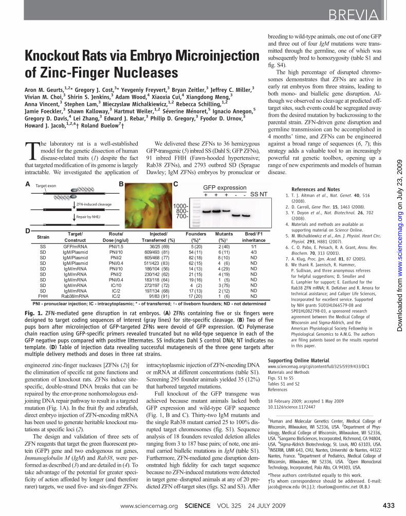
engineered zinc-finger nucleases [ZFNs (2)] forthe elimination of specific rat gene functions andgeneration of knockout rats. ZFNs induce site-specific, double-strand DNA breaks that can berepaired by the error-prone nonhomologous end-joining DNA repair pathway to result in a targetedmutation (Fig. 1A). In the fruit fly and zebrafish,direct embryo injection of ZFN-encoding mRNAhas been used to generate heritable knockout mu-tations at specific loci (2).
The design and validation of three sets ofZFN reagents that target the green fluorescent pro-tein (GFP) gene and two endogenous rat genes,Immunoglobulin M (IgM) and Rab38, were per-formed as described (3) and are detailed in (4). Totake advantage of the potential for greater speci-ficity of action afforded by longer (and thereforerarer) targets, we used five- and six-finger ZFNs.
We delivered these ZFNs to 36 hemizygousGFP-transgenic (5) inbred SS (Dahl S;GFPZFNs),91 inbred FHH (Fawn-hooded hypertensive;Rab38 ZFNs), and 2793 outbred SD (SpragueDawley; IgM ZFNs) embryos by pronuclear or
intracytoplasmic injection of ZFN-encodingDNAor mRNA at different concentrations (table S1).Screening 295 founder animals yielded 35 (12%)that harbored targeted mutations.
Full knockout of the GFP transgene wasachieved because mutant animals lacked bothGFP expression and wild-type GFP sequence(Fig. 1, B and C). Thirty-two IgM mutants andthe single Rab38 mutant carried 25 to 100% dis-rupted target chromosomes (fig. S1). Sequenceanalysis of 18 founders revealed deletion allelesranging from 3 to 187 base pairs; of note, one ani-mal carried biallelic mutations in IgM (table S1).Furthermore, ZFN-mediated gene disruption dem-onstrated high fidelity for each target sequencebecause no ZFN-inducedmutations were detectedin target gene–disrupted animals at any of 20 pre-dicted ZFN off-target sites (figs. S2 and S3). After
breeding towild-type animals, one out of oneGFPand three out of four IgM mutations were trans-mitted through the germline, one of which wassubsequently bred to homozygosity (table S1 andfig. S4).
The high percentage of disrupted chromo-somes demonstrates that ZFNs are active inearly rat embryos from three strains, leading toboth mono- and biallelic gene disruption. Al-though we observed no cleavage at predicted off-target sites, such events could be segregated awayfrom the desired mutation by backcrossing to theparental strain. ZFN-driven gene disruption andgermline transmission can be accomplished in4 months’ time, and ZFNs can be engineeredagainst a broad range of sequences (6, 7); thisstrategy adds a valuable tool to an increasinglypowerful rat genetic toolbox, opening up arange of new experiments and models of humandisease.
References and Notes1. T. J. Aitman et al., Nat. Genet. 40, 516
(2008).2. D. Carroll, Gene Ther. 15, 1463 (2008).3. Y. Doyon et al., Nat. Biotechnol. 26, 702
(2008).4. Materials and methods are available as
supporting material on Science Online.5. M. Michalkiewicz et al., Am. J. Physiol. Heart Circ.
Physiol. 293, H881 (2007).6. C. O. Pabo, E. Peisach, R. A. Grant, Annu. Rev.
Biochem. 70, 313 (2001).7. A. Klug, Proc. Jpn. Acad. 81, 87 (2005).8. We thank R. Jaenisch, R. Hammer,
P. Sullivan, and three anonymous refereesfor helpful suggestions; D. Smoller andE. Lanphier for support; E. Eastlund for theRab38 ZFN mRNA; R. DeKelver and R. Amora fortechnical assistance; and Caliper Life Sciences,Incorporated for excellent service. Supportedby NIH grants 5U01HL066579-08 and5P01HL082798-03, a sponsored researchagreement between the Medical College ofWisconsin and Sigma-Aldrich, and theAmerican Physiological Society Fellowship inPhysiological Genomics to A.M.G. The authorsare filing patents based on the results reportedin this paper.
Supporting Online Materialwww.sciencemag.org/cgi/content/full/325/5939/433/DC1Materials and MethodsFigs. S1 to S5Tables S1 and S2References
18 February 2009; accepted 1 May 200910.1126/science.1172447
BREVIA
1Human and Molecular Genetics Center, Medical College ofWisconsin, Milwaukee, WI 52336, USA. 2Department of Phys-iology, Medical College of Wisconsin, Milwaukee, WI 52336,USA. 3Sangamo BioSciences, Incorporated, Richmond, CA 94804,USA. 4Sigma-Aldrich Biotechnology, St. Louis, MO 63103, USA.5INSERM, UMR 643, CHU, Nantes, Université de Nantes, 44322Nantes, France. 6Department of Pediatrics, Medical College ofWisconsin, Milwaukee, WI 52336, USA. 7Open MonoclonalTechnology, Incorporated, Palo Alto, CA 94303, USA.
*These authors contributed equally to this work.†To whom correspondence should be addressed. E-mail:[email protected] (H.J.J.); [email protected] (R.B.)
NT
Fig. 1. ZFN-mediated gene disruption in rat embryos. (A) ZFNs containing five or six fingers weredesigned to target coding sequences of interest (gray lines) for site-specific cleavage. (B) Two of fivepups born after microinjection of GFP-targeted ZFNs were devoid of GFP expression. (C) Polymerasechain reaction using GFP-specific primers revealed truncated but no wild-type sequence in each of theGFP negative pups compared with positive littermates. SS indicates Dahl S control DNA; NT indicates notemplate. (D) Table of injection data revealing successful mutagenesis of the three gene targets aftermultiple delivery methods and doses in three rat strains.
www.sciencemag.org SCIENCE VOL 325 24 JULY 2009 433
on
July
23,
200
9 w
ww
.sci
ence
mag
.org
Dow
nloa
ded
from
www.sciencemag.org/cgi/content/full/325/5939/433/DC1
Supporting Online Material for
Knockout Rats via Embryo Microinjection of Zinc-Finger Nucleases
Aron M. Geurts, Gregory J. Cost, Yevgeniy Freyvert, Bryan Zeitler, Jeffrey C. Miller, Vivian M. Choi, Shirin S. Jenkins, Adam Wood, Xiaoxia Cui, Xiangdong Meng, Anna Vincent, Stephen Lam, Mieczyslaw Michalkiewicz, Rebecca Schilling, Jamie Foeckler,
Shawn Kalloway, Hartmut Weiler, Séverine Ménoret, Ignacio Anegon, Gregory D. Davis, Lei Zhang, Edward J. Rebar, Philip D. Gregory, Fyodor D. Urnov, Howard J.
Jacob,* Roland Buelow*
*To whom correspondence should be addressed. E-mail: [email protected] (H.J.J.);
[email protected] (R.B.)
Published 24 July 2009, Science 325, 433 (2009) DOI: 10.1126/science.1172447
This PDF file includes:
Materials and Methods Figs. S1 to S5 Tables S1 and S2 References

1
Materials and Methods
Design, assembly, in vitro and in vivo validation of zinc-finger nucleases
The ZFNs were designed, assembled, and validated using strategies and procedures
described elsewhere (S1, S2); minor modifications to the procedures are detailed below.
ZFN design made use of an archive of pre-validated 2-finger and 1-finger modules (S1,
S3-5). The target region of eGFP, IgM, and Rab38 were scanned for positions where modules
exist in the archive that allow the fusion of 2 or 3 such modules to generate a 4, 5 or 6 finger
protein to recognize a 12-18 bp site on the top (“Watson”) strand, and the fusion of 2-3 different
modules to recognize on the bottom (“Crick”) strand of a 12-18 bp site that lies 5 or 6 bp away.
The ZFNs were assembled using a PCR-based procedure and cloned into yeast
expression vectors as described elsewhere (S1). Screening of ZFNs in the yeast-based proxy
system (Fig. S5A) was done as described (S1), with one modification: the target regions of all
ZFN pairs from a given gene were extracted and concatenated to yield a composite DNA stretch
that was then used to generate a disrupted reporter construct. Final candidate ZFN pairs were
subcloned into a CMV expression plasmid for testing in cultured rat cells (below, left).
Screening of ZFNs for gene disruption activity at the eGFP transgene, and at the IgM and
Rab38 loci was done by transfection of ZFN expression constructs into rat C6 cells (ATCC)
using an Amaxa nucleofector according to the manufacturer’s instructions. Measurements of
ZFN gene disruption activity were performed using the Surveyor endonuclease (Cel-1) assay
exactly as described previously (S6) and below (Fig. S5B). The DNA recognition helices for the
most active ZFN pairs that were used in all subsequent rat experiments are given below. The IgM
ZFNs were subcloned into an expression plasmid as two open reading frames linked by the T2A
peptide sequence as previously described (S1) (below, right) for the rat embryo plasmid DNA
injections described below.
2
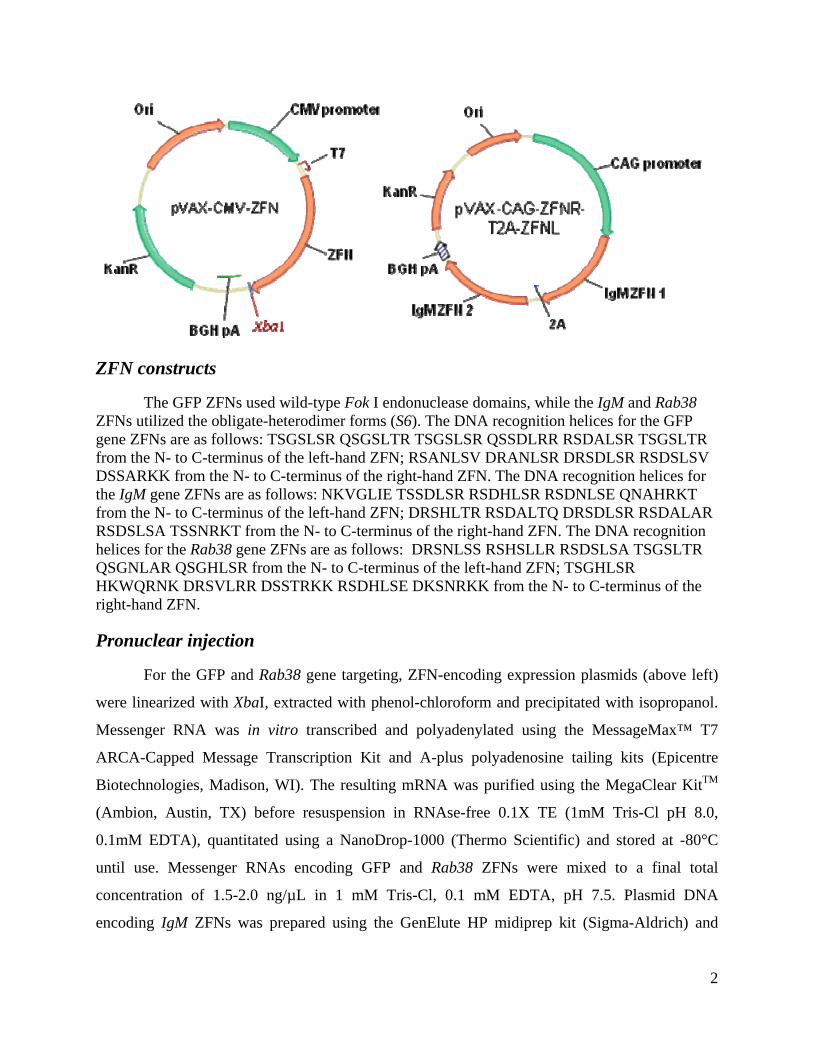
ZFN constructs
The GFP ZFNs used wild-type Fok I endonuclease domains, while the IgM and Rab38 ZFNs utilized the obligate-heterodimer forms (S6). The DNA recognition helices for the GFP gene ZFNs are as follows: TSGSLSR QSGSLTR TSGSLSR QSSDLRR RSDALSR TSGSLTR from the N- to C-terminus of the left-hand ZFN; RSANLSV DRANLSR DRSDLSR RSDSLSV DSSARKK from the N- to C-terminus of the right-hand ZFN. The DNA recognition helices for the IgM gene ZFNs are as follows: NKVGLIE TSSDLSR RSDHLSR RSDNLSE QNAHRKT from the N- to C-terminus of the left-hand ZFN; DRSHLTR RSDALTQ DRSDLSR RSDALAR RSDSLSA TSSNRKT from the N- to C-terminus of the right-hand ZFN. The DNA recognition helices for the Rab38 gene ZFNs are as follows: DRSNLSS RSHSLLR RSDSLSA TSGSLTR QSGNLAR QSGHLSR from the N- to C-terminus of the left-hand ZFN; TSGHLSR HKWQRNK DRSVLRR DSSTRKK RSDHLSE DKSNRKK from the N- to C-terminus of the right-hand ZFN.
Pronuclear injection
For the GFP and Rab38 gene targeting, ZFN-encoding expression plasmids (above left)
were linearized with XbaI, extracted with phenol-chloroform and precipitated with isopropanol.
Messenger RNA was in vitro transcribed and polyadenylated using the MessageMax™ T7
ARCA-Capped Message Transcription Kit and A-plus polyadenosine tailing kits (Epicentre
Biotechnologies, Madison, WI). The resulting mRNA was purified using the MegaClear KitTM
(Ambion, Austin, TX) before resuspension in RNAse-free 0.1X TE (1mM Tris-Cl pH 8.0,
0.1mM EDTA), quantitated using a NanoDrop-1000 (Thermo Scientific) and stored at -80°C
until use. Messenger RNAs encoding GFP and Rab38 ZFNs were mixed to a final total
concentration of 1.5-2.0 ng/µL in 1 mM Tris-Cl, 0.1 mM EDTA, pH 7.5. Plasmid DNA
encoding IgM ZFNs was prepared using the GenElute HP midiprep kit (Sigma-Aldrich) and

3
diluted for microinjection to either 10 ng/µL, 2ng/µL, or 0.4 ng/µL using 10 mM Tris-Cl, 0.1
mM EDTA, pH 7.5. Messenger RNAs encoding IgM ZFNs was prepared using the Ambion
mMessage mMachine kit (Ambion, Austin, TX) following the manufacturer protocol and diluted
at concentrations 0.4 ng/µL, 2 ng/µL, or 10 ng/µL with RNAseFree Water (Ambion) and stored
at -80°C until use. GFP and IgM ZFN mRNAs were kept on ice during all microinjection
procedures.
One goal of this study was to test the ability of ZFNs to induce mutations in a variety of
inbred and outbred rat strains. The GFP rat was previously produced on the SS (Dahl S) strain
background (S7), the SD (Sprague Dawley) rat was chosen to build the humanized monoclonal
antibody platform due to its robust breeding characteristics, and the FHH (Fawn Hooded
Hypertensive) rat is a model of hypertension where the Rab38 gene is thought to play a role in
end-stage renal disease (S8).
Inbred SS and FHH and outbred SD embryo pronuclear injections were performed at
different institutions. At the Medical College of Wisconsin, inbred strain animals are housed in
standard cages under approved animal care protocols in an American Association of Laboratory
Animal Care-approved facility. The rats are maintained on a 12 h light/dark cycle with ad libitum
access to food and water. Five to eight week old female SS female rats were injected with 20 IU
pregnant mare serum gonadotropin (PMSG) (Sigma-Aldrich or National Hormone and Peptide
Program) followed 48 hours later with 25 IU human chorionic gonadotropin (hCG) (Sigma-
Aldrich). Wild-type SS females were bred to homozygous SS-Tg(CAG-eGFP)1Mcwi males (7)
to generate hemizygous GFP embryos for microinjection. Wild-type FHH females, harboring the
natural knockout allele of Rab38 were superovulated for breeding to FHH.BN-Rab38 congenic
males harboring a wild-type copy of the Rab38 gene from the BN (Brown Norway) strain which
has been introgressed into the FHH inbred background (S8). The Rab38 ZFNs can target both
alleles equally. Embryos were microinjected with ZFNs into the pronucleus or cytoplasm using
an Eppendorf Microinjection system under standard conditions. Manipulated embryos were
transferred to pseudopregnant SD/Hsd female rats (Harlan Laboratories, Inc.) to be carried to
parturition.
At the Caliper Life Sciences (Xenogen Biosciences) facility, outbred SD/NTac strain
animals (Taconic) were housed in standard microisolator cages under approved animal care
protocols in animal facility that is accredited by the Association for the Assessment and

4
Accreditation for Laboratory Animal Care (AAALAC). The rats were maintained on a 14-10 h
light/dark cycle with ad libitum access to food and water. Four to five week old SD/Hsd female
rats were injected with 20-25 IU PMSG (Sigma-Aldrich, St. Louis, MO) followed 48 hours later
with 20-25 IU hCG (Sigma-Aldrich, St. Louis, MO) before breeding to outbred SD/Hsd males.
Fertilized 1-cell stage embryos were collected for subsequent IgM plasmid DNA microinjection.
Manipulated embryos were transferred to pseudopregnant SD/NTac female rats to be carried to
parturition.
At the INSERM UMR 643 Transgenic Rat Facility
(http://www.ifr26.nantes.inserm.fr/ITERT/transgenese-rat/), Sprague-Dawley (SD/Crl) outbred
strain animals (Charles River France, L’Arbresle, France) were housed in standard cages and
protocols were conducted in accordance with the guidelines for animal experiments of the
French Veterinary Services and were performed by officially authorized personnel in a certified
animal facility. The rats are maintained on a 12 h light/dark cycle with ad libitum access to food
and water. Females, 26-30 days old SD/Crl were injected with 30 IU pregnant mare serum
gonadotropin (PMSG, Intervet, France) and followed 48 hours later with 20 IU human chorionic
gonadotropin (hCG) (Intervet, France) before breeding. Fertilized 1-cell stage embryos were
collected for subsequent microinjection using previously published procedures (S9). The male
pronucleus or cytoplasm of one-cell stage embryos were microinjected with ZFN mRNA and
surviving embryos were implanted on the same day in the oviduct of pseudo-pregnant SD/Crl
females and allowed to develop to full term following described procedures (S9). The
microinjection pipettes for mRNA IgM ZFNs of pronuclear or cytoplasmic injections were
changed after injections of 20-30 embryos.
Interestingly, as is shown in Table 1, lower birth rates were observed after microinjection
of the highest doses of both plasmid DNA or mRNA encoding IgM ZFNs at10 ng/µL. While this
is suggestive of toxicity due to ZFN activity in the genome, this dosage of nucleic acid delivered
to an embryo is high by some laboratory standards (S9, S10), and could be due to cytotoxicity of
the nucleic acid itself and therefore unrelated to ZFN activity.
Analysis of genome editing at ZFN target sites
For genome editing of the GFP locus, tail biopsy DNA was amplified in a 32P-body-
labeled reaction using the primers eGF_out_F1 (5’-GTTGTGCTGTCTCATCATTTTGG-3’) and

5
eGFP_out_R4 (5’ ACATAGCGTAAAAGGAGCAACAT-3’) as previously described (S11).
Genotyping of F1 offspring from founder GFPm2 was performed on Proteinase K-extracted
DNA under standard PCR conditions using the primers GFP-F3 (5’-
CAGTGCTTCAGCCGCTACC-3’) and GFP-R5 (5’-TTGGGGTCTTTGCTCAGGGC-3’). For
IgM targeted genome editing on founder generation animals and F1 genotyping, tail biopsy
DNA from liveborn neonates was extracted following treatment with Proteinase K, and the
extracted DNA was PCR amplified using Accuprime DNA polymerase (Invitrogen, Calrsbad,
CA) and the primers GJC 153F (5’-GGAGGCAAGAAGATGGATTC-3’) and GJC 154R (5’-
GAATCGGCACATGCAGATCT-3’). For Rab38 genomic DNA was extracted amplified and
amplified in a similar manner using the primers Rab38-F4 (5’- GTAATCGGCGACCTAGGTG-
3’) and Rab38-R4 (5’-TCCATTCCCGGAACCTTCAC-3’). The amplified DNA for both
genomic targets was assayed for mutations using Surveyor nuclease (Transgenomic) as described
below (6). GFP, Rab38, and IgM PCR products were directly subcloned into the pCR4-TOPO
vector (Invitrogen, Carlsbad, CA), and plasmid DNA was prepared and sequenced using
standard methods.
Off-target site prediction
The prediction of off-target sites of ZFN cleavage is described in detail (S1, S2). Briefly,
an in vitro SELEX strategy involving binding of the zinc-finger domain to a randomized pool of
target sequences is used to identify a consensus binding site. Bioinformatics tools were used to
scan the most current assembly of the rat genome (RGSCv3.4) to identify putative off target
sequences based on this consensus. PCR primers were designed flanking the most likely off
target sites based on the number of nucleotide differences and these regions were amplified in
the founder animals and tested for ZFN cleavage using the Surveyor nuclease assay as described
below.
Surveyor nuclease assay
The Surveryor nuclease assay for detection of ZFN mediated genome editing is described
(S6). Briefly, 150-300 ng PCR product (5-15 µL) is transferred to a fresh tube, denatured and re-
annealed according to the following thermocycler program: (95º for 2 min, 95º to 85º -2ºC per
second, 85º to 25º -0.1º per second, 4º indefinitely). 0.5 µL of the Surveyor nuclease was added

6
and incubated at 42ºC for 20 minutes. After immediately placing the reactions on ice, 6X
Surveyor nuclease stop buffer and 0.25% Orange G (Sigma) is added and the sample is
electrophoresed on a 10% polyacrylamide gel (BioRad, Hercules, CA) at 10-15 V/cm. The
percentage of cleavage was estimated as previously described (S6). While Surveyor nuclease
digestion occasionally resulted in partial digestion, all Surveyor digestion reactions that were
quantitated proceeded to completion.

Fig. S1 – Surveyor nuclease (CEL-I) assay detection of targeted mutations in IgM and Rab38
IgM mutant foundersA8 4619 70 816 7 11
9
128
122
130
147
107
116
161
167
1.1
3.3
1.2
4.1
5.3
174
212
8.1
WT
C6
9.1
9.2
mRNA cytoplasm mRNA PNIDNA PNIDelivery Method:
300
400-500-
13.2
13.4
15.5
16.3
16.6
18.1
100-
200-
300-
Dose (ng/µL) 10 2 0.4 10 2 10 2 0.4
Rab38 founders 1-17
SS
*
B
100-
200-
300-
C
(-) WT
WT
+ pl
asm
id
6 7 8 19 46
Fig. S1. Surveyor nuclease (Cel-1) analysis of mutant founders for IgM and Rab38 ZFN injections. (A) A total of 295 founder animals were screened to identify 32 mutant founder animals. Animals are shown by delivery method (above gel) and dose
-1650
-1000
animals were screened to identify 32 mutant founder animals. Animals are shown by delivery method (above gel) and dose (below). A positive control from transfected C6 cells and negative control from unmodified wild-type DNA (WT) are shown at either end. (B) Seventeen founders born after Rab38 ZFN mRNA injections were screened using the Surveyor nuclease assay to reveal a putative mutation in founder 16 (asterisk). SS – control parental SS strain. (C) Of note, disruption of IgM after injection of ZFN-encoding plasmid is due to transient expression as no evidence for integration of the ZFN plasmid was detected by PCR for plasmid sequences among five mutant founders injected with the highest dose of plasmid (10ng/µL) (see Table 1). WT control DNA spiked with 1 copy per genome of the injected plasmid was used as a positive control. (-) – no template.

Fig. S2 - GFP ZFN off target analysis
Fig. S2. Twelve off-target sites were identified as described in the materials and methods (Table). Bases differing from the consensus target sequence are shown as lowercase. The Surveyor nuclease detection method was used to screen these predicted off-target sites (see methods) in the five pups shown in Fig. 1B. In all cases we could not detect any evidence of new bands of the predicted sizes appearing in the test samples compared to wild-type SS parental strain control (C). All samples were treated with the Surveyor nuclease. Lanes 1-3 are GFP-expressing founders and lanes 4 and 5 correspond to GFP-null mutant founders GFPm1 and GFPm2, respectively. Primer sequences are found in Table S2.

Site# (ranked) Chromosome Location Sequence Mismatches
Homodimer (+)/ Heterodimer (-) Gene?
1 chr8 92436990 AGtcAGCttCCTGTCTAGAAGAGAAcTgGGTGtCtATGGGCC 8
Fig. S3 – IgM ZFN off target analysis
1 chr8 92436990 AGtcAGCttCCTGTCTAGAAGAGAAcTgGGTGtCtATGGGCC 8 -
2 chr1 170756822 CaAatGCCaCCTGTCTGAATGGttTaTGcTGGCaATGGGCT 9 -
3 chr11 36783404 GGtGAGaCCCCTGTCTTAACAAAAgaTGGgGGggtTGGGaA 9 -
4 chr11 66707758 GatCCAaGGCCACCAAcTgGAGTTTAAGACAaaGGGCTCTgC 8 -
5 chr2 40914750 TGtCCATGGCCtCCtccTcTTTGCTAGAgcGGtGGCTCTCA 9 - Pde4d
6 chr3 141510596 GGAttGCCCCCTGTCaGTCACAGcATaTGGTGGCCATaGatG 8 - LOC499913
7 chr4 35317164 GGAGAagCCCaTGTgTACTCTTtAgTTGGTGGCtcTGGGaG 9 -
8 chr6 103216965 GcCCataGGCCAaCAAcTcTCAGGCTAGACAacGGGCTCTCA 9 - Actn1
WT C6 6 7 8 19 46 WT C6 6 7 8 19 46 WT C6 6 7 8 19 46 500 -
300 -
Site 1 Site 2 Site 3
Site 1
300
100 -
* * *
WT C6 6 7 8 19 46 WT C6 6 7 8 19 46 WT C6 6 7 8 19 46 500 -
300 -
100 -
Site 4 Site 5 Site 6#
WT C6 6 7 8 19 46 WT C6 6 7 8 19 46
500 -300 -
Site 7 Site 8
Fig. S3. Eight off-target sites were identified as described in the materials and methods (Table). Bases differing from the consensustarget sequence are shown as lowercase. The Surveyor nuclease detection method was used to screen these predicted off-targetsites (see methods) in five mutant founders. Site 1, which shared the most sequence similarity to the consensus target site,
100 -
demonstrated cleavage at approximately 3% of chromosomes in 3 out of 5 founders (*). This cleavage was due to an A→G singlenucleotide polymorphism (SNP) found in the heterogeneous SD rat strain seven base pairs upstream of Site 1 in these 3 foundersby sequencing this region in all 5 animals (right panel). Since this SNP would cause heteroduplex formation, the observed cleavageis not due to off target ZFN activity, further supported by the sequence traces which show no mutation in Site 1 in any of thesefounders. Site 5 in founder 7 demonstrated Cel-1 cleavage, but the resulting fragments were not of the correct size to be the resultof ZFN activity (#) and is likely due to a SNP. In all samples, in all other cases we could not detect any evidence of new bands of thepredicted sizes appearing in the test samples, including tranfected cells (C6), compared to wild-type (WT) control DNA. Primersequences are found in Table S2.

Fig. S4 – Germline transmission of GFP and IgM mutant alleles
A
BC6 6 19 46
Founder
CEL-I
Pup: 295
296
297
229
(F1)
No
tem
p.
300
301
302
298
299
306
307
304
305
303
WT-Δ64-
C
- 350
Female Male
- 200
Fig. S4. Germline transmission of ZFN induced mutations. (A) Founder GFPm2 was bred to a wild-type SS female and 4 of 13 offspring inherited the mutant GFP sequence, but none inherited the wild-type GFP sequence as detected by genotyping PCR (see methods). SS-GFP – positive control from SS/Tg(CAG-eGFP)1Mcwi strain. (B) Three founders transmitted mutant IgM alleles to their offspring after backcross to wild-type (WT) or intercross as determined by PCR (founder #19) or CEL-I (founders #6, #46) (see methods). F1 animal #251 inherited the mutant allele from both founders 6 ( ) ( , ) ( )and 19. The inheritance of both alleles causes a subtle, but detectable mobility shift in the upper band (arrow) due to the 9-bp deletion transmitted by founder #6 (All genotypes were confirmed by Sanger sequencing). The lower migrating minor bands are cleavage products from the Surveyor nuclease. C6 – positive control from transfected C6 cells; WT – control wild-type DNA from the SD parental strain. (C) PCR genotyping 13 offspring from an intercross of F1 heterozygotesdescendent of founder #19 reveals six male and female homozygous offspring carrying only the ∆64 allele, two wild-type (WT) and five heterozygotes.

Fig. S5 – Pathway to ZFN validation in yeast and cultured rat C6 cells
A
B
7.3%NHEJ 3.6 11.1
Fig. S5. Pathway to validation of GFP-targeted ZFNs. Methods are described and referenced in the supplemental methods. (A) Three out of seventeen pairs of designed ZFNs demonstrated significant activity in a yeast reporter assay compared to untransformed control. (B) These same pairs demonstrate activity in cultured rat C6 cells as determined using the Surveyor nuclease (CEL-I) assay as described in the methods. GFP mRNA was used as a negative control. Pair 15 was chosen for all subsequent rat experiments as described in the text and methods. ZFN reagents for the other genomic targets were validated in a similar fashion.
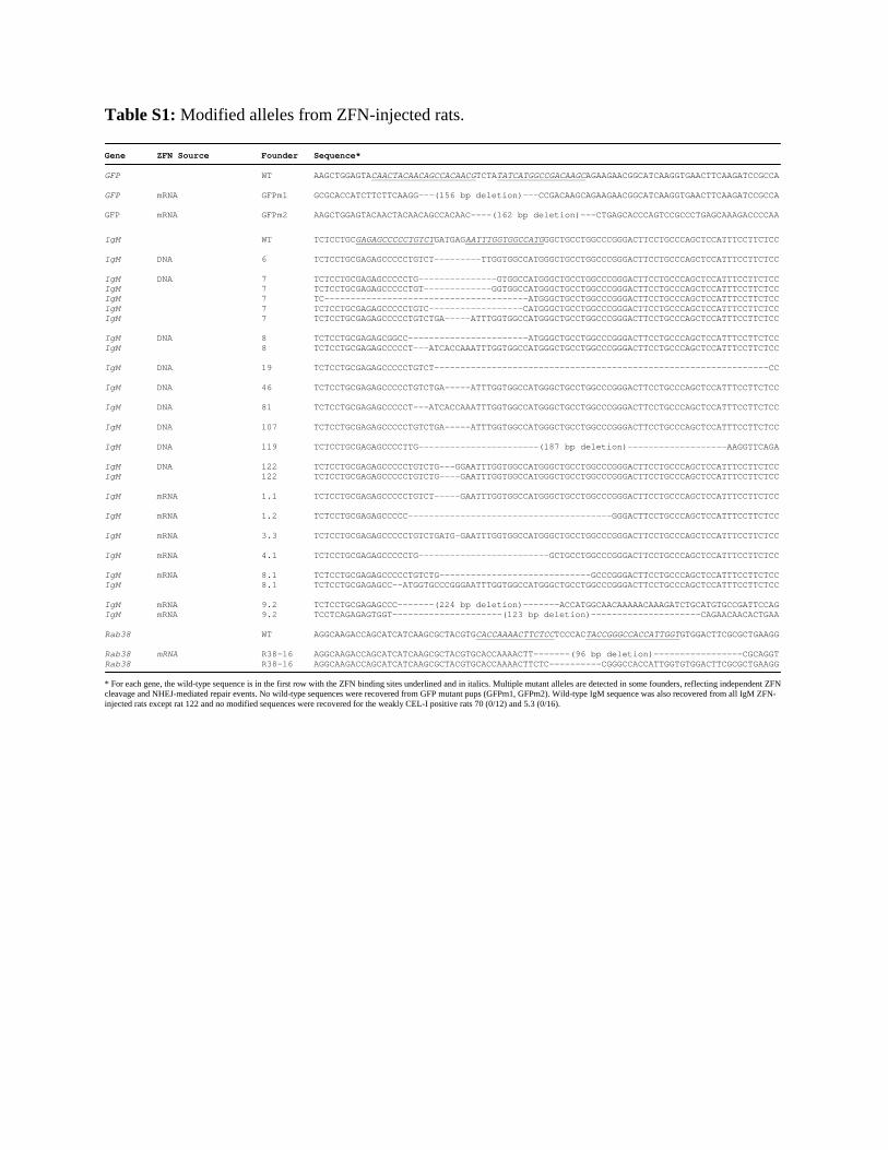
Table S1: Modified alleles from ZFN-injected rats. Gene ZFN Source Founder Sequence* GFP WT AAGCTGGAGTACAACTACAACAGCCACAACGTCTATATCATGGCCGACAAGCAGAAGAACGGCATCAAGGTGAACTTCAAGATCCGCCA GFP mRNA GFPm1 GCGCACCATCTTCTTCAAGG---(156 bp deletion)---CCGACAAGCAGAAGAACGGCATCAAGGTGAACTTCAAGATCCGCCA GFP mRNA GFPm2 AAGCTGGAGTACAACTACAACAGCCACAAC----(162 bp deletion)---CTGAGCACCCAGTCCGCCCTGAGCAAAGACCCCAA
IgM WT TCTCCTGCGAGAGCCCCCTGTCTGATGAGAATTTGGTGGCCATGGGCTGCCTGGCCCGGGACTTCCTGCCCAGCTCCATTTCCTTCTCC IgM DNA 6 TCTCCTGCGAGAGCCCCCTGTCT---------TTGGTGGCCATGGGCTGCCTGGCCCGGGACTTCCTGCCCAGCTCCATTTCCTTCTCC IgM DNA 7 TCTCCTGCGAGAGCCCCCTG---------------GTGGCCATGGGCTGCCTGGCCCGGGACTTCCTGCCCAGCTCCATTTCCTTCTCC IgM 7 TCTCCTGCGAGAGCCCCCTGT-------------GGTGGCCATGGGCTGCCTGGCCCGGGACTTCCTGCCCAGCTCCATTTCCTTCTCC IgM 7 TC---------------------------------------ATGGGCTGCCTGGCCCGGGACTTCCTGCCCAGCTCCATTTCCTTCTCC IgM 7 TCTCCTGCGAGAGCCCCCTGTC------------------CATGGGCTGCCTGGCCCGGGACTTCCTGCCCAGCTCCATTTCCTTCTCC IgM 7 TCTCCTGCGAGAGCCCCCTGTCTGA-----ATTTGGTGGCCATGGGCTGCCTGGCCCGGGACTTCCTGCCCAGCTCCATTTCCTTCTCC IgM DNA 8 TCTCCTGCGAGAGCGGCC-----------------------ATGGGCTGCCTGGCCCGGGACTTCCTGCCCAGCTCCATTTCCTTCTCC IgM 8 TCTCCTGCGAGAGCCCCCT---ATCACCAAATTTGGTGGCCATGGGCTGCCTGGCCCGGGACTTCCTGCCCAGCTCCATTTCCTTCTCC IgM DNA 19 TCTCCTGCGAGAGCCCCCTGTCT----------------------------------------------------------------CC IgM DNA 46 TCTCCTGCGAGAGCCCCCTGTCTGA-----ATTTGGTGGCCATGGGCTGCCTGGCCCGGGACTTCCTGCCCAGCTCCATTTCCTTCTCC IgM DNA 81 TCTCCTGCGAGAGCCCCCT---ATCACCAAATTTGGTGGCCATGGGCTGCCTGGCCCGGGACTTCCTGCCCAGCTCCATTTCCTTCTCC IgM DNA 107 TCTCCTGCGAGAGCCCCCTGTCTGA-----ATTTGGTGGCCATGGGCTGCCTGGCCCGGGACTTCCTGCCCAGCTCCATTTCCTTCTCC IgM DNA 119 TCTCCTGCGAGAGCCCCTTG-----------------------(187 bp deletion)-------------------AAGGTTCAGA IgM DNA 122 TCTCCTGCGAGAGCCCCCTGTCTG---GGAATTTGGTGGCCATGGGCTGCCTGGCCCGGGACTTCCTGCCCAGCTCCATTTCCTTCTCC IgM 122 TCTCCTGCGAGAGCCCCCTGTCTG----GAATTTGGTGGCCATGGGCTGCCTGGCCCGGGACTTCCTGCCCAGCTCCATTTCCTTCTCC IgM mRNA 1.1 TCTCCTGCGAGAGCCCCCTGTCT-----GAATTTGGTGGCCATGGGCTGCCTGGCCCGGGACTTCCTGCCCAGCTCCATTTCCTTCTCC IgM mRNA 1.2 TCTCCTGCGAGAGCCCCC---------------------------------------GGGACTTCCTGCCCAGCTCCATTTCCTTCTCC IgM mRNA 3.3 TCTCCTGCGAGAGCCCCCTGTCTGATG–GAATTTGGTGGCCATGGGCTGCCTGGCCCGGGACTTCCTGCCCAGCTCCATTTCCTTCTCC IgM mRNA 4.1 TCTCCTGCGAGAGCCCCCTG-------------------------GCTGCCTGGCCCGGGACTTCCTGCCCAGCTCCATTTCCTTCTCC IgM mRNA 8.1 TCTCCTGCGAGAGCCCCCTGTCTG-----------------------------GCCCGGGACTTCCTGCCCAGCTCCATTTCCTTCTCC IgM 8.1 TCTCCTGCGAGAGCC—-ATGGTGCCCGGGAATTTGGTGGCCATGGGCTGCCTGGCCCGGGACTTCCTGCCCAGCTCCATTTCCTTCTCC IgM mRNA 9.2 TCTCCTGCGAGAGCCC-------(224 bp deletion)-------ACCATGGCAACAAAAACAAAGATCTGCATGTGCCGATTCCAG IgM mRNA 9.2 TCCTCAGAGAGTGGT---------------------(123 bp deletion)---------------------CAGAACAACACTGAA Rab38 WT AGGCAAGACCAGCATCATCAAGCGCTACGTGCACCAAAACTTCTCCTCCCACTACCGGGCCACCATTGGTGTGGACTTCGCGCTGAAGG Rab38 mRNA R38-16 AGGCAAGACCAGCATCATCAAGCGCTACGTGCACCAAAACTT-------(96 bp deletion)-----------------CGCAGGT Rab38 R38-16 AGGCAAGACCAGCATCATCAAGCGCTACGTGCACCAAAACTTCTC----------CGGGCCACCATTGGTGTGGACTTCGCGCTGAAGG * For each gene, the wild-type sequence is in the first row with the ZFN binding sites underlined and in italics. Multiple mutant alleles are detected in some founders, reflecting independent ZFN cleavage and NHEJ-mediated repair events. No wild-type sequences were recovered from GFP mutant pups (GFPm1, GFPm2). Wild-type IgM sequence was also recovered from all IgM ZFN-injected rats except rat 122 and no modified sequences were recovered for the weakly CEL-I positive rats 70 (0/12) and 5.3 (0/16).

Table S2: Primer sequences for off-target analysis.
Name SEQUENCE (5'-3')eGFP-OT1_F GCCCAAGTCTCCTTTACTGCeGFP-OT1_R TGGGTTTATTGTGTGCCCTAeGFP-OT2_F TGGTCCTAGGTCCTTTGGAAeGFP-OT2_R TCTGCATGCTGACCAGTCTCeGFP-OT3_F CCACTCATGCACATCGAAAGeGFP-OT3_R CACTTGATATAAGCTGGGGTCACeGFP-OT4_F TAAAATCCACGCAGCCAAAGeGFP-OT4_R GGTGATGATGCCATGAGATGeGFP-OT5_F CCAGCTAGGAGAAAGCAAGAeGFP-OT5_R AACTGCTGCCCTACCTCTCCeGFP-OT6_F AACAAGGCCATCCTCTGCTAeGFP-OT6_R AACCAAAGTGCATAGGCTTCAeGFP-OT7_F ATGGTTCCGTGCAACAATTTeGFP-OT7_R TTTGCTCATTAGAGCCCTTCAeGFP-OT8_F GGTCACTTGGAAGCTCTTGGeGFP-OT8_R TATTGAAGGCCTGGGTTGACeGFP-OT9_F CCCTGATGACAGAGCAAAGGeGFP-OT9_R CTCTTGGGAAAATGCCCTTAeGFP-OT10_F GGTTAGCATTGTGGCCTGTTeGFP-OT10_R TGATCATTGGCTTTGAGCAGeGFP-OT11_F GCTCAGGAATTTGCCTCTCAeGFP-OT11_R CAAGCACAAAGACCTAAACTTGTeGFP-OT12_F TTTGCTGGAAAGCGTAGAGACeGFP-OT12_R CCTCTCCAGTTTCCTCCACTCeGFP-OTC_F TCGTGACCACCCTGACCTACeGFP-OTC_R GAACTCCAGCAGGACCATGTIgM-OT#1 F GGTCAATCAGTAGGAAGTTTIgM-OT#1 R GCTTCTCCAGCCTAAAAGCTIgM-OT#2 F GGTGTAGTTACATTGTTGTTGCIgM-OT#2 R TTAATAAACGGCCAGGAACCAIgM-OT#3 F AGGGAGAAGAGTGTCACTGIgM-OT#3 R CTGGAGACTAGAAGTCCAAGIgM-OT#4 F TGAGAGTGTGTAGACTCACAIgM-OT#4 R CAAAGTTTTCTAGGGAAGTTCCIgM-OT#5 F CAGCATTCTCCTAATTTTCACAIgM-OT#5 R GCTACTCAGTCTGTGGGTTGIgM-OT#6 F GATAGTGAAGACACAGGTGAGIgM-OT#6 R GTTAGTTTTACATCATGCACCCIgM-OT#7 F CTCAACTCGTTCTTCTTATTCAGIgM-OT#7 R CATCTCTTATGTAGAGACACCIgM-OT#8 F GTCTCTTGGGATAAAAGACACIgM-OT#8 R CATGCCGTCTTCTCTTGCTT

Supplemental References:
S1. Y. Doyon et al., Nat Biotechnol 26, 702 (Jun, 2008).
S2. E. E. Perez et al., Nat Biotechnol 26, 808 (Jul, 2008).
S3. M. Isalan, Y. Choo, Methods Enzymol 340, 593 (2001).
S4. Y. Santiago et al., Proc Natl Acad Sci U S A 105, 5809 (Apr 15, 2008).
S5. L. Zhang et al., J Biol Chem 275, 33850 (2000).
S6. J. C. Miller et al., Nat Biotechnol 25, 778 (Jul, 2007).
S7. M. Michalkiewicz et al., Am J Physiol Heart Circ Physiol 293, H881 (Jul, 2007).
S8. A. Rangel-Filho et al., J Am Soc Nephrol 16, 852 (Apr, 2005).
S9. L. Tesson et al., Transgenic Res 14, 531 (Oct, 2005).
S10. W. E. Filipiak, T. L. Saunders, Transgenic Res 15, 673 (December 2006, 2006).
S11. E. Bitoun, K. E. Davies, Cerebellum 4, 250 (2005).

www.sciencemag.org/cgi/content/full/325/5939/433/DC1
Supporting Online Material for
Knockout Rats via Embryo Microinjection of Zinc-Finger Nucleases
Aron M. Geurts, Gregory J. Cost, Yevgeniy Freyvert, Bryan Zeitler, Jeffrey C. Miller, Vivian M. Choi, Shirin S. Jenkins, Adam Wood, Xiaoxia Cui, Xiangdong Meng, Anna Vincent, Stephen Lam, Mieczyslaw Michalkiewicz, Rebecca Schilling, Jamie Foeckler,
Shawn Kalloway, Hartmut Weiler, Séverine Ménoret, Ignacio Anegon, Gregory D. Davis, Lei Zhang, Edward J. Rebar, Philip D. Gregory, Fyodor D. Urnov, Howard J.
Jacob,* Roland Buelow*
*To whom correspondence should be addressed. E-mail: [email protected] (H.J.J.);
[email protected] (R.B.)
Published 24 July 2009, Science 325, 433 (2009) DOI: 10.1126/science.1172447
This PDF file includes:
Materials and Methods Figs. S1 to S5 Tables S1 and S2 References

1
Materials and Methods
Design, assembly, in vitro and in vivo validation of zinc-finger nucleases
The ZFNs were designed, assembled, and validated using strategies and procedures
described elsewhere (S1, S2); minor modifications to the procedures are detailed below.
ZFN design made use of an archive of pre-validated 2-finger and 1-finger modules (S1,
S3-5). The target region of eGFP, IgM, and Rab38 were scanned for positions where modules
exist in the archive that allow the fusion of 2 or 3 such modules to generate a 4, 5 or 6 finger
protein to recognize a 12-18 bp site on the top (“Watson”) strand, and the fusion of 2-3 different
modules to recognize on the bottom (“Crick”) strand of a 12-18 bp site that lies 5 or 6 bp away.
The ZFNs were assembled using a PCR-based procedure and cloned into yeast
expression vectors as described elsewhere (S1). Screening of ZFNs in the yeast-based proxy
system (Fig. S5A) was done as described (S1), with one modification: the target regions of all
ZFN pairs from a given gene were extracted and concatenated to yield a composite DNA stretch
that was then used to generate a disrupted reporter construct. Final candidate ZFN pairs were
subcloned into a CMV expression plasmid for testing in cultured rat cells (below, left).
Screening of ZFNs for gene disruption activity at the eGFP transgene, and at the IgM and
Rab38 loci was done by transfection of ZFN expression constructs into rat C6 cells (ATCC)
using an Amaxa nucleofector according to the manufacturer’s instructions. Measurements of
ZFN gene disruption activity were performed using the Surveyor endonuclease (Cel-1) assay
exactly as described previously (S6) and below (Fig. S5B). The DNA recognition helices for the
most active ZFN pairs that were used in all subsequent rat experiments are given below. The IgM
ZFNs were subcloned into an expression plasmid as two open reading frames linked by the T2A
peptide sequence as previously described (S1) (below, right) for the rat embryo plasmid DNA
injections described below.

2
ZFN constructs
The GFP ZFNs used wild-type Fok I endonuclease domains, while the IgM and Rab38 ZFNs utilized the obligate-heterodimer forms (S6). The DNA recognition helices for the GFP gene ZFNs are as follows: TSGSLSR QSGSLTR TSGSLSR QSSDLRR RSDALSR TSGSLTR from the N- to C-terminus of the left-hand ZFN; RSANLSV DRANLSR DRSDLSR RSDSLSV DSSARKK from the N- to C-terminus of the right-hand ZFN. The DNA recognition helices for the IgM gene ZFNs are as follows: NKVGLIE TSSDLSR RSDHLSR RSDNLSE QNAHRKT from the N- to C-terminus of the left-hand ZFN; DRSHLTR RSDALTQ DRSDLSR RSDALAR RSDSLSA TSSNRKT from the N- to C-terminus of the right-hand ZFN. The DNA recognition helices for the Rab38 gene ZFNs are as follows: DRSNLSS RSHSLLR RSDSLSA TSGSLTR QSGNLAR QSGHLSR from the N- to C-terminus of the left-hand ZFN; TSGHLSR HKWQRNK DRSVLRR DSSTRKK RSDHLSE DKSNRKK from the N- to C-terminus of the right-hand ZFN.
Pronuclear injection
For the GFP and Rab38 gene targeting, ZFN-encoding expression plasmids (above left)
were linearized with XbaI, extracted with phenol-chloroform and precipitated with isopropanol.
Messenger RNA was in vitro transcribed and polyadenylated using the MessageMax™ T7
ARCA-Capped Message Transcription Kit and A-plus polyadenosine tailing kits (Epicentre
Biotechnologies, Madison, WI). The resulting mRNA was purified using the MegaClear KitTM
(Ambion, Austin, TX) before resuspension in RNAse-free 0.1X TE (1mM Tris-Cl pH 8.0,
0.1mM EDTA), quantitated using a NanoDrop-1000 (Thermo Scientific) and stored at -80°C
until use. Messenger RNAs encoding GFP and Rab38 ZFNs were mixed to a final total
concentration of 1.5-2.0 ng/µL in 1 mM Tris-Cl, 0.1 mM EDTA, pH 7.5. Plasmid DNA
encoding IgM ZFNs was prepared using the GenElute HP midiprep kit (Sigma-Aldrich) and

3
diluted for microinjection to either 10 ng/µL, 2ng/µL, or 0.4 ng/µL using 10 mM Tris-Cl, 0.1
mM EDTA, pH 7.5. Messenger RNAs encoding IgM ZFNs was prepared using the Ambion
mMessage mMachine kit (Ambion, Austin, TX) following the manufacturer protocol and diluted
at concentrations 0.4 ng/µL, 2 ng/µL, or 10 ng/µL with RNAseFree Water (Ambion) and stored
at -80°C until use. GFP and IgM ZFN mRNAs were kept on ice during all microinjection
procedures.
One goal of this study was to test the ability of ZFNs to induce mutations in a variety of
inbred and outbred rat strains. The GFP rat was previously produced on the SS (Dahl S) strain
background (S7), the SD (Sprague Dawley) rat was chosen to build the humanized monoclonal
antibody platform due to its robust breeding characteristics, and the FHH (Fawn Hooded
Hypertensive) rat is a model of hypertension where the Rab38 gene is thought to play a role in
end-stage renal disease (S8).
Inbred SS and FHH and outbred SD embryo pronuclear injections were performed at
different institutions. At the Medical College of Wisconsin, inbred strain animals are housed in
standard cages under approved animal care protocols in an American Association of Laboratory
Animal Care-approved facility. The rats are maintained on a 12 h light/dark cycle with ad libitum
access to food and water. Five to eight week old female SS female rats were injected with 20 IU
pregnant mare serum gonadotropin (PMSG) (Sigma-Aldrich or National Hormone and Peptide
Program) followed 48 hours later with 25 IU human chorionic gonadotropin (hCG) (Sigma-
Aldrich). Wild-type SS females were bred to homozygous SS-Tg(CAG-eGFP)1Mcwi males (7)
to generate hemizygous GFP embryos for microinjection. Wild-type FHH females, harboring the
natural knockout allele of Rab38 were superovulated for breeding to FHH.BN-Rab38 congenic
males harboring a wild-type copy of the Rab38 gene from the BN (Brown Norway) strain which
has been introgressed into the FHH inbred background (S8). The Rab38 ZFNs can target both
alleles equally. Embryos were microinjected with ZFNs into the pronucleus or cytoplasm using
an Eppendorf Microinjection system under standard conditions. Manipulated embryos were
transferred to pseudopregnant SD/Hsd female rats (Harlan Laboratories, Inc.) to be carried to
parturition.
At the Caliper Life Sciences (Xenogen Biosciences) facility, outbred SD/NTac strain
animals (Taconic) were housed in standard microisolator cages under approved animal care
protocols in animal facility that is accredited by the Association for the Assessment and

4
Accreditation for Laboratory Animal Care (AAALAC). The rats were maintained on a 14-10 h
light/dark cycle with ad libitum access to food and water. Four to five week old SD/Hsd female
rats were injected with 20-25 IU PMSG (Sigma-Aldrich, St. Louis, MO) followed 48 hours later
with 20-25 IU hCG (Sigma-Aldrich, St. Louis, MO) before breeding to outbred SD/Hsd males.
Fertilized 1-cell stage embryos were collected for subsequent IgM plasmid DNA microinjection.
Manipulated embryos were transferred to pseudopregnant SD/NTac female rats to be carried to
parturition.
At the INSERM UMR 643 Transgenic Rat Facility
(http://www.ifr26.nantes.inserm.fr/ITERT/transgenese-rat/), Sprague-Dawley (SD/Crl) outbred
strain animals (Charles River France, L’Arbresle, France) were housed in standard cages and
protocols were conducted in accordance with the guidelines for animal experiments of the
French Veterinary Services and were performed by officially authorized personnel in a certified
animal facility. The rats are maintained on a 12 h light/dark cycle with ad libitum access to food
and water. Females, 26-30 days old SD/Crl were injected with 30 IU pregnant mare serum
gonadotropin (PMSG, Intervet, France) and followed 48 hours later with 20 IU human chorionic
gonadotropin (hCG) (Intervet, France) before breeding. Fertilized 1-cell stage embryos were
collected for subsequent microinjection using previously published procedures (S9). The male
pronucleus or cytoplasm of one-cell stage embryos were microinjected with ZFN mRNA and
surviving embryos were implanted on the same day in the oviduct of pseudo-pregnant SD/Crl
females and allowed to develop to full term following described procedures (S9). The
microinjection pipettes for mRNA IgM ZFNs of pronuclear or cytoplasmic injections were
changed after injections of 20-30 embryos.
Interestingly, as is shown in Table 1, lower birth rates were observed after microinjection
of the highest doses of both plasmid DNA or mRNA encoding IgM ZFNs at10 ng/µL. While this
is suggestive of toxicity due to ZFN activity in the genome, this dosage of nucleic acid delivered
to an embryo is high by some laboratory standards (S9, S10), and could be due to cytotoxicity of
the nucleic acid itself and therefore unrelated to ZFN activity.
Analysis of genome editing at ZFN target sites
For genome editing of the GFP locus, tail biopsy DNA was amplified in a 32P-body-
labeled reaction using the primers eGF_out_F1 (5’-GTTGTGCTGTCTCATCATTTTGG-3’) and

5
eGFP_out_R4 (5’ ACATAGCGTAAAAGGAGCAACAT-3’) as previously described (S11).
Genotyping of F1 offspring from founder GFPm2 was performed on Proteinase K-extracted
DNA under standard PCR conditions using the primers GFP-F3 (5’-
CAGTGCTTCAGCCGCTACC-3’) and GFP-R5 (5’-TTGGGGTCTTTGCTCAGGGC-3’). For
IgM targeted genome editing on founder generation animals and F1 genotyping, tail biopsy
DNA from liveborn neonates was extracted following treatment with Proteinase K, and the
extracted DNA was PCR amplified using Accuprime DNA polymerase (Invitrogen, Calrsbad,
CA) and the primers GJC 153F (5’-GGAGGCAAGAAGATGGATTC-3’) and GJC 154R (5’-
GAATCGGCACATGCAGATCT-3’). For Rab38 genomic DNA was extracted amplified and
amplified in a similar manner using the primers Rab38-F4 (5’- GTAATCGGCGACCTAGGTG-
3’) and Rab38-R4 (5’-TCCATTCCCGGAACCTTCAC-3’). The amplified DNA for both
genomic targets was assayed for mutations using Surveyor nuclease (Transgenomic) as described
below (6). GFP, Rab38, and IgM PCR products were directly subcloned into the pCR4-TOPO
vector (Invitrogen, Carlsbad, CA), and plasmid DNA was prepared and sequenced using
standard methods.
Off-target site prediction
The prediction of off-target sites of ZFN cleavage is described in detail (S1, S2). Briefly,
an in vitro SELEX strategy involving binding of the zinc-finger domain to a randomized pool of
target sequences is used to identify a consensus binding site. Bioinformatics tools were used to
scan the most current assembly of the rat genome (RGSCv3.4) to identify putative off target
sequences based on this consensus. PCR primers were designed flanking the most likely off
target sites based on the number of nucleotide differences and these regions were amplified in
the founder animals and tested for ZFN cleavage using the Surveyor nuclease assay as described
below.
Surveyor nuclease assay
The Surveryor nuclease assay for detection of ZFN mediated genome editing is described
(S6). Briefly, 150-300 ng PCR product (5-15 µL) is transferred to a fresh tube, denatured and re-
annealed according to the following thermocycler program: (95º for 2 min, 95º to 85º -2ºC per
second, 85º to 25º -0.1º per second, 4º indefinitely). 0.5 µL of the Surveyor nuclease was added

6
and incubated at 42ºC for 20 minutes. After immediately placing the reactions on ice, 6X
Surveyor nuclease stop buffer and 0.25% Orange G (Sigma) is added and the sample is
electrophoresed on a 10% polyacrylamide gel (BioRad, Hercules, CA) at 10-15 V/cm. The
percentage of cleavage was estimated as previously described (S6). While Surveyor nuclease
digestion occasionally resulted in partial digestion, all Surveyor digestion reactions that were
quantitated proceeded to completion.
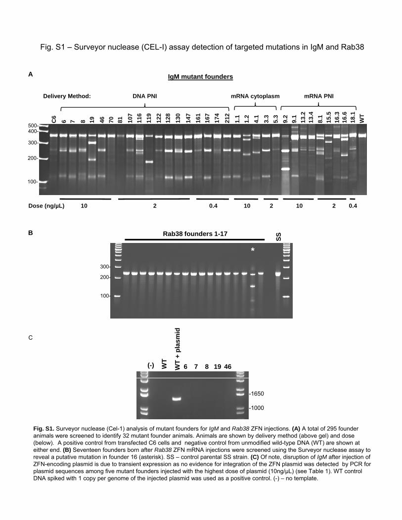
Fig. S1 – Surveyor nuclease (CEL-I) assay detection of targeted mutations in IgM and Rab38
IgM mutant foundersA8 4619 70 816 7 11
9
128
122
130
147
107
116
161
167
1.1
3.3
1.2
4.1
5.3
174
212
8.1
WT
C6
9.1
9.2
mRNA cytoplasm mRNA PNIDNA PNIDelivery Method:
300
400-500-
13.2
13.4
15.5
16.3
16.6
18.1
100-
200-
300-
Dose (ng/µL) 10 2 0.4 10 2 10 2 0.4
Rab38 founders 1-17
SS
*
B
100-
200-
300-
C
(-) WT
WT
+ pl
asm
id
6 7 8 19 46
Fig. S1. Surveyor nuclease (Cel-1) analysis of mutant founders for IgM and Rab38 ZFN injections. (A) A total of 295 founder animals were screened to identify 32 mutant founder animals. Animals are shown by delivery method (above gel) and dose
-1650
-1000
animals were screened to identify 32 mutant founder animals. Animals are shown by delivery method (above gel) and dose (below). A positive control from transfected C6 cells and negative control from unmodified wild-type DNA (WT) are shown at either end. (B) Seventeen founders born after Rab38 ZFN mRNA injections were screened using the Surveyor nuclease assay to reveal a putative mutation in founder 16 (asterisk). SS – control parental SS strain. (C) Of note, disruption of IgM after injection of ZFN-encoding plasmid is due to transient expression as no evidence for integration of the ZFN plasmid was detected by PCR for plasmid sequences among five mutant founders injected with the highest dose of plasmid (10ng/µL) (see Table 1). WT control DNA spiked with 1 copy per genome of the injected plasmid was used as a positive control. (-) – no template.

Fig. S2 - GFP ZFN off target analysis
Fig. S2. Twelve off-target sites were identified as described in the materials and methods (Table). Bases differing from the consensus target sequence are shown as lowercase. The Surveyor nuclease detection method was used to screen these predicted off-target sites (see methods) in the five pups shown in Fig. 1B. In all cases we could not detect any evidence of new bands of the predicted sizes appearing in the test samples compared to wild-type SS parental strain control (C). All samples were treated with the Surveyor nuclease. Lanes 1-3 are GFP-expressing founders and lanes 4 and 5 correspond to GFP-null mutant founders GFPm1 and GFPm2, respectively. Primer sequences are found in Table S2.

Site# (ranked) Chromosome Location Sequence Mismatches
Homodimer (+)/ Heterodimer (-) Gene?
1 chr8 92436990 AGtcAGCttCCTGTCTAGAAGAGAAcTgGGTGtCtATGGGCC 8
Fig. S3 – IgM ZFN off target analysis
1 chr8 92436990 AGtcAGCttCCTGTCTAGAAGAGAAcTgGGTGtCtATGGGCC 8 -
2 chr1 170756822 CaAatGCCaCCTGTCTGAATGGttTaTGcTGGCaATGGGCT 9 -
3 chr11 36783404 GGtGAGaCCCCTGTCTTAACAAAAgaTGGgGGggtTGGGaA 9 -
4 chr11 66707758 GatCCAaGGCCACCAAcTgGAGTTTAAGACAaaGGGCTCTgC 8 -
5 chr2 40914750 TGtCCATGGCCtCCtccTcTTTGCTAGAgcGGtGGCTCTCA 9 - Pde4d
6 chr3 141510596 GGAttGCCCCCTGTCaGTCACAGcATaTGGTGGCCATaGatG 8 - LOC499913
7 chr4 35317164 GGAGAagCCCaTGTgTACTCTTtAgTTGGTGGCtcTGGGaG 9 -
8 chr6 103216965 GcCCataGGCCAaCAAcTcTCAGGCTAGACAacGGGCTCTCA 9 - Actn1
WT C6 6 7 8 19 46 WT C6 6 7 8 19 46 WT C6 6 7 8 19 46 500 -
300 -
Site 1 Site 2 Site 3
Site 1
300
100 -
* * *
WT C6 6 7 8 19 46 WT C6 6 7 8 19 46 WT C6 6 7 8 19 46 500 -
300 -
100 -
Site 4 Site 5 Site 6#
WT C6 6 7 8 19 46 WT C6 6 7 8 19 46
500 -300 -
Site 7 Site 8
Fig. S3. Eight off-target sites were identified as described in the materials and methods (Table). Bases differing from the consensustarget sequence are shown as lowercase. The Surveyor nuclease detection method was used to screen these predicted off-targetsites (see methods) in five mutant founders. Site 1, which shared the most sequence similarity to the consensus target site,
100 -
demonstrated cleavage at approximately 3% of chromosomes in 3 out of 5 founders (*). This cleavage was due to an A→G singlenucleotide polymorphism (SNP) found in the heterogeneous SD rat strain seven base pairs upstream of Site 1 in these 3 foundersby sequencing this region in all 5 animals (right panel). Since this SNP would cause heteroduplex formation, the observed cleavageis not due to off target ZFN activity, further supported by the sequence traces which show no mutation in Site 1 in any of thesefounders. Site 5 in founder 7 demonstrated Cel-1 cleavage, but the resulting fragments were not of the correct size to be the resultof ZFN activity (#) and is likely due to a SNP. In all samples, in all other cases we could not detect any evidence of new bands of thepredicted sizes appearing in the test samples, including tranfected cells (C6), compared to wild-type (WT) control DNA. Primersequences are found in Table S2.

Fig. S4 – Germline transmission of GFP and IgM mutant alleles
A
BC6 6 19 46
Founder
CEL-I
Pup: 295
296
297
229
(F1)
No
tem
p.
300
301
302
298
299
306
307
304
305
303
WT-Δ64-
C
- 350
Female Male
- 200
Fig. S4. Germline transmission of ZFN induced mutations. (A) Founder GFPm2 was bred to a wild-type SS female and 4 of 13 offspring inherited the mutant GFP sequence, but none inherited the wild-type GFP sequence as detected by genotyping PCR (see methods). SS-GFP – positive control from SS/Tg(CAG-eGFP)1Mcwi strain. (B) Three founders transmitted mutant IgM alleles to their offspring after backcross to wild-type (WT) or intercross as determined by PCR (founder #19) or CEL-I (founders #6, #46) (see methods). F1 animal #251 inherited the mutant allele from both founders 6 ( ) ( , ) ( )and 19. The inheritance of both alleles causes a subtle, but detectable mobility shift in the upper band (arrow) due to the 9-bp deletion transmitted by founder #6 (All genotypes were confirmed by Sanger sequencing). The lower migrating minor bands are cleavage products from the Surveyor nuclease. C6 – positive control from transfected C6 cells; WT – control wild-type DNA from the SD parental strain. (C) PCR genotyping 13 offspring from an intercross of F1 heterozygotesdescendent of founder #19 reveals six male and female homozygous offspring carrying only the ∆64 allele, two wild-type (WT) and five heterozygotes.

Fig. S5 – Pathway to ZFN validation in yeast and cultured rat C6 cells
A
B
7.3%NHEJ 3.6 11.1
Fig. S5. Pathway to validation of GFP-targeted ZFNs. Methods are described and referenced in the supplemental methods. (A) Three out of seventeen pairs of designed ZFNs demonstrated significant activity in a yeast reporter assay compared to untransformed control. (B) These same pairs demonstrate activity in cultured rat C6 cells as determined using the Surveyor nuclease (CEL-I) assay as described in the methods. GFP mRNA was used as a negative control. Pair 15 was chosen for all subsequent rat experiments as described in the text and methods. ZFN reagents for the other genomic targets were validated in a similar fashion.

Table S1: Modified alleles from ZFN-injected rats. Gene ZFN Source Founder Sequence* GFP WT AAGCTGGAGTACAACTACAACAGCCACAACGTCTATATCATGGCCGACAAGCAGAAGAACGGCATCAAGGTGAACTTCAAGATCCGCCA GFP mRNA GFPm1 GCGCACCATCTTCTTCAAGG---(156 bp deletion)---CCGACAAGCAGAAGAACGGCATCAAGGTGAACTTCAAGATCCGCCA GFP mRNA GFPm2 AAGCTGGAGTACAACTACAACAGCCACAAC----(162 bp deletion)---CTGAGCACCCAGTCCGCCCTGAGCAAAGACCCCAA
IgM WT TCTCCTGCGAGAGCCCCCTGTCTGATGAGAATTTGGTGGCCATGGGCTGCCTGGCCCGGGACTTCCTGCCCAGCTCCATTTCCTTCTCC IgM DNA 6 TCTCCTGCGAGAGCCCCCTGTCT---------TTGGTGGCCATGGGCTGCCTGGCCCGGGACTTCCTGCCCAGCTCCATTTCCTTCTCC IgM DNA 7 TCTCCTGCGAGAGCCCCCTG---------------GTGGCCATGGGCTGCCTGGCCCGGGACTTCCTGCCCAGCTCCATTTCCTTCTCC IgM 7 TCTCCTGCGAGAGCCCCCTGT-------------GGTGGCCATGGGCTGCCTGGCCCGGGACTTCCTGCCCAGCTCCATTTCCTTCTCC IgM 7 TC---------------------------------------ATGGGCTGCCTGGCCCGGGACTTCCTGCCCAGCTCCATTTCCTTCTCC IgM 7 TCTCCTGCGAGAGCCCCCTGTC------------------CATGGGCTGCCTGGCCCGGGACTTCCTGCCCAGCTCCATTTCCTTCTCC IgM 7 TCTCCTGCGAGAGCCCCCTGTCTGA-----ATTTGGTGGCCATGGGCTGCCTGGCCCGGGACTTCCTGCCCAGCTCCATTTCCTTCTCC IgM DNA 8 TCTCCTGCGAGAGCGGCC-----------------------ATGGGCTGCCTGGCCCGGGACTTCCTGCCCAGCTCCATTTCCTTCTCC IgM 8 TCTCCTGCGAGAGCCCCCT---ATCACCAAATTTGGTGGCCATGGGCTGCCTGGCCCGGGACTTCCTGCCCAGCTCCATTTCCTTCTCC IgM DNA 19 TCTCCTGCGAGAGCCCCCTGTCT----------------------------------------------------------------CC IgM DNA 46 TCTCCTGCGAGAGCCCCCTGTCTGA-----ATTTGGTGGCCATGGGCTGCCTGGCCCGGGACTTCCTGCCCAGCTCCATTTCCTTCTCC IgM DNA 81 TCTCCTGCGAGAGCCCCCT---ATCACCAAATTTGGTGGCCATGGGCTGCCTGGCCCGGGACTTCCTGCCCAGCTCCATTTCCTTCTCC IgM DNA 107 TCTCCTGCGAGAGCCCCCTGTCTGA-----ATTTGGTGGCCATGGGCTGCCTGGCCCGGGACTTCCTGCCCAGCTCCATTTCCTTCTCC IgM DNA 119 TCTCCTGCGAGAGCCCCTTG-----------------------(187 bp deletion)-------------------AAGGTTCAGA IgM DNA 122 TCTCCTGCGAGAGCCCCCTGTCTG---GGAATTTGGTGGCCATGGGCTGCCTGGCCCGGGACTTCCTGCCCAGCTCCATTTCCTTCTCC IgM 122 TCTCCTGCGAGAGCCCCCTGTCTG----GAATTTGGTGGCCATGGGCTGCCTGGCCCGGGACTTCCTGCCCAGCTCCATTTCCTTCTCC IgM mRNA 1.1 TCTCCTGCGAGAGCCCCCTGTCT-----GAATTTGGTGGCCATGGGCTGCCTGGCCCGGGACTTCCTGCCCAGCTCCATTTCCTTCTCC IgM mRNA 1.2 TCTCCTGCGAGAGCCCCC---------------------------------------GGGACTTCCTGCCCAGCTCCATTTCCTTCTCC IgM mRNA 3.3 TCTCCTGCGAGAGCCCCCTGTCTGATG–GAATTTGGTGGCCATGGGCTGCCTGGCCCGGGACTTCCTGCCCAGCTCCATTTCCTTCTCC IgM mRNA 4.1 TCTCCTGCGAGAGCCCCCTG-------------------------GCTGCCTGGCCCGGGACTTCCTGCCCAGCTCCATTTCCTTCTCC IgM mRNA 8.1 TCTCCTGCGAGAGCCCCCTGTCTG-----------------------------GCCCGGGACTTCCTGCCCAGCTCCATTTCCTTCTCC IgM 8.1 TCTCCTGCGAGAGCC—-ATGGTGCCCGGGAATTTGGTGGCCATGGGCTGCCTGGCCCGGGACTTCCTGCCCAGCTCCATTTCCTTCTCC IgM mRNA 9.2 TCTCCTGCGAGAGCCC-------(224 bp deletion)-------ACCATGGCAACAAAAACAAAGATCTGCATGTGCCGATTCCAG IgM mRNA 9.2 TCCTCAGAGAGTGGT---------------------(123 bp deletion)---------------------CAGAACAACACTGAA Rab38 WT AGGCAAGACCAGCATCATCAAGCGCTACGTGCACCAAAACTTCTCCTCCCACTACCGGGCCACCATTGGTGTGGACTTCGCGCTGAAGG Rab38 mRNA R38-16 AGGCAAGACCAGCATCATCAAGCGCTACGTGCACCAAAACTT-------(96 bp deletion)-----------------CGCAGGT Rab38 R38-16 AGGCAAGACCAGCATCATCAAGCGCTACGTGCACCAAAACTTCTC----------CGGGCCACCATTGGTGTGGACTTCGCGCTGAAGG * For each gene, the wild-type sequence is in the first row with the ZFN binding sites underlined and in italics. Multiple mutant alleles are detected in some founders, reflecting independent ZFN cleavage and NHEJ-mediated repair events. No wild-type sequences were recovered from GFP mutant pups (GFPm1, GFPm2). Wild-type IgM sequence was also recovered from all IgM ZFN-injected rats except rat 122 and no modified sequences were recovered for the weakly CEL-I positive rats 70 (0/12) and 5.3 (0/16).

Table S2: Primer sequences for off-target analysis.
Name SEQUENCE (5'-3')eGFP-OT1_F GCCCAAGTCTCCTTTACTGCeGFP-OT1_R TGGGTTTATTGTGTGCCCTAeGFP-OT2_F TGGTCCTAGGTCCTTTGGAAeGFP-OT2_R TCTGCATGCTGACCAGTCTCeGFP-OT3_F CCACTCATGCACATCGAAAGeGFP-OT3_R CACTTGATATAAGCTGGGGTCACeGFP-OT4_F TAAAATCCACGCAGCCAAAGeGFP-OT4_R GGTGATGATGCCATGAGATGeGFP-OT5_F CCAGCTAGGAGAAAGCAAGAeGFP-OT5_R AACTGCTGCCCTACCTCTCCeGFP-OT6_F AACAAGGCCATCCTCTGCTAeGFP-OT6_R AACCAAAGTGCATAGGCTTCAeGFP-OT7_F ATGGTTCCGTGCAACAATTTeGFP-OT7_R TTTGCTCATTAGAGCCCTTCAeGFP-OT8_F GGTCACTTGGAAGCTCTTGGeGFP-OT8_R TATTGAAGGCCTGGGTTGACeGFP-OT9_F CCCTGATGACAGAGCAAAGGeGFP-OT9_R CTCTTGGGAAAATGCCCTTAeGFP-OT10_F GGTTAGCATTGTGGCCTGTTeGFP-OT10_R TGATCATTGGCTTTGAGCAGeGFP-OT11_F GCTCAGGAATTTGCCTCTCAeGFP-OT11_R CAAGCACAAAGACCTAAACTTGTeGFP-OT12_F TTTGCTGGAAAGCGTAGAGACeGFP-OT12_R CCTCTCCAGTTTCCTCCACTCeGFP-OTC_F TCGTGACCACCCTGACCTACeGFP-OTC_R GAACTCCAGCAGGACCATGTIgM-OT#1 F GGTCAATCAGTAGGAAGTTTIgM-OT#1 R GCTTCTCCAGCCTAAAAGCTIgM-OT#2 F GGTGTAGTTACATTGTTGTTGCIgM-OT#2 R TTAATAAACGGCCAGGAACCAIgM-OT#3 F AGGGAGAAGAGTGTCACTGIgM-OT#3 R CTGGAGACTAGAAGTCCAAGIgM-OT#4 F TGAGAGTGTGTAGACTCACAIgM-OT#4 R CAAAGTTTTCTAGGGAAGTTCCIgM-OT#5 F CAGCATTCTCCTAATTTTCACAIgM-OT#5 R GCTACTCAGTCTGTGGGTTGIgM-OT#6 F GATAGTGAAGACACAGGTGAGIgM-OT#6 R GTTAGTTTTACATCATGCACCCIgM-OT#7 F CTCAACTCGTTCTTCTTATTCAGIgM-OT#7 R CATCTCTTATGTAGAGACACCIgM-OT#8 F GTCTCTTGGGATAAAAGACACIgM-OT#8 R CATGCCGTCTTCTCTTGCTT

Supplemental References:
S1. Y. Doyon et al., Nat Biotechnol 26, 702 (Jun, 2008).
S2. E. E. Perez et al., Nat Biotechnol 26, 808 (Jul, 2008).
S3. M. Isalan, Y. Choo, Methods Enzymol 340, 593 (2001).
S4. Y. Santiago et al., Proc Natl Acad Sci U S A 105, 5809 (Apr 15, 2008).
S5. L. Zhang et al., J Biol Chem 275, 33850 (2000).
S6. J. C. Miller et al., Nat Biotechnol 25, 778 (Jul, 2007).
S7. M. Michalkiewicz et al., Am J Physiol Heart Circ Physiol 293, H881 (Jul, 2007).
S8. A. Rangel-Filho et al., J Am Soc Nephrol 16, 852 (Apr, 2005).
S9. L. Tesson et al., Transgenic Res 14, 531 (Oct, 2005).
S10. W. E. Filipiak, T. L. Saunders, Transgenic Res 15, 673 (December 2006, 2006).
S11. E. Bitoun, K. E. Davies, Cerebellum 4, 250 (2005).