Embed Size (px)
Citation preview

1 / 10
V0807
DC-3 Diagnostic Ultrasound System
A General-Purpose
Color Doppler Ultrasound System
DC-3, a compact design with best flexibility, provides convincing image performance with
smooth work flow to accommodate a wide range of applications in different working
environment. Equipped with a variety of transducers and complete software packages,
DC-3 expands its application to extended fields, such as emergency care, Urology,
Vascular, and etc.

2 / 10
Professional Clinical Applications & imaging modes
DC-3 incorporates complete imaging modes to supply a wide range of clinical
applications, meeting strict clinical requirements for accurate and professional diagnosis.
Preset exam modes: Users can execute exams including but not limit to
Abdomen
Cardiology
Gynecology
Obstetrics
Urology
Small Part
Pediatrics
Musculoskeletal
Orthopaedics
Intraoperative
Peripheral Vascular
Transcranial Doppler
FAST (Focused Abdominal Sonography For Trauma)
Nerve and tendon
Standard imaging modes: high level standard feature including
B, dual B, quad B, B+M, M
Color Doppler
Power, Directional Power Doppler
PW mode, HPRF
Tissue harmonic imaging
Trapezoid imaging
CW Doppler – with continuous wave Doppler to measure high blood velocity to meet
more professional cardiology applications.
iScape™ View Imaging – also called panoramic imaging, extending the field of view
to display intact structure within one image.
Smart3D™ – also called freehand 3D, showing three-dimensional structure with
all-round view. Free rotation and powerful post-processing allow users to obtain
more intuitive information in exams.
Compact Appearance and Intuitive Operation Flow
DC-3 offers a compact design with guaranteed comfort and convenience. The intuitive
and comfortable working experience with intelligent work flow components facilitate
greatly your efficiency during scanning:
iStation™ – One button integrated patient information management system,
supporting patients information review, archive, search, edit and export
3 / 10
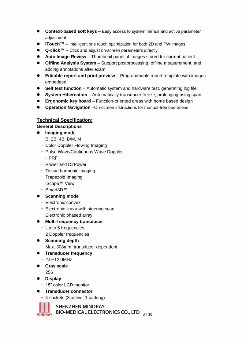
Context-based soft keys – Easy access to system menus and active parameter
adjustment
iTouch™ – Intelligent one touch optimization for both 2D and PW images
Q-click™ – Click and adjust on-screen parameters directly
Auto Image Review – Thumbnail panel of images stored for current patient
Offline Analysis System – Support postprocessing, offline measurement, and
adding annotations after exam
Editable report and print preview – Programmable report template with images
embedded
Self test function – Automatic system and hardware test, generating log file
System Hibernation – Automatically transducer freeze, prolonging using span
Ergonomic key board – Function-oriented areas with home based design
Operation Navigation –On-screen instructions for manual-free operations
Technical Specification:
General Descriptions
Imaging mode
- B, 2B, 4B, B/M, M
- Color Doppler Flowing Imaging
- Pulse Wave/Continuous Wave Doppler
- HPRF
- Power and DirPower
- Tissue harmonic imaging
- Trapezoid Imaging
- iScape™ View
- Smart3D™
Scanning mode
- Electronic convex
- Electronic linear with steering scan
- Electronic phased array
Multi-frequency transducer
- Up to 5 frequencies
- 2 Doppler frequencies
Scanning depth
- Max. 308mm, transducer dependent
Transducer frequency
- 2.0~12.0MHz
Gray scale
- 256
Display
- 15” color LCD monitor
Transducer connector
- 4 sockets (3 active, 1 parking)
4 / 10
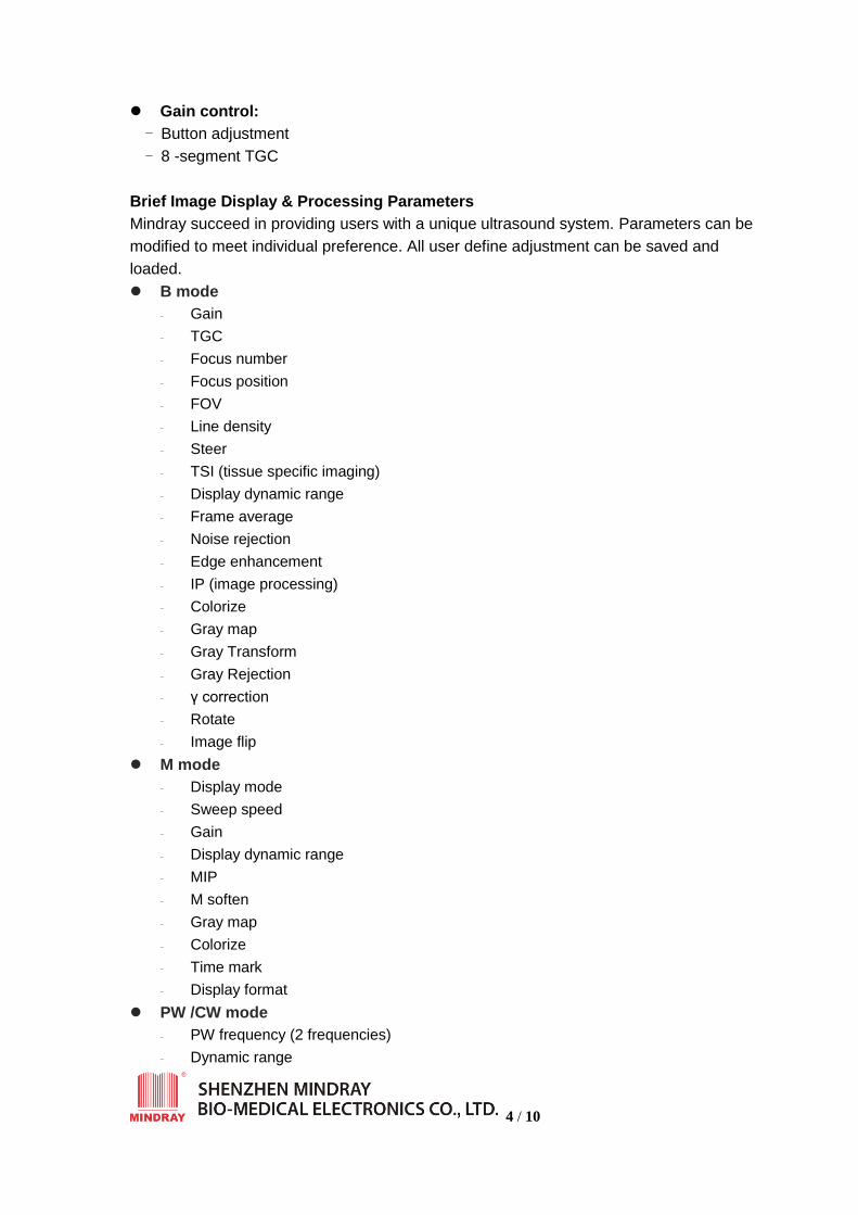
Gain control:
- Button adjustment
- 8 -segment TGC
Brief Image Display & Processing Parameters
Mindray succeed in providing users with a unique ultrasound system. Parameters can be
modified to meet individual preference. All user define adjustment can be saved and
loaded.
B mode
- Gain
- TGC
- Focus number
- Focus position
- FOV
- Line density
- Steer
- TSI (tissue specific imaging)
- Display dynamic range
- Frame average
- Noise rejection
- Edge enhancement
- IP (image processing)
- Colorize
- Gray map
- Gray Transform
- Gray Rejection
- γ correction
- Rotate
- Image flip
M mode
- Display mode
- Sweep speed
- Gain
- Display dynamic range
- MIP
- M soften
- Gray map
- Colorize
- Time mark
- Display format
PW /CW mode
- PW frequency (2 frequencies)
- Dynamic range

5 / 10
- Scale
- Baseline
- Sweep speed
- Sample volume
- Sample depth
- Steer
- Angle correlation
- Colorize
- Wall filter
- Auto Trace and auto calculation
- Duplex
- Triplex
- Threshold
- Trace Area
- Trace smooth
- Trace sensitivity
- Audio
- Full screen
- Time mark
- Display format
- HPRF
Color mode
- Gain
- Frequency (2 frequencies)
- Steer
- Scale
- Color IP
- Baseline
- Color map
- Wall filter
- Line density
- Packet size
- Flow state
- Smooth
- Persistence
- Contrast
- Priority
- Map invert
- Focus position
- B/C wide (automatically adjust the 2D image size according to the color ROI)
- ROI color (off, red, green, blue, cyan, MAG, yellow, white)
- B/C dual live
- Image display
6 / 10
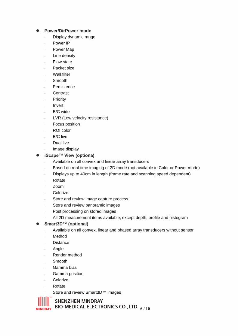
Power/DirPower mode
- Display dynamic range
- Power IP
- Power Map
- Line density
- Flow state
- Packet size
- Wall filter
- Smooth
- Persistence
- Contrast
- Priority
- Invert
- B/C wide
- LVR (Low velocity resistance)
- Focus position
- ROI color
- B/C live
- Dual live
- Image display
iScape™ View (optiona)
- Available on all convex and linear array transducers
- Based on real-time imaging of 2D mode (not available in Color or Power mode)
- Displays up to 40cm in length (frame rate and scanning speed dependent)
- Rotate
- Zoom
- Colorize
- Store and review image capture process
- Store and review panoramic images
- Post processing on stored images
- All 2D measurement items available, except depth, profile and histogram
Smart3D™ (optional)
- Available on all convex, linear and phased array transducers without sensor
- Method
- Distance
- Angle
- Render method
- Smooth
- Gamma bias
- Gamma position
- Colorize
- Rotate
- Store and review Smart3D™ images

7 / 10
- Cut
- Adjust VOI
Functions
Cine loop
- 2D mode (B, Color, Power, DirPower), max: >1200-frame
- Time line mode (M, PW, CW), 131s max
Zoom
- *RAZ (regional acoustic zoom)
- Pan zoom
- PIP (picture in picture)
- For real-time or frozen images
Image storage
- BMP
- JPG
- CIN
- FRM
- AVI
- DCM
Image archive
- Built-in 80G hard disk
- DVD-R/W
- USB
- DVD recorder
- VCR
- Video printer
- DICOM3.0
Advanced Imaging Technology
Benefiting from MINDRAY’s advanced image process technologies, DC-3 provides
brilliant color Doppler images and precise anatomic 2D imaging.
Powerful Multi-beam Parallel Imaging (MBP) increases temporal resolution and
real-time frame rate, while collecting useful information to re-build high quality
images.
Fine Tissue Optimization (FTO) eliminates noise, improves signal-to-noise ratio
and emphasizes boundary imaging.
Transmitting Spectrum Focusing (TSF) greatly decreases side lobe while
improving spatial and temporal resolution.
Innovative Transmitting Apodization (ITA) minimizes artifact through specific
apodized transmission,reducing near-field clutter and enhancing precise sound
beam for more predictable results.
Accurate Vessel Imaging (AVI) automatically distinguishes subtle tissue Doppler
signal from blood cell to form high resolution images.
8 / 10
iBeamTM Spatial compounding imaging for linear probes – innovative new idea of
spatial compounding imaging to rebuild high quality image without compromising frame
rates
Measurement & Calculation
General B mode measurement:
General M mode measurement
General Spectral Doppler mode measurement
General Color mode measurement
Clinical Analysis Packages
Obstetrics
Cardiac
Gynecology
Small Parts
Urology
Orthopedics
Peripheral Vascular
Standard configuration
High resolution 15 inch LCD display
Pulse Wave Doppler
HPRF
Color Doppler Flow Imaging
Power Doppler Flow Imaging
Directional Power Doppler Flow Imaging
Tissue Harmonic Imaging
Trapezoid Imaging
iBeam (Spatial compounding imaging for linear probe)
iTouch™ (Automatic image optimization by pressing one button)
80G integrated hard disk
iStation™
USB ports
Ethernet port
S-video out port and cable
Measurement and calculation software packages
Multi-language screen display
Convex transducer 3C5A(2.5/3.5/5.0/H5.0/H6.0MHz)
Linear array transducer 7L4A (5.0/7.5/10.0MHz)
DVD-RW
Software options
DICOM 3.0 software
iScape™ View (Panoramic imaging)
9 / 10
Smart3D™ (Freehand 3D)
Hardware options
Transducers
CW
Needle guide brackets
ECG module with electrode and cables (AHA/IEC)
Water-resistant footswitch
Multi-language
Screen display, keyboard layout* and user manuals* support
English
French
German
Spanish
Portuguese
Italian
Russian
Chinese
Others Parameters
Inputs and outputs
Serial port: 1
Parallel port: 1
S-video in: 1
S-video out: 1
Video in: 1
Video out: 1
Audio in: 2
Audio out: 2
VGA in: 1
VGA out:1
RGB in: 1
RGB out: 1
USB port: 4
Ethernet: 1
Remote control: 1
Footswitch port: 1
System power in: 1
Auxiliary power out: 3
Ground pole: 1
Equal potential pole: 1
Microphone port: 1

10 / 10
Reset button: 1
Power supply
Power supply voltage: 100 ~ 127 VAC or 220 ~ 240 VAC
Power supply frequency: 50/60 Hz
Power consumption: 600 VA
Dimensions and weight
Height:1209~1570 mm (47.6~61.8 in) Panel and LCD dependent
Width: 460mm ( 18.1 in)
Depth: 730mm ( 28.7 in)
Weight: approx. 91.5kg (201.7 lb.)
Transducer Specification
Model name
Array type Multi-frequency
(MHz)
Doppler
frequency
(MHz)
Scanning
Angle
/length
3C5A Convex 2.5/3.5/5.0
H5.0/H6.0 2.5/3.3 68°
7L4A Linear 5.0/7.5/10.0 5.0/5.7 35mm
6CV1 Endocavity 5.0/6.5/8.0 4.4/5.0 140°
6LB7 Biplanar 5.0/6.5/8.0 Convex 4.4/5.0
Linear 3.7/4.6
Convex 168°
Linear 62mm
10L4 Linear 8.0/10.0/12.0 7.3/8.0 35mm
7L6 Linear 5.0/7.5/10.0 5.0/5.7 56mm
6LE7 Intrarectal 5.0/6.5/8.0 4.4/5.0 62mm
6C2 Micro-convex 5.0/6.5/8.0 4.4/5.0 93°
2P2 Phased
array
2.5/3.0/3.5/
H3.5/H4.0 2.0/2.5 90°
*3C1 Micro-conve
x
2.5/3.5/5.0
H4.6/H6.0 2.5/3.3 136°
*7LT4 T-type linear 5.0/7.5/10.0 5.0/5.7 37mm
*To be released soon.