Embed Size (px)
Citation preview

King’s Research Portal
DOI:10.1016/j.bpc.2017.08.006
Document VersionPeer reviewed version
Link to publication record in King's Research Portal
Citation for published version (APA):Basher, M. A., Rahman, K. M., Jackson, P. J. M., Thurston, D. E., & Fox, K. R. (2017). Sequence-selectivebinding of C8-conjugated pyrrolobenzodiazepines (PBDs) to DNA. Biophysical Chemistry, 230, 53-61.https://doi.org/10.1016/j.bpc.2017.08.006
Citing this paperPlease note that where the full-text provided on King's Research Portal is the Author Accepted Manuscript or Post-Print version this maydiffer from the final Published version. If citing, it is advised that you check and use the publisher's definitive version for pagination,volume/issue, and date of publication details. And where the final published version is provided on the Research Portal, if citing you areagain advised to check the publisher's website for any subsequent corrections.
General rightsCopyright and moral rights for the publications made accessible in the Research Portal are retained by the authors and/or other copyrightowners and it is a condition of accessing publications that users recognize and abide by the legal requirements associated with these rights.
•Users may download and print one copy of any publication from the Research Portal for the purpose of private study or research.•You may not further distribute the material or use it for any profit-making activity or commercial gain•You may freely distribute the URL identifying the publication in the Research Portal
Take down policyIf you believe that this document breaches copyright please contact [email protected] providing details, and we will remove access tothe work immediately and investigate your claim.
Download date: 23. Apr. 2020

Accepted Manuscript
Sequence-selective binding of C8-conjugatedpyrrolobenzodiazepines (PBDs) to DNA
Mohammad A. Basher, Khondaker Miraz Rahman, Paul J.M.Jackson, David E. Thurston, Keith R. Fox
PII: S0301-4622(17)30330-7DOI: doi: 10.1016/j.bpc.2017.08.006Reference: BIOCHE 6038
To appear in: Biophysical Chemistry
Received date: 27 July 2017Revised date: 18 August 2017Accepted date: 18 August 2017
Please cite this article as: Mohammad A. Basher, Khondaker Miraz Rahman, Paul J.M.Jackson, David E. Thurston, Keith R. Fox , Sequence-selective binding of C8-conjugatedpyrrolobenzodiazepines (PBDs) to DNA, Biophysical Chemistry (2017), doi: 10.1016/j.bpc.2017.08.006
This is a PDF file of an unedited manuscript that has been accepted for publication. Asa service to our customers we are providing this early version of the manuscript. Themanuscript will undergo copyediting, typesetting, and review of the resulting proof beforeit is published in its final form. Please note that during the production process errors maybe discovered which could affect the content, and all legal disclaimers that apply to thejournal pertain.

ACC
EPTE
D M
ANU
SCR
IPT
1
Sequence-selective binding of C8-conjugated pyrrolobenzodiazepines (PBDs) to DNA
Mohammad A. Basher1,2, Khondaker Miraz Rahman3, Paul J.M. Jackson3, David E. Thurston3 and
Keith R. Fox1*
1Biological Sciences, Life Sciences Building 85, University of Southampton, Southampton SO17
1BJ, UK
2Present Address Department of Pharmacy, Noakhali Science and Technology University,
Sonapur-3814, Noakhali, Bangladesh.
3Institute of Pharmaceutical Science, King’s College London, Franklin-Wilkins Building, Stamford
Street, London SE1 9NH, UK
*Corresponding author:
Email [email protected]
Tel +44 23 8059 4374
Fax +44 23 8059 5159
Keywords: Conjugate, Anthramycin, PBD, Polyamide, Footprinting, DNA melting.
Research Highlights
C8-conjugates of pyrrolobenzodiazepines with benzofused rings modulate sequence-selective
DNA binding
The conjugates bind best to guanines flanked by A/T-rich sequences.
Bi- and tri-phasic melting curves show that 14-mer oligonucleotides can bind more than one
ligand
ABSTRACT
DNA footprinting and melting experiments have been used to examine the sequence-specific
binding of C8-conjugates of pyrrolobenzodiazepines (PBDs) and benzofused rings including
benzothiophene and benzofuran, which are attached using pyrrole- or imidazole-containing linkers.
The conjugates modulate the covalent attachment points of the PBDs, so that they bind best to
guanines flanked by A/T-rich sequences on either the 5'- or 3'- side. The linker affects the binding,
and pyrrole produces larger changes than imidazole. Melting studies with 14-mer oligonucleotide
duplexes confirm covalent attachment of the conjugates, which show a different selectivity to
anthramycin and reveal that more than one ligand molecule can bind to each duplex.
ACCEPTED MANUSCRIPT

ACC
EPTE
D M
ANU
SCR
IPT
2
1. Introduction
Pyrrolobenzodiazepines [1] are sequence selective DNA minor groove binding ligands, of which
anthramycin (1; Fig. 1) was the first compound to be isolated in the 1960s from cultures of
Streptomyces [2]. These compounds have received considerable attention because of their potent
antitumour activity, which results from covalent binding of the C11 position of the diazepine ring to
the 2-amino group of a guanine [3, 4]. Naturally occurring PBDs such as anthramycin, tomaymycin,
neothramycin and sibiromycin form adducts that span three base pairs, with guanine in the central
position [5, 6], and their preferred binding site is 5'-AGA-3' [7], although more recent data suggest
that they have a kinetic preference for 5′-YGR-3′ sequences [8] (R = purine, Y = pyrimidine).
Synthetic PBD monomers with C2 (pyrrolidine ring)-aryl substitution exhibit nanomolar to picomolar
cytotoxic potency against a number of tumour cell lines [9]. In an attempt to enhance their binding
selectivity, bis-PBDs have been developed in which two PBD monomers are joined by a suitable
linker and SJG-136, which forms interstrand cross-links at 5'-RGATCY-3' as well as intrastrand
cross-links and mono-adducts [10, 11], has reached Phase II clinical trial [12, 13]. Another
approach for targeting longer DNA sequences is to prepare PBD C8-conjugates, in which
heterocyclic rings such as pyrrole and imidazole are linked to the PBD anthranilate ring through
amide bonds [14-17].
Polyamides composed of pyrrole and imidazole heterocycles are able to form antiparallel
dimers in the DNA minor groove and are widely known for their ability to recognize AT and TA
base pairs with pyrrole-pyrrole pairs, and CG and GC with pyrrole-imidazole and imidazole-pyrrole
pairs, respectively [18-20]. The conjugation of a PBD with a polyamide requires a suitable linker
between these two entities. By comparing the binding energy of different linkers, a trimethylene
group has been shown to provide a good isohelical fit in the DNA minor groove [15]. A conjugate of
a PBD linked with a polyamide composed of two pyrroles was found to recognise the sequence
(CCATT) in the promoter of DNA Topo IIα and to inhibit the binding of NF-γ, which is involved in
cell cycle progression [17]. The combination of pyrrole and thiazole showed better DNA binding
affinity than di-pyrroles [16], while addition of an imidazole offered superior sequence selectivity
[14]. In addition, several other PBD conjugates have shown significant antibacterial [21] and
antitubercular activity [22]. More recently, members of the PBD family have been developed as
cytotoxic payloads for attachment to antibodies to form antibody-drug conjugates (ADCs) and a
number of these are presently in the clinic for the treatment of various leukaemias and lung cancer
[1].
There has been significant interest in trying to understand the molecular basis of the selectivity
of known DNA-targeting agents with a view to designing novel molecules targeted to specific
sequences within genes to allow selective modulation of expression for both research and
therapeutic purposes. We previously reported a series of C8-linked pyrrolobenzodiazepine (PBD)-
ACCEPTED MANUSCRIPT

ACC
EPTE
D M
ANU
SCR
IPT
3
poly(N-methylpyrrole) conjugates (e.g., GWL-78) which demonstrated the synergistic effect of
joining a GC-specific PBD unit to an AT-recognizing polypyrrole fragments [15]. More recently we
identified 4-(1-methyl-1H-pyrrol-3-yl)benzenamine (MPB) motif as a GC-selective building block
(Figure 1) and coupling MPB motif to PBDs through C8-position resulted in molecule with with sub-
nanomolar to femtomolar IC50 across 65 cell lines tested [23]. As part of our aim to develop
pyrrolobenzodiazepine based selective antitumour compounds, and to understand the role of
benzofused building blocks in sequence selectivity, we linked benzofuran, benzothiophene or
benzopyrrole (indole) groups to the unsubstituted pyrrolobenzodiazepine core through a C4 linker.
The benzofused groups were either directly linked to the PBD core through the C4 linker or
separated by a further pyrrole/imidazole heterocycle to assess the impact of pyrrole/imidazole
heterocycles on the DNA stabilisation potential of PBD-Benzofused conjugates.
In this paper, we describe the DNA-binding properties of five C8-PBD-polyamide conjugates
with benzofused rings (2-6; Fig. 1), and have examined their DNA binding sequence selectivity by
DNase I footprinting and fluorescence melting.
ACCEPTED MANUSCRIPT

ACC
EPTE
D M
ANU
SCR
IPT
4
2. Materials and methods
2.1 C8-PBD-conjugates
The structures of the C8-PBD-conjugates used in this work are shown in Fig. 1. Their synthesis
will be reported elsewhere and the purity data are presented in the Supplementary material. The
compounds were dissolved in dimethylsulfoxide at a concentration of 10 mM and stored at -20 °C.
These were diluted to working concentrations in 10 mM Tris-HCl pH 7.5 containing 10 mM NaCl
immediately before use.
2.2 DNase I footprinting.
Footprinting reactions were performed as previously described [24] using the DNA fragments
HexA and HexB, which together contain all 64 symmetrical hexanucleotide sequences [25], and
MS1 that contains all possible 134 tetranucleotide sequences [26]. The DNA fragments were
obtained by cutting the parent plasmids with HindIII and SacI (for HexA and MS1) or EcoRI and
PstI (for HexB), and were labelled at the 3'-end of the HindIII or EcoRI sites with [α-32P]dATP using
reverse transcriptase or exo- Klenow fragment. After gel purification, the radiolabelled DNA was
dissolved in 10 mM Tris-HCl pH 7.5 containing 0.1 mM EDTA, at a concentration of about 10 c.p.s
per μL as determined on a hand held Geiger counter. 1.5 μL of radiolabelled DNA was mixed with
1.5 μL ligand that had been freshly diluted in 10 mM Tris-HCl pH 7.5, containing 10 mM NaCl. The
complexes were left to equilibrate for at least 12 hours before digesting with 2 μL DNase I (final
concentration about 0.01 units/mL). The reactions were stopped after 1 minute by adding 4 μL of
formamide containing 10 mM EDTA and bromophenol blue (0.1% w/v). The samples were then
heated at 100 °C for 3 minutes before loading onto 8% denaturing polyacrylamide gels containing
8 M urea. Gels were fixed in 10% acetic acid, transferred to 3MM paper, dried and exposed to a
phosphor screen overnight, before analysing with a Typhoon phosphorimager.
2.3 DNA thermal denaturation studies
Fluorescence melting curves were determined using a Roche LightCycler in a total reaction
volume of 20 μL as previously described [27]. For each reaction the final oligonucleotide
concentration was 0.25 μM, diluted in 10 mM sodium phosphate pH 7.5 containing 100 mM NaCl,
with ligand concentrations between 0.1 – 10 µM. The experiments used the duplexes shown in
Table 1, in which one strand contained 5’-fluorescein (F), with a 3’-dabcyl quencher (Q) on the
complementary strand. In a typical experiment the samples, which had been equilibrated overnight,
were first denatured by heating to 95 °C at a rate of 0.1 °C.s-1. The samples were then maintained
at 95 °C for 5 min before annealing by cooling to 30 °C at 0.1 °C.s-1. They were held at 25 °C for a
further 5 min and then melted by heating to 95 °C at 0.1 °C.s-1. Recordings of the fluorescence
emission at 520 nm were taken during both the melting steps as well as during annealing. The data
were normalized to show the fractional change in fluorescence for each sample between the
ACCEPTED MANUSCRIPT

ACC
EPTE
D M
ANU
SCR
IPT
5
starting and final values. Tm values were determined from the first derivatives of the melting profiles
using the Roche LightCycler software.
2.4 Molecular Modelling
The ligand 2 was generated using ChemBioOffice and energy minimized using the
MMFF94force-field [28]. The structure was then imported into AMBER (v11) [29] and the structure
was manipulated using the AMBER modules antechamber and parmchk. Antechamber was used
to convert the structure to mol2 format (through the application of Gasteiger charges) and missing
parameters were generated using parmchk. The DNA sequence (5'-AAAAGTTAACTTTT-3') was
then built using the AMBER module nuc and the parm99bsc0 [30] and gaff.dat force-fields were
loaded. A further AMBER module xleap was used to align the ligand in the minor groove of DNA,
with the N10 of the PBD either orientated within 2 Å of the exocyclic amine of the relevant guanine
residue (in the case of non-covalent simulations) or N10-C11 covalently bound to DNA in the case
of covalent simulations. The initial orientation was undertaken in the case of non-covalent
simulations as PBDs are thought to form a reaction-mediating H-bond with DNA, which in turn pulls
the molecule into the minor groove.
Each adduct was then minimized in a stepwise manner to facilitate accommodation in the minor
groove. In this procedure, positional restraints were initially used on the DNA atoms to keep their
positions fixed, and the ligands were then energy minimized alone. This was followed by full
minimization of the system (without restraints) to ensure ligands were accommodated deep in the
minor groove.
Production simulations were undertaken in implicit solvent using the Generalised Born solvation
method. This is equivalent to the Poisson Boltzmann method but includes a surface area term to
enable accuracy in the simulation of macromolecules [23, 31]. A term to allow for monovalent
electrostatic ion screening was also employed to simulate the effect of Na+ ions in the surrounding
environment. The affinity of the ligand for DNA was then assessed using free energy of binding
calculations; a detailed explanation of this established methodology can be found in our previous
publications [23, 31].
ACCEPTED MANUSCRIPT

ACC
EPTE
D M
ANU
SCR
IPT
6
3. Results
3.1 DNase I footprinting
DNase I footprinting was used to identify the binding sites of the PBD-conjugates using the DNA
fragments HexA and HexB, which together contain all 64 symmetrical hexanucleotide sequences
[25] and MS1 that contains all 134 tetranucleotide sequences [26]. DNase I overestimates the size
of ligand binding sites and produces footprints that are staggered towards the 3’-end of actual
binding site. In the following description, for the sake of clarity, we refer to the central region of the
sequence(s) that are protected in each of the footprints. The concentration dependence of the
footprints for conjugates 2-6 with HexA and MS1 are shown in Fig. 2 and 3 and, for each DNA
template, are compared with the cleavage pattern in the presence of anthramycin (1). Similar
experiments with HexB are shown in Fig. S1. As expected, most of the footprints with anthramycin
are located at the sequence RGR/YCY. For example, the two central footprints with HexA are in
the sequences AGATCTAGAATTCCGGA and AGGCCT (RGR and YCY underlined). It is clear
that the footprinting patterns with anthramycin are different to those produced by any of the
conjugates, demonstrating that addition of the appending groups has affected the preferred binding
sites. In general, the conjugates produce large footprints at higher concentrations (10 μM, 5μM and
2 μM) which resolve into shorter regions of protection at lower concentrations. The footprinting
patterns for 2, 3 and 4 are similar, while 5 and 6 appear to protect only a subset of these sites.
With DNA fragment HexA (Fig. 2), 2, 3 and 4 produced four main footprints at low
concentrations (1 μM). Each of these footprints occurs close to a block of AT residues, in
sequences such as CGATATATATG, CAAATTTAG, GAATTC, GTTTAAAC and CTTAAG. If we
assume that covalent attachment of the PBD moiety must occur at a guanine, then it appears that
this can be located at either the 3’- or 5’- end of the footprint, with any arrangement of adjacent
(A/T)n residues, where n is at least 4. The footprint at 5'-GAATTC has the reactive guanine at the
5’-end of an AT-tract. A similar effect is seen at 5'-GTTTAAAC, with the guanine at the 5'-end of an
AT-tract, though the footprint at 5'-CTTAAG would place the reactive G at the 3'-end of the AT-
tract. 5 binds to a greater number of sites at high concentrations (10 μM), generating large regions
of protection, though there is little protection at lower ligand concentrations. 6 produces fewer
footprints with the clearest footprint around GAATTC.
Conjugates 2, 3 and 4 produced similar footprints with DNA fragment HexB (Fig. S1), with three
or four sites persisting to 1 μM ligand concentration. These are again located close to AT-tracts
and can be seen around CATATG, CTTATAAG, CAATTG and CTAATTAG. As for HexA, if we
assume that the PBD moiety attaches to guanine, then this can be at either the 3'- or 5'-end of an
AT-tract. No strong footprints are evident at guanines that are adjacent to only 2 AT base pairs,
although since the HexA and HexB fragments were designed to contain symmetrical
hexanucleotides, they do not contain any examples of only three consecutive AT pairs. 5 again
generates long footprints at 10 μM, but produces fewer clear footprints at lower concentrations,
which are strongest at CATATG, CAATTG and AATTAG. 6 also binds to only a subset of these
ACCEPTED MANUSCRIPT

ACC
EPTE
D M
ANU
SCR
IPT
7
sites with clearest footprints around CTAATTA, AATTG and TAAGCGC and a weaker footprint at
ATGCAT (Fig. S1).
Similar DNase I footprinting patterns for the interaction of these compounds with MS1 are
presented in Fig. 3. Once again 2, 3 and 4 generate similar footprints at three or four sites, which
are clearest around GATTT, GTTAA, CTAT, ATAG, CAATTA, GTTATGTAAAG and GTTTTG. As
noted for the other fragments, these all cluster around A/T tracts. The footprint around CTATC,
which, although one of the weaker sites, is stronger for 2 and 3 than the other compounds,
suggests that the AT-tract adjacent to the guanine only needs to be 3 base pairs long. 5 again
produces very large footprints at the highest concentration (10 μM) with only a few sites around
CAATTAG and CTATC that persist at 5 and 2 µM. As noted for the other fragments 6 produces
fewer footprints, the strongest of which are around CAATTAG and AATGC.
3.2 DNA melting studies
The interaction of these PBD-conjugates with DNA was further evaluated by examining their
effects on the melting temperature of a series of short 14-mer oligonucleotide duplexes. These
duplexes contained a central GGTACC, GGATCC, GCTAGC, GCTTGC, GTATAC or GTTAAC
embedded within an An.Tn tract in which the A-strand was labelled with 5’-fluorescein and the T-
strand with 3’-dabcyl (see Table 1). On duplex formation, the fluorescein and dabcyl are close
together and the fluorescence is quenched. When the temperature is increased, the duplex melts
and the fluorophore and quencher move apart and there is a large increase in the fluorescence.
These studies were performed with ligand concentrations between 0.1-10 µM. Since the
concentration of the duplex is only 0.25 µM there is an excess DNA over ligand at the lowest
concentration.
The effects of 1 μM of each ligand on the melting of the six oligonucleotides are shown in Fig.
S2 and the data derived from these melting curves (and those with 10 µM ligand) are summarized
in Table 2. The five conjugates (Fig. S2) each produced two elevated melting transitions; one
around 65 °C and another above 85 °C. Since the ligands are covalently bound to the DNA, rather
than in dynamic equilibrium, the Tm values of each of these transitions did not change with ligand
concentration. However, the relative proportions of the different transitions varied, such that a
greater fraction of the melting curve appeared at higher temperatures with higher ligand
concentrations. Each of the duplexes contains more than one guanine to which the conjugates
could attach (i.e. two guanines for GTATAC and GTTAAC, and four each for GGTACC, GGATCC,
GCTAGC and GCTTGC) and further transitions at higher temperatures were observed for most of
the complexes as second (and third) PBDs attach to the oligonucleotides. The fraction of the
melting transition that has shifted to the higher temperature therefore indicates the proportion of
the duplex that has been modified within the incubation period, while the absolute values of the
melting temperatures indicate the duplex stabilization that is imparted by the covalently bound
ligands.
ACCEPTED MANUSCRIPT

ACC
EPTE
D M
ANU
SCR
IPT
8
All five of these compounds clearly bind to each of these duplexes, though they have noticeably
different relative effects. At a concentration of 1 μM, 4 shifted the entire melting curve to higher
temperatures with all six duplexes. A similar effect was produced by 1 µM 3, for which no
transitions corresponding to the uncomplexed duplexes are evident, except for GGTACC and
GCTAGC, which showed a small amount that had not reacted with the ligand. For most of the
other ligand-duplex combinations a significant amount of the unmodified transition was still
observed at 1 µM. 6 showed the weakest effect, with lower melting temperatures and a greater
amount of uncomplexed duplex. All five conjugates had the greatest effect on the sequence with a
central GGATCC, for which little or no unmodified duplex remained with 1 µM ligand. 5 produced
smaller changes in melting temperature with GGTACC and GTTAAC than 2, 3 and 4, though it
showed a strong interaction with GCTTGC, producing a single melting transition in the presence of
1 µM with a ΔTm of 31 °C, the highest shift for any of the ligands with any of the duplexes.
While there are subtle differences between these PBD-conjugates, their effects are very
different from those of anthramycin itself. The concentration dependence of the melting curves of
the six oligonucleotide duplexes in the presence of anthramycin are compared with those for 2 in
Fig. 4. It can be seen that anthramycin produces much larger effects with GGTACC and GGATCC
than the other oligonucleotide duplexes, and it had little or no effect on GTTAAC. In addition, with
the exception of GGATCC, anthramycin only produced one elevated melting transition with ΔTm
values that are smaller than with most of the conjugates (see Table 2), although similar to those
seen with 6. GGTACC and GGATCC are the only oligonucleotides that contain the sequence
RGR; GGTACC contains a single AGG site, while GGATCC contains AGGA (with two overlapping
RGR sites) and TGGA on the two duplex strands.
Although the PBDs form covalent links to their target guanines, these bonds are broken at high
temperatures and the ligand dissociates from the DNA [32]. The fluorescence was therefore
followed during the annealing curves as the samples were cooled back to 30 °C. The ligands only
slowly re-attach to the DNA, and in each case, these annealing profiles were similar to those of the
free uncomplexed DNA. After leaving at 30 °C for 5 minutes the samples were melted for a second
time, recording the new melting transitions. Some of the ligands had reattached to the DNA during
the annealing phase and so stabilised the duplexes relative to the drug-free controls. While the
ΔTm values for this second melt were similar to those for when the complexes were first melted, the
proportion of the profile showing an elevated melting temperature was always much lower than for
the first melt. The results for these second melting experiments are shown in Table 3. Anthramycin
had no effect when its complexes were melted for a second time and all these transitions were
identical to those of the drug-free control. In general, the proportion of the transition corresponding
to the free DNA in the presence of 1 µM of the conjugates was lowest for GGATCC, suggesting
that they re-associated fastest with this sequence. In contrast to the first melt, all of the complexes
produced only one elevated melting transition with 1 µM ligand, with no evidence of a second
melting transition around 85 °C. However, this higher transition was seen with some of the ligands
ACCEPTED MANUSCRIPT

ACC
EPTE
D M
ANU
SCR
IPT
9
at a concentration of 10 µM and was most pronounced for 4. 6 produced almost no changes in the
melting temperature when the complexes were melted for the second time, with only a small effect
for GCTAGC and GGATCC. No higher transitions were produced with 5 when the complexes were
melted for the second time, and this ligand had no effect on both GTATAC and GTTAAC even at a
concentration of 10 µM. However, 1 µM of 5 showed some of the greatest changes with GGTACC
and GGATCC.
3.3 Molecular Modelling
Molecular modelling studies were undertaken on 2 in an effort to understand the interaction of
the compound with DNA and to establish a rationale behind the biphasic curves observed in the
FRET melting studies. The compound consists of a G-alkylating PBD coupled to a pyrrole-
benzofuran tail. The lack of hydrogen-bonding acceptors in the DNA-interactive face of ligand (i.e.
within the pyrrole-benzofuran tail) suggests that the molecule should interact with AT tracts.
Molecules of this type containing pyrrole chains have been previously shown to interact with
inverted CCAAT boxes of DNA [14], and a similar interactivity pattern would be predicted for 2. As
GTTAAC (containing an extended AT tract) would be expected to be an excellent substrate for 2
was chosen for analysis
In initial in silico positioning of 2 in the DNA minor groove suggests that the molecule can span 5
base pairs and, as the sequences used in the FRET experiments contain fourteen base pairs and
more than one guanine, two ligands may be able to bind to the same duplex (i.e., dual-occupancy
binding). Molecular dynamics simulations were undertaken on 2 in a variety of orientations in one
sequence (GTTAAC) to ascertain whether dual occupancy was possible. Analysis of the extended
sequence (AAAAGTTAACTTTT) suggests a number of potential configurations for the interaction
of 2 with GTTAAC. Firstly, a single ligand may be able to bind to G5 or G19 (with the A-ring
oriented in the 5' or 3' direction) or two ligands might bind to either G5 (with the A-ring oriented 5')
in combination with G19 (with the A-ring in a 3' orientation on the lower strand, see Fig. 5), or to G5
(with the A-ring oriented 3') in combination with G19 (with the A-ring oriented 5’ on the lower
strand).
Free energy of binding calculations (Table 4) conducted on the four potential adducts in single
occupancy form show that a 3’-orientation of the ligand’s A-ring is preferred over a 5’-orientation by
~-2 kcal/mol. This is exemplified on visual analysis of these non-covalent simulations whereby an
H-bond can be formed between the N10 proton of the PBD and the exocyclic amine of a guanine
residue (i.e. G5). The H-bond is thought to facilitate nucleophilic attack of the guanine by pulling
the PBD into the minor groove [23]. In contrast, this H-bond is not present in simulations of the 5'
orientation of the A-ring of either G5 or G19 adducts, presumably due to inappropriate orientation
of the N10 proton, and as a result is likely to be a less preferred adduct.
Non-covalent simulations were conducted on the two possible dual-occupancy orientations of 2
in the minor groove where the ligands do not overlap (i.e., concurrently covalently bound to G5 in
ACCEPTED MANUSCRIPT

ACC
EPTE
D M
ANU
SCR
IPT
10
A-ring 5’ and G19 in A-ring 3’ or G5 in A-ring 3’ and G19 in A-ring 5’). Simulations suggest that
either of these orientations is viable, and this hypothesis would explain the biphasic curves
observed in the FRET studies. There is little difference between alkylation of G5 with the A-ring in
5’ orientation and G19 with the A-ring in a 3’ orientationas the PBD will be bound to the sequence
5’-GTTAAC-3’ in both instances, with alkylation of the top strand for G5 and the bottom strand for
G19. The second adduct would then either be at 5’-AAAG-3’ (with the A-ring oriented 5’ to G5) or
5’-TTTG-3’ (with the A-ring oriented 5’ to G19). The free energy of binding calculations undertaken
suggest that these adducts are equally likely (Table 4).
Furthermore, the potential of 2 to bind in an overlapping 2:1 (ligand:DNA) manner was also
investigated.Simulations of 2 covalently bound to G5 and G19 in A-ring 3’ orientation (on the upper
and lower strands respectively, see Fig. 5 lower panel) suggest that DNA interaction of this type is
less favourable. Binding of two molecules of 2 in a 2:1 ratio (where an overlap in the same
sequence is necessary) would require substantial widening of the minor groove as the exocyclic
amines of the guanines are positioned towards the centre of the DNA, and once alkylated, a
molecule of 2 would sterically prevent a second molecule binding. Furthermore, the van der Waals
surface of the ligand is sufficient to occupy the full width of the DNA in 1:1 binding ratio, meaning a
second binding event in this instance would be less favourable.
4. Discussion
PBDs are known to bind to the N2 position of guanine residues within the DNA minor groove
and simple PBD monomers, such as anthramycin, are known to bind best to the sequence RGR,
as confirmed by the footprinting and fluorescence melting experiments described here. When the
PBD core is conjugated to other components through the C8 position, the sequence selectivity will
be determined by the combined properties of the PBD, the linker and the side chain, though
covalent attachment will still require the presence of a guanine residue. The profiles of the
fluorescence melting curves confirm that these conjugates are indeed still covalently attached to
the six oligonucleotides that we have studied, while the footprinting and melting experiments
demonstrate that the conjugates have very different sequence binding properties to a simple PBD
such as anthramycin, and the sequence RGR alone is no longer sufficient to define a good binding
site.
The footprinting data with the five PBD-conjugates show that the preferred sites all contain AT-
rich regions to the 5’- or 3’-side of the reactive guanine residues. Pyrrole and imidazole rings have
been extensively studied as components of minor groove-binding ligands, in which imidazole is
used to recognize GC base pairs in the 2:1 mode [33] although in the 1:1 mode it shows AT-
selectivity similar, but weaker to that of pyrrole [34, 35]. This is consistent with the observation that
3 and 5 bind to similar sequences, though 5 displays a weaker interaction.
ACCEPTED MANUSCRIPT

ACC
EPTE
D M
ANU
SCR
IPT
11
The fact that the C8-side chain affects the interaction suggests that this must bind first, before
covalent attachment of the PBD. If the kinetics of binding were driven by the PBD, then the
conjugates would have a similar selectivity to anthramycin and the initial covalent attachment
would lock the complex onto its preferred RGR sites, irrespective of the flanking sequences.
The presence of pyrrole or imidazole in the linker is important as compound 6, which lacks
either of these rings, only binds to a few short sequences. Modelling studies suggest that 2 has an
ideal shape for interactivity with the DNA minor groove, and the pyrrole unit is key in providing its
isohelicity. As 2, 3, 4 and 5 all contain a five-membered heterocycle followed by a benzofused
group, both their span and their ability to bind to DNA would be expected to be similar, and all
compounds would be expected to form extensive van der Waals interactions with the DNA minor
groove, thereby resulting in a similar sequence interactivity pattern. The DNA melting results are
in agreement with footprinting studies and modelling-based analysis. Compounds 2-6 form strong
complexes and produce large changes in the melting curves of the 14-mer DNA duplexes, and
molecular dynamics simulations show that the strong binding may be promoted by dual occupancy
of the compounds in the DNA minor groove. The ΔTm values for the first melting transitions are
significantly greater than anthramycin for all these conjugates (except 6), which again confirms the
important role of the C8-side chains in promoting the interaction.
The footprinting results suggest that the flanking A/T tract can be towards the 3'- or 5'-side of
the guanine to which the PBD is attached. In contrast, previous structural studies have shown that
the PBD phenyl ring (A ring) is oriented towards the 3’-end of the bound guanine [36, 37], from
which we would predict that these conjugates should bind best to RG(A/T)n. In the melting
experiments, all the putative central six base pair sites are flanked by An.Tn tracts. If the tail is
oriented to the 3’-side of the guanine (i.e. towards the centre of the oligonucleotides), then we
would expect GTATAC and GTTAAC to generate the best sites. If it is oriented in the opposite
direction, then the conjugates should all have similar effects on the melting curves, as all the
targets contain the sequence A4G on one strand (and T4G on the opposing complementary strand).
For these melting experiments, the only sequences that contain the preferred RGR binding site are
GGATCC, which has three such sites (AGGA, AGAA and TGAA (on the complementary strand))
and GGTACC, which contains only one such site (AGGT). As all the sequences are affected to
some degree, the PBD portion of these ligands is clearly not restricted to interacting with RGR,
though the sequence-dependent effects suggest that the central base pairs affect the binding
affinity.
The experiments in which the complexes were melted for a second time provide information on
the kinetics of drug interaction. Only those PBD-oligonucleotide complexes with the fastest rates of
covalent attachment will be affected in these second melts. In these experiments, sequences
GGATCC and GGTACC show the lowest proportion of uncomplexed DNA, indicating faster rates
of re-attachment, while GTTAAC and GATATC are the least affected. Under these conditions,
there is no evidence for a second melting transition in the presence of 1 µM ligand. It is especially
ACCEPTED MANUSCRIPT

ACC
EPTE
D M
ANU
SCR
IPT
12
noteworthy that, despite its weaker binding, 10 µM 5 produced clear monophasic melting curves
with a single elevated melting transition, with all the oligonucleotides except GTATAC and
GTTAAC. 6 did not affect the melting profiles in this second melt, which all resembled the ligand-
free transition, indicating that this molecule is slow to re-attach to its binding sites. A similar effect
was seen with anthramycin, which also did not affect the second melting transitions.
Acknowledgments. Mohammad Basher was supported by a Commonwealth Scholarship
ACCEPTED MANUSCRIPT

ACC
EPTE
D M
ANU
SCR
IPT
13
REFERENCES
[1] J. Mantaj, P.J.M. Jackson, K.M. Rahman, D.E. Thurston, From Anthramycin to Pyrrolobenzodiazepine (PBD)-Containing Antibody-Drug Conjugates (ADCs), Angew. Chem. Int. Ed. Engl., 56 (2017) 462-488.
[2] W. Leimgruber, V. Staefanovic, F. Schenker, A. Karr, J. Berger, J. Am. Chem. Soc., 87 (1965) 5791-5793.
[3] L.H. Hurley, Pyrrolo(1,4)benzodiazepine antitumor antibiotics - comparative aspects of anthramycin, tomaymycin and sibiromycin, J. Antibiot., 30 (1977) 349-370.
[4] F.L. Boyd, D. Stewart, W.A. Remers, M.D. Barkley, L.H. Hurley, Characterization of a unique tomaymycin-d(CICGAATTCICG)2 adduct containing 2 drug molecules per duplex by NMR, fluorescence, and molecular modeling studies, Biochemistry, 29 (1990) 2387-2403.
[5] R.P. Hertzberg, S.M. Hecht, V.L. Reynolds, I.J. Molineux, L.H. Hurley, DNA-sequence specificity of the pyrrolo 1,4 benzodiazepine antitumor antibiotics - Methidiumpropyl-EDTA-iron(ii) footprinting analysis of DNA-binding sites for anthramycin and related drugs, Biochemistry, 25 (1986) 1249-1258.
[6] L.H. Hurley, T. Reck, D.E. Thurston, D.R. Langley, K.G. Holden, R.P. Hertzberg, J.R.E. Hoover, G. Gallagher, L.F. Faucette, S.M. Mong, R.K. Johnson, Pyrrolo 1,4 benzodiazepine antitumor antibiotics - relationship of DNA alkylation and sequence specificity to the biological-activity of natural and synthetic compounds, Chem. Res. Toxicol., 1 (1988) 258-268.
[7] M.S. Puvvada, S.A. Forrow, J.A. Hartley, P. Stephenson, I. Gibson, T.C. Jenkins, D.E. Thurston, Inhibition of bacteriophage T7 RNA polymerase in vitro transcription by DNA-binding pyrrolo[2,1-c][1,4]benzodiazepines, Biochemistry, 36 (1997) 2478-2484.
[8] K.M. Rahman, H. Vassoler, C.H. James, D.E. Thurston, DNA Sequence Preference and Adduct Orientation of Pyrrolo [2,1-c][1,4] benzodiazepine Antitumor Agents, ACS Med. Chem. Lett., 1 (2010) 427-432.
[9] N. Cooper, D.R. Hagan, A. Tiberghien, T. Ademefun, C.S. Matthews, P.W. Howard, D.E. Thurston, Synthesis of novel C2-aryl pyrrolobenzodiazepines (PBDs) as potential antitumour agents, Chem. Commun., (2002) 1764-1765.
[10] S.R. Hopton, A.S. Thompson, Nuclear Magnetic Resonance solution structures of inter- and intrastrand adducts of DNA cross-linker SJG-136, Biochemistry, 50 (2011) 4720-4732.
[11] K.M. Rahman, A.S. Thompson, C.H. James, M. Narayanaswamy, D.E. Thurston, The Pyrrolobenzodiazepine Dimer SJG-136 Forms Sequence-Dependent Intrastrand DNA Cross-Links and Monoalkylated Adducts in Addition to Interstrand Cross-Links, J. Am. Chem. Soc., 131 (2009) 13756-13766.
[12] D. Hochhauser, T. Meyer, V.J. Spanswick, J. Wu, P.H. Clingen, P. Loadman, M. Cobb, L. Gumbrell, R.H. Begent, J.A. Hartley, D. Jodrell, Phase I study of sequence-selective minor groove DNA binding agent SJG-136 in patients with advanced solid tumors, Clin. Cancer. Res., 15 (2009) 2140-2147.
[13] Y.Y. Janjigian, W. Lee, M.G. Kris, V.A. Miller, L.M. Krug, C.G. Azzoli, E. Senturk, M.W. Calcutt, N.A. Rizvi, A phase I trial of SJG-136 (NSC#694501) in advanced solid tumors, Cancer Chemother. Pharmacol., 65 (2010) 833-838.
[14] F. Brucoli, R.M. Hawkins, C.H. James, P.J.M. Jackson, G. Wells, T.C. Jenkins, T. Ellis, M. Kotecha, D. Hochhauser, J.A. Hartley, P.W. Howard, D.E. Thurston, An extended pyrrolobenzodiazepine-polyamide conjugate with selectivity for a DNA sequence containing the ICB2 transcription factor binding site, J. Med. Chem., 56 (2013) 6339-6351.
[15] G. Wells, C.R.H. Martin, P.W. Howard, Z.A. Sands, C.A. Laughton, A. Tiberghien, C.K. Woo, L.A. Masterson, M.J. Stephenson, J.A. Hartley, T.C. Jenkins, S.D. Shnyder, P.M. Loadman, M.J. Waring, D.E. Thurston, Design, synthesis, and biophysical and biological evaluation of a series of Pyrrolobenzodiazepine - Poly(N-methylpyrrole) conjugates, J. Med. Chem., 49 (2006) 5442-5461.
[16] F. Brucoli, R.M. Hawkins, C.H. James, G. Wells, T.C. Jenkins, T. Ellis, J.A. Hartley, P.W. Howard, D.E. Thurston, Novel C8-linked pyrrolobenzodiazepine (PBD)-heterocycle conjugates that recognize DNA sequences containing an inverted CCAAT box, Bioorg. Med. Chem. Lett., 21 (2011) 3780-3783.
[17] M. Kotecha, J. Kluza, G. Wells, C.C. O'Hare, C. Forni, R. Mantovani, P.W. Howard, P. Morris, D.E. Thurston, J.A. Hartley, D. Hochhauser, Inhibition of DNA binding of the NF-Y transcription factor by the pyrrolobenzodiazepine-polyamide conjugate GWL-78, Molecular Cancer Therapeutics, 7 (2008) 1319-1328.
ACCEPTED MANUSCRIPT

ACC
EPTE
D M
ANU
SCR
IPT
14
[18] M. Mrksich, P.B. Dervan, Antiparallel side-by-side heterodimer for sequence-specific recognition in the minor groove of DNA by a distamycin/1- methylimidazole-2-carboxamide-netropsin pair, J. Am. Chem. Soc, 115 (1993) 2572-2576.
[19] W.S. Wade, M. Mrksich, P.B. Dervan, Design of peptides that bind in the minor groove of DNA at 5'-(A,T)G(A,T)C(A,T)-3' sequences by a dimeric side-by-side motif, J. Am. Chem. Soc., 114 (1992) 8783-8794.
[20] P.B. Dervan, B.S. Edelson, Recognition of the DNA minor groove by pyrrole-imidazole polyamides, Curr. Opin. Struct. Biol., 13 (2003) 284-299.
[21] K.M. Rahman, H. Rosado, J.B. Moreira, E.A. Feuerbaum, K.R. Fox, E. Stecher, P.W. Howard, S.J. Gregson, C.H. James, M. de la Fuente, D.E. Waldron, D.E. Thurston, P.W. Taylor, Antistaphylococcal activity of DNA-interactive pyrrolobenzodiazepine (PBD) dimers and PBD-biaryl conjugates, J. Antimicrob. Chemother., 67 (2012) 1683-1696.
[22] F. Brucoli, G. J.D., M.A. Basher, D. Evangelopoulos, E. McMahon, T. Munshi, T.D. McHugh, K.R. Fox, S. Bhakta, DNA sequence-selective C8-linked pyrrolobenzodiazepine–heterocyclic polyamide conjugates show anti-tubercular-specific activities, J. Antibiot., 69 (2016) 843-849.
[23] K.M. Rahman, P.J.M. Jackson, C.H. James, B.P. Basu, J.A. Hartley, M. de la Fuente, A. Schatzlein, M. Robson, R.B. Pedley, C. Pepper, K.R. Fox, P.W. Howard, D.E. Thurston, GC-Targeted C8-Linked Pyrrolobenzodiazepine-Biaryl Conjugates with Femtomolar in Vitro Cytotoxicity and in Vivo Antitumor Activity in Mouse Models, J. Med. Chem., 56 (2013) 2911-2935.
[24] A.J. Hampshire, D.A. Rusling, V.J. Broughton-Head, K.R. Fox, Footprinting: A method for determining the sequence selectivity, affinity and kinetics of DNA-binding ligands, Methods, 42 (2007) 128-140.
[25] A.J. Hampshire, K.R. Fox, Preferred binding sites for the bifunctional intercalator TANDEM determined using DNA fragments that contain every symmetrical hexanucleotide sequence, Anal. Biochem., 374 (2008) 298-303.
[26] M. Lavesa, K.R. Fox, Preferred binding sites for [N-MeCys3,N-MeCys7]TANDEM determined using a universal footprinting substrate, Anal. Biochem., 293 (2001) 246-250.
[27] R.A.J. Darby, M. Sollogoub, C. McKeen, L. Brown, A. Risitano, N. Brown, C. Barton, T. Brown, K.R. Fox, High throughput measurement of duplex, triplex and quadruplex melting curves using molecular beacons and a LightCycler, Nucleic Acids Res., 30 (2002).
[28] T.A. Halgren, Merck molecular force field .1. Basis, form, scope, parameterization, and performance of MMFF94, J. Comput. Chem., 17 (1996) 490-519.
[29] D.A. Case, T.E. Cheatham, T. Darden, H. Gohlke, R. Luo, K.M. Merz, A. Onufriev, C. Simmerling, B. Wang, R.J. Woods, The Amber biomolecular simulation programs, J. Comput. Chem., 26 (2005) 1668-1688.
[30] A. Perez, I. Marchan, D. Svozil, J. Sponer, T.E. Cheatham, III, C.A. Laughton, M. Orozco, Refinenement of the AMBER force field for nucleic acids: Improving the description of alpha/gamma conformers, Biophys. J., 92 (2007) 3817-3829.
[31] P.J.M. Jackson, K.M. Rahman, D.E. Thurston, The use of molecular dynamics simulations to evaluate the DNA sequence-selectivity of G-A cross-linking PBD-duocarmycin dimers, Bioorg. Med. Chem. Lett., 27 (2017) 102-108.
[32] K.M. Rahman, C.H. James, D.E. Thurston, Observation of the reversibility of a covalent pyrrolobenzodiazepine (PBD) DNA adduct by HPLC/MS and CD spectroscopy, Org. Biomol. Chem., 9 (2011) 1632-1641.
[33] P.B. Dervan, Molecular recognition of DNA by small molecules, Bioorg. Med. Chem., 9 (2001) 2215-2235.
[34] D.S. Goodsell, H.L. Ng, M.L. Kopka, J.W. Lown, R.E. Dickerson, Structure of a dicationic monoimidazole lexitropsin bound to DNA, Biochemistry, 34 (1995) 16654-16661.
[35] J.W. Lown, K. Krowicki, U.G. Bhat, A. Skorobogaty, B. Ward, J.C. Dabrowiak, Molecular recognition between oligopeptides and nucleic-acids - novel imidazole-containing oligopeptides related to netropsin that exhibit altered DNA-sequence specificity, Biochemistry, 25 (1986) 7408-7416.
[36] M.L. Kopka, D.S. Goodsell, I. Baikalov, K. Grzeskowiak, D. Cascio, R.E. Dickerson, Crystal structure of a covalent DNA-drug adduct: Anthramycin bound to C-C-A-A-C-G-T-T-G-G and a molecular explanation of specificity, Biochemistry, 33 (1994) 13593-13610.
[37] D. Antonow, T. Barata, T.C. Jenkins, G.N. Parkinson, P.W. Howard, D.E. Thurston, M. Zloh, Solution structure of a 2:1 C2-(2-Naphthyl) Pyrrolo 2,1-c 1,4 benzodiazepine DNA adduct:
ACCEPTED MANUSCRIPT

ACC
EPTE
D M
ANU
SCR
IPT
15
Molecular basis for unexpectedly high DNA helix stabilization, Biochemistry, 47 (2008) 11818-11829.
ACCEPTED MANUSCRIPT

ACC
EPTE
D M
ANU
SCR
IPT
16
Name Sequence
GGTACC 5’-F-AAAAGGTACCAAAA
3’-Q-TTTTCCATGGTTTT
GGATCC 5’-F-AAAAGGATCCAAAA
3’-Q-TTTTCCTAGGTTTT
GCTAGC 5’-F-AAAAGCTAGCAAAA
3’-Q-TTTTCGATCGTTTT
GCTTGC 5’-F-AAAAGCTTGCAAAA
3’-Q-TTTTCGTTCGTTTT
GTTAAC 5’-F-AAAAGTTAACAAAA
3’-Q-TTTTCAATTGTTTT
GTATAC 5’-F-AAAAGTATACAAAA
3’-Q-TTTTCATATGTTTT
Table 1. Sequences of the fluorescently labelled oligonucleotides that were used for the fluorescence melting experiments. F = fluorescein, Q = dabcyl
ACCEPTED MANUSCRIPT

ACC
EPTE
D M
ANU
SCR
IPT
17
1 2 3 4 5 6
GGTACC
Tm = 41.4 °C
ΔTm1 (ΔTm2) 20 28 (47) 29 (47) 28 (47) 24 (-) 22 (37)
%free 70 (0) 20 (0) 10 (0) 0 (0) 20 (0) 50 (0)
%T1 30 (100) 80 (50) 90 (50) 85 (25) 80 (100) 50 (45)
%T2 0 (50) 0 (50) 15 (75) 0 (0) 0 (55)
GTATAC
Tm = 36.5 °C
ΔTm1 (ΔTm2) 21 27 (48) 28 (50) 29 (50) 26 (47) 21 (38)
%free 100 (80) 10 (0) 0 (0) 0 (0) 10 (0) 25 (0)
%T1 0 (20) 45 (35) 50 (20) 45 (*) 75 (50) 50 (20)
%T2 45 (65) 50 (80) 55 (*) 15 (50) 25 (80)
GGATCC
Tm = 42.3 °C
ΔTm1 (ΔTm2) 18 (36) 29 (49) 29 (48) 30 (50) 30 (45) 22 (38)
%free 20 (0) 0 (0) 0 (0) 0 (0) 10 (0) 10 (0)
%T1 80 (75) 85 (40) 80 (20) 55 (25) 90 (55) 70 (35)
%T2 0 (25) 15 (60) 20 (80) 45 (75) 0 (45) 20 (65)
GTTAAC
Tm = 37.8 °C
ΔTm1 (ΔTm2) 26 (48) 26 (48) 27 (49) 24 (50) 20 (37)
%free 100 15 (0) 0 (0) 0 (0) 25 (10) 45 (0)
%T1 0 (0) 60 (40) 55 (20) 65 (30) 60 (60) 50 (20)
%T2 25 (60) 45 (80) 35 (70) 15 (30) 5 (80)
GCTAGC
Tm = 43.9 °C
ΔTm1 (ΔTm2) 22 29 (46) 29 (48) 29 (49) 28 (45) 23 (39)
%free 100 (80) 0 (0) 5 (0) 0 (0) 30 (0) 15 (0)
%T1 0 (20) 25 (35) 75 (35) 65 (40) 70 (55) 65 (40)
%T2 75 (65) 20 (65) 35 (60) 0 (45) 20 (60)
GCTTGC
Tm = 44.6 °C
ΔTm1 (ΔTm2) 20 29 (46) 29 (46) 29 (47) 31 (45) 23 (38)
%free 100 (70) 0 (0) 0 (0) 0 (0) 0 (0) 20 (0)
%T1 0 (30) 80 (40) 75 (40) 55 (40) 100 (70) 65 (35)
%T2 20 (60) 25 (60) 45 (60) 0 (30) 15 (65)
Table 2. Effects of 1 µM (and 10 µM) of the compounds on the fluorescence melting
curves of the fluorescently labelled oligonucleotides. ΔTm1 and ΔTm2 correspond to the
change in melting temperature of the first (and second) ligand-induced transition, relative
to each uncomplexed oligonucleotide. Melting temperatures were reproducible to with less
than 1 °C. %free, %T1, and %T2 correspond to the proportion of the transition that is
represented by the uncomplexed DNA, first and second melting transitions respectively,
with the values in the presence of 10 µM ligand in bracketsThese percentages are
rounded to the nearest 5%. * indicates that the two transitions could not be clearly
resolved.
ACCEPTED MANUSCRIPT

ACC
EPTE
D M
ANU
SCR
IPT
18
2 3 4 5 6
GGTACC
%free 100 (40) 95 (40) 85 (30) 45 (0) 100 (100)
%T1 0 (60) 5 (60) 15 (60) 55 (100) 0 (0)
%T2 0 (0) 0 (0) 0 (10) 0 (0) 0 (0)
GTATAC
%free 100 (55) 90 (55) 85 (35) 100 (95) 100 (100)
%T1 0 (25) 10 (30) 15 ( 35) 0 (5) 0 (0)
%T2 0 (20) 0 (15) 0 (30) 0 (0) 0 (0)
GGATCC
%free 80 (25) 75 (0) 45 (0) 50 (0) 100 (90)
%T1 15 (75) 25 (90) 55 (70) 50 (100) 0 (10)
%T2 0 (0) 0 (10) 0 (30) 0 (0) 0 (0)
GTTAAC
%free 100 (75) 100 (80) 80 (55) 100 (100) 100 (100)
%T1 0 (25) 0 (20) 20 (45) 0 (0) 0 (0)
%T2 0 (5) 0 (0) 0 (0) 0 (0) 0 (0)
GCTAGC
%free 80 (30) 80 (15) 70 (10) 85 (0) 100 (90)
%T1 20 (55) 20 (60) 30 (50) 15 (100) 0 (10)
%T2 0 (15) 0 (25) 0 (40) 0 (0) 0 (0)
GCTTGC
%free 90 (25) 85 (15) 60 (15) 70 (0) 100 (90)
%T1 10 (65) 15 (65) 40 (60) 30 (100) 0 (10)
%T2 0 (10) 0 (20) 0 (25) 0 (0) 0 (0)
Table 3. Effects of 1 µM (and 10 µM) of the compounds on the fluorescence melting
curves when the complexes were melted for the second time (i.e. after melting and
annealing) . %free, %T1, and %T2 correspond to the proportion of the transition that is
represented by the uncomplexed DNA, first and second melting transitions respectively.
These percentages are rounded to the nearest 5%.
DNA Base contact and orientation Free Energy of
Binding (kcal/mol)
G5 (A-ring 5’ orientation) -46.27
G5 (A-ring 3’ orientation) -48.29
G19 (A-ring 5’ orientation on the lower strand) -46.47
G19 (A-ring 3’ orientation on the lower strand) -48.18
Table 4. Free energy of binding calculations of 2 in various binding orientations in the sequence 5’-AAAAGTTAACTTTT-3’.Figures
ACCEPTED MANUSCRIPT

ACC
EPTE
D M
ANU
SCR
IPT
19
Fig. 1. Structures of PBD-polyamide conjugates. (1 = anthramycin)
ACCEPTED MANUSCRIPT

ACCEPTED MANUSCRIPT
ACCEPTED MANUSCRIPT

ACCEPTED MANUSCRIPT
21
Fig. 2. DNase I footprinting of anthramycin (1) and conjugates 2-6 with the HexA DNA fragment. ‘Con’ shows DNase I cleavage in the absence of any
added ligand and tracks labelled GA are Maxam-Gilbert markers specific for purines. The boxes indicate the locations of the main regions that are
protected from DNase I cleavage, as determined by visual inspection of the gels; unfilled boxes show protection with 10 µM ligand while filled boxes
show the location of footprints with low concentration (1 µM and lower). The right hand panel shows the sequence of the labelled DNA strand
indicating the locations of the footprints produced by each of the conjugates. Filled boxes indicate the strongest footprints, while hatched boxes show
the footprints that are observed with 10 µM ligand.
ACCEPTED MANUSCRIPT

ACCEPTED MANUSCRIPT
22
Fig. 3. DNase I footprinting of anthramycin (1) and conjugates 2-6 with the MS1 DNA fragment. ‘Con’ shows DNase I cleavage in the absence of any
added ligand and tracks labelled GA are Maxam-Gilbert markers specific for purines. The boxes indicate the locations of the main regions that are
protected from DNase I cleavage, as determined by visual inspection of the gels; unfilled boxes show protection with 10 µM ligand while filled boxes
show the location of footprints with low concentration (1 µM and lower). The right hand panel shows the sequence of the labelled DNA strand
indicating the locations of the footprints produced by each of the conjugates. Filled boxes indicate the strongest footprints, while hatched boxes show
the footprints that are observed with 10 µM ligand.
ACCEPTED MANUSCRIPT

ACC
EPTE
D M
ANU
SCR
IPT
23
Fig. 4. Fluorescence melting profiles for the six DNA duplexes in the presence of different
concentrations of anthramycin (1) (upper panels) and 2 (lower panels). The identity of the
central six bases is indicated above each panel. The target duplex concentration was 0.25
µM.
ACCEPTED MANUSCRIPT
ACCEPTED MANUSCRIPT
24
ACCEPTED MANUSCRIPT

ACCEPTED MANUSCRIPT
25
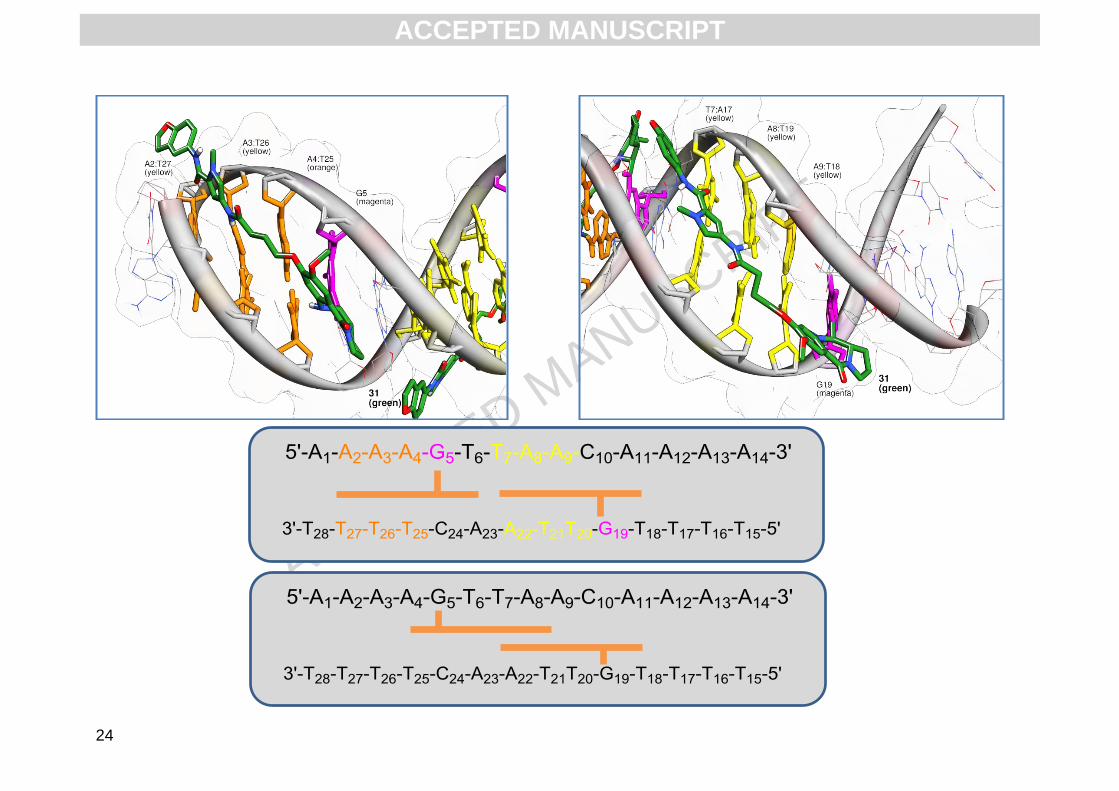
Fig. 5. Low energy snapshot of molecular dynamics simulation of 2 (green) covalently bound to both G5 (magenta) and G19 (magenta) of the
sequence 5'-AAAAGTTAACAAAA-3’. The G5 adduct is in A-ring 5’ orientation and binds to the sequence 5’-AAAG-3’ (where AT base pairs are
shown as orange sticks) and the G19 adduct is in A-ring 3’ orientation on the sequence 5’-GTTAA-3’ (top panel schematic). The potential
second, less favourable orientation of 2 shows the molecule bound in A-ring 3’ orientation alkylated to both G5 and G19, and positioned in the
DNA in an overlapping 2:1 (ligand:DNA) manner.
ACCEPTED MANUSCRIPT

ACC
EPTE
D M
ANU
SCR
IPT
ACCEPTED MANUSCRIPT

ACC
EPTE
D M
ANU
SCR
IPT
27
Table of Contents Entry
C8-conjugates of pyrrolobenzodiazepines bind to guanines that are flanked by A/T-rich sequences
on either the 5'- or 3'- side.
ACCEPTED MANUSCRIPT

ACC
EPTE
D M
ANU
SCR
IPT
28
Research Highlights
C8-conjugates of pyrrolobenzodiazepines with benzofused rings modulate sequence-selective
DNA binding
The conjugates bind best to guanines flanked by A/T-rich sequences.
Bi- and tri-phasic melting curves show that 14-mer oligonucleotides can bind more than one
ligand
ACCEPTED MANUSCRIPT