Embed Size (px)
Citation preview
King’s Research Portal
DOI:10.1371/journal.pone.0178682
Document VersionPublisher's PDF, also known as Version of record
Link to publication record in King's Research Portal
Citation for published version (APA):Jansen, C. H. P., Reimann, C., Brangsch, J., Botnar, R. M., & Makowski, M. R. (2017). In vivo MR-angiographyfor the assessment of aortic aneurysms in an experimental mouse model on a clinical MRI scanner: Comparisonwith high-frequency ultrasound and histology. PloS one, 12(6), e0178682. DOI: 10.1371/journal.pone.0178682
Citing this paperPlease note that where the full-text provided on King's Research Portal is the Author Accepted Manuscript or Post-Print version this maydiffer from the final Published version. If citing, it is advised that you check and use the publisher's definitive version for pagination,volume/issue, and date of publication details. And where the final published version is provided on the Research Portal, if citing you areagain advised to check the publisher's website for any subsequent corrections.
General rightsCopyright and moral rights for the publications made accessible in the Research Portal are retained by the authors and/or other copyrightowners and it is a condition of accessing publications that users recognize and abide by the legal requirements associated with these rights.
•Users may download and print one copy of any publication from the Research Portal for the purpose of private study or research.•You may not further distribute the material or use it for any profit-making activity or commercial gain•You may freely distribute the URL identifying the publication in the Research Portal
Take down policyIf you believe that this document breaches copyright please contact [email protected] providing details, and we will remove access tothe work immediately and investigate your claim.
Download date: 06. Nov. 2017

RESEARCH ARTICLE
In vivo MR-angiography for the assessment of
aortic aneurysms in an experimental mouse
model on a clinical MRI scanner: Comparison
with high-frequency ultrasound and histology
Christian H. P. Jansen1☯, Carolin Reimann2☯*, Julia Brangsch2, Rene M. Botnar1,3,4,5,6,
Marcus R. Makowski1,2,3
1 King’s College London, Division of Imaging Sciences and Biomedical Engineering, London, United
Kingdom, 2 Department of Radiology, Charite, Berlin, Germany, 3 BHF Centre of Excellence, King’s College
London, London, United Kingdom, 4 Wellcome Trust and EPSRC Medical Engineering Center, King’s
College London, London, United Kingdom, 5 NIHR Biomedical Research Centre, King’s College London,
London, United Kingdom, 6 School of Engineering, Pontificia Universidad Catolica de Chile, Santiago, Chile
☯ These authors contributed equally to this work.
Abstract
Background
MR-angiography currently represents one of the clinical reference-standards for the assess-
ment of aortic-dimensions. For experimental research in mice, dedicated preclinical high-
field MRI scanners are used in most studies. This type of MRI scanner is not available in
most institutions. The aim of this study was to evaluate the potential of MR-angiography per-
formed on a clinical MR scanner for the assessment of aortic aneurysms in an experimental
mouse model, compared to a preclinical high-resolution ultrasound imaging system and
histopathology.
Methods
All in vivo MR imaging was performed with a clinical 3T MRI system (Philips Achieva)
equipped with a clinical gradient system in combination with a single-loop surface-coil (47
mm). All MR sequences were based on clinically used sequences. For ultrasound, a dedi-
cated preclinical high-resolution system (30 MHz linear transducer, Vevo770, VisualSonics)
was used. All imaging was performed with an ApoE knockout mouse-model for aortic aneu-
rysms. Histopathology was performed as reference-standard at all stages of aneurysm
development.
Results
MR-angiography on a clinical 3T system enabled the clear visualization of the aortic lumen
and aneurysmal dilation at different stages of aneurysm development. A close correlation
(R2 = 0.98; p < 0.001) with histological area measurements was found. Additionally, a good
agreement between MR and ultrasound area measurements in systole (R2 = 0.91; p <
PLOS ONE | https://doi.org/10.1371/journal.pone.0178682 June 5, 2017 1 / 14
a1111111111
a1111111111
a1111111111
a1111111111
a1111111111
OPENACCESS
Citation: Jansen CHP, Reimann C, Brangsch J,
Botnar RM, Makowski MR (2017) In vivo MR-
angiography for the assessment of aortic
aneurysms in an experimental mouse model on a
clinical MRI scanner: Comparison with high-
frequency ultrasound and histology. PLoS ONE 12
(6): e0178682. https://doi.org/10.1371/journal.
pone.0178682
Editor: Davide Pacini, Universita degli Studi di
Bologna, ITALY
Received: January 20, 2017
Accepted: May 17, 2017
Published: June 5, 2017
Copyright: © 2017 Jansen et al. This is an open
access article distributed under the terms of the
Creative Commons Attribution License, which
permits unrestricted use, distribution, and
reproduction in any medium, provided the original
author and source are credited.
Data Availability Statement: All relevant data are
within the paper.
Funding: The study was funded by the British
Heart Foundation www.bhf.org.uk (PG/09/061) and
the Deutsche Forschungsgemeinschaft [http://
www.dfg.de/] (MA 5943/3-1/ and MA 5943/4-1/9-
1). The funders had no role in study design, data
collection and analysis, decision to publish, or
preparation of the manuscript. Otherwise, there are
0.001) and diastole (R2 = 0.94; p < 0.001) were measured. Regarding interobserver repro-
ducibility, MRI measurements yielded a smaller 95% confidence interval and a closer inter-
reader correlation compared to ultrasound measurements (-0.37–0.46; R2 = 0.97 vs. -0.78–
0.88; R2 = 0.87).
Conclusion
This study demonstrates that MR-angiography, performed on a clinical 3T MR scanner,
enables the reliable detection and quantification of the aortic dilatation at different stages of
aneurysm development in an experimental mouse model.
Introduction
Cardiovascular diseases, including aortic aneurysms, currently represent the main cause of
death in Western societies. Especially the incidence of abdominal aortic aneurysms (AAAs) is
steadily increasing, especially in the last 20 years [1, 2]. One of the main factors for this increase
in incidence is the progressive aging of the general population. Currently the incidence of
abdominal aortic aneurysms is estimated to be around 5% in the general population older than
50 years [3, 4]. The development of abdominal aortic aneurysms is associated with different
causes, which include aortic infection, disorders of connective tissues and traumatic events [5,
6]. However, in most cases, the exact initiating event and pathophysiology, underlying the
development is not fully understood yet [7]. In most cases, abdominal aortic aneurysms are
associated with a progressive dilation of the aortic lumen. If this process continues, abdominal
aortic rupture with fatal consequences can be the result [8].
In clinical practice, the screening for and the evaluation of abdominal aortic aneurysms can
be performed using different imaging modalities [9]. These imaging modalities include magnetic
resonance imaging (MRI), computed tomography (CT) and ultrasound (US). Each imaging
technique is associated with specific advantages and disadvantages. MRI is unique in a sense that
it enables the high-resolution 3D visualization of the aorta without the need for contrast agent or
ionizing radiation. The main disadvantage of MRI is the relatively long scan time, compared to
e.g. CT. The main advantage of CT is that imaging can be performed with a relatively high spatial
resolution in a short time, however CT angiography is dependent on the use of iodinated con-
trast agents. Ultrasound has the advantage, that it is a widely distributed imaging technique avail-
able in most clinical centers. One of its main disadvantages is the operator dependence, which is
especially relevant in the context of follow up examinations.
In magnetic resonance imaging, different techniques can be used for the visualization of
vessels [9]. As MR contrast agents have been recently linked to side effects such as nephrogenic
systemic fibrosis (NSF), non-contrast enhanced techniques are gaining in popularity [10]. In
an experimental setting with small animals, such as mice, non-contrast enhanced techniques
have several advantages. The main advantage is that imaging can be repeated longitudinally at
limitless timepoints in a single animal without the need for intervention.
Experimental mouse models are the most widely used animal models for the preclinical
investigation of diseases [11]. These models enable researchers to investigate the development
of diseases and the influence of genetic modulation or pharmacological therapies on disease
development. Additionally, most novel drugs are initially tested and validated in animal mod-
els prior to clinical trials. For most preclinical studies, histological analysis is performed to
evaluate and quantify in vivo changes. However, noninvasive imaging techniques are gaining
In vivo MR angiography on a clinical MR scanner for the assessment of aortic aneurysms
PLOS ONE | https://doi.org/10.1371/journal.pone.0178682 June 5, 2017 2 / 14
no financial or other relations that could lead to a
conflict of interest.
Competing interests: The authors have declared
that no competing interests exist.
in importance. The main advantage of such an approach is that the number of required ani-
mals for a study is dramatically reduced.
In this context morphological as well as molecular imaging methods, including PET (posi-
tron emission tomography), SPECT (single-photon emission computed tomography), MRI,
CT and ultrasound are the most frequently used techniques. In this group of modalities, MRI
has several advantages including a unique soft tissue contrast in combination with the 3D
acquisition of morphology and function [12, 13]. The majority of MRI studies in mice are per-
formed with dedicated preclinical scanners with an ultra-high field strength (4.7–16.4 Tesla).
Many institutions do not have these kind of preclinical imaging systems available as it usually
requires dedicated personnel, including MR physicists, to run and maintain these systems.
Advantages in hardware development and sequence design have made it possible to perform
small animal imaging in clinical MRI scanners.
In this study, we evaluated the potential of a widely available clinical 3T the MRI system for
the assessment of aortic aneurysm development in a mouse model. We used the most fre-
quently investigated and best validated mouse model, which is based on the ApoE-/- mouse in
combination with angiotensin II infusion [14–17]. Such a mouse model is highly relevant as
abdominal aortic aneurysms represent a cardiovascular disease with severe complications.
Many aspects of this experimental model are comparable to human disease including an
increased incidence of hyperlipidemia [14]. Besides hyperlipidemia, other factors such as
hypertension and cystic necrosis of the aortic wall also play an important role during the devel-
opment of aortic aneurysms. Especially in the context of thoracic aortic aneurysms, a potential
association with cystic medial necrosis has been described by previous studies [18, 19].
The aim of this study was to test the potential and reliability of a clinical 3T MRI system for
the performance of MR-angiography in a mouse model of aortic aneurysm, compared to a
dedicated preclinical high resolution ultrasound imaging system. Histological analysis was
used as reference standard.
Methods
Setup of animal experiments
This study was carried out in strict accordance with the recommendations in the Guide for the
Care and Use of Laboratory Animals of the United Kingdom Home Office and is regulated
under the Animals Scientific Procedures Act 1986 (ASPA). ASPA has recently been revised to
transpose to the European Directive 2010/63/EU on the protection of animals used for scien-
tific purposes. The protocol was approved by the Committee on the Ethics of Animal Experi-
ments of the King’s College London. All animal experiments in this study were performed in
accordance with these international regulations. All intervention was performed with a combi-
nation anesthesia (Medetomidin, Midazolam, Fentanyl), and all efforts were made to minimize
suffering. Osmotic minipumps (Alzet model 2004, Durect Corporation, Cupertino, CA, USA)
were implanted into eight weeks old mice. For this study, homozygous eight weeks old C57BL/
6J ApoE-knockout mice (male) from the Charles Rivers Laboratories were used. The animals
were fed with a standard lab diet and housed in a clean barrier. The minipumps were loaded
(loading was performed as suggested by the manufacturer) ex vivo with AngII (Sigma-Aldrich,
Saint Louis, MO, USA,), implanted subcutaneously in the dorsal region under a combination
anesthesia (500 μg/kg Medetomidin, 50 μg/kg Fentanyl, 5 mg/kg Midazolam) and infused a
continuously dose of 1 microgram kg-1 min-1 into the mice [15, 20]. At week one, two, three
and four after AngII infusion MRI imaging was performed and vessels were harvested for his-
tological analysis each week (n = 8 per group). A sham-operated group (control group, n = 6)
were also implanted minipumps which infused saline for four weeks. Eight mice were scanned
In vivo MR angiography on a clinical MR scanner for the assessment of aortic aneurysms
PLOS ONE | https://doi.org/10.1371/journal.pone.0178682 June 5, 2017 3 / 14
by MRI at each time point. all animals were sacrificed for further histopathological analysis
after the final imaging sessions. For the imaging session, mice were anesthetized with an intra-
muscular application [21, 22] of the same combination of Medetomidin, Fentanyl, Midazolam
as mentioned above. In all mice with abdominal aortic aneurysm, an exsanguination in ante-
rior perfusion with phosphate buffered saline (100 mm Hg) was performed following the MR
imaging session. This was followed by a perfusion with 10% formalin if vessel samples were
used for histology. Aorta, right renal artery and the last pair of intercostal arteries was excised
to allow anatomical matching during histopathological processing of the samples.
Animal handling and in vivo magnetic resonance imaging
Each animal was in anesthesia (as described earlier) and placed in a prone position on a surface
microscopy coil (Philips healthcare, Best, the Netherlands). All imaging was performed using a
clinical 3T Achieva MR system (Philips Healthcare, Best, The Netherlands). A gradient system
with a gradient strength of 30 mT/m and a slew rate of 200 T/m/s was used. For the sequence
acquisition, a dedicated software package for cardiac imaging was available. The signal was
gained using a microscopy single loop coil with an inner diameter of 47 mm. The coil was
placed in the magnetic center of the bore of the MRI. An MR compatible body temperature
monitoring and heating system was used to maintain the temperature (37º degrees Celsius) of
all animals during the entire acquisition of the MR data sets (Model 1025, SA Instruments
Inc., Stony Brook, NY). All MR imaging sequences were based on clinical MR sequences. The
imaging protocol included the following sequences. At the beginning of a MR imaging proto-
col a low-resolution scout sequence (three-dimensional gradient echo sequence) was used to
for an anatomical overview and localization of the abdominal aorta. The scout scan was per-
formed in the coronal and transverse orientation using the following parameters: field-of-view
(FOV) = 200 mm, matrix = 320, slice thickness = 2 mm, TR/TE = 20/5.8ms, flip angle = 30˚
and slices = 9. Following a transverse orientation of a two-dimensional time-of-flight angiog-
raphy (2D TOF) was executed for a precise visualization of the abdominal aorta. The 2D time-
of-flight sequence was planned to include the renal arteries as anatomical landmarks in all
scans. The image parameters included: Slice thickness = 0.5 mm, inplane spatial resolution = 0.3
x 0.3 mm (reconstructed 0.13 x 0.13 mm), imaging matrix = 160 x 160, field of view = 20 x 20 x
10 mm, flip angle = 60˚, echo time (TE) 7.7 ms and repetition time (TR) sequence = 37 ms.
Fold-over suppression was activated. Fold-over direction was right to left. A cartesian acquisi-
tion mode was used. A maximum intensity projection (MIP) in a 360-degree reconstruction
was automatically reconstructed based on the time-of-flight angiography (Fig 1).
Magnetic resonance image analysis
Signal to noise measurements (SNR) were performed directly proximal to the right renal artery
in the control group (sham group, Fig 1). The right renal artery was clearly visible in all MR
scans. If an aneurysm was present, signal to noise measurements (SNR) were performed at the
location of the maximal area size (Fig 2). MR image analysis of DICOM images was performed
using the open source version of OsiriX (version 7.1, OsiriX foundation). Time of flight (TOF)
images were used to localize aortic aneurysms. Region of interests (ROIs) were measured as
areas of signal enhancement on TOF images for the evaluation of signal intensity. Region of
interests were drawn to delineate the complete vascular lumen for a reproducible measure-
ment. For the ROIs signal to noise ratio (SNR) was calculated with following formula: Signal
to noise ratio MR-angiography (MRA) = ((aneurysmal) aortic lumen signal) / (standard devia-
tion lumen signal). Such an approach was chosen to allow for comparability between the anal-
ysis methods for MR and ultrasound.
In vivo MR angiography on a clinical MR scanner for the assessment of aortic aneurysms
PLOS ONE | https://doi.org/10.1371/journal.pone.0178682 June 5, 2017 4 / 14

In vivo high-frequency ultrasound imaging
High resolution ultrasound imaging was performed with animals placed on a automatically
heated table (37˚) in supine position. Prior to ultrasound imaging, all animals were depilated
with hair removal cream. For ultrasound measurements, a Vevo 770 ultrasound imaging sys-
tem (VisualSonics, Toronto, Canada) with a 30 MHz linear signal transducer was used. For
Fig 1. Visualization of the abdominal aorta in a control mouse by MR-angiography on a clinical MR
system in comparison to high-frequency ultrasound. Images demonstrate the visualization of the
abdominal aorta in a sham-operated ApoE-/- mouse by in vivo MR-angiography on a clinical MR system (A)
and dedicated high-frequency ultrasound (C) in comparison to histology (B). The reconstructed TOF
angiogram (A1, maximum intensity projection (MIP)) of the suprarenal part of the nondilated abdominal aorta
is shown. Red lines indicate the orientation of subsequently performed transverse MRI sequences (A2, A3,
A4). Corresponding ex vivo histological sections (B1- B6), Elastica van Gieson (EvG) stain (B1, B2, B3),
hematoxylin eosin (HE) stain (B4, B5, B6) demonstrate a nondilated abdominal aorta at different levels (red
lines). High-frequency ultrasound images (C) of an abdominal aorta using a dedicated imaging system (Vevo
770). Corresponding longitudinal (C4) and transversal imaging planes (C1, C2, C3) are shown.
https://doi.org/10.1371/journal.pone.0178682.g001
Fig 2. Evaluation of the abdominal aorta in an ApoE-/- mouse by MR-angiography on a clinical MR
system and high-frequency ultrasound. Evaluation of an abdominal aortic aneurysm in an ApoE-/- mouse
by in vivo MRI and ultrasound 4 week after continuous infusion of angiotensin II (4-week group). The maximum
intensity projection (MIP) of the time-of-flight (TOF) angiogram (A1) demonstrates a significantly dilated aortic
lumen. The location of transverse slices (A2, A3) are depicted by the red lines in A1. Corresponding ex vivo
histological sections (Elastica van Gieson (EvG) stain (B1, B2), hematoxylin eosin (HE) stain (B3, B4) confirm
the dilation of the aortic lumen. Magnifications of B2 and B4 highlight the site of rupture of the elastic laminae in
the tunica media of the aorta in EvG stain and HE stain. Corresponding ultrasound images of abdominal aorta
using the dedicated high-frequency US imaging system (Vevo 770) in sagittal (C3) and transversal orientation
(C1, C2).
https://doi.org/10.1371/journal.pone.0178682.g002
In vivo MR angiography on a clinical MR scanner for the assessment of aortic aneurysms
PLOS ONE | https://doi.org/10.1371/journal.pone.0178682 June 5, 2017 5 / 14
imaging and anatomical colocalization the suprarenal abdominal aorta more than 20 dynamic
2D-transverse and sagittal images were acquired in all animals. Cine transversal and sagittal
images were reconstructed by the ECG-based kilohertz visualization (EKV) technique. The
resulting images were analyzed during end-diastole and during end-systole.
Analysis of in vivo high-frequency ultrasound imaging
Signal to noise measurements (SNR) were performed in the same animal at the same location as
MR-angiography measurements. If an aneurysm was present, measurements were performed at
the location of the maximal area size. If no aneurysm was present, measurements were per-
formed directly proximal to the right renal artery. To obtain comparable measurements be-
tween MRI and ultrasound data sets comparable techniques for the assessment of the SNR were
applied. Region of interests (ROIs) were measured as areas of signal enhancement of luminal
aortic signal. The region of interest were drawn to delineate the complete vessel. For the ROIs
signal to noise ratio (SNR) was calculated with following formula: Signal to noise ratio ultra-
sound (US) = ((aneurysmal) aortic lumen signal) / (standard deviation lumen signal).
Aortic aneurysm morphometry
Aorta, right renal artery and the last pair of intercostal arteries was excised to allow precise
anatomical matching between MRI, ultrasound and histopathology. The left renal artery and
the last pair of intercostal artery were the main landmarks for the co-registration. The mor-
phometrical analysis was performed using elastin-stained sections (Miller’s Elastica van Gieson
stain) and ImageProPlus software (ImageProPlus, MediaCybernetics).
Histological analysis of aortic aneurysms
Histological analysis was performed in the same region of aorta that was imaged with magnetic
resonance imaging and ultrasound. In vivo and ex vivo morphometric data could therefore be
directly compared. Surgically removed aortic aneurysms were processed overnight for further
histological processing. Segmented aortic aneurysms were embedded in paraffin and were cut
from the proximal end of the aneurysm every 40 μm into 6 μm thick serial sections. After
dewaxing and rehydration, the sections were stained using Miller’s Elastica van Gieson stain
(EvG) and hematoxylin and eosin (HE).
Interobserver agreements magnetic resonance angiography and
ultrasound measurements
For the assessment of the interreader variability two investigators performed the aortic mea-
surements. All images were analyzed independently in a randomized order and blinded to the
according other imaging modalities. Area sizes were recorded for each measurement.
Statistical analysis
Data are expressed as mean ± standard deviation. A Student’s t test (two–tailed, unpaired) was
used to compare continuous variables and verify the statistical significance between sham
(control) and treated aortas. If more than two groups were investigated, a variance analysis
(ANOVA) and Bonferroni correction was performed for statistical comparison. Interobserver
agreements for ex vivo and in vivo measurements were assessed using Bland-Altman plots,
which were generated for the raw volume data to display the spread of data and the limits of
agreement. Linear regression was applied to determine the relationship between are measure-
ments on MRI, ultrasound and histology.
In vivo MR angiography on a clinical MR scanner for the assessment of aortic aneurysms
PLOS ONE | https://doi.org/10.1371/journal.pone.0178682 June 5, 2017 6 / 14
Results
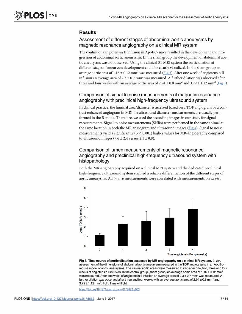
Assessment of different stages of abdominal aortic aneurysms by
magnetic resonance angiography on a clinical MR system
The continuous angiotensin II infusion in ApoE-/- mice resulted in the development and pro-
gression of abdominal aortic aneurysms. In the sham group the development of abdominal aor-
tic aneurysms was not observed. Using the clinical 3T MRI system the aortic dilation at
different stages of aneurysm development could be clearly visualized. In the sham group an
average aortic area of 1.16 ± 0.12 mm2 was measured (Fig 3). After one week of angiotensin II
infusion an average area of 2.3 ± 0.7 mm2 was measured. A further dilation was observed after
three and four weeks with an average aortic area of 2.94 ± 0.8 mm2 and 3.79 ± 1.12 mm2 (Fig 3).
Comparison of signal to noise measurements of magnetic resonance
angiography with preclinical high-frequency ultrasound system
In clinical practice, the luminal area/diameter is assessed based on a TOF angiogram or a con-
trast enhanced angiogram in MRI. In ultrasound diameter measurements are usually per-
formed in the B-mode. Therefore, we used the according images in our study for signal
measurements. Signal to noise measurements (SNRs) were performed in the same animal at
the same location in both the MR angiogram and ultrasound images (Fig 4). Signal to noise
measurements yield a significantly (p< 0.001) higher values for MR-angiography compared
to ultrasound images (7.6 ± 2.4 versus 2.1 ± 0.9).
Comparison of lumen measurements of magnetic resonance
angiography and preclinical high-frequency ultrasound system with
histopathology
Both the MR-angiography acquired on a clinical MRI system and the dedicated preclinical
high-frequency ultrasound system enabled a reliable differentiation of the different stages of
aortic aneurysms. All in vivo measurements were correlated with measurements on ex vivo
Fig 3. Time course of aortic dilatation assessed by MR-angiography on a clinical MR system. In vivo
assessment of the dimensions of abdominal aortic aneurysm measured in the TOF angiography in an ApoE-/-
mouse model of aortic aneurysms. The luminal aortic areas were measured in vivo after one, two, three and four
weeks of angiotensin II infusion. In the control group (sham group) an average aortic area of 1.16 ± 0.12 mm2
was measured. After one week of angiotensin II infusion an average area of 2.3 ± 0.7 mm2 was measured. A
further dilation was observed after three and four weeks with an average aortic area of 2.94 ± 0.8 mm2 and
3.79 ± 1.12 mm2. ToF: Time of flight.
https://doi.org/10.1371/journal.pone.0178682.g003
In vivo MR angiography on a clinical MR scanner for the assessment of aortic aneurysms
PLOS ONE | https://doi.org/10.1371/journal.pone.0178682 June 5, 2017 7 / 14

histology (Elastica-van-Giesson stain, reference standard). Area measurements on in vivo MR
angiograms showed the closest correlation with ex vivo measurements (R2 = 0.98; p< 0.001,
Table 1), while in vivo measurements slightly and systemically overestimated the size of the
aneurysmal area (Fig 5). This can be explained by the shrinkage of the histological specimens
following the processing of the tissue samples. In high-frequency ultrasound, systolic and
diastolic area measurements showed a strong, however slightly lower, correlation with ex vivohistology compared to the MR-angiography. The correlation coefficient for systolic area mea-
surements was R2 = 0.91 (p< 0.001) and for diastolic area measurements R2 = 0.93 (p<
0.001). Comparable to MR measurements, ultrasound measurements in both cardiac phases
resulted in a slight systemic overestimation of the area of the aortic aneurysm. This can be
explained by the shrinkage of the histological specimens following tissue processing.
Fig 4. Signal to noise measurements of the aortic lumen on a clinical MR system and on a dedicated
high-frequency ultrasound system. This figure shows that magnetic resonance angiography (MRA, black
bar) demonstrated a significantly (p < 0.001) higher signal to noise ratio (SNR) compared to ultrasound (US,
grey bar). MRI and ultrasound measurements were performed at comparable locations of the aorta. The time-
of-flight technique in MR and the B-mode in ultrasound are techniques which are also frequently used in a
clinical setting.
https://doi.org/10.1371/journal.pone.0178682.g004
Table 1. Summary of results from in vivo magnetic resonance imaging, ultrasound and ex vivo histology.
Mean SD 95% CI R2 p value
MRI vs US systole -0.26 0.34 -0.94 to 0.42 0.91 <0.001
MRI vs US diastole 0.03 0.26 -0.49 to 0.54 0.94 <0.001
MRI vs Histology -0.50 0.26 -1.01 to 0.01 0.98 <0.001
US systole vs Histology -0.25 0.31 -0.86 to 0.37 0.91 <0.001
US diastole vs Histology -0.53 0.33 -1.20 to 0.14 0.93 <0.001
Interobserver MRI 0.04 0.21 -0.37 to 0.46 0.96 <0.001
Interobserver US 0.05 0.42 -0.78 to 0.88 0.87 <0.001
https://doi.org/10.1371/journal.pone.0178682.t001
In vivo MR angiography on a clinical MR scanner for the assessment of aortic aneurysms
PLOS ONE | https://doi.org/10.1371/journal.pone.0178682 June 5, 2017 8 / 14
This table summarizes the results from the different in vivo imaging modalities (MRI, ultra-
sound in systole and diastole, including interobserver variability) and ex vivo histology.
Regarding in vivo measurements, the closest correlation was found between MRI and ultra-
sound measurements in diastole. MRI showed the closest correlation with area measurements
on ex vivo histology. Interobserver variation was smaller for MRI compared to ultrasound.
95% CI: 95% confidence interval.
Comparison of lumen measurements of magnetic resonance
angiography with high-frequency ultrasound system
Luminal area measurements on TOF MR-angiography showed a close correlation with measure-
ments derived from the dedicated preclinical high-frequency ultrasound system in systole and
diastole (Fig 6). However, the correlation was slightly higher in diastole (R2 = 0.94; p< 0.001)
compared to the systole (R2 = 0.91; p< 0.001). As the TOF angiography continuously acquires
images throughout systole and diastole, the resulting image reflects the larger diameter acquired
in diastole, as summation effects occur. Therefore, a slightly better correlation was found between
TOF angiography and ultrasound images acquired in diastole.
Interobserver agreements magnetic resonance angiography and
ultrasound measurements
Interobserver correlation for area measurements in MR-angiography showed a close correla-
tion between both image readers (R2 = 0.96; p< 0.001) (Fig 7). The associated 95% confidence
interval (CI) for the correlation range was -0.73 to 0.46. Interobserver correlation for area mea-
surements for high-frequency ultrasound also showed a strong correlation between both
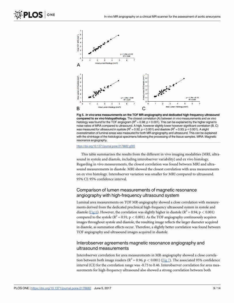
Fig 5. In vivo area measurements on the TOF MR-angiography and dedicated high-frequency ultrasound
compared to ex vivo histopathology. The closest correlation (A) between in vivo measurements and ex vivo
histology was found for the TOF angiogram (R2 = 0.98; p < 0.001). This can be explained by the higher signal to
noise ratios of MRA compared to ultrasound. A high, however slightly lower however significant correlation (B, C)
was measured for ultrasound in systole (R2 = 0.92; p < 0.001) and diastole (R2 = 0.93; p < 0.001). A slight
overestimation of luminal areas was measured for both MR-angiography and ultrasound. This can be explained
with the shrinkage of the histological specimens following the processing of the tissue samples. MRA: Magnetic
resonance angiography.
https://doi.org/10.1371/journal.pone.0178682.g005
In vivo MR angiography on a clinical MR scanner for the assessment of aortic aneurysms
PLOS ONE | https://doi.org/10.1371/journal.pone.0178682 June 5, 2017 9 / 14
readers (R2 = 0.87; y = 0.98x + 0.11; p< 0.001). The associated 95% confidence interval (CI)
for the correlation range was 0.88 to -0.78. The interobserver correlation for MR-angiography
measurements was slightly higher compared to the interobserver correlation for high-fre-
quency ultrasound measurements (Fig 7).
Discussion
In this imaging study, we report that MR-angiography (MRA), performed on a clinical 3T MR
scanner, enables the reliable detection and quantification of the different stages of aneurysm
development in an experimental mouse model.
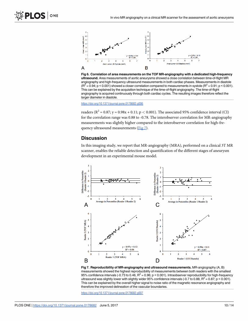
Fig 6. Correlation of area measurements on the TOF MR-angiography with a dedicated high-frequency
ultrasound. Area measurements of aortic aneurysms showed a close correlation between time-of-flight MR
angiography and high-frequency ultrasound measurements in both cardiac phases. Measurements in diastole
(R2 = 0.94; p < 0.001) showed a closer correlation compared to measurements in systole (R2 = 0.91; p < 0.001).
This can be explained by the acquisition technique of the time-of-flight angiography. The time-of-flight
angiography is acquired continuously through both cardiac cycles. The resulting images therefore reflect the
larger diameter in diastole.
https://doi.org/10.1371/journal.pone.0178682.g006
Fig 7. Reproducibility of MR-angiography and ultrasound measurements. MR-angiography (A, B)
measurements showed the highest reproducibility of measurements between both readers with the smallest
95% confidence intervals (-0.73 to 0.46, R2 = 0.96; p < 0.001). Intraobserver reproducibility for high-frequency
ultrasound was slightly lower with slightly wider 95% confidence intervals (-0.7 to 0.88, R2 = 0.87; p < 0.001).
This can be explained by the overall higher signal to noise ratio of the magnetic resonance angiography and
therefore the improved delineation of the vascular boundaries.
https://doi.org/10.1371/journal.pone.0178682.g007
In vivo MR angiography on a clinical MR scanner for the assessment of aortic aneurysms
PLOS ONE | https://doi.org/10.1371/journal.pone.0178682 June 5, 2017 10 / 14
Imaging techniques for the assessment of aortic aneurysms
Different imaging modalities such as MRI and ultrasound can be used for the assessment of
the size of the aortic lumen [9]. In MRI, different techniques can be used for the visualization
of vessels. These include non-contrast enhanced and contrast-enhanced techniques [9]. In an
experimental setting with small animals, such as mice, non-contrast enhanced techniques have
several advantages. The main advantage is that imaging can be repeated at limitless timepoints
longitudinally in a single animal. A further advantage is, that TOF imaging sequences do not
rely on a first pass of the contrast agent. Furthermore, if a study with e.g. a specific or unspe-
cific MR probe or contrast agent is performed, no additional contrast agent for the angiogra-
phy is required. Such an additional administration of contrast agent could e.g. interfere with
the biokinetics of the evaluated MR probe. The used TOF MR imaging technique is based on
the use of relatively short MR repetition times [23]. At each acquisition step, fresh or unsatu-
rated nuclear spins flow into the imaging slice driven by the arterial blood stream. This leads
to the bright signal in this MR angiographic technique. In the clinical setting, this imaging
technique is routinely used for the visualization of e.g. the cerebral arteries [23]. In preclinical
models, this technique is especially useful for the assessment of the vascular system, as it is a
relatively fast imaging sequence enabling high throughput preclinical serial imaging, e.g. for
pharmacological studies.
A further technique which is used in the preclinical and clinical setting is ultrasound of
the aorta. Ultrasound has the advantage that it enables image acquisition with a high temporal
resolution which goes beyond the temporal resolution of standard MR imaging sequences. In
this study, ultrasound images could therefore be analyzed in end-diastole and end-systole. Var-
ious clinical imaging studies have shown that ultrasound is associated with potential limita-
tions [24]. This imaging technique is dependent on the level of experience the operator has.
Additionally, image quality depends on the acoustic window. Compared to MR imaging, the
luminal boundaries are more challenging to delineate on conventional ultrasound images
compared to MR-angiography. This preclinical study confirmed what clinical studies have
suggested, the interobserver reproducibility is higher in MR measurements compared to ultra-
sound measurements [9].
Besides magnetic resonance imaging and ultrasound, computed tomography (CT) can also
be used for the evaluation of aortic aneurysms. The main advantage of computed tomography
is that image acquisition is relatively fast compared to MRI and ultrasound. The acquisition
of the complete aorta is usually performed with in one minute. The main disadvantage of CT
however is, that it relies on the use of ionizing radiation. This is especially important in the
context of frequent follow up examinations, which are usually required for the evaluation of
aortic aneurysms. Additionally, a CT angiography is in clinical practice performed in combi-
nation with iodinated contrast agents. In contrast, MRI and ultrasound enable the visualiza-
tion of the aorta without the need for contrast agents or ionizing radiation.
In summary, in this study we could demonstrate that MR-angiography in an experimental
mouse model can reliably be performed on a clinical 3T MR system. In vivo area measure-
ments derived from MRI showed a good correlation with measurements from high-frequency
ultrasound and histopathology. Area measurements in MR slightly overestimated the luminal
area, compared to histopathology. This can be explained by the tissue shrinkage resulting from
the processing of histological samples. The interobserver variability of MR measurements was
lower compared to ultrasound measurements. This could be explained by the 3D MR acquisi-
tion. This is of relevance, as a high interobserver reproducibility is important, especially for
follow-up measurements during the development of aortic aneurysms. To the best of our
knowledge, this is the first study that reports the reliable assessment of aortic aneurysms in a
In vivo MR angiography on a clinical MR scanner for the assessment of aortic aneurysms
PLOS ONE | https://doi.org/10.1371/journal.pone.0178682 June 5, 2017 11 / 14

mouse model using a clinical 3T system with a close correlation to ultrasound and histological
area measurements.
Relevance of experimental models in the context of aortic aneurysms
In the group of cardiovascular diseases, the 3rd most common cause of sudden death is the
rupture of aortic aneurysms. Overall the pathophysiology of the development of aortic aneu-
rysms is not fully understood yet [25]. This is reflected by the fact that most aortic aneurysms
are still classified as nonspecific [26]. Regardless of the underlying reason, aortic aneurysms
are recognized by a progressive dilation of the aortic lumen. If this process is not detected,
progressive dilation can lead to aortic rupture with potentially deadly consequences for the
patient. The degree of dilation of the aorta is currently the best established parameter to assess
the risk of rupture [27, 28]. To further develop our understanding of the etiology of the onset,
development and progression of aortic aneurysms different animal models have been used in
previous studies [29–31]. Both pharmacological and surgical techniques have been applied to
induce the reproducible development of aortic aneurysms [32]. The most widely used model is
based on an ApoE-/- mouse in which angiotensin II is continuously administered [17]. This
leads to the spontaneous, reliable and reproducible onset and progression of aortic aneurysms
without the need for a direct surgical manipulation of the aorta. For an accurate and reproduc-
ible assessment of aneurysm development, measurements on histopathological slides are usu-
ally performed ex vivo. The drawback of such an ex vivo approach is the high number of
animals required to achieve a sufficient statistical power.
MR angiography performed on a clinical 3T MR scanner could give more research groups
access to a reliable in vivo MR imaging technique for the investigation of aortic aneurysms
in experimental models. Additionally, based on in vivo MR imaging, the number of research
animals required for the investigation of the different stages of aortic aneurysms could be
reduced.
Limitations
Clinical MRI scanners with a clinically used field strength (1.5–3.0 T) yield a lower signal-to-
noise ratio (SNR) compared scanner with a high field strength (4.7–16.4 T), we however did
not perform a comparison between ultrahigh field dedicated preclinical MR imaging systems
and clinical MR systems. We did not perform Doppler color velocity ultrasound imaging as it
was not available on our ultrasound system. However, area measurements in ultrasound are in
clinical practice usually performed on standard B-mode images, as they offer the highest spa-
tial resolution.
Conclusion
This study demonstrates that MR-angiography performed on a clinical 3T MR scanner enables
the reliable detection and quantification of aortic dilatation at the different stages of aneurysm
development in an experimental mouse model. Interobserver analysis demonstrated a higher
reproducibility of MR measurements compared to ultrasound measurements. Both MR-angi-
ography and ultrasound showed a close correlation with histology.
Author Contributions
Conceptualization: CHPJ CR JB RMB MRM.
Data curation: CHPJ.
In vivo MR angiography on a clinical MR scanner for the assessment of aortic aneurysms
PLOS ONE | https://doi.org/10.1371/journal.pone.0178682 June 5, 2017 12 / 14
Formal analysis: CHPJ MRM.
Funding acquisition: RMB MRM.
Investigation: CHPJ MRM.
Methodology: CHPJ RMB MRM.
Project administration: RMB MRM.
Supervision: CHPJ RMB MRM.
Validation: CHPJ RMB MRM.
Writing – original draft: CHPJ CR JB RMB MRM.
Writing – review & editing: CHPJ CR JB RMB MRM.
References1. Gillum RF. Epidemiology of aortic aneurysm in the United States. J Clin Epidemiol. 1995; 48
(7490591):1289–98.
2. Bengtsson H, Sonesson B, Bergqvist D. Incidence and prevalence of abdominal aortic aneurysms, esti-
mated by necropsy studies and population screening by ultrasound. Ann N Y Acad Sci. 1996; 800:1–
24. Epub 1996/11/18.
3. Kniemeyer HW, Kessler T, Reber PU, Ris HB, Hakki H, Widmer MK. Treatment of ruptured abdominal
aortic aneurysm, a permanent challenge or a waste of resources? Prediction of outcome using a multi-
organ-dysfunction score. Eur J Vasc Endovasc Surg. 2000; 19(10727370):190–6.
4. Hallett JW Jr. Management of abdominal aortic aneurysms. Mayo Clin Proc. 2000; 75(4):395–9. Epub
2000/04/13. https://doi.org/10.4065/75.4.395 PMID: 10761495
5. Erentug V, Bozbuga N, Omeroglu SN, Ardal H, Eren E, Guclu M, et al. Rupture of abdominal aortic
aneurysms in Behcet’s disease. Ann Vasc Surg. 2003; 17(14738093):682–5.
6. Matsumura K, Hirano T, Takeda K, Matsuda A, Nakagawa T, Yamaguchi N, et al. Incidence of aneu-
rysms in Takayasu’s arteritis. Angiology. 1991; 42(1673052):308–15.
7. Johnston KW, Rutherford RB, Tilson MD, Shah DM, Hollier L, Stanley JC. Suggested standards for
reporting on arterial aneurysms. Subcommittee on Reporting Standards for Arterial Aneurysms, Ad Hoc
Committee on Reporting Standards, Society for Vascular Surgery and North American Chapter, Inter-
national Society for Cardiovascular Surgery. J Vasc Surg. 1991; 13(1999868):452–8.
8. Sakalihasan N, Limet R, Defawe OD. Abdominal aortic aneurysm. Lancet. 2005; 365(9470):1577–89.
Epub 2005/05/04. https://doi.org/10.1016/S0140-6736(05)66459-8 PMID: 15866312
9. Evangelista A. Imaging aortic aneurysmal disease. Heart. 2014; 100(12):909–15. https://doi.org/10.
1136/heartjnl-2013-305048 PMID: 24842834
10. Todd DJ, Kay J. Gadolinium-Induced Fibrosis. Annu Rev Med. 2016; 67:273–91. https://doi.org/10.
1146/annurev-med-063014-124936 PMID: 26768242
11. Trollope A, Moxon JV, Moran CS, Golledge J. Animal models of abdominal aortic aneurysm and their
role in furthering management of human disease. Cardiovascular pathology: the official journal of the
Society for Cardiovascular Pathology. 2011; 20(2):114–23.
12. Buijs RV, Willems TP, Tio RA, Boersma HH, Tielliu IF, Slart RH, et al. Current state of experimental
imaging modalities for risk assessment of abdominal aortic aneurysm. J Vasc Surg. 2013; 57(3):851–9.
https://doi.org/10.1016/j.jvs.2012.10.097 PMID: 23357517
13. Forsythe RO, Newby DE, Robson JM. Monitoring the biological activity of abdominal aortic aneurysms
Beyond Ultrasound. Heart. 2016; 102(11):817–24. PubMed Central PMCID: PMC4893091. https://doi.
org/10.1136/heartjnl-2015-308779 PMID: 26879242
14. Daugherty A, Manning MW, Cassis LA. Angiotensin II promotes atherosclerotic lesions and aneurysms
in apolipoprotein E-deficient mice. The Journal of clinical investigation. 2000; 105(11):1605–12.
PubMed Central PMCID: PMC300846. https://doi.org/10.1172/JCI7818 PMID: 10841519
15. Saraff K, Babamusta F, Cassis LA, Daugherty A. Aortic dissection precedes formation of aneurysms
and atherosclerosis in angiotensin II-infused, apolipoprotein E-deficient mice. Arteriosclerosis, thrombo-
sis, and vascular biology. 2003; 23(9):1621–6. https://doi.org/10.1161/01.ATV.0000085631.76095.64
PMID: 12855482
In vivo MR angiography on a clinical MR scanner for the assessment of aortic aneurysms
PLOS ONE | https://doi.org/10.1371/journal.pone.0178682 June 5, 2017 13 / 14
16. Daugherty A, Cassis LA. Mouse models of abdominal aortic aneurysms. Arteriosclerosis, thrombosis,
and vascular biology. 2004; 24(3):429–34. https://doi.org/10.1161/01.ATV.0000118013.72016.ea
PMID: 14739119
17. Cao RY, Amand T, Ford MD, Piomelli U, Funk CD. The Murine Angiotensin II-Induced Abdominal Aortic
Aneurysm Model: Rupture Risk and Inflammatory Progression Patterns. Frontiers in pharmacology.
2010; 1:9. PubMed Central PMCID: PMC3112241. https://doi.org/10.3389/fphar.2010.00009 PMID:
21713101
18. El-Hamamsy I, Yacoub MH. Cellular and molecular mechanisms of thoracic aortic aneurysms. Nat Rev
Cardiol. 2009; 6(12):771–86. https://doi.org/10.1038/nrcardio.2009.191 PMID: 19884902
19. Moritz AR. Medionecrosis Aortae Idiopathica Cystica. The American Journal of Pathology. 1932; 8
(6):717–34.3. PMID: 19970043
20. Daugherty Manning MW, Cassis LA. Angiotensin II promotes atherosclerotic lesions and aneurysms in
apolipoprotein E-deficient mice. The Journal of clinical investigation. 2000; 105(10841519):1605–12.
21. Botnar RM, Wiethoff AJ, Ebersberger U, Lacerda S, Blume U, Warley A, et al. In vivo assessment of
aortic aneurysm wall integrity using elastin-specific molecular magnetic resonance imaging. Circulation
Cardiovascular imaging. 2014; 7(4):679–89. https://doi.org/10.1161/CIRCIMAGING.113.001131
PMID: 24871347
22. Makowski MR, Wiethoff AJ, Blume U, Cuello F, Warley A, Jansen CH, et al. Assessment of atheroscle-
rotic plaque burden with an elastin-specific magnetic resonance contrast agent. Nature medicine. 2011;
17(3):383–8. https://doi.org/10.1038/nm.2310 PMID: 21336283
23. MacDonald ME, Frayne R. Cerebrovascular MRI: a review of state-of-the-art approaches, methods and
techniques. NMR Biomed. 2015; 28(7):767–91. https://doi.org/10.1002/nbm.3322 PMID: 26010775
24. Barisione C, Charnigo R, Howatt DA, Moorleghen JJ, Rateri DL, Daugherty A. Rapid dilation of the
abdominal aorta during infusion of angiotensin II detected by noninvasive high-frequency ultrasonogra-
phy. J Vasc Surg. 2006; 44(2):372–6. https://doi.org/10.1016/j.jvs.2006.04.047 PMID: 16890871
25. Schouten O, Poldermans D. Statins in the prevention of perioperative cardiovascular complications.
Current opinion in anaesthesiology. 2005; 18(1):51–5. PMID: 16534317
26. Johnston KW, Rutherford RB, Tilson MD, Shah DM, Hollier L, Stanley JC. Suggested standards for
reporting on arterial aneurysms. Subcommittee on Reporting Standards for Arterial Aneurysms, Ad Hoc
Committee on Reporting Standards, Society for Vascular Surgery and North American Chapter, Inter-
national Society for Cardiovascular Surgery. Journal of vascular surgery. 1991; 13(3):452–8. PMID:
1999868
27. Kent KC. Clinical practice. Abdominal aortic aneurysms. N Engl J Med. 2014; 371(22):2101–8. https://
doi.org/10.1056/NEJMcp1401430 PMID: 25427112
28. Wanhainen A, Mani K, Golledge J. Surrogate Markers of Abdominal Aortic Aneurysm Progression.
Arteriosclerosis, thrombosis, and vascular biology. 2016; 36(2):236–44. https://doi.org/10.1161/
ATVBAHA.115.306538 PMID: 26715680
29. Tsui JC. Experimental models of abdominal aortic aneurysms. The open cardiovascular medicine jour-
nal. 2010; 4:221–30. PubMed Central PMCID: PMC3026392. https://doi.org/10.2174/
1874192401004010221 PMID: 21270944
30. Argenta R, Pereira AH. Animal models of aortic aneurysm. Jornal Vascular Brasileiro. 2009; 8:148–53.
31. Yoo YS, Park HS, Choi GH, Lee T. Recent Advances in the Development of Experimental Animal Mod-
els Mimicking Human Aortic Aneurysms. Vascular specialist international. 2015; 31(1):1–10. PubMed
Central PMCID: PMC4480291. https://doi.org/10.5758/vsi.2015.31.1.1 PMID: 26217637
32. Daugherty A, Rateri DL, Cassis LA. Role of the renin-angiotensin system in the development of abdomi-
nal aortic aneurysms in animals and humans. Ann N Y Acad Sci. 2006; 1085:82–91. https://doi.org/10.
1196/annals.1383.035 PMID: 17182925
In vivo MR angiography on a clinical MR scanner for the assessment of aortic aneurysms
PLOS ONE | https://doi.org/10.1371/journal.pone.0178682 June 5, 2017 14 / 14