Embed Size (px)
Citation preview
395
Kinetic and kinematic gait analysis in a spastic hemiplegic patient after selective tibial neurotomy: a case report1Ippei Kitade RPT PhD, 2Hidetaka Arishima MD PhD, 2Ken-ichiro Kikuta MD PhD
1Division of Rehabilitation Medicine, University of Fukui Hospital, Fukui; 2Department of Neurosurgery, University of Fukui, Japan Abstract
Kinematics-based studies before and after selective tibial neurotomy (STN) gait have not been performed. It is very important for spastic patients before and after STN to evaluate quality of gait motion. We examined the quantitative changes in kinetic and kinematic parameters in the gait of a hemiplegic patient after STN. A patient with stroke-related hemiplegia who did not require aids to walk underwent a three-dimensional gait analysis (3DGA) before and after STN. 3DGA system was used to obtain spatiotemporal, kinetic and kinematic parameters of the lower extremities. Postoperative increases in walking speed and the single leg support ratio were detected in the paralyzed limb. Kinetic and kinematic analyses of the stance phase performed after STN detected dorsiflexion in the ankle, the appearance of generation power during plantar flexion, an extension of the range of hip movement during the gait cycle, and the disappearance of genu recurvatum. The acquisition of a normalized ankle joint gait pattern after STN might result in coordinated improvements in the kinetic and kinematic parameters of other joints. The measurement of spatiotemporal, kinetic, and kinematic gait parameters using 3DGA systems might aid decisions regarding the optimal post-STN rehabilitation strategies for spastic patients who hope to improve their gaits.
Neurology Asia 2015; 20(4) : 395 – 399
Address correspondence to: Ippei Kitade, RPT, PhD, Division of Rehabilitation Medicine, University of Fukui Hospital, Shimoaizuki 23, Matsuoka, Eiheiji-cho,Yoshida-gun, Fukui 910-1193, Japan. Tel: +81-776-61-8676, Fax: +81-776-61-8480, E-mail: [email protected]
INTRODUCTION
Stroke-induced equinovarus foot deformities have a negative influence on physical performance.1 Selective tibial neurotomy (STN) is useful for treating patients with localized spasticity. STN was indicated in cases of disabling spastic equinovarus foot deformities without associated musculotendinous shortening.2,3 Most previous reports on the effects of STN were based on clinical studies in the static position.3-8 Previous research on changes after STN in gait mainly focused on changes in spatiotemporal parameters, such as walking speed or stride length.3,6 It is important for spastic patients after surgery to evaluate quality of gait. However, no detailed quantitative investigations (including kinetic/kinematic parameters) in gait after STN have been performed. We showed a case with spastic pain during gait disappeared after STN and rehabilitation, and discussed the change of kinetic/kinematic parameters in gait of a hemiplegic patient with an equinovarus foot deformity using three-dimensional gait analysis (3DGA) systems.
CASE REPORT
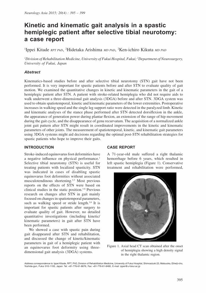
A 71-year-old male suffered a right thalamic hemorrhage before 6 years, which resulted in left spastic hemiplegia (Figure 1). Conservative treatment and rehabilitation were performed,
Figure 1. Axial head CT scan obtained after the onset of hemiplegia showing a high density signal in the right thalamic region.

Neurology Asia December 2015
396
which enabled the patient to walk independently with an ankle foot orthosis and cane. As the patient’s spasticity-induced equinovarus foot deformity deteriorated. Because a block test for the tibial nerve involving injecting 5 cc of xylocaine was effective, STN was performed under general anesthesia. During the procedure, a skin incision was made below the popliteal fossa. The tibial nerve was then dissected, and the motor nerve branches extending to the soleus, gastrocnemius, tibialis posterior, and flexor hallucis longus muscles were identified via muscle palpation by a physical therapist and
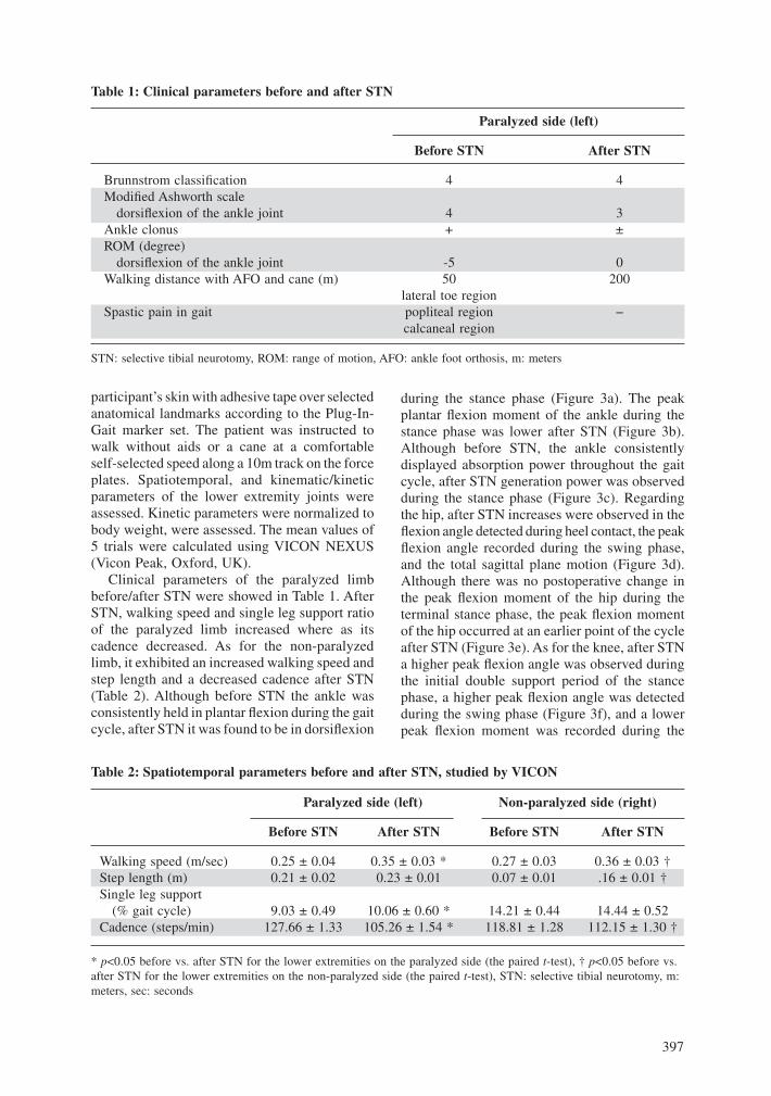
intraoperative electrical stimulation.9 The selected motor nerve branches were partially sectioned over a distance of 10 mm under a microscope. From the day after surgery, the patient benefited from a 40-minute daily rehabilitation program, which included stretching, muscle strengthening, and gait exercises. We performed gait analysis before and 1 week after STN using a VICON MX with 10-camera (Vicon Peak, Oxford, UK) and 4-force plate (Advanced Mechanical Technology, Watertown, MA, USA) (Figure 2). Thirty-five 15mm retro-reflective markers were attached to the
Figure 2. The measurement scene using a three-dimensional gait analysis system. The three-dimensional motion analysis system consists of VICON and force plates. VICON is a system
for motion capturing. Force plates are a system to measure ground reaction force, kinetic and kinematic parameter in motion. Spheres covered with reflective tape, known as markers, are placed on visual reference points on different parts of the body. The VICON system consists of 10 cameras and is designed to track and reconstruct these markers in 3-dimensional space.
397
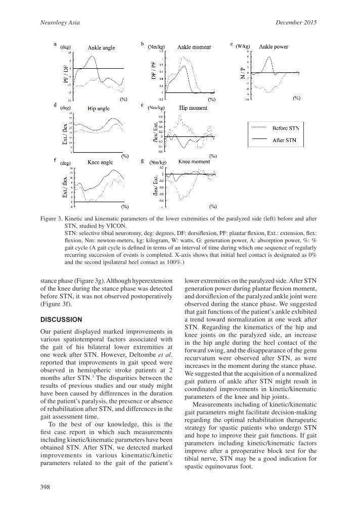
participant’s skin with adhesive tape over selected anatomical landmarks according to the Plug-In-Gait marker set. The patient was instructed to walk without aids or a cane at a comfortable self-selected speed along a 10m track on the force plates. Spatiotemporal, and kinematic/kinetic parameters of the lower extremity joints were assessed. Kinetic parameters were normalized to body weight, were assessed. The mean values of 5 trials were calculated using VICON NEXUS (Vicon Peak, Oxford, UK). Clinical parameters of the paralyzed limb before/after STN were showed in Table 1. After STN, walking speed and single leg support ratio of the paralyzed limb increased where as its cadence decreased. As for the non-paralyzed limb, it exhibited an increased walking speed and step length and a decreased cadence after STN (Table 2). Although before STN the ankle was consistently held in plantar flexion during the gait cycle, after STN it was found to be in dorsiflexion
during the stance phase (Figure 3a). The peak plantar flexion moment of the ankle during the stance phase was lower after STN (Figure 3b). Although before STN, the ankle consistently displayed absorption power throughout the gait cycle, after STN generation power was observed during the stance phase (Figure 3c). Regarding the hip, after STN increases were observed in the flexion angle detected during heel contact, the peak flexion angle recorded during the swing phase, and the total sagittal plane motion (Figure 3d).Although there was no postoperative change in the peak flexion moment of the hip during the terminal stance phase, the peak flexion moment of the hip occurred at an earlier point of the cycle after STN (Figure 3e). As for the knee, after STN a higher peak flexion angle was observed during the initial double support period of the stance phase, a higher peak flexion angle was detected during the swing phase (Figure 3f), and a lower peak flexion moment was recorded during the
Paralyzed side (left)
Before STN After STN
Brunnstrom classification 4 4Modified Ashworth scale dorsiflexion of the ankle joint 4 3 Ankle clonus + ± ROM (degree) dorsiflexion of the ankle joint -5 0 Walking distance with AFO and cane (m) 50 200 lateral toe region Spastic pain in gait popliteal region − calcaneal region
Table 1: Clinical parameters before and after STN
STN: selective tibial neurotomy, ROM: range of motion, AFO: ankle foot orthosis, m: meters
Table 2: Spatiotemporal parameters before and after STN, studied by VICON
Paralyzed side (left) Non-paralyzed side (right)
Before STN After STN Before STN After STN
Walking speed (m/sec) 0.25 ± 0.04 0.35 ± 0.03 * 0.27 ± 0.03 0.36 ± 0.03 †Step length (m) 0.21 ± 0.02 0.23 ± 0.01 0.07 ± 0.01 .16 ± 0.01 †Single leg support (% gait cycle) 9.03 ± 0.49 10.06 ± 0.60 * 14.21 ± 0.44 14.44 ± 0.52Cadence (steps/min) 127.66 ± 1.33 105.26 ± 1.54 * 118.81 ± 1.28 112.15 ± 1.30 †
* p<0.05 before vs. after STN for the lower extremities on the paralyzed side (the paired t-test), † p<0.05 before vs. after STN for the lower extremities on the non-paralyzed side (the paired t-test), STN: selective tibial neurotomy, m: meters, sec: seconds
Neurology Asia December 2015
398
stance phase (Figure 3g). Although hyperextension of the knee during the stance phase was detected before STN, it was not observed postoperatively (Figure 3f).
DISCUSSION
Our patient displayed marked improvements in various spatiotemporal factors associated with the gait of his bilateral lower extremities at one week after STN. However, Deltombe et al.
reported that improvements in gait speed were observed in hemispheric stroke patients at 2 months after STN.3 The disparities between the results of previous studies and our study might have been caused by differences in the duration of the patient’s paralysis, the presence or absence of rehabilitation after STN, and differences in the gait assessment time. To the best of our knowledge, this is the first case report in which such measurements including kinetic/kinematic parameters have been obtained STN. After STN, we detected marked improvements in various kinematic/kinetic parameters related to the gait of the patient’s
lower extremities on the paralyzed side. After STN generation power during plantar flexion moment, and dorsiflexion of the paralyzed ankle joint were observed during the stance phase. We suggested that gait functions of the patient’s ankle exhibited a trend toward normalization at one week after STN. Regarding the kinematics of the hip and knee joints on the paralyzed side, an increase in the hip angle during the heel contact of the forward swing, and the disappearance of the genu recurvatum were observed after STN, as were increases in the moment during the stance phase. We suggested that the acquisition of a normalized gait pattern of ankle after STN might result in coordinated improvements in kinetic/kinematic parameters of the knee and hip joints. Measurements including of kinetic/kinematic gait parameters might facilitate decision-making regarding the optimal rehabilitation therapeutic strategy for spastic patients who undergo STN and hope to improve their gait functions. If gait parameters including kinetic/kinematic factors improve after a preoperative block test for the tibial nerve, STN may be a good indication for spastic equinovarus foot.
Figure 3. Kinetic and kinematic parameters of the lower extremities of the paralyzed side (left) before and after STN, studied by VICON.
STN: selective tibial neurotomy, deg: degrees, DF: dorsiflexion, PF: plantar flexion, Ext.: extension, flex: flexion, Nm: newton-meters, kg: kilogram, W: watts, G: generation power, A: absorption power, %: % gait cycle (A gait cycle is defined in terms of an interval of time during which one sequence of regularly recurring succession of events is completed. X-axis shows that initial heel contact is designated as 0% and the second ipsilateral heel contact as 100%.)

399
DISCLOSURE
Conflicts of interest: None
REFERENCES 1. Levin MF, Hui-Chan C. Ankle spasticity is inversely
correlated with antagonist voluntary contraction in hemiparetic subjects. Electromyogr Clin Neurophysiol 1994; 34:415-25.
2. Deltombe T, Detrembleur C, Hanson P, et al. Selective tibial neurotomy in the treatment of spastic equinovarus foot: a 2-year follow-up of three cases. Am J Phys Med Rehabil 2006; 85:82-8.
3. Deltombe T, Gustin T. Selective tibial neurotomy in the treatment of spastic equinovarus foot in hemiplegic patients: A 2-year longitudinal follow-up of 30 cases. Arch Phys Med Rehabil 2010; 91:1025-30.
4. Feve A, Decq P, Filipetti P, et al. Physiological effects of selective tibial neurotomy on lower limb spasticity. J Neurol Neurosurg Psychiatry 1997; 63:575-8.
5. Rousseaux M, Buisset N, Daveluy W, et al. Comparison of botulinum toxin injection and neurotomy in patients with distal lower limb spasticity. Eur J Neurol 2008; 15:506-11.
6. Roujeau T, Lefaucheur JP, Slavov V, et al. Long term course of the H reflex after selective tibial neurotomy. J Neurol Neurosurg Psychiatry 2003; 74:913-7.
7. Fouad W. Selective neurotomy of the tibial nerve for treatment of spastic foot. Alexandria Journal of Medicine 2011; 47:325-31.
8. Rousseaux M, Buisset N, Daveluy W, et al. Long term effect of tibial nerve neurotomy in stroke patients with lower limb spasticity. J Neurol Sci 2009; 278:71-6.
9. Sindou M, Mertens P. Selective neurotomy of the tibial nerve for treatment of spastiv foot. Neurosurgery 1998; 23:738-44.


![[PPT]Hemiplegic Shoulder Pain: Approach to Diagnosis & …f45ebd178a369304538a-da09e9363888411f910f2103a3cb9db6.r58... · Web viewHemiplegic Shoulder Pain:Approach to Diagnosis &](https://img.dokumen.tips/doc/110x75/5aadbe627f8b9a9c2e8eb879/ppthemiplegic-shoulder-pain-approach-to-diagnosis-f45ebd178a369304538a-da09e9363888411f910f2103a3cb9db6r58web.jpg)