Embed Size (px)
Citation preview
1
Kinematics of the Shoulder Joint in Tennis Players
Lädermann A,1,2 Chagué S,3 Kolo FC,4 Charbonnier C3
1 Division of Orthopaedics and Trauma Surgery, La Tour Hospital, Geneva,
Switzerland
2 Faculty of Medicine, University of Geneva, Geneva, Switzerland
3 Artanim Foundation, Medical Research Department, Geneva, Switzerland
4 Rive Droite Radiology Center, Geneva, Switzerland

2
ABSTRACT 1
Background: Shoulder pain and injury are common in tennis players. The precise 2
causes for such pain remain unclear. Impingement at critical tennis positions and 3
glenohumeral instability have never been dynamically evaluated in-vivo. The purpose 4
of this study was to evaluate the different types of impingement and stability during 5
tennis movements. 6
Methods: Type and frequency of impingement as well as percentage of subluxation 7
were evaluated in 10 tennis players through a novel dedicated patient-specific 8
measurement technique based on optical motion capture and Magnetic Resonance 9
Imaging (MRI). 10
Results: All volunteers, nine male and one female, had a clinically functional rotator 11
cuff. MRI revealed 11 rotator cuff lesions in six subjects and six labral lesions in five 12
subjects. Lateral subacromial, anterior subacromial, internal anterosuperior, and 13
internal posterosuperior impingements were observed in four, three, two and seven 14
subjects, respectively. No instability could be demonstrated in this population. 15
Conclusion: Tennis players presented frequent radiographic signs of structural 16
lesions that could mainly be related to posterosuperior impingements due to 17
repetitive abnormal motion contacts. This is the first study demonstrating that a 18
dynamic and precise motion analysis of the entire kinematic chain of the shoulder is 19
possible through a non-invasive method of investigation. This premier kinematic 20
observation offers novel insights into the analysis of shoulder impingement and 21
instability that could, with future studies, be generalized to other shoulder pathologies 22
and sports. This original method may open new horizons leading to improvement in 23
impingement comprehension. 24

3
Keywords: Shoulder kinematics modeling; Biomechanics, Tennis players; Overhead 25
athletes; Impingement; Magnetic resonance imaging. 26

4
INTRODUCTION 27
Shoulder pain and injury are common in tennis players, with a prevalence of 50% for 28
certain categories of age.1 A majority of shoulder pain is caused by impingement and 29
instability due to repetitive lifting and overhead arm movements. Two types of 30
impingement have been described: external and internal. External types include 31
subacromial impingement of the rotator cuff between the anterior acromion2 or lateral 32
acromion3 and the superior humeral head that could occur with serves and overhead 33
shots. Another type of external impingement is the less common subcoracoid 34
impingement4 of the subscapularis or biceps tendon. It results from contact between 35
the coracoid process against the lesser tuberosity of the humeral head and is more 36
likely to occur at the backhand preparation phase and the late follow-through phase 37
of the forehand. Internal impingement consists of (1) posterosuperior impingement5 38
of the supraspinatus and infraspinatus tendons between the greater tuberosity of the 39
humeral head and the posterosuperior aspect of the glenoid when the arm is in 40
extreme abduction, extension and external rotation during the late cocking stage of 41
the serve; and (2) anterosuperior impingement6 of the deep surface of the 42
subscapularis tendon and the reflection pulley on the anterosuperior glenoid rim that 43
could also occur at the backhand preparation phase and the late follow-through 44
phase of forehand. 45
The precise causes for these impingements remain unclear, but it is believed that 46
repetitive contact (Figure 1A and 1B), glenohumeral instability (Figure 1C), scapular 47
orientation, rotator cuff dysfunction, and posteroinferior capsular contracture with 48
resultant glenohumeral internal rotation deficit (GIRD) may play a role in the 49
development of symptomatic impingement.5,7,8 Measuring the dynamic in-vivo 50
shoulder kinematics seems crucial to better understand these pathologies and to 51

5
propose an adequate treatment. Indeed, a patient with an internal impingement will 52
be treated differently if the etiology is a posteroinferior capsular contracture with 53
resultant GIRD (that generally responds positively to a compliant posteroinferior 54
capsular stretching program or to an arthroscopic selective posteroinferior 55
capsulotomy and concomitant partial articular sided tendon avulsion (SLAP) lesion 56
repair9) or a repetitive contact of the undersurface of the rotator cuff on the 57
posterosuperior glenoid labrum (that can respond to debridement, glenoidplasty or 58
derotational humeral osteotomy).10-12 However, such kinematic measurements 59
remain a challenging problem due to the complicated anatomy and large range of 60
motion of the shoulder. To our knowledge, impingements at critical tennis positions 61
and glenohumeral stability have never been dynamically evaluated. Unfortunately, 62
the motion of the shoulder cannot be explored with standard Magnetic Resonance 63
Imaging (MRI) or Computed Tomography (CT) because they are limited by space 64
and the velocity of the movement and might therefore miss dynamic motion. 65
Fluoroscopy-based measurements provide sufficient accuracy for dynamic shoulder 66
analysis,13 but they use ionizing radiation. Motion capture systems using skin-67
mounted markers provide a non-invasive method to determine shoulder kinematics 68
during dynamic movements.14 However, none of the current motion capture 69
techniques have reported translation values at the glenohumeral joint. One reason 70
that might explain this void is that current techniques have either concentrated their 71
efforts on the analysis of a single shoulder bone (e.g., scapula) or focused on the 72
description of humeral motion relative to the thorax rather than to its proximal bone. 73
The purpose of the study was thus: (1) to develop a dedicated patient-specific 74
measurement technique based on optical motion capture and MRI to accurately 75
determine glenohumeral kinematics (rotations and translations) taking into account 76

6
the whole kinematic chain of the shoulder complex from the thorax to the humerus 77
through the clavicle and scapula, (2) to evaluate impingement, stability, and other 78
motion-related disorders during dynamic movements in high-level tennis players. 79
80
Figure 1: (A) Gilles Walch’s theory: the deep layer of the posterosuperior rotator cuff 81
impinged with the posterior labrum and glenoid. (B) Christopher Jobe’s theory: the 82
impingement is mainly due to hyperextension of the humerus relative to the scapula. (C) 83
Frank Jobe’s theory: lesions in throwing athletes are related to subtle anterior instability. 84
85
METHODS 86
Ten volunteers who were intermediate or ex-professional tennis players were 87
recruited for this study. Ethical approval was gained from the local Institutional 88
Review Board, and all participants gave their written informed consent prior to taking 89
part in the study. Exclusion criteria were reported previous shoulder injuries, shoulder 90
surgery or contraindications for MRI. 91
The outcomes of interest were the prevalence of internal and external 92
impingement and glenohumeral instability in this particular population. Furthermore, 93
the prevalence of other radiographic pathologies was evaluated in relation to the 94
main outcomes of interest. The following baseline characteristics were assessed: 95
age, sex, body mass index, shoulder side, and limb dominance. 96
Rotator cuff examination included the belly-press, bear hug, Jobe tests, and 97
external rotation strength again resistance. Constant score,15 American Shoulder and 98
Elbow Surgeons (ASES) score,16 a single assessment numeric evaluation (SANE) 99

7
score,17 and a visual analog scale (VAS) pain score graded from 0 points (no pain) to 100
10 points (maximal pain) were recorded. 101
All volunteers underwent an MR shoulder arthrography. The MRI examinations 102
were conducted after a fluoroscopically guided arthrography with a contrast agent 103
and with an anterior approach. MRI was performed with a 1.5 T HDxT system 104
(General Electric Healthcare, Milwaukee WI, USA). A dedicated shoulder surface coil 105
was used. A sagittal T1 weighted fast spin echo sequence, a coronal and sagittal T2 106
weighted fast spin echo sequence with fat saturation, a coronal and axial T1 107
weighted fast spin echo sequence with fat saturation, and three 3D fast gradient echo 108
(Cosmic® and Lava®) sequences were achieved. Table 1 details the imaging 109
parameters of each MRI sequence. 110
MR arthrograms were assessed by a musculoskeletal radiologist for shoulder 111
pathology including rotator cuff, labral or ligament (HAGL) lesion and bony changes. 112
Based on the 3D MR images, patient-specific 3D models of the shoulder bones 113
(humerus, scapula, clavicle and sternum) were reconstructed for each volunteer 114
using ITK-SNAP software (Penn Image Computing and Science Laboratory, 115
Philadelphia, PA). 116
Kinematic data was recorded using a Vicon MX T-Series motion capture system 117
(Vicon, Oxford Metrics, UK) consisting of 24 cameras (24 × T40S) sampling at 240 118
Hz. The volunteers were equipped with spherical retroreflective markers placed 119
directly onto the skin using double-sided adhesive tape (Figure 2). Four markers (Ø 120
14 mm) were attached to the thorax (sternal notch, xyphoid process, C7 and T8 121
vertebra). Four markers (Ø 6.5 mm) were placed on the clavicle. Four markers (Ø 14 122
mm) were fixed on the upper arm, two placed on anatomical landmarks (lateral and 123
medial epicondyles) and two as far as possible from the deltoid. For the scapula, one 124

8
marker (Ø 14 mm) was fixed on the acromion. In addition, the scapula was covered 125
with 56 markers (Ø 6.5 mm) to form a 7×8 regular grid. Finally, additional markers 126
were distributed over the body (non-dominant arm and legs). 127
128
Figure 2: Markers placement. 129
After appropriate warm-up, participants were asked to perform the following tennis 130
movements: forehand, backhand, flat and kick serves. They were also instructed to 131
perform three motor tasks: internal-external rotation of the arm with 90° abduction 132
and the elbow flexed 90°, flexion of the arm from neutral to maximum flexion, and 133
empty-can abduction from neutral to maximum abduction in the scapular plane. 134
Three trials of each motion were recorded. The same investigators attached all 135
markers and performed all measurements. 136
Shoulder kinematics were computed with custom-made software using the 137
recorded markers' trajectories. The major drawback with optical motion capture 138
systems is the soft tissue deformation due to muscle contractions and skin sliding, 139
causing marker movements with respect to the underlying bones. In the upper 140
extremity, the scapula is particularly affected. To solve this issue, it was 141
demonstrated that the use of global optimization could help reduce soft tissue 142
artifacts (STA) errors globally.18 Therefore, we developed a patient-specific kinematic 143

9
chain model of the shoulder complex (including the thorax, clavicle, scapula and 144
humerus) using the subject’s 3D bony models19. The shoulder joints were each 145
modeled as a ball-and-socket joint (3 degrees of freedom) with loose constraints on 146
joint translations. The optimal pose of the kinematic chain was then obtained using a 147
global optimization algorithm. To verify its accuracy, kinematic data was collected 148
simultaneously from an X-ray fluoroscopy unit (MultiDiagnost Eleva, Philips Medical 149
Systems, The Netherlands) and the motion capture system during clinical motion 150
patterns (flexion, abduction and internal-external rotation of the arm) in a validation 151
test. Glenohumeral kinematics were derived from the marker position data and 152
compared with the one obtained with the fluoroscopy gold-standard.13,19 The 153
accuracy of the model for glenohumeral orientation was within 4° for each anatomical 154
plane and between 1.9 and 3.3 mm in average for glenohumeral translation. 155
Moreover, the results showed that the translation patterns computed with the model 156
were in good agreement with previous research.20 157
Finally, the computed motions were applied to the tennis player’s shoulder 3D 158
models reconstructed from their MRI data. Figure 3 shows examples of computed 159
tennis positions. A ball and stick representation of the overall skeleton was also 160
added to improve the analysis and visualization of the motion. The method is 161
summarized in video 1. 162
To permit motion description of the shoulder kinematic chain, local coordinate 163
systems (Figure 4) were established based on the definitions suggested by the 164
International Society of Biomechanics21 to represent the thorax, clavicle, scapula and 165
humerus segments using anatomical landmarks identified on the subject’s bony 3D 166
models. The glenohumeral joint center was calculated based on a sphere fitting 167

10
method22 that fits the optimal sphere to the humeral head using the points of the 3D 168
humeral model. 169
170
Figure 3: Computed tennis positions (here the right shoulder) according to the three main 171
phases, showing the markers setup (small colored spheres) and the virtual skeleton. Top: 172
serve shot. Position 4, 7 and 8 are commonly known as the cocking, deceleration and finish 173
stages, respectively. Middle: forehand shot. Bottom: backhand shot. 174
Glenohumeral range of motion (ROM) was quantified for flexion, abduction and 175
internal-external rotation movements. This was obtained by calculating the relative 176
orientation between the scapula and humerus coordinate systems at each point of 177
movement and then expressed in clinically recognizable terms (flex/ext, abd/add and 178
IR/ER) by decomposing the relative orientation into three successive rotations. It is 179
important to note that these computations were performed independently from the 180
major anatomical planes (i.e., sagittal, transverse, frontal planes). To facilitate clinical 181

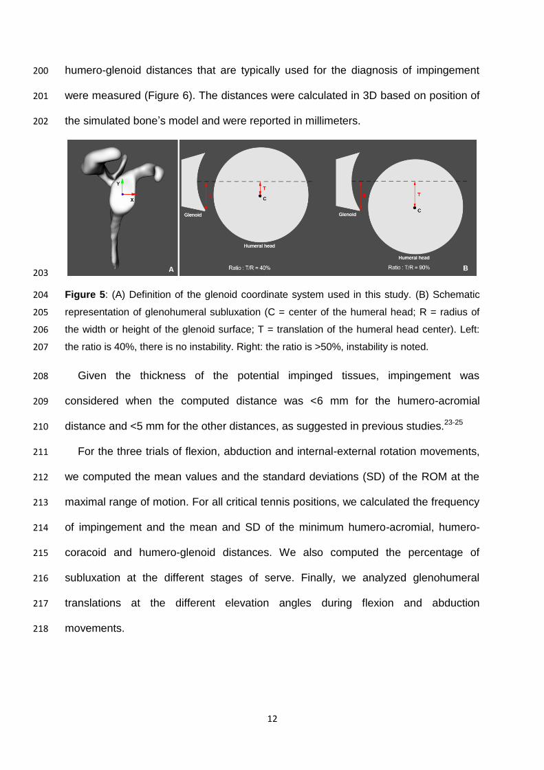
11
comprehension and comparison, motion of the humerus with respect to the thorax 182
was also calculated. This was achieved with the same method but using the thorax 183
and humerus coordinate systems. 184
185
Figure 4: Bone coordinate systems for the thorax (Xt Yt Zt), clavicle (Xc Yc Zc), scapula (Xs Ys 186
Zs) and humerus (Xh Yh Zh). 187
Glenohumeral stability was assessed during flexion and abduction movements 188
and during flat and kick serves at the late cocking, deceleration and finish stages. 189
Glenohumeral translation was defined as anterior-posterior and superior-inferior 190
motion of the humeral head center relative to the glenoid coordinate system. This 191
coordinate system was determined by an anterior-posterior X-axis and a superior-192
inferior Y-axis with origin placed at the intersection of the anteroposterior aspects and 193
superoinferior aspects of the glenoid rim (Figure 5A). Subluxation was defined as the 194
ratio (in %) between the translation of the humeral head center and the radius of 195
width (anteroposterior subluxation) or height (superoinferior subluxation) of the 196
glenoid surface (Figure 5B). Instability was defined as subluxation >50%. 197
Impingement was evaluated at critical tennis positions. While visualizing the tennis 198
player’s shoulder joint in motion, minimum humero-acromial, humero-coracoid and 199
12
humero-glenoid distances that are typically used for the diagnosis of impingement 200
were measured (Figure 6). The distances were calculated in 3D based on position of 201
the simulated bone’s model and were reported in millimeters. 202
203
Figure 5: (A) Definition of the glenoid coordinate system used in this study. (B) Schematic 204
representation of glenohumeral subluxation (C = center of the humeral head; R = radius of 205
the width or height of the glenoid surface; T = translation of the humeral head center). Left: 206
the ratio is 40%, there is no instability. Right: the ratio is >50%, instability is noted. 207
Given the thickness of the potential impinged tissues, impingement was 208
considered when the computed distance was <6 mm for the humero-acromial 209
distance and <5 mm for the other distances, as suggested in previous studies.23-25 210
For the three trials of flexion, abduction and internal-external rotation movements, 211
we computed the mean values and the standard deviations (SD) of the ROM at the 212
maximal range of motion. For all critical tennis positions, we calculated the frequency 213
of impingement and the mean and SD of the minimum humero-acromial, humero-214
coracoid and humero-glenoid distances. We also computed the percentage of 215
subluxation at the different stages of serve. Finally, we analyzed glenohumeral 216
translations at the different elevation angles during flexion and abduction 217
movements. 218

13
219
Figure 6: Visualization of the humero-acromial, humero-coracoid and humero-glenoid 220
distances during motion. The red lines represent the minimum distances. 221
222
RESULTS 223
The ten volunteers, nine male and one female, had all been playing tennis for more 224
than 17 years. The mean ± SD age, weight, height and body mass index of the 225
subjects were 39.7 ± 8.9 years, 180.2 ± 7.1 cm, 76.7 ± 8.62 kg, and 23.5 ± 1.9 kg/m2, 226
respectively. Nine volunteers were right-handed. 227
None of the tennis players displayed sudden loss of serving ability during the late 228
cocking stage (so-called “dead arm”). All subjects had a competent rotator cuff. The 229
mean Constant, ASES, SANE and VAS pain scores were 99.2 ± 1.4 points (range, 230
96 to 100 points), 99.5 ± 1.6 points (range, 95 to 100 points), 95.0 ± 7.5 points 231
(range, 80 to 100 points) and 0.6 ± 1.3 points (range, 0 to 4 points), respectively. 232
Only 2 of the 10 subjects reported shoulder pain at the time of the examination. Nine 233
had a history of shoulder pain during their career. Shoulder ROM determined by 234
motion capture during clinical motor tasks are shown in Table 2. None of the tennis 235
players had 180° ROM in internal-external rotation. 236
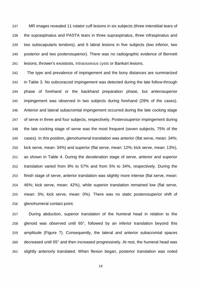
14
MR images revealed 11 rotator cuff lesions in six subjects (three interstitial tears of 237
the supraspinatus and PASTA tears in three supraspinatus, three infraspinatus and 238
two subscapularis tendons), and 6 labral lesions in five subjects (two inferior, two 239
posterior and two posterosuperior). There was no radiographic evidence of Bennett 240
lesions, thrower's exostosis, intraosseous cysts or Bankart lesions. 241
The type and prevalence of impingement and the bony distances are summarized 242
in Table 3. No subcoracoid impingement was detected during the late follow-through 243
phase of forehand or the backhand preparation phase, but anterosuperior 244
impingement was observed in two subjects during forehand (29% of the cases). 245
Anterior and lateral subacromial impingement occurred during the late cocking stage 246
of serve in three and four subjects, respectively. Posterosuperior impingement during 247
the late cocking stage of serve was the most frequent (seven subjects, 75% of the 248
cases). In this position, glenohumeral translation was anterior (flat serve, mean: 34%; 249
kick serve, mean: 34%) and superior (flat serve, mean: 12%; kick serve, mean: 13%), 250
as shown in Table 4. During the deceleration stage of serve, anterior and superior 251
translation varied from 8% to 57% and from 5% to 34%, respectively. During the 252
finish stage of serve, anterior translation was slightly more intense (flat serve, mean: 253
46%; kick serve, mean: 42%), while superior translation remained low (flat serve, 254
mean: 3%; kick serve, mean: 0%). There was no static posterosuperior shift of 255
glenohumeral contact point. 256
During abduction, superior translation of the humeral head in relation to the 257
glenoid was observed until 65°, followed by an inferior translation beyond this 258
amplitude (Figure 7). Consequently, the lateral and anterior subacromial spaces 259
decreased until 65° and then increased progressively. At rest, the humeral head was 260
slightly anteriorly translated. When flexion began, posterior translation was noted 261

15
until 70° followed by a return to a more anterior translation (Figure 8). There was no 262
posterior subluxation at any degree of flexion. 263
264
Figure 7: Superior-inferior translations of the humeral head center relative to the glenoid 265
during abduction. Means and standard deviations for all 10 shoulders. 266
267
Figure 8: Anterior-posterior translations of the humeral head center relative to the glenoid 268
during flexion. Means and standard deviations for all 10 shoulders. 269
Also, based on the visual assessment of the 3D simulations, we noticed in six 270
subjects that the arm in abduction was beyond the scapular plane during the cocking 271
16
stage of serve, resulting in hyperextension. 272
273
DISCUSSION 274
Shoulder pain and pathologic lesions are common in overhead athletes. In the 275
present study, 9 of 10 tennis players presented with radiographic signs of structural 276
lesions that could be related to impingement syndrome that occurred with overhead 277
arm movements. However, the precise causes for these lesions remain unclear. It 278
might result from several factors (e.g., repetitive contact, subtle glenohumeral 279
instability, torsional overload with repetitive hypertwisting, scapular orientation and 280
dyskinesis, etc.). The theory of internal impingement in these athletes, which occurs 281
with the arm in the cocked position of 90° abduction, full external rotation and 282
extension,26 holds that repeated contact between the rotator cuff insertion and the 283
posterosuperior glenoid rim lead to articular-sided partial thickness rotator cuff tears 284
and superior labral lesions.5,26 If the contact is physiologic, repetitive contact applied 285
at a rate exceeding tissue repair or torsional and shear stresses9 may be responsible 286
for rotator cuff or labral damages. 287
This article evaluated dynamically and in-vivo the different aforementioned causes 288
of lesions in tennis players. As shown by the results of this study, anterosuperior and 289
subacromial impingement remain occasional in this particular population. No 290
shoulder instability could be noted during tennis movements. However, 291
posterosuperior impingement was frequent when serving. Thus, as expected, this 292
shot seems to be the most harmful for the tennis player’s shoulder. Regarding this 293
type of impingement, repetitive contact could be the cause of posterior and 294
posterosuperior labral lesions, as well as PASTA lesions of the posterosuperior 295
cuff.5,27 Indeed, we were not able, as other authors,28 to confirm the role in the 296
17
impingement development from other culprits like (1) static posterosuperior shifts of 297
glenohumeral contact point leading to torsional overload,9 or (2) instability due to 298
gradual repetitive stretching of the anterior capsuloligamentous structures.8,26 299
Nevertheless, this could be explained by the fact that there are many kinds of 300
overhead athletes, and tennis players do not have the same external rotation in 301
abduction and arm speed as do, for example, throwers which have previously been 302
studied. In addition, this could also reflect the efficiency of injury prevention programs 303
that have been established in many tennis clubs (e.g. promotion of compact serve). 304
Concerning subacromial impingement during abduction, superior translation of the 305
humeral head in relation to the glenoid was observed, followed by inferior translation 306
beyond 65°. Such superior and inferior translation confirms previous 307
observations.20,29 Consequently, subacromial space decreased until 65° and then 308
increased progressively. Anterior2 and lateral3 impingement could hence occur at the 309
beginning of abduction and not at or above 90° like previously believed.30 310
Regarding motion of the glenohumeral joint, the range in internal and external 311
rotation should remain constant between the dominant and the non-dominant arm, 312
with a shift in the external rotation sector of the dominant arm in overhead throwers.9 313
We could not confirm the 180° rotation rule in tennis players, as the mean values of 314
the ROM computed in this study were approximately two times smaller than similar 315
measurements found in handball players.31 We are, therefore, not convinced that a 316
contracted posterior band, evoking the posterior cable to shorten with resultant 317
GIRD, is a theory that can be extrapolated in tennis players. This theory might be 318
specific to baseball players. 319
Finally, we also evaluated posterior humeral head translation in relation to the 320
glenoid during flexion. An hypothesis of the development of posterior static 321

18
subluxation described by Walch et al.32 could be posterior subluxation during normal 322
anterior elevation. At rest, the humeral head was slightly anteriorly translated. When 323
forward flexion began, slight posterior translation was noted until 70° followed by a 324
return to a more anterior translation. There was no posterior subluxation at any 325
degree of flexion. Therefore, since no dynamic or physiologic posterior instability was 326
observed, it is probably not responsible (at term) for static instability in these subjects 327
without hyperlaxity. 328
We acknowledge the following limitations in our study: (1) the accuracy of the 329
kinematics computation from motion capture data, which was only validated for low 330
velocity movements. Glenohumeral orientation errors were within 4° for each 331
anatomical plane, which is acceptable for clinical use in the study of shoulder 332
pathology. There is potential for difficulty in the calculation of glenohumeral 333
translation from skin markers due to the high mobility of the shoulder. Although the 334
translations could be significant with our model, we demonstrated in the validation 335
work and in this study that the computed translation patterns and amplitudes were in 336
good agreement with published data. To our knowledge, this non-invasive method is 337
the first attempt to calculate both rotations and translations at the glenohumeral joint 338
based on skin markers. (2) The use of bone-to-bone distances to assess 339
impingement which do not take into account precise measurements of the thickness 340
of the impinged soft tissues. One improvement could be to perform a more advanced 341
simulation accounting for the 3D shapes and movements of cartilages, the labrum 342
and the rotator cuff. (3) The findings may not be generalizable. This was a relatively 343
small sample size of primary males in a single sport and skill level, with a narrow age 344
range. (4) The use of 1.5 T MRI, as stronger magnet strengths would enhance image 345
resolution. Moreover, MRI is not a gold standard to demonstrate bony changes. This 346
19
study may hence underestimate bony lesions such as Bennett exostosis, and (5) as 347
volunteers were not known for any pathology, a criticism could be to have tested 348
healthy players that would prevent extrapolation of results to complaining patients. 349
However, 9 out of the 10 volunteers reported previous symptoms, so we think that 350
they were a good representation. Despite these limitations, we do believe that they 351
did not call into question the results of this study. 352
353
CONCLUSION 354
Tennis players presented frequent radiographic signs of structural lesions that could 355
mainly be related to posterosuperior impingements due to repetitive abnormal motion 356
contacts. This is the first study demonstrating that a dynamic and precise motion 357
analysis of the entire kinematic chain of the shoulder is possible through a non-358
invasive method of investigation. This premier observation offers novel insights into 359
the analysis of shoulder impingement and instability that could, with future studies, be 360
generalized to other shoulder pathologies and sports. This original method may open 361
new horizons leading to improvement in impingement comprehension. 362
363
Practical implications 364
Anterior and lateral subacromial and posterosuperior impingements are 365
frequent in overhead athletes. 366
Repetitive contact in extreme abduction, extension and external rotation could 367
be the cause of posterior and posterosuperior labral lesions, as well as 368
PASTA lesions of the posterosuperior cuff. 369
Coaches and medical staff should consider promotion of compact serve. 370
20
This study has highlighted the benefits of a non-invasive, dynamic and in-vivo 371
evaluation of shoulder pathologies. 372
373
ACKNOWLEDGMENTS 374
The authors thank the European Society for Surgery of the Shoulder and the Elbow 375
for their financial support. 376

21
REFERENCES
1. Abrams GD, Renstrom PA, Safran MR. Epidemiology of musculoskeletal injury
in the tennis player. Br J Sports Med. 2012; 46(7):492-498.
2. Neer CS, 2nd. Anterior acromioplasty for the chronic impingement syndrome
in the shoulder: a preliminary report. J Bone Joint Surg Am. 1972; 54(1):41-50.
3. Nyffeler RW, Werner CM, Sukthankar A, et al. Association of a large lateral
extension of the acromion with rotator cuff tears. J Bone Joint Surg Am. 2006;
88(4):800-805.
4. Gerber C, Terrier F, Ganz R. The role of the coracoid process in the chronic
impingement syndrome. J Bone Joint Surg Br. 1985; 67(5):703-708.
5. Walch G, Boileau P, Noel E, et al. Impingement of the deep surface of the
supraspinatus tendon on the posterosuperior glenoid rim: An arthroscopic
study. J Shoulder Elbow Surg. 1992; 1(5):238-245.
6. Gerber C, Sebesta A. Impingement of the deep surface of the subscapularis
tendon and the reflection pulley on the anterosuperior glenoid rim: a
preliminary report. J Shoulder Elbow Surg. 2000; 9(6):483-490.
7. Burkhart SS, Morgan CD, Kibler WB. The disabled throwing shoulder:
spectrum of pathology Part III: The SICK scapula, scapular dyskinesis, the
kinetic chain, and rehabilitation. Arthroscopy. 2003; 19(6):641-661.
8. Jobe CM. Posterior superior glenoid impingement: expanded spectrum.
Arthroscopy. 1995; 11(5):530-536.
9. Burkhart SS, Morgan CD, Kibler WB. The disabled throwing shoulder:
spectrum of pathology Part I: Pathoanatomy and biomechanics. Arthroscopy.
2003; 19(4):404-420.
10. Riand N, Levigne C, Renaud E, et al. Results of derotational humeral
osteotomy in posterosuperior glenoid impingement. Am J Sports Med. 1998;
26(3):453-459.
11. Riand N, Boulahia A, Walch G. Posterosuperior impingement of the shoulder
in the athlete: results of arthroscopic debridement in 75 patients. Rev Chir
Orthop Reparatrice Appar Mot. 2002; 88(1):19-27.
12. Levigne C, Garret J, Grosclaude S, et al. Surgical technique arthroscopic
posterior glenoidplasty for posterosuperior glenoid impingement in throwing
athletes. Clin Orthop Relat Res. 2012; 470(6):1571-1578.

22
13. Zhu Z, Massimini DF, Wang G, et al. The accuracy and repeatability of an
automatic 2D-3D fluoroscopic image-model registration technique for
determining shoulder joint kinematics. Medical Eng & Phys. 2012; 34(9):1303-
1309.
14. Klotz MC, Kost L, Braatz F, et al. Motion capture of the upper extremity during
activities of daily living in patients with spastic hemiplegic cerebral palsy. Gait
& Posture. 2013; 38(1):148-152.
15. Constant CR, Murley AH. A clinical method of functional assessment of the
shoulder. Clin Orthop Relat Res. 1987(214):160-164.
16. Michener LA, McClure PW, Sennett BJ. American Shoulder and Elbow
Surgeons Standardized Shoulder Assessment Form, patient self-report
section: reliability, validity, and responsiveness. J Shoulder Elbow Surg. 2002;
11(6):587-594.
17. Williams GN, Gangel TJ, Arciero RA, et al. Comparison of the Single
Assessment Numeric Evaluation method and two shoulder rating scales.
Outcomes measures after shoulder surgery. Am J Sports Med. 1999;
27(2):214-221.
18. Roux E, Bouilland S, Godillon-Maquinghen AP, et al. Evaluation of the global
optimisation method within the upper limb kinematics analysis. J Biomech.
2002; 35(9):1279-1283.
19. Charbonnier C, Chagué S, Kolo F, et al. A patient-specific measurement
technique to model the kinematics of the glenohumeral joint. Orthop &
Traumatol: Surg & Res. 2014; 100(7):715-719.
20. Matsuki K, Matsuki KO, Yamaguchi S, et al. Dynamic in vivo glenohumeral
kinematics during scapular plane abduction in healthy shoulders. J Orthop
Sports Phys Ther. 2012; 42(2):96-104.
21. Wu G, van der Helm FC, Veeger HE, et al. ISB recommendation on definitions
of joint coordinate systems of various joints for the reporting of human joint
motion - Part II: Shoulder, elbow, wrist and hand. J Biomech. 2005; 38(5):981-
992.
22. Schneider P, Eberly DH. Geometric Tools for Computer Graphics (The
Morgan Kaufmann Series in Computer Graphics), San Francisco, Morgan
Kaufmann; 2002.

23
23. Chopp JN, Dickerson CR. Resolving the contributions of fatigue-induced
migration and scapular reorientation on the subacromial space: an
orthopaedic geometric simulation analysis. Hum Mov Sci. 2012; 31(2):448-
460.
24. De Maeseneer M, Van Roy P, Shahabpour M. Normal MR imaging anatomy of
the rotator cuff tendons, glenoid fossa, labrum, and ligaments of the shoulder.
Radiol Clin North Am. 2006; 44(4):479-487, vii.
25. Zumstein V, Kraljevic M, Muller-Gerbl M. Glenohumeral relationships:
Subchondral mineralization patterns, thickness of cartilage, and radii of
curvature. J Orthop Res. 2013; 31(11):1704-1707.
26. Davidson PA, Elattrache NS, Jobe CM, et al. Rotator cuff and posterior-
superior glenoid labrum injury associated with increased glenohumeral motion:
a new site of impingement. J Shoulder Elbow Surg. 1995; 4(5):384-390.
27. Jobe CM. Superior glenoid impingement. Current concepts. Clin Orthop Relat
Res. 1996; 330:98-107.
28. Halbrecht JL, Tirman P, Atkin D. Internal impingement of the shoulder:
comparison of findings between the throwing and nonthrowing shoulders of
college baseball players. Arthroscopy. 1999; 15(3):253-258.
29. Massimini DF, Boyer PJ, Papannagari R, et al. In-vivo glenohumeral
translation and ligament elongation during abduction and abduction with
internal and external rotation. J Orthop Surg Res. 2012; 7:29.
30. Harrison AK, Flatow EL. Subacromial impingement syndrome. J Am Acad
Orthop Surg. 2011; 19(11):701-708.
31. Almeida GP, Silveira PF, Rosseto NP, et al. Glenohumeral range of motion in
handball players with and without throwing-related shoulder pain. J Shoulder
Elbow Surg. 2013; 22(5):602-607.
32. Walch G, Ascani C, Boulahia A, et al. Static posterior subluxation of the
humeral head: an unrecognized entity responsible for glenohumeral
osteoarthritis in the young adult. J Shoulder Elbow Surg. 2002; 11(4):309-314.

24
TABLES
TABLE 1
MRI sequences and their imaging parameters
MRI Sequence Imaging Parameters
Sagittal T1 weighted fast spin echo without fat saturation
Section thickness 3.5 cm; intersection gap 0.5 cm
TR/TE 380/11; FOV 16 x 16 cm
Coronal T2 weighted fast spin echo with fat saturation
Section thickness 4 mm; intersection gap 0.5 cm
TR/TE 1920/101,6; FOV 16 x 16cm
Sagittal T2 weighted fast spin echo with fat saturation
Section thickness 3.5 cm; intersection gap 0.5 cm
TR/TE 5680/103.5; FOV 16 x 16cm
Coronal T1 weighted fast spin echo with fat saturation
Section thickness 4 mm; intersection gap 0.5 cm
TR/TE 320/13; FOV 16 x 16cm
Axial T1 weighted fast spin echo with fat saturation
Section thickness 4 mm; intersection gap 0.5 cm
TR/TE 640/26,8; FOV 16 x 16 cm
Axial Cosmic 3D fast gradient echo with fat saturation
Section thickness 1.8 mm; no intersection gap;
TR/TE 6.1/3.0; FOV 28 x 28cm
Axial Cosmic 3D fast gradient echo without fat saturation
Section thickness 4 mm; no intersection gap;
TR/TE 5.7/2.8; FOV 28 x 28cm
Axial Lava 3D fast gradient echo with fat saturation
Section thickness 5.2 mm; no intersection gap;
TR/TE 3.7/1.7; FOV 35 x 35cm

25
TABLE 2
Shoulder range of motion (deg) determined by motion capture during flexion, empty-can abduction and internal-external rotation
with 90° abduction according to the two referentials (n = 30; 10 subjects, 3 trials)
Motion
Humerus motion relative to the thorax Glenohumeral motion
Mean ± SD Range Mean ± SD Range
Flexion 144.8 ± 8.0 125 - 157 98.7 ± 9.7 83 - 116
Abduction 139.4 ± 10.9 119 - 161 88.8 ± 11.8 65 - 108
Internal rotation (IR) 44.0 ± 9.8 30 - 70 22.3 ± 11.1 11 - 45
External rotation (ER) 52.6 ± 10.8 36 - 77 58.6 ± 10.3 43 - 79
Total IR-ER 96.6 ± 17.5 74 - 147 80.8 ± 14.9 60 - 107

26
TABLE 3
Frequency of impingement and minimum humero-acromial, humero-coracoid and humero-glenoid distances (mm) at critical tennis
positions (n = 30; 10 subjects, 3 trials)
Distances
Flat serve Kick serve Forehand Backhand
Frequency Mean ± SD
Frequency Mean ± SD
Frequency Mean ± SD
Frequency Mean ± SD
Lateral humero-acromial 29%
7.5 ± 3.2 42%
6.8 ± 3.7 - -
Anterior humero-acromial 29%
7.4 ± 2.9 29%
7.0 ± 3.1 - -
Humero-coracoid - - 0%
15.9 ± 1.6 0%
15.0 ± 2.7
Anterosuperior humero-glenoid - - 29%
5.5 ± 1.2 0%
6.9 ± 1.3
Posterosuperior humero-glenoid 76%
3.6 ± 1.4 75%
3.3 ± 1.8 - -

27
TABLE 4
Pourcentage of subluxation of the glenohumeral joint during tennis serves (n = 30; 10 subjects, 3 trials)
Shot, position
Anterior-posterior subluxation* Superior-inferior subluxation†
Mean ± SD Range Mean ± SD Range
Flat serve, late cocking stage 34% ± 9% 14% - 47% 12% ± 6% -1% - 21%
Kick serve, late cocking stage 34% ± 6% 22% - 44% 13% ± 9% 0% - 32%
Flat serve, deceleration stage 34% ± 14% 8% - 57% 18% ± 7% 8% - 34%
Kick serve, deceleration stage 37% ± 9% 20% - 56% 19% ± 7% 5% - 32%
Flat serve, finish stage 46% ± 15% 18% - 68% 3% ± 5% -5% - 14%
Kick serve, finish stage 42% ± 13% 17% - 67% 10% ± 8% 0% - 30%
* A positive value means that the subluxation is anterior, otherwise it is posterior.
† A positive value means that the subluxation is superior, otherwise it is inferior.


![Complications of Surgical Treatment of Anterior …shoulder kinematics, resulting in shoulder anterior instability and additional re-dislocation [5] [6]. The paradigm of these lesions](https://img.dokumen.tips/doc/110x75/5f216fb3eb8fed0579410d58/complications-of-surgical-treatment-of-anterior-shoulder-kinematics-resulting-in.jpg)