Embed Size (px)
Citation preview
RESEARCH ARTICLE
Kif4 Is Essential for Mouse Oocyte Meiosis
Nicole J. Camlin1,2*, Eileen A. McLaughlin1,2,3*, Janet E. Holt2,4
1 School of Environmental and Life Sciences, University of Newcastle, Callaghan, NSW, Australia, 2 Priority
Research Centre for Reproductive Science, University of Newcastle, Callaghan, NSW, Australia, 3 School of
Biological Sciences, University of Auckland, Auckland, New Zealand, 4 School of Biomedical Sciences and
Pharmacy, University of Newcastle, Callaghan, NSW, Australia
* [email protected] (NJC); [email protected] (EAM)
Abstract
Progression through the meiotic cell cycle must be strictly regulated in oocytes to generate
viable embryos and offspring. During mitosis, the kinesin motor protein Kif4 is indispensable
for chromosome condensation and separation, midzone formation and cytokinesis. Addi-
tionally, the bioactivity of Kif4 is dependent on phosphorylation via Aurora Kinase B and
Cdk1, which regulate Kif4 function throughout mitosis. Here, we examine the role of Kif4 in
mammalian oocyte meiosis. Kif4 localized in the cytoplasm throughout meiosis I and II, but
was also observed to have a dynamic subcellular distribution, associating with both microtu-
bules and kinetochores at different stages of development. Co-localization and proximity
ligation assays revealed that the kinetochore proteins, CENP-C and Ndc80, are potential
Kif4 interacting proteins. Functional analysis of Kif4 in oocytes via antisense knock-down
demonstrated that this protein was not essential for meiosis I completion. However, Kif4
depleted oocytes displayed enlarged polar bodies and abnormal metaphase II spindles, indi-
cating an essential role for this protein for correct asymmetric cell division in meiosis I. Fur-
ther investigation of the phosphoregulation of meiotic Kif4 revealed that Aurora Kinase and
Cdk activity is critical for Kif4 kinetochore localization and interaction with Ndc80 and CENP-
C. Finally, Kif4 protein but not gene expression was found to be upregulated with age, sug-
gesting a role for this protein in the decline of oocyte quality with age.
Introduction
The fidelity of chromosome segregation during meiosis is key to producing high quality
oocytes, capable of creating healthy offspring. In mammalian oocytes, errors in chromosome
separation result in embryonic aneuploidy, which may lead to spontaneous abortion or tri-
somy births such as Downs Syndrome. A known risk factor associated with oocyte aneuploidy
is maternal ageing, with the aneuploidy incidence approximately 2% for women in their 20s
increasing to 35% for women in their 40s [1–3]. In recent years, a growing body of evidence
suggests that alterations in chromosome architecture with age results in abnormal kinetochore
structure, aberrant kinetochore microtubule (KT-MT) interaction and ultimately aneuploidy
[4–6]. To further elucidate how such events arise, we investigated a kinesin motor protein,
Kif4 which has known roles in microtubule flux [7].
PLOS ONE | DOI:10.1371/journal.pone.0170650 January 26, 2017 1 / 17
a1111111111
a1111111111
a1111111111
a1111111111
a1111111111
OPENACCESS
Citation: Camlin NJ, McLaughlin EA, Holt JE
(2017) Kif4 Is Essential for Mouse Oocyte Meiosis.
PLoS ONE 12(1): e0170650. doi:10.1371/journal.
pone.0170650
Editor: Qing-Yuan Sun, Institute of Zoology
Chinese Academy of Sciences, CHINA
Received: November 22, 2016
Accepted: January 9, 2017
Published: January 26, 2017
Copyright: © 2017 Camlin et al. This is an open
access article distributed under the terms of the
Creative Commons Attribution License, which
permits unrestricted use, distribution, and
reproduction in any medium, provided the original
author and source are credited.
Data Availability Statement: All relevant data are
within the paper and its Supporting Information
files.
Funding: This work was supported by Australian
Research Council (ARC) grants to E A McLaughlin
(DP110100418) and J E Holt (DP120100946/
DE120101242) and an Australian Postgraduate
award to N J Camlin.
Competing Interests: The authors have declared
that no competing interests exist.

The kinesin motor protein Kif4 (human homologue KIF4A) is a member of the Kinesin-4
subfamily, consisting of KIF4A and B in humans and Kif4 in mice [8]. Studies in mitotic sys-
tems have revealed key roles for Kif4 in chromosome condensation and separation, metaphase
and midzone spindle formation as well as cytokinesis [7,9–18]. Such functions are achieved
through the ability of Kif4 to interact with condensin I, influence kinetochore protein loading,
regulate microtubule length throughout metaphase and telophase and control microtubule
kinetochore flux [7,9,11,19]. A growing body of evidence suggests that Kif4 function is medi-
ated by cell cycle kinases Cyclin-dependent kinase 1(Cdk1) and Aurora Kinase B (AurB)
[14,19–21]. Loss of Kif4 has also been associated with multiple tumours and is evident in can-
cer cell lines, indicating an important role for this protein throughout the cell cycle [18,22,23].
To date the role of Kif4 in mammalian fertility has not been investigated. However, the Dro-sophila homologue Klp3A is essential for male and female fertility [24,25]. In males, mutations
in Klp3A results in midzone instability and cytokinesis failure in meiotically dividing spermato-
cytes [24]. In contrast, females are able to successfully complete meiosis, however, oocytes are
unable to support embryogenesis [25,26]. In meiotic Xenopus egg extracts, the Xenopus homo-
logue XKlp1, limits microtubule growth and is important for correct spindle shape [27,28].
Thus, in the present study we sought to characterize the localization and role of mammalian
Kif4 in female meiosis. Additionally, we present evidence indicating Kif4 is involved in kineto-
chore dynamics under the control of AurB/C and Cdk1.
Materials and Methods
All reagents were obtained from Sigma-Aldrich unless otherwise specified.
Animals and ethics approval
Animal use for this study was approved by the University of Newcastle Animal Care and Ethics
Committee. C57BL/6 x CBA F1 hybrid cross were obtained from the University of Newcastle
Animal Resources and housed with ad libitum water and food under a 12hrs light: 12hrs dark
cycle.
Oocyte collection and maturation
4–6 week old female mice were intraperitoneally (i.p) injected with 7.5 IU of pregnant
mares’ gonadotropin (Intervet) 44-52hrs prior to a second i.p injection of 5 IU human cho-
rionic gonadotropin (Intervet) or germinal vesicle (GV) oocyte collection from the ovary.
12hrs after the second injection MII stage eggs were collected from the ampulla. Oocytes
were collected into M2 media containing BSA. For GV collections media was supplemented
with 1mM milrinone (M4659) to prevent meiotic resumption. Cumulus cells were then
removed enzymatically with hyaluronidase (300μg/ml; H4272) for MII or mechanically via
repeated pipetting for GV oocytes. For collection from aged (12–19 months) females and
corresponding young controls, GV oocytes were retrieved directly from the ovary without
prior hormonal stimulation.
To allow meiotic maturation in vitro, GV oocytes were washed into milrinone free M2
media at 37˚C (GVB/MI) or MEM (MII; 11900024, Gibco) with 20% FCS at 37˚C in 5% CO2.
GV oocytes underwent IVM for 1.5hrs for GVB stage, 7.5hrs for MI stage or 16hrs for MII
stage oocytes. For inhibitor experiments 10μM ZM447439 (2458, Tocris), 100μM Roscovitine
(R7772), 100nM BI 2539 (S1109, Selleck) or DMSO (0.2% final concentration; D2650) was
added to media at 3.5hrs post washout. Inhibitor experiments were performed in triplicate
with oocytes from age matched individual animals.
Kif4 and Oocyte Meiosis
PLOS ONE | DOI:10.1371/journal.pone.0170650 January 26, 2017 2 / 17
Immunocytochemistry and proximity ligation assay
For all immunocytochemistry oocytes were fixed in 2% paraformaldehyde in PBS with 0.5%
triton X for 30mins. Oocytes were blocked in 7% goat serum PBS-0.1% Tween for 1hr prior to
overnight incubation with primary antibody (see Supplementary S1 Table). Secondary anti-
bodies were conjugated with Alexa-488, Alexa-555 and Alexa-633 (see Supplementary S1
Table) and incubated with oocytes for 1hr at room temperature prior to Hoechst (20μg/ml)
counter staining and mounting in Citifluor (Citifluor Ltd). All immunocytochemistry was per-
formed in triplicate with oocytes from age matched individual animals. Secondary only nega-
tive controls were also performed to confirm the specificity of immunocytochemistry.
Proximity ligation assay (DUO92101; PLA) was performed as per manufactures instruc-
tions to detect proteins within 40nm of each other, and therefore potential protein-protein
interactions. Oocytes were then counterstained in Hoechst and mounted in Citifluor as above.
Specificity of PLA was confirmed through the use of negative controls including Kif4 antibody
only, secondary antibody only or Kif4 antibody with testis specific protein Piwil1 antibody. All
PLA was performed in triplicate with oocytes from aged matched individual animals.
Imaging was performed using an Olympus FV1000 confocal using a 60x/1.2 NA UPLSAPO
oil immersion objective lens (Olympus, Australia). ImageJ (freeware; National Institute of
Health) was used to measure fluorescent intensity and count PLA foci as previously described
[29]. Briefly, the area and integrated density of each oocyte was measured. The average of the
mean background was determined for secondary antibody only probed oocytes. Fluorescent
intensity for each oocyte was then determined by subtracting the oocytes area times the mean
background from the integrated density prior to normalizing to 1. For PLA ImageJ particle
counting function was used to determine number of PLA foci per oocyte.
Oocyte qRT-PCR
Five denuded oocytes were used per reaction. The zona pellucida was removed with Acid Tyr-
odes solution (T1788) prior to being washed in PBS/ polyvinylpyrrolidone. Following lysis,
cDNA was prepared using the TaqMan Gene Expression Cells-toCT Kit (AM1728, Ambion)
as per manufactures instructions. A whole cDNA sample was used per one reaction. qRT-PCR
was performed with a Light Cycle 96 SW 1.1 (Roche) using TaqMan mRNA assay (4369016,
ThermoFisher Scientific) as per manufactures instructions. Ppia was employed as an endoge-
nous control to normalize the expression levels of Kif4. The relative expression levels of Kif4
were calculated using the ΔCt method.
Oocyte microinjection
GV oocytes were microinjected as previously described by Holt et al., with either Kif4 knock-
down or mis-match morpholino oligo (Gene-tools; see Supplementary S2 Table for targeting
sequence) [30,31]. Oocytes were then incubated in MEM (11900024, Gibco) with 20% FCS at
37˚C in 5% CO2 for 24hrs to allow protein knock-down.
Phosphorylation site prediction
Potential AurB and Cdk1 phosphorylation sites for Kif4 (accession BAA02167.1) were pre-
dicted using GPS 3.0 (freeware, The Cuckoo Workgroup [32–34].
Statistics
GraphPad Prism 6.0 software (GraphPad Software, Inc) was used for statistical analysis. For
numerical data D’Agnostino-Pearson omnibus normality test was performed. For normally
Kif4 and Oocyte Meiosis
PLOS ONE | DOI:10.1371/journal.pone.0170650 January 26, 2017 3 / 17

distributed data Student’s t-test was performed. For data that was not normally disturbed Mann-
Whitney test or Kruskal-Wallis Test with Dunn’s post-hoc statistical test was performed. For cate-
gorical data Fisher’s Exact Test was used. A p value<0.05 was considered statistically significant.
Results
Kif4 has dynamic localization throughout meiosis
The localization of Kif4 through four distinct stages of female meiosis was investigated, using
an antibody directed against the N-terminal motor domain. At each stage examined Kif4 was
found dispersed throughout the cytoplasm and in cytoplasm aggregates (Fig 1A). Additionally,
it displayed distinct changes in subcellular localization dependent upon meiotic stage. In GV
arrested oocytes, Kif4 was found to be enriched around the nuclear envelope and chromatin
(Fig 1A). At GVB, Kif4 was distributed around the chromosomes, presumably associated with
the microtubule ball, which forms as the meiotic spindle develops (Fig 1A). By MI, Kif4 had
relocated and was now associated with the chromosomes where it co-localized with centro-
mere/kinetochore marker ACA (Fig 1B.) To study this change in localization more closely, we
analyzed fixed oocytes every 30 minutes throughout late prometaphase-MI and confirmed
that, as Kif4 was lost from the microtubules, it become associated with the kinetochore region
of the chromosomes (Fig 1C).
Once the polar body was extruded and oocytes had reached MII arrest, Kif4 was once again
associated with the microtubules on the metaphase II spindle (Fig 1A). To confirm Kif4 localiza-
tion throughout meiosis, immunocytochemistry was performed using a second antibody directed
against the C-terminal cargo domain with a similar pattern observed (Supplementary S1A Fig).
Kif4 interacts with both inner and outer kinetochore proteins
Having established that Kif4 was co-located with the centromere/kinetochore at MI, we next
sought to investigate whether this kinesin might interact directly with key kinetochore pro-
teins. Immunolocalization revealed Kif4 signal overlap with the inner kinetochore protein,
CENP-C at all meiotic stages examined. CENP-C associated with Kif4 aggregates in GV and
MII oocytes (Fig 2A) and as expected, CENP-C and Kif4 foci overlapped at the kinetochore, at
both MI and MII (Fig 2A). To determine whether these proteins interacted, we performed a
proximity ligation assay (PLA) on fixed oocytes using the CENP-C and Kif4 antibodies, and
observed positive PLA signal throughout the cytoplasm, at all three stages of meiosis examined
(Fig 2B). Furthermore, PLA foci were also associated with the chromosomes, and therefore
presumably the kinetochore at MI and MII (Fig 2B and Supplementary S1B Fig).
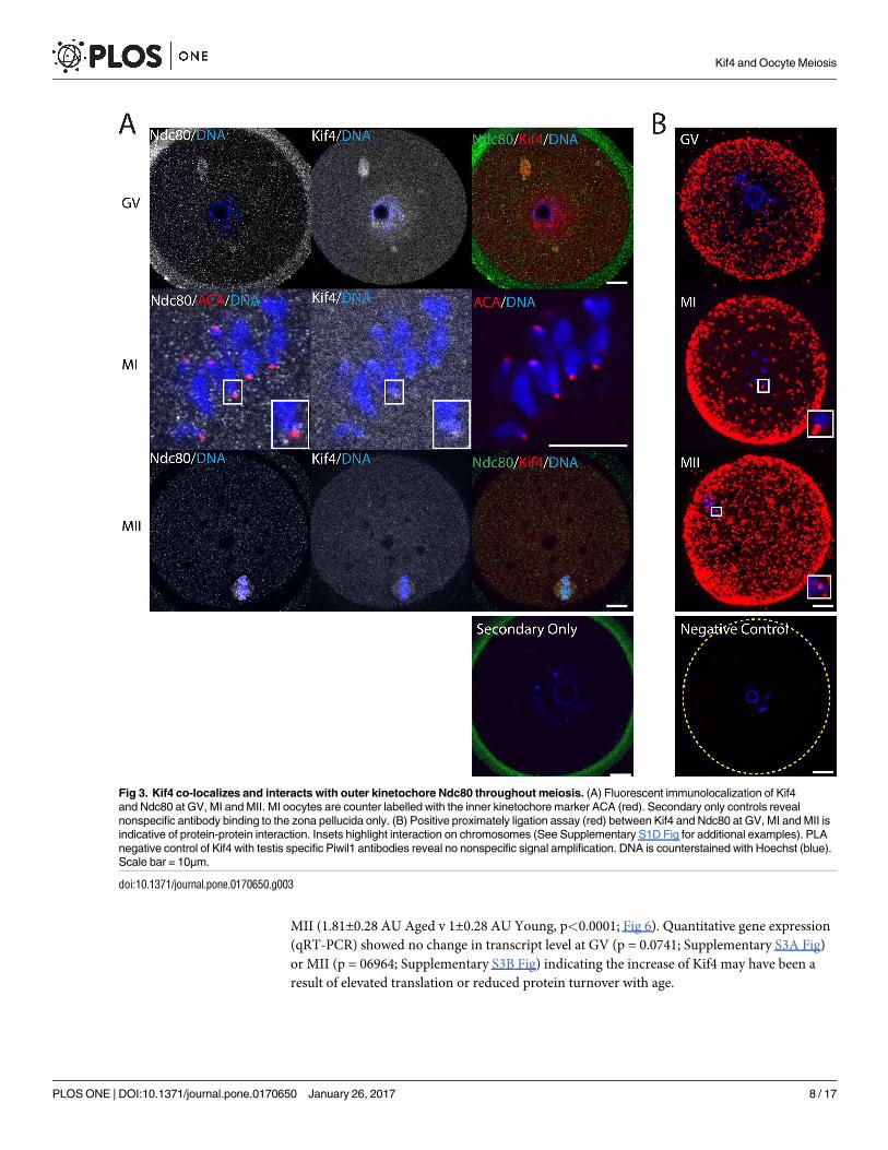
To gain further insight into the role of Kif4 at the kinetochores, we next investigated its
interaction with the outer kinetochore protein Ndc80. As with CENP-C, Ndc80 co-localized
with Kif4 aggregates throughout GV oocytes (Fig 3A). At MI Kif4 foci overlapped with
ACA fluorescence, however, Ndc80 foci flanked either side, indicative of its role as an outer
kinetochore protein (Fig 3A and Supplementary S1C Fig). By MII, both Ndc80 and Kif4 were
found on the metaphase spindle. Confirmation of a potential protein-protein interaction was
achieved via PLA, with proteins interacting cytoplasmically throughout meiosis (Fig 3B).
Additionally, as observed previously, these PLA foci were associated with chromosomes at MI
and MII (Fig 3B and Supplementary S1D Fig).
Kif4 is essential for normal oocyte meiosis
The spindle/kinetochore localization and potential binding partners of Kif4 suggested it may
be important for meiotic progression. To examine its role more closely Kif4 knock-down (KD)
Kif4 and Oocyte Meiosis
PLOS ONE | DOI:10.1371/journal.pone.0170650 January 26, 2017 4 / 17

Fig 1. Kif4 has dynamic localization throughout oocyte meiosis. (A) Fluorescent immunolocalization of Kif4 (grey) at GV, GVB, MI and
MII. Inserts highlight the localization of Kif4 to the nuclear membrane (yellow arrows) and microtubules (green arrow). Secondary only
controls reveal no nonspecific antibody binding. Scale bar = 10μm. (B) Fluorescent immunolocalization of Kif4 (grey/green) to the kinetochore
at MI (red arrow). Kinetochores are counter labelled with the inner kinetochore marker ACA (red). Scale bar = 2μm. (C) Fluorescent
Kif4 and Oocyte Meiosis
PLOS ONE | DOI:10.1371/journal.pone.0170650 January 26, 2017 5 / 17
was performed via oligo morpholino translational repression. Using immunocytochemistry
we confirmed Kif4 protein expression was reduced 2.5 fold 24hrs post morpholino injection
when compared to mis-match morpholino injected controls (0.39±0.25 AU KD v 1±0.07 AU
control, p<0.0001; Fig 4A). IVM of KD oocytes found no abnormalities in meiosis timing or
maturation rates (data not shown). However, investigation of MII oocyte quality found KD
oocytes had a significantly higher proportion of abnormal MII spindles compared to controls
(48% knock-down v 12% controls, p = 0.0017; Fig 4B). Spindle abnormalities included mild
and severe chromosome misalignment, and microtubule aggregates attached to spindles.
Additionally, KD oocytes were observed to have abnormal cytokinesis as determined by a sig-
nificant increase in polar body size (5489±1692μm2 KD v 2661±522μm2 control, p<0.0001;
Fig 4C).
Kif4 and kinetochore protein interaction is mediated via kinase activity
Since meiosis is driven by the activity of several important kinases, we next sought to deter-
mine if the function of Kif4 was regulated in this manner. Previous research has found that
Cdk1 and AurB mediate the interaction of Kif4 with condensin I [19]. Furthermore, AurB has
been found to directly interact with Kif4 at mitotic midzones [14]. Group based prediction of
AurB and Cdk1 phosphorylation sites on Kif4 found 9 potential Cdk1 sites and 13 potential
AurB sites located throughout the three major domains; motor, coiled-coil and cargo (Fig 5A).
Of note is that 4 of these sites, serine 816, 1224, 1230 and threonine 800, are known Kif4 phos-
phorylation residues [35]. Additionally threonine 800 is a well categorized AurB phosphoryla-
tion site [14,21].
To examine whether Cdk1 and AurB kinase activity did in fact regulate Kif4, oocytes were
allowed to mature in the presence of either the pan Aurora Kinase inhibitor ZM 447439 (ZM)
or the pan Cdk inhibitor Roscovitine (Ros) for 4hrs from prometaphase to MI. Kif4 cytosolic
immunolocalization was unchanged with Cdk and Aurora Kinase inhibition. However, immu-
nolocalization of Kif4 to the kinetochore was lost after treatment with both ZM and Ros sug-
gesting an inability of Kif4 to relocalize to the kinetochore with Cdk and/or Aurora Kinase
inhibition (Fig 5B). Furthermore, PLA analysis found that both treatments significantly
reduced Kif4/CENP-C and Kif4/Ndc80 interaction compared to the vehicle control (CENP-C:
0.58±0.27 PLA foci ZM v 0.54±0.37 PLA foci Ros v 1±0.50 PLA foci Veh, p = 0.0013; Fig 5C
and Ndc80: 0.44±0.12 PLA foci ZM v 0.60±0.19 PLA foci Ros v 1±0.25 PLA foci Veh,
p<0.0001; Fig 5D).
To confirm that these results were the consequence of specific Aurora Kinase or Cdk regu-
lation oocytes were allowed to mature in the presence of the Plk1 inhibitor BI 2536. Unlike
Ros or ZM treatment, BI 2536 had no effect on Kif4 kinetochore localization consistent with
the lack of Plk1 and Kif4 co-localization off the kinetochores (Supplementary S2 Fig).
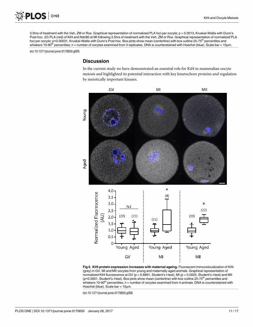
Kif4 expression increases with maternal ageing
Finally, we sought to determine if Kif4 protein was altered with maternal age in oocytes which
might be associated with their reduced quality. Immunocytochemistry revealed that Kif4
expression was unchanged in young vs aged GV stage oocytes (0.95±0.39 AU Aged v 1±0.28
AU Young, p = 0.6891; Fig 6). However, by MI there was a significant increase in protein
expression (1.73±0.93 AU Aged v 1±0.26 AU Young, p = 0.0025; Fig 6) which continued into
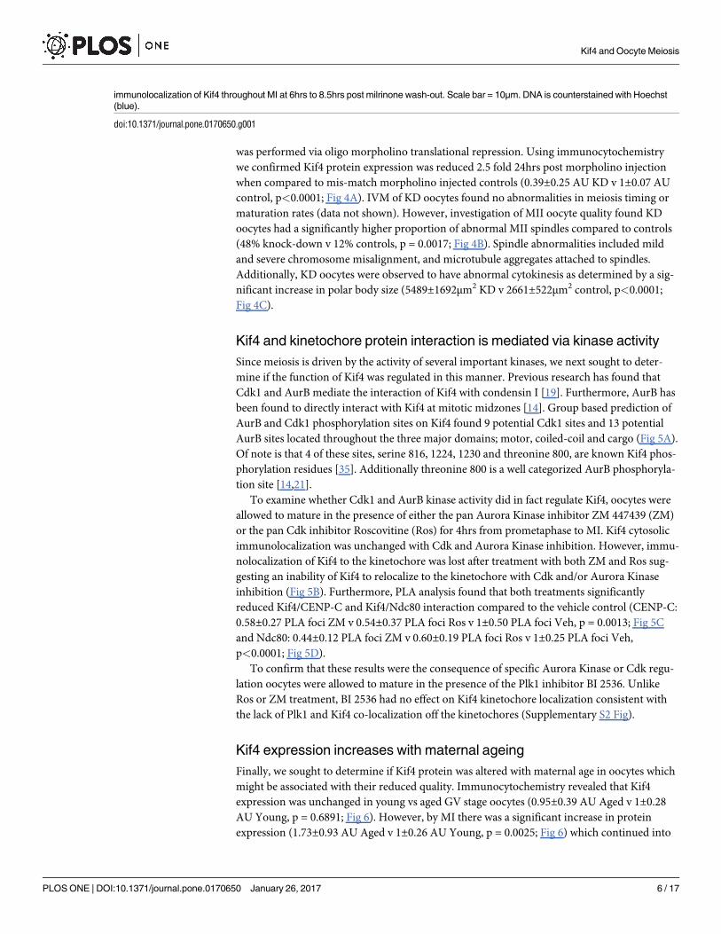
immunolocalization of Kif4 throughout MI at 6hrs to 8.5hrs post milrinone wash-out. Scale bar = 10μm. DNA is counterstained with Hoechst
(blue).
doi:10.1371/journal.pone.0170650.g001
Kif4 and Oocyte Meiosis
PLOS ONE | DOI:10.1371/journal.pone.0170650 January 26, 2017 6 / 17

Fig 2. Kif4 co-localizes and interacts with inner kinetochore protein CENP-C throughout meiosis. (A) Fluorescent immunolocalization of
Kif4 and CENP-C at GV, MI and MII. MI and MII oocytes are counter labelled with the inner kinetochore marker ACA (red). Red arrows highlight
Kif4 and CENP-C aggregates. Secondary antibody only controls reveal nonspecific antibody binding to the zona pellucida only. (B) Positive
proximately ligation assay (red) between Kif4 and CENP-C at GV, MI and MII is indicative of protein-protein interaction. Insets highlight interaction
on chromosomes (See Supplementary S1B Fig for additional examples). PLA negative control of Kif4 with testis specific Piwil1antibodies reveal
no nonspecific signal amplification. DNA is counterstained with Hoechst (blue). Scale bar = 10μm.
doi:10.1371/journal.pone.0170650.g002
Kif4 and Oocyte Meiosis
PLOS ONE | DOI:10.1371/journal.pone.0170650 January 26, 2017 7 / 17
MII (1.81±0.28 AU Aged v 1±0.28 AU Young, p<0.0001; Fig 6). Quantitative gene expression
(qRT-PCR) showed no change in transcript level at GV (p = 0.0741; Supplementary S3A Fig)
or MII (p = 06964; Supplementary S3B Fig) indicating the increase of Kif4 may have been a
result of elevated translation or reduced protein turnover with age.
Fig 3. Kif4 co-localizes and interacts with outer kinetochore Ndc80 throughout meiosis. (A) Fluorescent immunolocalization of Kif4
and Ndc80 at GV, MI and MII. MI oocytes are counter labelled with the inner kinetochore marker ACA (red). Secondary only controls reveal
nonspecific antibody binding to the zona pellucida only. (B) Positive proximately ligation assay (red) between Kif4 and Ndc80 at GV, MI and MII is
indicative of protein-protein interaction. Insets highlight interaction on chromosomes (See Supplementary S1D Fig for additional examples). PLA
negative control of Kif4 with testis specific Piwil1 antibodies reveal no nonspecific signal amplification. DNA is counterstained with Hoechst (blue).
Scale bar = 10μm.
doi:10.1371/journal.pone.0170650.g003
Kif4 and Oocyte Meiosis
PLOS ONE | DOI:10.1371/journal.pone.0170650 January 26, 2017 8 / 17

Fig 4. Kif4 is essential for correct spindle formation and cytokinesis. (A) Fluorescent immunolocalization of Kif4 (grey) in control
and knock-down (KD) oocytes. Graphical representation of normalized Kif4 fluorescence; p<0.0001, Mann-Whitney test. (B) Fluorescent
immunolocalization of α-tubulin at MII in control and Kif4 KD. Kif4 KD oocytes were found to have a higher percentage of abnormal
spindles (orange arrow) and chromosome misalignment (red arrow). Graphical representation of abnormal spindle percentages per
group; p = 0.0017, Fishers Exact test. (C) Phase contrast images of control and Kif4 KD MII oocytes. Graphical representation of PB1
size between control and Kif4 KD oocytes; p<0.0001, Mann-Whitney test. Box plots show mean (centerline) with box outline 25-75th
percentiles and whiskers 10-90th percentiles, bar graphs show mean. n = number of oocytes examined from 3 replicates. DNA is
counterstained with Hoechst (blue), scale bar = 10μm.
doi:10.1371/journal.pone.0170650.g004
Kif4 and Oocyte Meiosis
PLOS ONE | DOI:10.1371/journal.pone.0170650 January 26, 2017 9 / 17
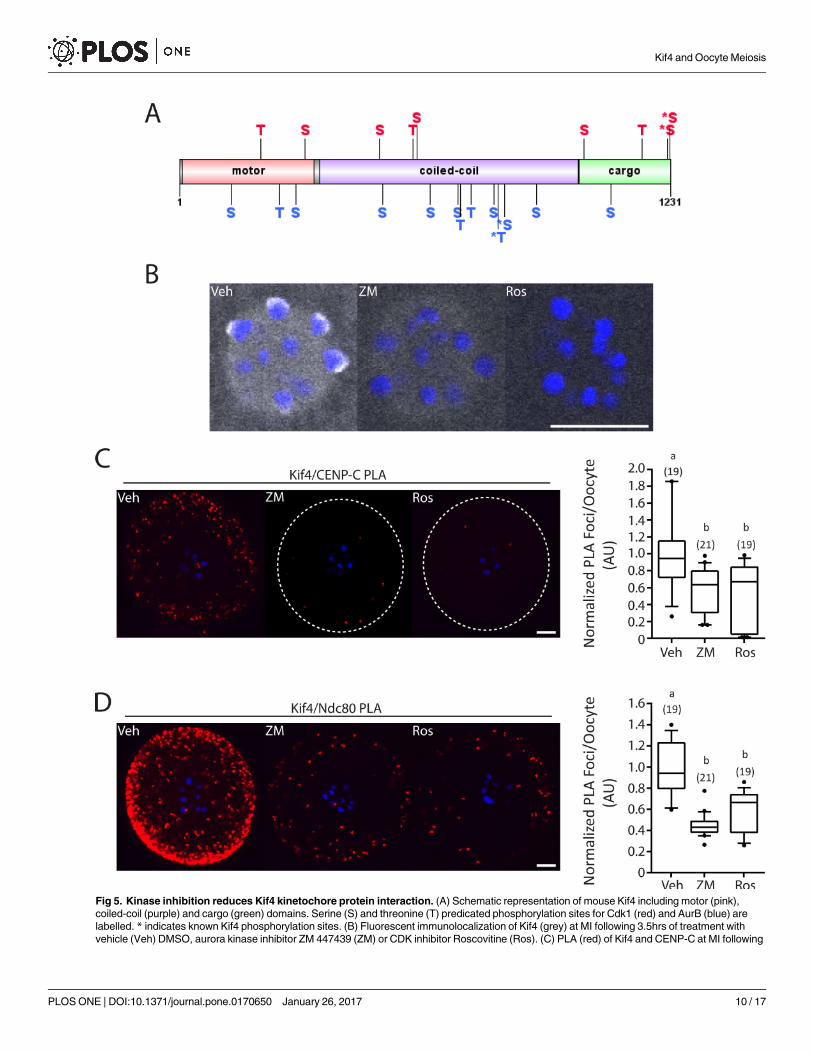
Fig 5. Kinase inhibition reduces Kif4 kinetochore protein interaction. (A) Schematic representation of mouse Kif4 including motor (pink),
coiled-coil (purple) and cargo (green) domains. Serine (S) and threonine (T) predicated phosphorylation sites for Cdk1 (red) and AurB (blue) are
labelled. * indicates known Kif4 phosphorylation sites. (B) Fluorescent immunolocalization of Kif4 (grey) at MI following 3.5hrs of treatment with
vehicle (Veh) DMSO, aurora kinase inhibitor ZM 447439 (ZM) or CDK inhibitor Roscovitine (Ros). (C) PLA (red) of Kif4 and CENP-C at MI following
Kif4 and Oocyte Meiosis
PLOS ONE | DOI:10.1371/journal.pone.0170650 January 26, 2017 10 / 17
Discussion
In the current study we have demonstrated an essential role for Kif4 in mammalian oocyte
meiosis and highlighted its potential interaction with key kinetochore proteins and regulation
by meiotically important kinases.
3.5hrs of treatment with the Veh, ZM or Ros. Graphical representation of normalized PLA foci per oocyte; p = 0.0013, Kruskal-Wallis with Dunn’s
Post-hoc. (D) PLA (red) of Kif4 and Ndc80 at MI following 3.5hrs of treatment with the Veh, ZM or Ros. Graphical representation of normalized PLA
foci per oocyte; p<0.00031, Kruskal-Wallis with Dunn’s Post-hoc. Box plots show mean (centerline) with box outline 25-75th percentiles and
whiskers 10-90th percentiles; n = number of oocytes examined from 3 replicates. DNA is counterstained with Hoechst (blue). Scale bar = 10μm.
doi:10.1371/journal.pone.0170650.g005
Fig 6. Kif4 protein expression increases with maternal ageing. Fluorescent immunolocalization of Kif4
(grey) in GV, MI and MII oocytes from young and maternally aged animals. Graphical representation of
normalized Kif4 fluorescence at GV (p = 0.6891, Student’s t-test), MI (p = 0.0025, Student’s t-test) and MII
(p<0.0001, Student’s t-test). Box plots show mean (centerline) with box outline 25-75th percentiles and
whiskers 10-90th percentiles; n = number of oocytes examined from 4 animals. DNA is counterstained with
Hoechst (blue). Scale bar = 10μm.
doi:10.1371/journal.pone.0170650.g006
Kif4 and Oocyte Meiosis
PLOS ONE | DOI:10.1371/journal.pone.0170650 January 26, 2017 11 / 17

The localization of Kif4 in oocytes is dynamic: we observed that as meiosis I progressed
Kif4 left the metaphase I spindle and became enriched on the chromosomes and kinetochores.
This enrichment at the kinetochore towards the end of metaphase I occurs at time when kinet-
ochore-microtubule (KT-MT) attachments are stabilized and so could indicate a role for Kif4
in this stabilization. In line with this, is the known role of Kif4 in microtubule stabilization in
mitotic cells and Xenopus egg extracts. Ablation of Kif4 from cells lines or XKlp1 from Xenopusegg extracts significantly altered microtubule dynamics including microtubule overgrowth,
decreased KT-MT flux and altered kinetochore oscillations [7,27,36,37]. These altered micro-
tubule dynamics ultimately lead to abnormal spindle formation and misaligned chromosomes,
cumulating in aneuploidy with misaligned chromosomes as observed in our KD oocytes
[9,36,37]. Evidence in mitotic extracts suggests that the abnormal microtubule dynamics is a
likely cause of XKlp1 limiting microtubule growth via allosteric inhibition of microtubule
dynamic instability [38]. Alterations in microtubule stability, growth and kinetochore oscilla-
tions could account for the metaphase spindle abnormalities observed in KD oocytes.
Increased microtubule length, and reduced kinetochore oscillations would be expected to
cause the chromosome misalignment we observed. Further to this, microtubule overgrowth
was detected in Kif4 ablated oocytes, with the addition of microtubule spheres attached to
spindle poles.
Cytokinesis abnormalities were also a common feature in Kif4 KD oocytes. Ablation of
Kif4, XKlp1 or Klp3A mutation results in cytokinesis failure in HeLa cells, mitotic Xenopusegg extracts or Drosophila spermatocytes respectively [15,24,37]. Conversely, we have shown
that depletion of Kif4 in oocytes results in abnormal cytokinesis as demonstrated via enlarged
polar bodies. This indicates that Kif4 is not essential for cytokinesis but is necessary for asym-
metric cytokinesis in oocytes.
Interestingly, enlarged polar bodies have previously also been observed in oocytes depleted
of Ndc80 [39]. Of note is that Ndc80 is crucial for correct spindle formation and chromosome
alignment at metaphase I and II in mouse and porcine oocytes [39–41]. The similar phenotype
observed between Ndc80 and Kif4 ablated oocytes in conjunction with their continued co-
localization and potential interaction throughout meiosis highlights a probable role for Kif4 in
correct Ndc80 function throughout the cell cycle. Interestingly, Kif4 was also found to interact
with CENP-C throughout meiosis. KD of CENP-C in DT40 chicken cells results in abnormal
chromosome alignment similar to that seen in our Kif4 KD oocytes [42]. It is therefore likely
that Kif4 depletion from oocytes results in alterations to both CENP-C and Ndc80 localization,
which may be responsible for the observed abnormal metaphase II oocyte phenotype.
It is also tempting to speculate that CENP-C and Ndc80 are potential cargo proteins of
Kif4, with Kif4 involved in the correct shuttling of these proteins throughout the oocyte. In
support of this theory, KD of Kif4 in DT40 cells significantly altered the expression of a large
number of kinetochore proteins, including CENP-C, Ndc80 and AurB specifically on chromo-
somes [11]. We found that interaction of kinetochore proteins CENP-C or Ndc80 with Kif4
was not restricted to chromosomes, but was also found throughout the cytoplasm of meioti-
cally cycling oocytes. As kinesins have traditionally been associated with roles in cargo shut-
tling, it is possible that Kif4 is at least partially responsible for shuttling CENP-C/Ndc80 to and
from the chromosomes. Ablation of Kif4, therefore, could result in incorrect loading of
CENP-C/Ndc80 to the kinetochore which might account for the chromosome misalignment
observed.
Intriguingly, the movement of Kif4 to the kinetochores or its interaction with CENP-C and
Ndc80 appears to be cell cycle dependent and under the control of Aurora Kinase and Cdk
activity. CENP-C and Ndc80 expansion has been found to be AurB dependent and essential
for correct KT-MT attachments [43]. Further to this, AurB is responsible for phosphorylation
Kif4 and Oocyte Meiosis
PLOS ONE | DOI:10.1371/journal.pone.0170650 January 26, 2017 12 / 17

of human Kif4 T799 (mouse T800), with an additional12 predicted AurB phosphorylation
sites identified [21]. Oocytes express Aurora Kinase B and C, with both inhibited with ZM.
AurB/C is essential in meiosis I for correct KT-MT attachment and spindle formation, with
AurB/C destabilizing KT-MT attachments to allow error correction [44–46]. The phosphatase
PP2A is a known antagonist of AurB and has also been found to interact with Kif4, reversing
T799 phosphorylation [21]. Furthermore, Kif4 has been found to regulate PP2A localization
throughout mitosis [21]. In contrast, Cdk1 appears to regulate KT-MT attachment stabiliza-
tion [47]. Cdk1 activity increases throughout meiosis I with Cdk1 inhibition during prometa-
phase/metaphase I leading to a reduction in stable KT-MT attachments [47]. Conversely, over
activation of Cdk1 leads to accelerated stabilization of KT-MT during meiosis I [47]. It is
therefore possible that, AurB and Cdk1 recruits Kif4 to the kinetochore during the later stages
of prometaphase when microtubule stabilization is occurring. This accumulation, could in
turn recruit PP2A. Stabilization of correct KT-MT attachments via Kif4 directly, and PP2A
dephosphorylation of AurB/C substrates could then occur. In support of this, KD of Kif4 in
oocytes led to a significant increase in spindle abnormalities including misaligned chromo-
somes, likely a result of incorrect KT-MT attachments. In addition, the localization of Kif4 to
the kinetochore and its interaction with kinetochore proteins appears to be under kinase
control.
It is important to note, however, that inhibitors used throughout this study are pan Cdk
and Aurora Kinase inhibitors. Research into the role of other cyclin dependent kinases in
oocyte meiosis is limited, however it appears probable that Cdk1 is the key Cdk in oocyte mei-
osis [48]. As Cdk1 has been found in numerous studies to interact with Kif4, it is also the most
likely candidate for Kif4 regulation in oocytes [19,20]. Additionally, to date there is evidence
for AurB but not AurC, directed regulation of Kif4. ZM inhibits both AurB and AurC and
unlike somatic cells AurC regulation has been found to have essential roles in oocyte meiosis,
with multiple proteins being controlled via both AurB and C kinases [49]. Therefore it cannot
be ruled out that AurC may be involved in Kif4 functioning throughout meiosis, although this
requires further investigation.
Finally, maternal ageing is a well-established cause of reduced oocyte quality, which ulti-
mately leads to subfertility [2]. Considering the overexpression of Kif4 at the protein level in
reproductively aged mice at the MI/MII stage, this indicates a potential role for Kif4 in age
related oocyte quality decline. Interestingly, aged GV oocytes had normal Kif4 levels, suggest-
ing that Kif4 upregulation occurs as a result of its increased translation or reduced turnover
post meiotic resumption. This increased expression of Kif4, a known microtubule stabilizing
protein, could result in increased microtubule stabilization throughout meiosis in aged
oocytes. In support of this, Kif4 overexpression in migrating fibroblasts has been shown to
increase the number of microtubules resistant to the microtubule destabilizing agent nocoda-
zole—consistent with a role for Kif4 in microtubule stabilization [50]. Furthermore, mater-
nally aged oocytes are less sensitive to nocodazole treatment, with aged oocytes having
increased ability to complete meiosis I in the presence of nocodazole compared to young
oocytes [51]. Additionally, maternally aged oocytes have a higher frequency of KT-MT attach-
ment errors, ultimately leading to aneuploidy [4]. It is therefore possible that over-expression
of Kif4 with age partially desensitizes oocytes to spindle abnormalities and incorrect KT-MT
attachment. However, further investigation is needed to determine the potential cause and
consequence of Kif4 upregulation with maternal age.
In conclusion we have found that Kif4 is expressed throughout oocyte meiosis and has
essential roles in cytokinesis and spindle formation. Furthermore, Kif4 localization and inter-
action with kinetochore proteins appears to be regulated via AurB and Cdk1. In addition, its
Kif4 and Oocyte Meiosis
PLOS ONE | DOI:10.1371/journal.pone.0170650 January 26, 2017 13 / 17
upregulation with age makes Kif4 a promising lead protein in our understanding of age-related
oocyte quality decline.
Supporting Information
S1 Fig. (A) Immunolocalization of Kif4 at GV, GVB, MI and MII with C-terminal direct
antibody. (B) Kif4 and CENP-C PLA foci in MI oocytes were found throughout the cytoplasm
and associated with chromosomes (yellow arrow). (C) Fluorescent immunolocalization of Kif4
and Ndc80 at MI. Oocytes are counter labelled with the inner kinetochore marker ACA (red).
(D) Kif4 and Ndc80 PLA foci in MI oocytes were found throughout the cytoplasm and associ-
ated with chromosomes (yellow arrow). DNA is counterstained with Hoechst (blue). Scale
bar = 10μm (A, B & D) or 5μm (C).
(TIF)
S2 Fig. (A) Immunolocalization of Kif4 and Plk1 at MI at the kinetochores and spindle poles
(B) Fluorescent immunolocalization of Kif4 (grey) at MI following 3.5hrs of treatment with
the vehicle (Veh) DMSO or Plk1 inhibitor BI 2536. Scale bar = 10μm.
(TIF)
S3 Fig. (A) Relative expression (ΔCt) of Kif4 mRNA between young and aged females in GV
oocytes; p = 0.0741, Student’s t-test. (B) Relative expression (ΔCt) of Kif4 mRNA between
young and aged females in MII oocytes; p = 06964, Student’s t-test. Bar graphs show mean
with SD marked, n = number of animals.
(TIF)
S1 Table. Antibodies used for immunocytochemistry and proximity ligation.
(DOCX)
S2 Table. Morpholino target sequence.
(DOCX)
Acknowledgments
The authors would like to thank Bettina P. Mihalas for contributions to the manuscript.
Author Contributions
Conceptualization: NJC EAM JEH.
Data curation: NJC.
Formal analysis: NJC.
Funding acquisition: EAM JEH.
Investigation: NJC.
Methodology: NJC EAM JEH.
Project administration: NJC EAM JEH.
Resources: EAM JEH.
Supervision: EAM JEH.
Visualization: NJC.
Writing – original draft: NJC.
Kif4 and Oocyte Meiosis
PLOS ONE | DOI:10.1371/journal.pone.0170650 January 26, 2017 14 / 17
Writing – review & editing: NJC EAM JEH.
References1. Pan H, Ma P, Zhu W, Schultz RM (2008) Age-associated increase in aneuploidy and changes in gene
expression in mouse eggs. Developmental Biology 316: 397–407. doi: 10.1016/j.ydbio.2008.01.048
PMID: 18342300
2. Hassold T, Hunt P (2001) To err (meiotically) is human: the genesis of human aneuploidy. Nat Rev
Genet 2: 280–291. doi: 10.1038/35066065 PMID: 11283700
3. Hassold T, Hall H, Hunt P (2007) The origin of human aneuploidy: where we have been, where we are
going. Human Molecular Genetics 16: R203–R208. doi: 10.1093/hmg/ddm243 PMID: 17911163
4. Shomper M, Lappa C, FitzHarris G (2014) Kinetochore microtubule establishment is defective in
oocytes from aged mice. Cell Cycle 13: 1171–1179. doi: 10.4161/cc.28046 PMID: 24553117
5. Patel J, Tan SL, Hartshorne GM, McAinsh AD (2016) Unique geometry of sister kinetochores in human
oocytes during meiosis I may explain maternal age-associated increases in chromosomal abnormali-
ties. Biology Open 5: 178.
6. Zielinska AP, Holubcova Z, Blayney M, Elder K, Schuh M (2015) Sister kinetochore splitting and preco-
cious disintegration of bivalents could explain the maternal age effect. eLife 4: e11389. doi: 10.7554/
eLife.11389 PMID: 26670547
7. Wandke C, Barisic M, Sigl R, Rauch V, Wolf F, Amaro AC, et al. (2012) Human chromokinesins pro-
mote chromosome congression and spindle microtubule dynamics during mitosis. The Journal of Cell
Biology 198: 847–863. doi: 10.1083/jcb.201110060 PMID: 22945934
8. Hirokawa N, Noda Y, Tanaka Y, Niwa S (2009) Kinesin superfamily motor proteins and intracellular
transport. Nat Rev Mol Cell Biol 10: 682–696. doi: 10.1038/nrm2774 PMID: 19773780
9. Mazumdar M, Sundareshan S, Misteli T (2004) Human chromokinesin KIF4A functions in chromosome
condensation and segregation. The Journal of Cell Biology 166: 613–620. doi: 10.1083/jcb.200401142
PMID: 15326200
10. Mazumdar M, Sung M-H, Misteli T (2011) Chromatin maintenance by a molecular motor protein.
Nucleus 2: 591–600. doi: 10.4161/nucl.2.6.18044 PMID: 22130187
11. Samejima K, Samejima I, Vagnarelli P, Ogawa H, Vargiu G, Kelly DA, et al. (2012) Mitotic chromo-
somes are compacted laterally by KIF4 and condensin and axially by topoisomerase IIα. The Journal of
Cell Biology 199: 755–770. doi: 10.1083/jcb.201202155 PMID: 23166350
12. Hu C-K, Coughlin M, Field Christine M, Mitchison Timothy J (2011) KIF4 Regulates Midzone Length
during Cytokinesis. Current Biology 21: 815–824. doi: 10.1016/j.cub.2011.04.019 PMID: 21565503
13. Kurasawa Y, Earnshaw WC, Mochizuki Y, Dohmae N, Todokoro K (2004) Essential roles of KIF4 and
its binding partner PRC1 in organized central spindle midzone formation. 3237–3248 p.
14. Nunes Bastos R, Gandhi SR, Baron RD, Gruneberg U, Nigg EA, Barr FA, et al. (2013) Aurora B sup-
presses microtubule dynamics and limits central spindle size by locally activating KIF4A. The Journal of
Cell Biology 202: 605–621. doi: 10.1083/jcb.201301094 PMID: 23940115
15. Zhu C, Jiang W (2005) Cell cycle-dependent translocation of PRC1 on the spindle by Kif4 is essential
for midzone formation and cytokinesis. Proc Natl Acad Sci U S A 102: 343–348. doi: 10.1073/pnas.
0408438102 PMID: 15625105
16. Shrestha S, Wilmeth LJ, Eyer J, Shuster CB (2012) PRC1 controls spindle polarization and recruitment
of cytokinetic factors during monopolar cytokinesis. Molecular Biology of the Cell 23: 1196–1207. doi:
10.1091/mbc.E11-12-1008 PMID: 22323288
17. Hu C-K, Coughlin M, Mitchison TJ (2012) Midbody assembly and its regulation during cytokinesis.
Molecular Biology of the Cell 23: 1024–1034. doi: 10.1091/mbc.E11-08-0721 PMID: 22278743
18. Mazumdar M, Lee J-H, Sengupta K, Ried T, Rane S, et al. (2006) Tumor Formation via Loss of a Molec-
ular Motor Protein. Current Biology 16: 1559–1564. doi: 10.1016/j.cub.2006.06.029 PMID: 16890532
19. Takahashi M, Tanaka K, Wakai T, Hirota T (2016) Phosphoproteomic analysis of human mitotic chro-
mosomes identified a chromokinesin KIF4A. Biomedical Research 37: 161–165. doi: 10.2220/
biomedres.37.161 PMID: 27108885
20. Voets E, Marsman J, Demmers J, Beijersbergen R, Wolthuis R (2014) The lethal response to Cdk1 inhi-
bition depends on sister chromatid alignment errors generated by KIF4 and isoform 1 of PRC1. Scien-
tific reports 5: 14798–14798.
21. Bastos RN, Cundell MJ, Barr FA (2014) KIF4A and PP2A–B56 form a spatially restricted feedback loop
opposing Aurora B at the anaphase central spindle. The Journal of Cell Biology 207: 683–693. doi: 10.
1083/jcb.201409129 PMID: 25512391
Kif4 and Oocyte Meiosis
PLOS ONE | DOI:10.1371/journal.pone.0170650 January 26, 2017 15 / 17
22. Gao J, Sai N, Wang C, Sheng X, Shao Q, Zhou C, et al. (2011) Overexpression of chromokinesin KIF4
inhibits proliferation of human gastric carcinoma cells both in vitro and in vivo. Tumor Biology 32: 53–
61. doi: 10.1007/s13277-010-0090-0 PMID: 20711700
23. Cohen Y, Gutwein O, Garach-Jehoshua O, Bar-Haim A, Kornberg A (2014) The proliferation arrest of
primary tumor cells out-of-niche is associated with widespread downregulation of mitotic and transcrip-
tional genes. Hematology 19: 286–292. doi: 10.1179/1607845413Y.0000000125 PMID: 24074379
24. Williams BC, Riedy MF, Williams EV, Gatti M, Goldberg ML (1995) The Drosophila kinesin-like protein
KLP3A is a midbody component required for central spindle assembly and initiation of cytokinesis. The
Journal of Cell Biology 129: 709–723. PMID: 7730406
25. Williams BC, Dernburg AF, Puro J, Nokkala S, Goldberg ML (1997) The Drosophila kinesin-like protein
KLP3A is required for proper behavior of male and female pronuclei at fertilization. Development 124:
2365–2376. PMID: 9199363
26. Page SL, Hawley RS (2005) The Drosophila Meiotic Mutant mei-352 Is an Allele of klp3A and Reveals a
Role for a Kinesin-like Protein in Crossover Distribution. Genetics 170: 1797–1807. doi: 10.1534/
genetics.105.041194 PMID: 15965253
27. Mitchison TJ, Nguyen P, Coughlin M, Groen AC (2013) Self-organization of stabilized microtubules by
both spindle and midzone mechanisms in Xenopus egg cytosol. Molecular Biology of the Cell 24:
1559–1573. doi: 10.1091/mbc.E12-12-0850 PMID: 23515222
28. Bieling P, Kronja I, Surrey T (2010) Microtubule Motility on Reconstituted Meiotic Chromatin. Current
Biology 20: 763–769. doi: 10.1016/j.cub.2010.02.067 PMID: 20399095
29. Camlin NJ, Sobinoff AP, Sutherland JM, Beckett EL, Jarnicki AG, Vanders RL, et al. (2016) Maternal
Smoke Exposure Impairs the Long-Term Fertility of Female Offspring in a Murine Model. Biol Reprod
94: 39, 31–12.
30. Holt J, Lane SR, Jones K (2013) Time-Lapse Epifluorescence Imaging of Expressed cRNA to Cyclin B1
for Studying Meiosis I in Mouse Oocytes. In: Homer HA, editor. Mammalian Oocyte Regulation:
Humana Press. pp. 91–106.
31. Holt JE, Tran SM-T, Stewart JL, Minahan K, Garcı́a-Higuera I, Moreno S, et al. (2011) The APC/C acti-
vator FZR1 coordinates the timing of meiotic resumption during prophase I arrest in mammalian
oocytes. Development 138: 905–913. doi: 10.1242/dev.059022 PMID: 21270054
32. Xue Y, Zhou F, Zhu M, Ahmed K, Chen G, Yao X (2005) GPS: a comprehensive www server for phos-
phorylation sites prediction. Nucleic Acids Research 33: W184–W187. doi: 10.1093/nar/gki393 PMID:
15980451
33. Xue Y, Liu Z, Cao J, Ma Q, Gao X, Wang Q, et al. (2011) GPS 2.1: enhanced prediction of kinase-spe-
cific phosphorylation sites with an algorithm of motif length selection. Protein Engineering Design and
Selection 24: 255–260.
34. Xue Y, Ren J, Gao X, Jin C, Wen L, Yao X (2008) GPS 2.0, a Tool to Predict Kinase-specific Phosphory-
lation Sites in Hierarchy. Molecular & Cellular Proteomics 7: 1598–1608.
35. Huttlin EL, Jedrychowski MP, Elias JE, Goswami T, Rad R, Beausoleil SA, et al. (2010) A Tissue-Spe-
cific Atlas of Mouse Protein Phosphorylation and Expression. Cell 143: 1174–1189. doi: 10.1016/j.cell.
2010.12.001 PMID: 21183079
36. Castoldi M, Vernos I (2006) Chromokinesin Xklp1 Contributes to the Regulation of Microtubule Density
and Organization during Spindle Assembly. Molecular Biology of the Cell 17: 1451–1460. doi: 10.1091/
mbc.E05-04-0271 PMID: 16407411
37. Vernos I, Raats J, Hirano T, Heasman J, Karsenti E, Wylie C (1995) Xklp15 a chromosomal xenopus
kinesin-like protein essential for spindle organization and chromosome positioning. Cell 81: 117–127.
PMID: 7720067
38. Bringmann H, Skiniotis G, Spilker A, Kandels-Lewis S, Vernos I, Surrey T. (2004) A Kinesin-like Motor
Inhibits Microtubule Dynamic Instability. Science 303: 1519–1522. doi: 10.1126/science.1094838
PMID: 15001780
39. Sun S-C, Zhang D-X, Lee S-E, Xu Y-N, Kim N-H (2011) Ndc80 Regulates Meiotic Spindle Organization,
Chromosome Alignment, and Cell Cycle Progression in Mouse Oocytes. Microscopy and Microanalysis
17: 431–439. doi: 10.1017/S1431927611000274 PMID: 21600073
40. Gui L, Homer H (2013) Hec1-Dependent Cyclin B2 Stabilization Regulates the G2-M Transition and
Early Prometaphase in Mouse Oocytes. Developmental Cell 25: 43–54. doi: 10.1016/j.devcel.2013.02.
008 PMID: 23541922
41. Wei X, Gao C, Luo J, Zhang W, Qi S, Liang W, et al. (2014) Hec1 inhibition alters spindle morphology
and chromosome alignment in porcine oocytes. Molecular Biology Reports 41: 5089–5095. doi: 10.
1007/s11033-014-3374-4 PMID: 24752407
Kif4 and Oocyte Meiosis
PLOS ONE | DOI:10.1371/journal.pone.0170650 January 26, 2017 16 / 17
42. Kwon M-S, Hori T, Okada M, Fukagawa T (2007) CENP-C Is Involved in Chromosome Segregation,
Mitotic Checkpoint Function, and Kinetochore Assembly. Molecular Biology of the Cell 18: 2155–2168.
doi: 10.1091/mbc.E07-01-0045 PMID: 17392512
43. Wynne DJ, Funabiki H (2015) Kinetochore function is controlled by a phospho-dependent coexpansion
of inner and outer components. The Journal of Cell Biology 210: 899–916. doi: 10.1083/jcb.201506020
PMID: 26347137
44. Tang A, Shi P, Song A, Zou D, Zhou Y, Gu P, et al. (2016) PP2A regulates kinetochore-microtubule
attachment during meiosis I in oocyte. Cell Cycle: 00–00.
45. Lane SIR, Chang H-Y, Jennings PC, Jones KT (2010) The Aurora kinase inhibitor ZM447439 acceler-
ates first meiosis in mouse oocytes by overriding the spindle assembly checkpoint. Reproduction 140:
521–530. doi: 10.1530/REP-10-0223 PMID: 20660090
46. Yoshida S, Kaido M, Kitajima Tomoya S (2015) Inherent Instability of Correct Kinetochore-Microtubule
Attachments during Meiosis I in Oocytes. Developmental Cell 33: 589–602. doi: 10.1016/j.devcel.2015.
04.020 PMID: 26028219
47. Davydenko O, Schultz RM, Lampson MA (2013) Increased CDK1 activity determines the timing of
kinetochore-microtubule attachments in meiosis I. The Journal of Cell Biology 202: 221–229. doi: 10.
1083/jcb.201303019 PMID: 23857768
48. Adhikari D, Zheng W, Shen Y, Gorre N, Ning Y, Halet G, et al. (2012) Cdk1, but not Cdk2, is the sole
Cdk that is essential and sufficient to drive resumption of meiosis in mouse oocytes. Human Molecular
Genetics 21: 2476–2484. doi: 10.1093/hmg/dds061 PMID: 22367880
49. Balboula AZ, Schindler K (2014) Selective Disruption of Aurora C Kinase Reveals Distinct Functions
from Aurora B Kinase during Meiosis in Mouse Oocytes. PLoS Genet 10: e1004194. doi: 10.1371/
journal.pgen.1004194 PMID: 24586209
50. Morris EJ, Nader GPF, Ramalingam N, Bartolini F, Gundersen GG (2014) Kif4 Interacts with EB1 and
Stabilizes Microtubules Downstream of Rho-mDia in Migrating Fibroblasts. PLoS ONE 9: e91568. doi:
10.1371/journal.pone.0091568 PMID: 24658398
51. Yun Y, Holt JE, Lane SIR, McLaughlin EA, Merriman JA, Jones KT (2014) Reduced ability to recover
from spindle disruption and loss of kinetochore spindle assembly checkpoint proteins in oocytes from
aged mice. Cell Cycle 13: 1938–1947. doi: 10.4161/cc.28897 PMID: 24758999
Kif4 and Oocyte Meiosis
PLOS ONE | DOI:10.1371/journal.pone.0170650 January 26, 2017 17 / 17


![Journal of Reproduction and Development, Vol. 62, No 5 ... · induces the oocyte to resume meiosis [24]. Thereafter, the cAMP concentration decreases to basal levels at approximately](https://img.dokumen.tips/doc/110x75/5fa829506dc35e5ddf7bf349/journal-of-reproduction-and-development-vol-62-no-5-induces-the-oocyte-to.jpg)