Embed Size (px)
Citation preview
Kidney StonesKidney Stones
Dr. Usman Dr. Usman
GhaniGhani
Renal Renal
BlockBlock
OverviewOverviewOverviewOverview
IntroductionIntroduction Conditions causing kidney stone formationConditions causing kidney stone formation Types of kidney stonesTypes of kidney stones
– Calcium saltsCalcium salts– Uric acidUric acid– Mg ammonium POMg ammonium PO44
– CystineCystine– Other (xanthine, etc.)Other (xanthine, etc.)
Laboratory investigationsLaboratory investigations
What are kidney stones?What are kidney stones?What are kidney stones?What are kidney stones?
Renal calculi (kidney stones) are formed Renal calculi (kidney stones) are formed in renal tubules, ureter or bladderin renal tubules, ureter or bladder
Composed of metabolic products Composed of metabolic products present in glomerular filtratepresent in glomerular filtrate
These products are in high conc.These products are in high conc.– Near or above maximum solubility Near or above maximum solubility
Conditions causingConditions causingkidney stone formationkidney stone formation
Conditions causingConditions causingkidney stone formationkidney stone formation
High conc. of metabolic products in High conc. of metabolic products in glomerular filtrateglomerular filtrate
Changes in urine pHChanges in urine pH Urinary stagnationUrinary stagnation Deficiency of stone-forming inhibitors Deficiency of stone-forming inhibitors
in urinein urine
Conditions causingConditions causingkidney stone formationkidney stone formation
Conditions causingConditions causingkidney stone formationkidney stone formation
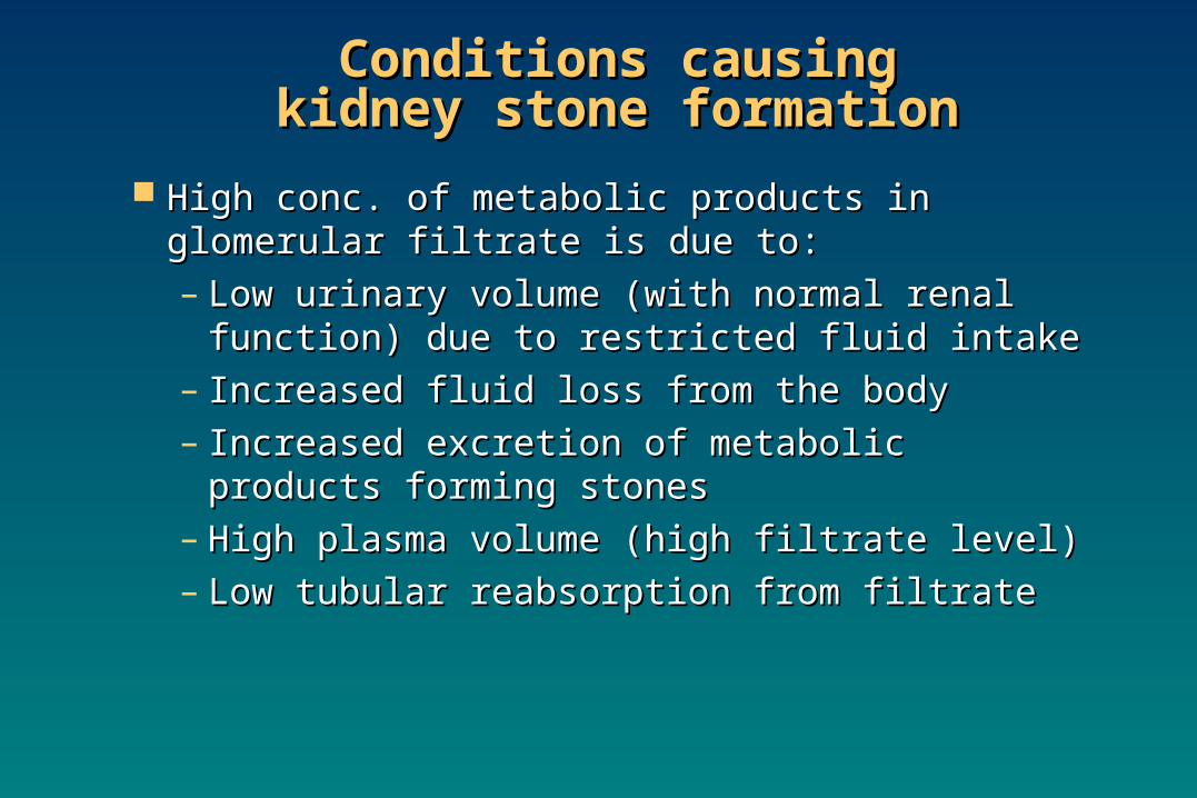
High conc. of metabolic products in High conc. of metabolic products in glomerular filtrate is due to:glomerular filtrate is due to:– Low urinary volume (with normal renal Low urinary volume (with normal renal
function) due to restricted fluid intakefunction) due to restricted fluid intake– Increased fluid loss from the bodyIncreased fluid loss from the body– Increased excretion of metabolic products Increased excretion of metabolic products
forming stonesforming stones– High plasma volume (high filtrate level)High plasma volume (high filtrate level)– Low tubular reabsorption from filtrateLow tubular reabsorption from filtrate
Conditions causingConditions causingkidney stone formationkidney stone formation
Conditions causingConditions causingkidney stone formationkidney stone formation
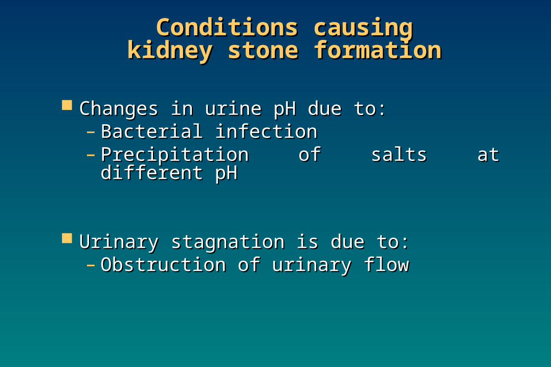
Changes in urine pH due to:Changes in urine pH due to:– Bacterial infectionBacterial infection– Precipitation of salts at different pHPrecipitation of salts at different pH
Urinary stagnation is due to:Urinary stagnation is due to:– Obstruction of urinary flow Obstruction of urinary flow
Conditions causingConditions causingkidney stone formationkidney stone formation
Conditions causingConditions causingkidney stone formationkidney stone formation
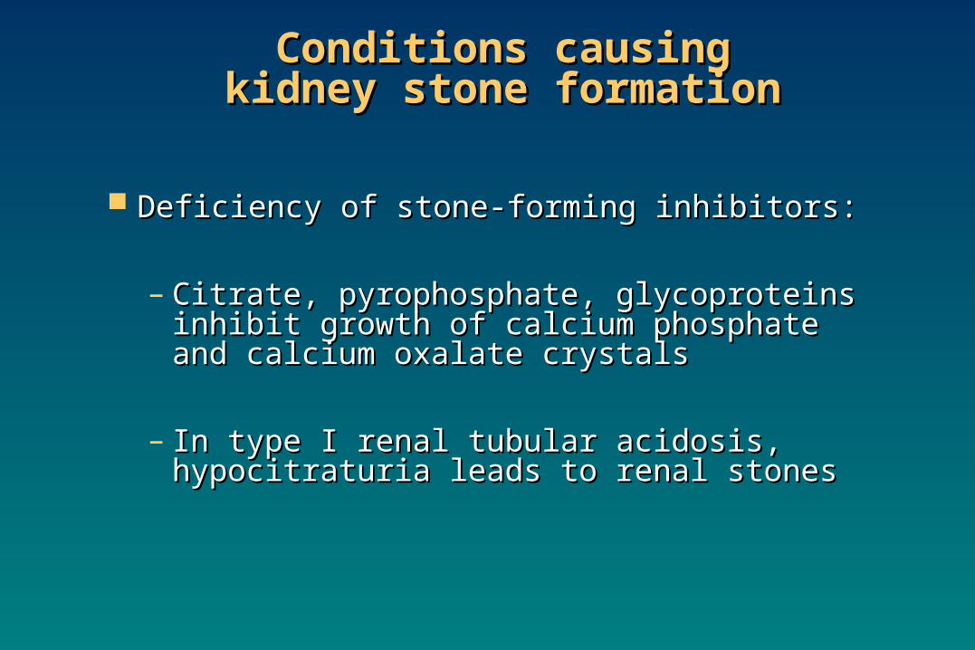
Deficiency of stone-forming inhibitors:Deficiency of stone-forming inhibitors:
– Citrate, pyrophosphate, glycoproteins Citrate, pyrophosphate, glycoproteins inhibit growth of calcium phosphate and inhibit growth of calcium phosphate and calcium oxalate crystalscalcium oxalate crystals
– In type I renal tubular acidosis, In type I renal tubular acidosis, hypocitraturia leads to renal stoneshypocitraturia leads to renal stones
Types of kidney stonesTypes of kidney stonesTypes of kidney stonesTypes of kidney stones
Calcium saltsCalcium salts Uric acidUric acid Mg ammonium POMg ammonium PO44
CystineCystine Other (xanthine, etc.)Other (xanthine, etc.)
Calcium salt stonesCalcium salt stonesCalcium salt stonesCalcium salt stones
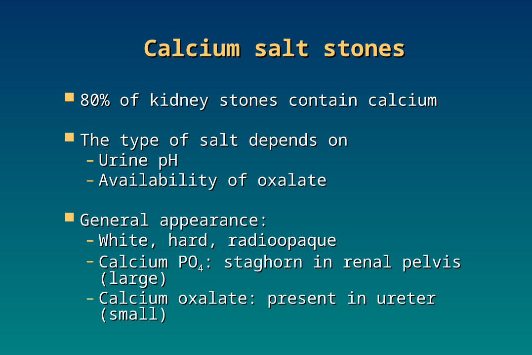
80% of kidney stones contain calcium80% of kidney stones contain calcium
The type of salt depends onThe type of salt depends on– Urine pHUrine pH– Availability of oxalateAvailability of oxalate
General appearance:General appearance:– White, hard, radioopaqueWhite, hard, radioopaque– Calcium POCalcium PO44: staghorn in renal pelvis : staghorn in renal pelvis
(large)(large)– Calcium oxalate: present in ureter (small)Calcium oxalate: present in ureter (small)
Calcium salt stonesCalcium salt stonesCalcium salt stonesCalcium salt stones
Causes of calcium salt stones:Causes of calcium salt stones:
Hypercalciuria:Hypercalciuria:– Increased urinary calcium excretionIncreased urinary calcium excretion– Men: > 7.5 mmols/dayMen: > 7.5 mmols/day– Women > 6.2 mmols/dayWomen > 6.2 mmols/day– May or may not be due to hypercalcemiaMay or may not be due to hypercalcemia
Calcium salt stonesCalcium salt stonesCalcium salt stonesCalcium salt stones
Hyperoxaluria:Hyperoxaluria:– Causes the formation of calcium oxalates Causes the formation of calcium oxalates
without hypercalciuriawithout hypercalciuria– Diet rich in oxalatesDiet rich in oxalates– Increased oxalate absorption in fat Increased oxalate absorption in fat
malabsorptionmalabsorption
Primary hyperoxaluria:Primary hyperoxaluria:– Due to inborn errorsDue to inborn errors– Urinary oxalate excretion: > 400 mmols/dayUrinary oxalate excretion: > 400 mmols/day
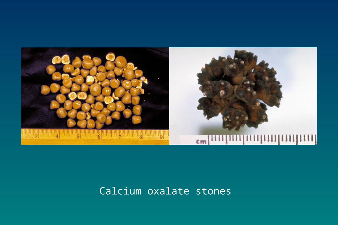
Calcium oxalate stones
Calcium salt stonesCalcium salt stonesCalcium salt stonesCalcium salt stones
Treatment:Treatment:– Treatment of primary causes such as Treatment of primary causes such as
infection, hypercalcemia, hyperoxaluriainfection, hypercalcemia, hyperoxaluria– Oxalate-restricted dietOxalate-restricted diet– Increased fluid intakeIncreased fluid intake– Acidification of urine (by dietary Acidification of urine (by dietary
changes)changes)Calcium salt stones are formed in Calcium salt stones are formed in
alkaline urinealkaline urine

Uric acid stonesUric acid stonesUric acid stonesUric acid stones
About 8% of renal stones contain uric acidAbout 8% of renal stones contain uric acid May be associated with hyperuricemia May be associated with hyperuricemia
(with or without gout) (with or without gout) Form in acidic urineForm in acidic urine General appearance:General appearance:
– Small, friable, yellowishSmall, friable, yellowish– May form staghornMay form staghorn– Radiolucent (plain x-rays cannot detect)Radiolucent (plain x-rays cannot detect)– Visualized by ultrasound or i.v. Visualized by ultrasound or i.v.
pyelogrampyelogram
Uric acid stonesUric acid stonesUric acid stonesUric acid stones
Treatment:Treatment:– Purine-restricted dietPurine-restricted diet– Alkalinization of urine (by dietary Alkalinization of urine (by dietary
changes)changes)– Increased fluid intakeIncreased fluid intake
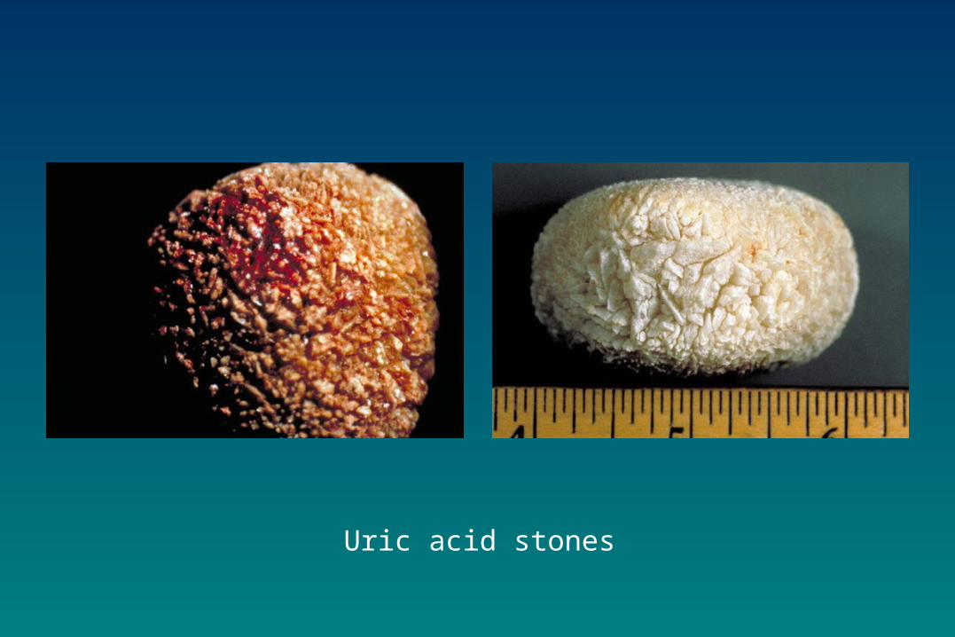
Uric acid stones
Mg ammonium POMg ammonium PO44 stones stonesMg ammonium POMg ammonium PO44 stones stones
About 10% of all renal stones contain Mg About 10% of all renal stones contain Mg amm. POamm. PO44
Also called struvite kidney stonesAlso called struvite kidney stones Associated with chronic urinary tract Associated with chronic urinary tract
infectioninfection– Microorganisms (such as from Proteus Microorganisms (such as from Proteus
genus) that metabolize urea into ammoniagenus) that metabolize urea into ammonia– Causing urine pH to become alkaline and Causing urine pH to become alkaline and
stone formationstone formation
Mg ammonium POMg ammonium PO44 stones stonesMg ammonium POMg ammonium PO44 stones stones
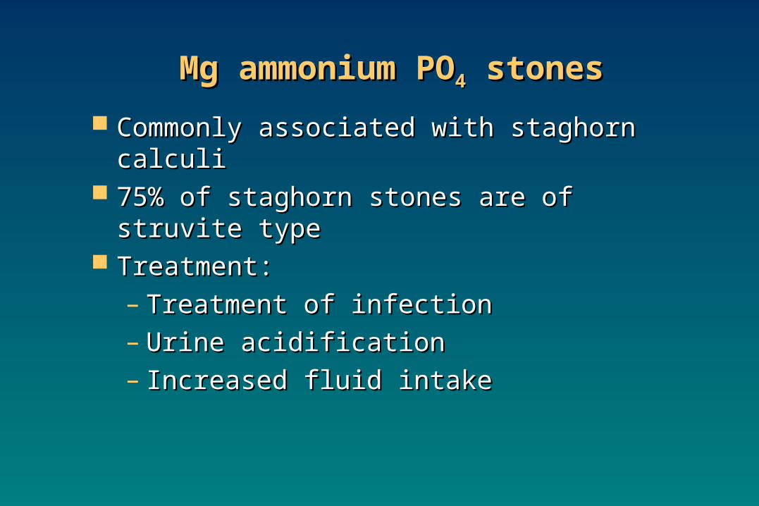
Commonly associated with staghorn calculiCommonly associated with staghorn calculi 75% of staghorn stones are of struvite type75% of staghorn stones are of struvite type Treatment:Treatment:
– Treatment of infectionTreatment of infection– Urine acidificationUrine acidification– Increased fluid intakeIncreased fluid intake
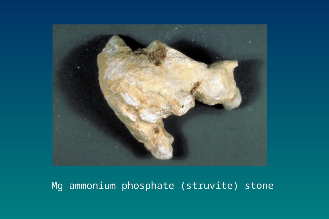
Mg ammonium phosphate (struvite) stone
Cystine stonesCystine stonesCystine stonesCystine stones
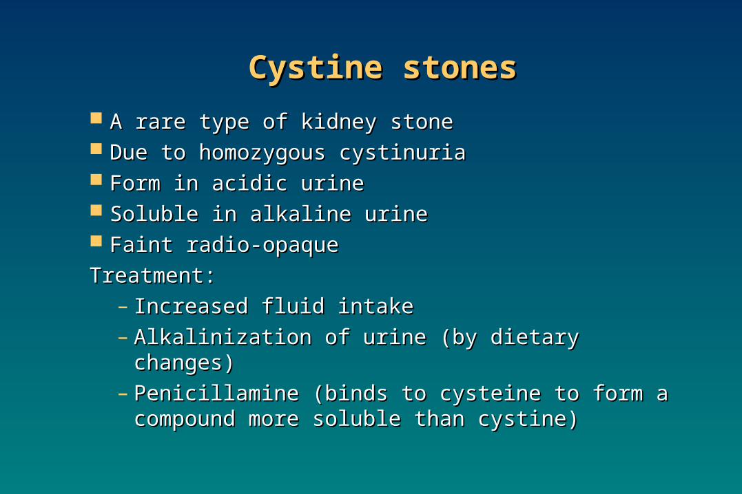
A rare type of kidney stoneA rare type of kidney stone Due to homozygous cystinuriaDue to homozygous cystinuria Form in acidic urineForm in acidic urine Soluble in alkaline urineSoluble in alkaline urine Faint radio-opaqueFaint radio-opaque
Treatment:Treatment:– Increased fluid intakeIncreased fluid intake– Alkalinization of urine (by dietary changes)Alkalinization of urine (by dietary changes)– Penicillamine (binds to cysteine to form a Penicillamine (binds to cysteine to form a
compound more soluble than cystine)compound more soluble than cystine)
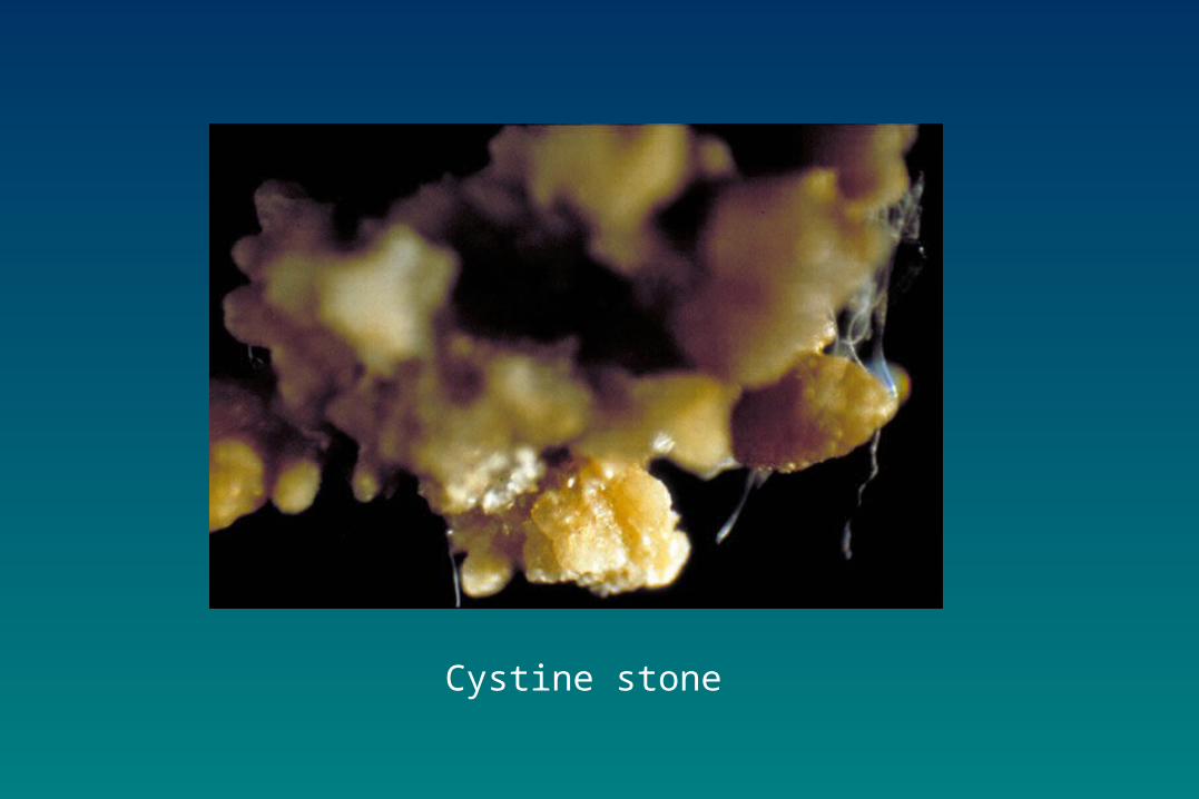
Cystine stone
Laboratory investigationsLaboratory investigationsof kidney stonesof kidney stones
Laboratory investigationsLaboratory investigationsof kidney stonesof kidney stones
If stone has formed and removed:If stone has formed and removed:
Chemical analysis of stone helps to:Chemical analysis of stone helps to:– Identify the causeIdentify the cause– Advise patient on prevention and future Advise patient on prevention and future
recurrencerecurrence
Laboratory investigationsLaboratory investigationsof kidney stonesof kidney stones
Laboratory investigationsLaboratory investigationsof kidney stonesof kidney stones
If stone has not formed:If stone has not formed: This type of investigation identifies This type of investigation identifies
causes that may contribute to stone causes that may contribute to stone formationformation– Serum calcium and uric acid analysisSerum calcium and uric acid analysis– Urinalysis: volume, calcium, oxalates Urinalysis: volume, calcium, oxalates
and cystine levelsand cystine levels– Urine pH > 8 suggests urinary tract Urine pH > 8 suggests urinary tract
infection (Mg amm. POinfection (Mg amm. PO44)) Urinary tract imaging:Urinary tract imaging:
– Ultrasound and i.v. pyelogramUltrasound and i.v. pyelogram


![Determination of Pb (Lead), Cd (Cadmium), Cr (Chromium ...harmful to the skin, liver, kidney, and respiratory organs [13], causing various diseases, such as dermatitis, renal tubular](https://img.dokumen.tips/doc/110x75/5fa6d8c32f1da633d06221bb/determination-of-pb-lead-cd-cadmium-cr-chromium-harmful-to-the-skin.jpg)


