Embed Size (px)
Citation preview

INSIGHT ANNUAL REPORTKECK SCHOOL OF MEDICINE OF USCDEPARTMENT OF OPHTHALMOLOGY
PRESENTED BY
2 eye.keckmedicine.org
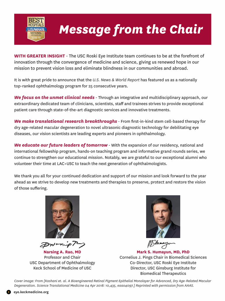
WITH GREATER INSIGHT - The USC Roski Eye Institute team continues to be at the forefront of innovation through the convergence of medicine and science, giving us renewed hope in our mission to prevent vision loss and eliminate blindness in our communities and abroad.
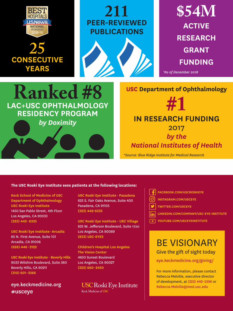
It is with great pride to announce that the U.S. News & World Report has featured us as a nationally top-ranked ophthalmology program for 25 consecutive years.
We focus on the unmet clinical needs - Through an integrative and multidisciplinary approach, our extraordinary dedicated team of clinicians, scientists, staff and trainees strives to provide exceptional patient care through state-of-the-art diagnostic services and innovative treatments.
We make translational research breakthroughs - From fi rst-in-kind stem cell-based therapy for dry age-related macular degeneration to novel ultrasonic diagnostic technology for debilitating eye diseases, our vision scientists are leading experts and pioneers in ophthalmology.
We educate our future leaders of tomorrow - With the expansion of our residency, national and international fellowship program, hands-on teaching program and informative grand rounds series, we continue to strengthen our educational mission. Notably, we are grateful to our exceptional alumni who volunteer their time at LAC+USC to teach the next generation of ophthalmologists.
We thank you all for your continued dedication and support of our mission and look forward to the year ahead as we strive to develop new treatments and therapies to preserve, protect and restore the visionof those suff ering.
Message from the Chair
Narsing A. Rao, MDProfessor and Chair
USC Department of OphthalmologyKeck School of Medicine of USC
Mark S. Humayun, MD, PhDCornelius J. Pings Chair in Biomedical Sciences
Co-Director, USC Roski Eye InstituteDirector, USC Ginsburg Institute for
Biomedical Therapeutics
Cover image: From [Kashani et. al. A Bioengineered Retinal Pigment Epithelial Monolayer for Advanced, Dry Age-Related Macular Degeneration. Science Translational Medicine 04 Apr 2018: 10,435, eaao4097.] Reprinted with permission from AAAS.

3Advertising Supplement
YOUR VISION is OUR MISSION
PRESERVEThe USC Roski Eye Institute
diagnoses, treats and manages the most complex eye conditions,
from in utero to advanced age.
PROTECTThe USC Roski Eye Instituteleads major research in the
diagnosis of eye disease with advanced imaging technology to
help prevent blindness.
RESTOREThe USC Roski Eye Institute
integrates and applies emerging technologies to develop new methods to restore sight to
the blind.
SPECIALIZED CARE for ADULTS & CHILDREN
The USC Roski Eye Institute treats the full spectrum of eye conditions - from the most common to the most complex.
▪ CATARACT▪ CORNEA & EXTERNAL DISEASES▪ GLAUCOMA▪ LASER VISION CORRECTION▪ LOW VISION REHABILITATION▪ NEURO-OPHTHALMOLOGY AND ADULT STRABISMUS▪ OCULAR ONCOLOGY
▪ OCULO-FACIAL PLASTIC SURGERY▪ OPHTHALMIC MOLECULAR AND IMMUNOPATHOLOGY▪ PEDIATRIC OPHTHALMOLOGY▪ SPECIALTY CONTACT LENSES AND PROSE▪ UVEITIS AND OCULAR INFLAMMATION▪ RETINA, VITREOUS AND MACULAR DISEASES & SURGERY

4 eye.keckmedicine.org
Notable Accolades & Achievements
HELEN KELLER LAUREATE AWARD
Presented to Dr. A Linn Murphree (left) for his discovery of the RB1 tumor suppressor gene
2018 IEEEBiomedical Engineering Award
Karen Bartelson, past IEEE president,presenting the Award to Dr. Mark Humayun
VOTED BEST OF THE BEST
Congratulations to Drs. Hossein Ameri, Mark Borchert, Mark Humayun,
Jonathan Kim, Linda Lam, Thomas Lee, Karen Morgan, Arlanna Moshfeghi, Bibiana Reiser, and Grace Richter
USC DEPARTMENT OF OPHTHALMOLOGY
#1IN RESEARCH FUNDING
2017by the National Institutes of Health
ASOPRS 2018 JAMES A. KATOWITZ PEDIATRIC AWARD
Dr. Jonathan Kim was honored for hiscontributions in oculoplastic surgery
LAC+USC OPHTHALMOLOGY RESIDENCY PROGRAM
by Doximity
RANKED

5Advertising Supplement
G L O B A L C O N N E C T I O NVision Care without Borders
Armenian Eye Care Project
Residency Rotation
7th Indo-China Ocular Inammation Meeting
International Society of Ocular Oncology
Restoring Vision with Argus II
Earthquake Relief E�ortsSurgical Skills Training
India
ArmeniaMongolia
NepalMyanmar
Clinical Outreach
Jamaica
South Korea
Australia

6 eye.keckmedicine.org
Advancing Vision Sciencewith Three New Integrative Cores
Mahnaz Shahidi, PhDPrincipal Investigator
▪ Cultivating opportunities for inter-disciplinary basic and translational vision research
▪ Enabling collaboration among outstanding scientists and clinicians
▪ Providing cutting-edge bioinstrumentation and expert technical personnel
Sarah Hamm-Alvarez, PhDDirector, Cell and Tissue Imaging Core
Jeannie Chen, PhDDirector, In Vivo Models and Imaging Core
Andrew MacKay, PhDDirector, Ophthalmic Therapeutics Engineering Core
Lacrimal gland acinus showing infi ltrating immune cells (purple) and therapeutic nanoparticle micelles.
National Eye Institute Funded Center Core Grant for Vision Research
Fostering Innovation in Ophthalmology
R01: "Protein-Polymer Nanomedicine for Sjogren's Syndrome"Principal Investigator: Sarah Hamm-Alvarez, PhD
R01: "Imaging of Retinal Oxygenation and Metabolism"Principal Investigator: Mahnaz Shahidi, PhD
Retinal oxygen metabolism is determined by imaging of arteriovenous oxygen content diff erence and blood fl ow. Left) Retinal vascular oxygen tension measurements displayed in pseudo color. Middle) Automatically detected retinal vessel boundaries are outlined on a red free image to visualize vessel diameter measurements. Right) A projection image generated from superimposed images of circulating fl uorescent microspheres over time to depict blood velocity measurements.
R01: "Corneal Biomechanical Analysis with Brillouin Microscopy"Principal Investigator: J. Bradley Randleman, MD
Comparison analysis between two diff erent cross-linking techniques, control and lens.
Three Integrative Research Cores
Ophthalmic Therapeutics Engineering Core
Cell and Tissue Imaging Core
In Vivo Models and Imaging Core

7Advertising Supplement
HOPE is in SIGHT for Dry AMD
Anna Kuehl, a USC alumna, was diagnosed with dry age-related macular degeneration (AMD), which caused her to lose the ability to drive, read or even recognize faces over time.
With a diminished quality of life, Anna turned to the USC Roski Eye Institute, where she learned of anopportunity to participate in a phase I/IIa clinical trial for dry AMD. Dr. Amir Kashani, principal investigator of the clinical trial and Assistant Professor of Clinical Ophthalmology, replaced the diseased portion of Anna’s eye with the first-in-kind retinal implant, a single layer of RPE-derived from stem cells grown on a synthetic scaffold.
“We are grateful to the California Institute for Regenerative Medicine, who supported this initiative and to people like Anna who will help us one day find a cure for this devastating disease”, says Dr. Mark Humayun, Director of the USC Ginsburg Institute for Biomedical Therapeutics and Co-Director of the USC Roski Eye Institute, who led the multidisciplinary team that developed the stem cell-based retinal implant at USC.
The results of a subset of subjects were published in April 2018 and featured on the cover of Science Translational Medicine. A phase II multi-center trial is on the horizon with the completion of the phase I/IIa trial.
“I got my independence back, and I’m just so happy about it.” - Anna Kuehl

8 eye.keckmedicine.org
ACTIVE RESEARCH FUNDING - DECEMBER 2018PRINCIPAL INVESTIGATOR PROJECT SOURCE
Amir Kashani, MD, PhD 3D Angiography for Quantitative Characterization NIH/NEI
Amir Kashani, MD, PhD Functional Imaging in Hypoxic-Ischemic Retinal Disease NIH/NEI
Amir Kashani, MD, PhD Imaging Cerebral and Retinal Microvasculature in Cerebral Small Vessel Disease NIH/NINDS
Amir Kashani, MD, PhD OCT Angiography Research Consortium (OARC) Carl Zeiss Meditec
Amir Kashani, MD, PhD Study of Subretinal Implantation of Human Embryonic Stem Cell-Derived RPE Cells in Advanced Dry AMD CIRM
Benjamin Xu, MD, PhD Automated Detection of Gonioscopic Angle Closure Based on Anterior Segment OCT Imaging AGS
Benjamin Xu, MD, PhD Development and Validation of a Quantitative Anterior Segment OCT-based Method to Evaluate Patients with Primary Angle Closure Disease NIH/NEI
Biju Thomas, PhD Studies on Functionality of iPS-RPE Transplanted in Immunodeficient RCS Rats BrightFocus Foundation
David Cobrinik, MD, PhD Human Specific Signaling Circuitry in Cone Precursor Development NIH/NEI
David Cobrinik, MD, PhD Regulation of NCL and RdCVF in Cone Photoreceptor and Retinoblastoma Development RPB
David Hinton, MD An Experimental Approach to Maculopathy NIH/NEI
Gianluca Lazzi, PhD, MBAConnectome-Derived Computational Models of Degenerated Retina for Retinal Prosthetic Design
NIH/NEI
Gianluca Lazzi, PhD, MBA EAGER: Bioelectronic Color Vision NSF
Gianluca Lazzi, PhD, MBA Predictive Modeling of Bioelectric Activity on Mammalian Multilayered Neuronal Structures in the Presence of Supraphysiological Electric Fields NIH/NIBIB
Grace Richter, MD, MPH Defining the Relationships of Retinal Microcirculation with Glaucoma, Systemic Disease, and Ocular Anatomic Factors in African Americans NIH/NEI
Grace Richter, MD, MPH Retinal Microcirculation Changes after Intraocular Pressure Reduction in Glaucoma AGS
Grace Richter, MD, MPH Role of Optical Coherence Tomography Angiography in Detecting Retinal Microcirculation Changes after Intraocular Pressure Reduction in Glaucoma USC
J. Bradley Randleman, MD Corneal Biomechanical Analysis with Brillouin Microscopy NIH/NEI
Jesse Berry, MD Development of a Surrogate Liquid Biopsy from the Aqueous Humor in Retinoblastoma Eyes NIH/NCI
Kimberly Gokoffski, MD, PhD Electrical Fields Direct Retinal Ganglion Cell Axon Growth by Modulating GTPase Signaling NANOS
Kimberly Gokoffski, MD, PhD Physiological Electrical Fields Direct Optic Nerve Regeneration NIH/NCATS
Kimberly Gokoffski, MD, PhD Molecular Signals that Underlie Electrical Field Directed Retinal Ganglion Cell Axon Growth USC
Mahnaz Shahidi, PhD Center Core Grant for Vision Research NIH/NEI
Mahnaz Shahidi, PhD Imaging of Retinal Oxygenation and Metabolism NIH/NEI
Mahnaz Shahidi, PhD Ocular Biomarkers of Microvascular, Neural and Metabolic Function in Diabetes NIH/NIDDK
Mark Humayun, MD, PhD Phase 1 Safety Assessment of CPCB-RPE1, hESC-derived RPE Cell Coated Parylene Membrane Implants, in Patients with Advanced Dry Age Related Macular Degeneration CIRM
Mark Humayun, MD, PhD Thermoresponsive Reversible Adhesive for Temporary Intervention of Ocular Trauma - II DoD/U.S. Army
Mark Humayun, MD, PhD USC Roski Eye K12 Clinician-Vision Scientist Training Program (USC Roski Eye K12) NIH/NEI
Narsing Rao, MD Research to Prevent Blindness Unrestricted Grant RPB
Qifa Zhou, PhD Combined OCT/US/PAT System for Intravascular Imaging NIH/NHLBI
Qifa Zhou, PhD High Resolution Elastographic Assessment of the Optic Nerve Head NIH/NEI
Qifa Zhou, PhD High Resolution Elastography of Retina Under Prosthetic Electrical Stimulation NIH/NEI
Qifa Zhou, PhD Large Aperture and Wideband Modular Ultrasound Arrays for the Diagnosis of Liver Cancer NIH/NCI
Qifa Zhou, PhD Novel Focused Ultrasound (NFU) for Transscleral Drug Delivery USC Zumberge
Qifa Zhou, PhD Phase Resolved ARF Optical Coherence Elastography for Intravascular Imaging NIH/NHLBI
Sandy Zhang-Nunes, MD Therapeutic Applications of Ultrasound for the Prevention and Treatment of Dry Eye Disease USC Zumberge
Sarah Hamm-Alvarez, PhD Identification of Tear Biomarkers for Parkinson's Disease Patients Michael J. Fox Foundation
Sarah Hamm-Alvarez, PhD Microtubule-Based Transport in Lacrimal Gland Function NIH/NEI
Sarah Hamm-Alvarez, PhD Protein-Polymer Nanomedicine for Sjogren's Syndrome NIH/NEI
Vivek Patel, MD Human Connectomes for Low Vision, Blindness, and Sight Restoration NIH/NEI
Vivek Patel, MD Retinal Prosthesis Neural Imaging: Measuring the Impact of Crossmodal Plasticity on Visual Restoration
Beckman Foundation
Xuejuan Jiang, PhD The Cumulative Effects of Sickle Cell Trait on the Eye Among Older African Americans NIH/NEI
AGS (American Glaucoma Society) • CIRM (California Institute for Regenerative Medicine) • DoD (Department of Defense) • NANOS (North American Neuro-Ophthalmology Society) • NCATS (National Center for Advancing Translational Sciences) • NCI (National Cancer Institute) • NEI (National Eye Institute) • NHLBI (National Heart, Lung and Blood Institute) • NIBIB (National Institute of Biomedical Imaging and Bioengineering) • NIDDK (National Institute of Diabetes and Digestive and Kidney Diseases) • NIH (National Institutes of Health) • NINDS (National Institute of Neurological Disorders and Stroke) • NSF (National Science Foundation) • RPB (Research to Prevent Blindness)
9Advertising Supplement
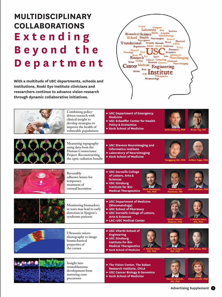
MULTIDISCIPLINARY COLLABORATIONSE x t e n d i n gB e y o n d t h e D e p a r t m e n tWith a multitude of USC departments, schools and institutions, Roski Eye Institute clinicians and researchers continue to advance vision research through dynamic collaborative initiatives.
Measuring topography using data from the Human Connectome Project: Reconstructing the optic radiation bundle
► USC Stevens Neuroimaging and Informatics Institute► Laboratory of Neuroimaging► Keck School of Medicine
Arthur Toga, PhD
Mark Humayun, MD, PhD
Juan Carlos Martinez, MD
Jack Whalen, PhD
Sarah Hamm-Alvarez, PhD
J. Martin Heur, MD, PhD
J. Martin Heur, MD, PhD
Mark Humayun, MD, PhD
Qifa Zhou, PhD
David Cobrinik, MD, PhD
Cheryl Craft, PhD
Reversibly adhesive lenses for temporary treatment of corneal laceration
► USC Dornsife College of Letters, Arts & Sciences► USC Ginsburg Institute for Bio- Medical Therapeutics
Monitoring biomarkers in tears may lead to early detection in Sjogren's syndrome patients
► USC Department of Medicine (Rheumatology)► USC School of Pharmacy► USC Dornsife College of Letters, Arts & Sciences► LAC+USC Medical Center
Ultrasonic micro- elastography to image biomechanical properties of the cornea
► USC Viterbi School of Engineering► USC Ginsburg Institute for Bio- Medical Therapeutics ► Keck School of Medicine
Insight into retinoblastoma development from maturing cone precursors
► The Vision Center, The Saban Research Institute, CHLA► USC Cancer Biology & Genomics ► Keck School of Medicine
Seth Seabury, PhD Brian Toy, MD
Combining policy-driven research with clinical insight to develop strategies to improve the health of vulnerable populations
► USC Department of Emergency Medicine► USC Schaeff er Center for Health Policy & Economics► Keck School of Medicine
Yonggang Shi, PhD

10 eye.keckmedicine.org
REVOLUTIONARY FIRST ON THE WEST COASTFDA-Approved Gene � erapy
After decades of declining vision, 44-year-old Toby Willis received the fi rst FDA-approved gene replacement therapy to treat aninherited form of vision loss.
Drs. Thomas Lee and Aaron Nagiel administered treatment by injecting it underneath the retina, the light sensing tissue of the eye. This treatment is for patients with Leber congenital amaurosis, who have mutations in the REP65 gene.
Leber congenital amaurosis aff ects children at a very young age and may lead to complete blindness over time.
"We are pleased to be able to off er this therapy that can truly impact a patient's quality of life, and potentially, help them see their future through 'new eyes'," said Thomas Lee, MD, Director of the Vision Center at Children's Hospital Los Angeles and Associate Professor of Ophthalmology at the USC Roski Eye Institute.
Through the Human Connectome Project (HCP), scientists developed advanced state-of-the-art brain mapping technologies allowing them to map the human brain in intricate detail. Through a multidisciplinary collaboration, led by Arthur W. Toga, PhD and collaborator Yonggang Shi, PhD from the USC Stevens Neuroimaging and Informatics Institute, researchers seek to elucidate brain circuitry as it relates to neurological function. The team has focused on the novel mathematical characterization of topographic regularity, which is the spatial organization of neurons when mapping brain connectivity.
“Detailed retinal mapping of the visual pathways may help scientists further their understanding of eye diseases," said Dr. Toga, Provost Professor of Ophthalmology.
Using the large-scale dataset from 215 HCP subjects, the team quantitatively reconstructed the optic radiation bundle with greater precision relative to other methods.
RECONSTRUCTING THE BRAIN Enhancing Retinal Mapping through a Novel Mathematical Model

11Advertising Supplement
Enhancing Daily Life with Vision Rehabilitation & PROSE
1 in 28 Americans over the age of 40 suffer from low vision, according to the National Federation of the Blind. Low vision is comprised of decreased visual acuity, contrast sensitivity, and/or visual field which creates an impingement on an individual's ability to effectively and independently carry out activities such as cooking, cleaning, reading the newspaper or watching television.
Vision rehabilitation services, customized by Dr. Rachel Young, are considered when patients no longer find improvement with prescription glasses, contact lenses, surgical or medical treatment. Those who suffer from low vision experience a dramatic decrease in their overall quality of life.
What Assistive Devices are Available?
► Hand held pocket magnifiers
► Stand magnifiers
► Monocular and binocular telescopes
► Applications for smart phones and tablets
► Solar shields/fit overs
► Portable electronic magnifiers
► Lighting recommendations
► Rehabilitation treatment plans
Prosthetic Replacement of the Ocular Surface Ecosystem (PROSE) Treatment
The USC Roski Eye Institute is one of only 12 sites in the nation to offer cutting-edge PROSE treatment. PROSE treatment involves specialty, scleral prosthetic devices that are custom-designed to help improve vision, comfort or support of the ocular surface.
Supervised by Dr. Gloria Chiu, PROSE treatment generally involves multiple visits, which include a consultation, training for proper device wear and maintenance, and follow up visits to design and evaluate fitting of custom devices. Literature has demonstrated that patients who have received this pioneering treatment experience a notable improvement in their quality of vision and quality of life as a result.
12 eye.keckmedicine.org
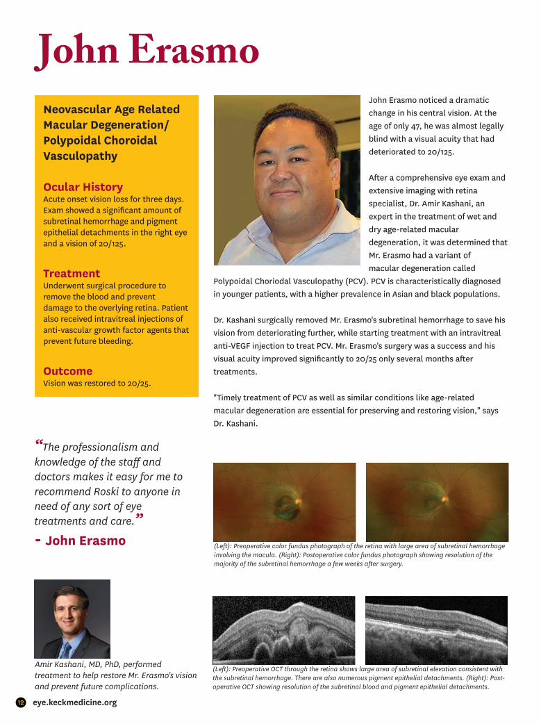
John ErasmoJohn Erasmo noticed a dramatic change in his central vision. At the age of only 47, he was almost legally blind with a visual acuity that had deteriorated to 20/125.
After a comprehensive eye exam and extensive imaging with retina specialist, Dr. Amir Kashani, an expert in the treatment of wet and dry age-related macular degeneration, it was determined that Mr. Erasmo had a variant of macular degeneration called
Polypoidal Choriodal Vasculopathy (PCV). PCV is characteristically diagnosed in younger patients, with a higher prevalence in Asian and black populations.
Dr. Kashani surgically removed Mr. Erasmo's subretinal hemorrhage to save his vision from deteriorating further, while starting treatment with an intravitreal anti-VEGF injection to treat PCV. Mr. Erasmo's surgery was a success and his visual acuity improved significantly to 20/25 only several months after treatments.
"Timely treatment of PCV as well as similar conditions like age-related macular degeneration are essential for preserving and restoring vision," says Dr. Kashani.
Neovascular Age Related Macular Degeneration/ Polypoidal Choroidal Vasculopathy
Ocular HistoryAcute onset vision loss for three days. Exam showed a significant amount of subretinal hemorrhage and pigment epithelial detachments in the right eye and a vision of 20/125.
TreatmentUnderwent surgical procedure to remove the blood and prevent damage to the overlying retina. Patient also received intravitreal injections of anti-vascular growth factor agents that prevent future bleeding.
OutcomeVision was restored to 20/25.
“The professionalism and knowledge of the staff and doctors makes it easy for me to recommend Roski to anyone in need of any sort of eye treatments and care.” - John Erasmo (Left): Preoperative color fundus photograph of the retina with large area of subretinal hemorrhage
involving the macula. (Right): Postoperative color fundus photograph showing resolution of the majority of the subretinal hemorrhage a few weeks after surgery.
Amir Kashani, MD, PhD, performed treatment to help restore Mr. Erasmo's vision and prevent future complications.
(Left): Preoperative OCT through the retina shows large area of subretinal elevation consistent with the subretinal hemorrhage. There are also numerous pigment epithelial detachments. (Right): Post-operative OCT showing resolution of the subretinal blood and pigment epithelial detachments.
13Advertising Supplement
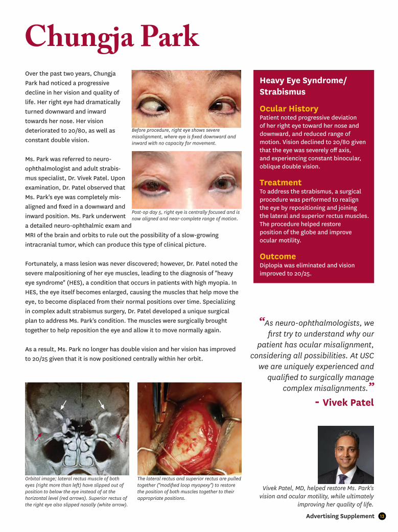
Over the past two years, Chungja Park had noticed a progressive decline in her vision and quality of life. Her right eye had dramatically turned downward and inward towards her nose. Her vision deteriorated to 20/80, as well as constant double vision.
Ms. Park was referred to neuro-ophthalmologist and adult strabis-mus specialist, Dr. Vivek Patel. Upon examination, Dr. Patel observed that Ms. Park's eye was completely mis-aligned and fixed in a downward and inward position. Ms. Park underwent a detailed neuro-ophthalmic exam and MRI of the brain and orbits to rule out the possibility of a slow-growing intracranial tumor, which can produce this type of clinical picture.
Fortunately, a mass lesion was never discovered; however, Dr. Patel noted the severe malpositioning of her eye muscles, leading to the diagnosis of "heavy eye syndrome" (HES), a condition that occurs in patients with high myopia. In HES, the eye itself becomes enlarged, causing the muscles that help move the eye, to become displaced from their normal positions over time. Specializing in complex adult strabismus surgery, Dr. Patel developed a unique surgical plan to address Ms. Park's condition. The muscles were surgically brought together to help reposition the eye and allow it to move normally again.
As a result, Ms. Park no longer has double vision and her vision has improved to 20/25 given that it is now positioned centrally within her orbit.
Chungja ParkHeavy Eye Syndrome/ Strabismus
Ocular HistoryPatient noted progressive deviation of her right eye toward her nose and downward, and reduced range of motion. Vision declined to 20/80 given that the eye was severely off axis, and experiencing constant binocular, oblique double vision.
TreatmentTo address the strabismus, a surgical procedure was performed to realign the eye by repositioning and joining the lateral and superior rectus muscles. The procedure helped restore position of the globe and improve ocular motility.
OutcomeDiplopia was eliminated and vision improved to 20/25.
Vivek Patel, MD, helped restore Ms. Park's vision and ocular motility, while ultimately
improving her quality of life.
“As neuro-ophthalmologists, we first try to understand why our
patient has ocular misalignment, considering all possibilities. At USC
we are uniquely experienced and qualified to surgically manage
complex misalignments.” - Vivek Patel
Before procedure, right eye shows severe misalignment, where eye is fixed downward and inward with no capacity for movement.
Post-op day 5, right eye is centrally focused and is now aligned and near-complete range of motion.
Orbital image; lateral rectus muscle of both eyes (right more than left) have slipped out of position to below the eye instead of at the horizontal level (red arrows). Superior rectus of the right eye also slipped nasally (white arrow).
The lateral rectus and superior rectus are pulled together ("modified loop myopexy") to restore the position of both muscles together to their appropriate positions.

14 eye.keckmedicine.org
MENTORING and INSPIRING Exceptional Clinician-Scientists
When ophthalmologists are trained in both medicine and science, they are uniquely prepared to advance treatment and open new avenues of discovery. The USC Roski Eye Institute mentors our clinician-scientists through K-Awards that provide support and protected time for intensive, supervised research-career development.
K-Awards are individualized to the recipients’ past training and career stage. The goal is to help our clinician-scientists independently conduct complex research that will earn them increased National Institutes of Health Funding for their work.
By inspiring talented, dedicated researchers to excel to their fullest, we actively pursue the eventual elimination of blindness.
Recent K-Awardees include:
▪ Jesse Berry, MD, “Development of a Surrogate Liquid Biopsy from the Aqueous Humor in Retinoblastoma Eyes”
▪ Kimberly Gokoffski, MD, PhD, “Physiologic Electrical Fields Direct Optic Nerve Regeneration”
▪ Amir H. Kashani, MD, PhD, “Functional Imaging in Hypoxic-Ischemic Retinal Disease”
▪ Grace Richter, MD, MPH, “Defining the Relationships of Retinal Microcirculation with Glaucoma, Systemic Disease, and Ocular Anatomic Factors in African Americans”
▪ Benjamin Xu, MD, PhD, “Development and Validation of a Quantitative Anterior Segment OCT-based Method to Evaluate Patients with Primary Angle Closure Disease”
15Advertising Supplement
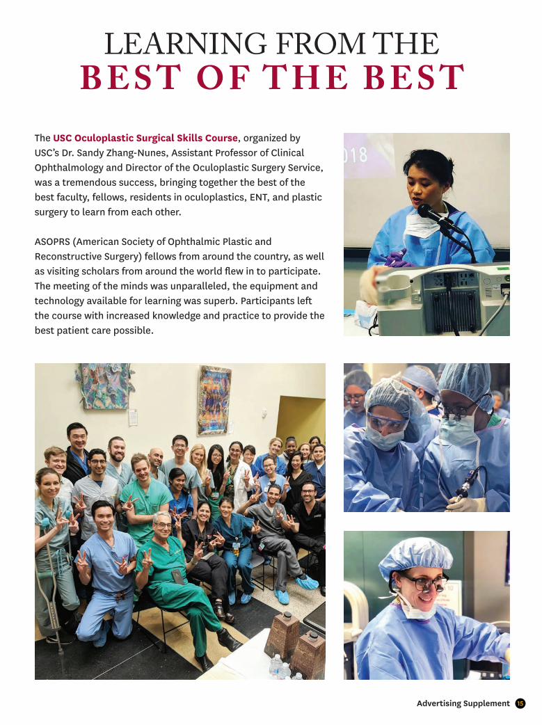
LEARNING FROM THE B E S T O F T H E B E S T
The USC Oculoplastic Surgical Skills Course, organized by USC’s Dr. Sandy Zhang-Nunes, Assistant Professor of Clinical Ophthalmology and Director of the Oculoplastic Surgery Service, was a tremendous success, bringing together the best of the best faculty, fellows, residents in oculoplastics, ENT, and plastic surgery to learn from each other.
ASOPRS (American Society of Ophthalmic Plastic and Reconstructive Surgery) fellows from around the country, as well as visiting scholars from around the world flew in to participate. The meeting of the minds was unparalleled, the equipment and technology available for learning was superb. Participants left the course with increased knowledge and practice to provide the best patient care possible.
16 eye.keckmedicine.org
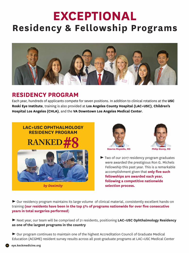
EXCEPTIONALResidency & Fellowship Programs
RESIDENCY PROGRAMEach year, hundreds of applicants compete for seven positions. In addition to clinical rotations at the USC Roski Eye Institute, training is also provided at Los Angeles County Hospital (LAC+USC), Children’s Hospital Los Angeles (CHLA), and the VA Downtown Los Angeles Medical Center.
LAC+USC OPHTHALMOLOGY RESIDENCY PROGRAM
by Doximity
► Two of our 2017 residency program graduates were awarded the prestigious Ron G. Michels Fellowship this past year. This is a remarkable accomplishment given that only five such fellowships are awarded each year, following a competitive nationwide selection process.
Stavros Moysidis, MD Philip Storey, MD
► Our residency program maintains its large volume of clinical material, consistently excellent hands-on training (our residents have been in the top 5% of programs nationwide for over five consecutive years in total surgeries performed)
► Next year, our team will be comprised of 21 residents, positioning LAC+USC Ophthalmology Residency as one of the largest programs in the country
► Our program continues to maintain one of the highest Accreditation Council of Graduate MedicalEducation (ACGME) resident survey results across all post-graduate programs at LAC+USC Medical Center

17Advertising Supplement
PROGRAM LEADERSHIP
Narsing Rao, MDProfessor and Chairman,
USC Department of Ophthalmology
Benjamin Xu, MD, PhDAssistant Program Director,
USC Department of Ophthalmology
Vivek Patel, MDEducation Director
Residency Program DirectorNeuro-Ophthalmology
Fellowship Director,USC Department of Ophthalmology
Malvin Anders, MDChief of Ophthalmology,LAC+USC Medical Center
FELLOWSHIP PROGRAMFellows at the USC Roski Eye Institute receive training in fi ve subspecialty areas:
▪ Cornea, External Disease & Refractive Surgery
▪ Glaucoma
▪ Neuro-Ophthalmology
▪ Oculofacial Plastic Surgery
▪ Vitreoretinal Surgery
► The fellowship experience spans three institutions: the USC Roski Eye Institute, CHLA, and LAC+USC
► In addition to working with their clinical and academic mentors, our fellows have the opportunity to run their own clinics and lead resident teaching rounds
► Fellows are given unprecedented responsibility in caring for the needs of patients at LAC+USC Medical Center as well as providing supervision of residents in both clinical and surgical settings

18 eye.keckmedicine.org
Grand Rounds Case Study“ P U S H F O R A S O L U T I O N ”
HISTORY• 62-year-old male, past medical history of hypertension, presents with binocular diplopia
• First presented to primary MD with left eye discomfort and diplopia; exam found EOM limitations OS
• Seen by neurology seven months later, MRI found sinus disease and mass extending into left orbit
• Seen by ENT; exam found mild-moderate s-shaped septal deviation
• No past ocular, surgical or family history
• Social history: worked as a MBA, former smoker, no alcohol use
EXAM FINDINGS• BCVA: 20/25 OD, 20/300 OS (was unremarkable at ENT visit)
• Pupils: round and reactive OU, +RAPD OS
• Color plates 10/10 OD, 6/10 OS
• IOP: 14 OD, 14 OS
• EOM:
• Anterior exam: remarkable for left eye proptosis, ptosis and medical conjunctival injection
• Posterior exam: Remarkable for congested and tortuous vessels OS
DIFFERENTIAL DIAGNOSIS• Benign growths (e.g. hemangioma)
• Malignancies (e.g. lymphoma)
• Idiopathic orbital infl ammatory syndrome
• Infectious (e.g. orbital abscess)
• Cysts (e.g. mucocele)
ADDITIONAL INVESTIGATIONS
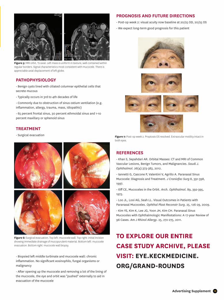
DIAGNOSIS• Ethmoid sinus mucocele
Figure 1: Extraocular motility on initial presentation. -4 abduction, -3 supraduction, -2 infraduction and adduction.
Sandy Zhang-Nunes, MDAssistant Professor of
Clinical OphthalmologyDirector, Oculofacial Plastic
Surgery [email protected]
Meghan Shan, MD, PhDjingmeghan.shan@
med.usc.eduPGY-3 ophthalmology
resident
DIAGNOSISTREATMENTCURRENT OPINIONSMODERN APPROACHES
CHALLENGING EYE CARE
Figure 3: Fundus photos show left eye vessels are congested and tortuous.
Figure 4: CT scan. Left mass measures 3cm in diameter. Smaller mass present in the right posterior ethmoid sinus. Mass is evident in the left orbit, aff ecting the medical rectus, inferior muscles as well as the optic nerve.
Figure 2: Worm's eye view showing left eye proptosis. Hertel measured 16.5mm OD, 20mm OS.
19Advertising Supplement
PATHOPHYSIOLOGY• Benign cysts lined with ciliated columnar epithelial cells that secrete mucous
• Typically occurs in 3rd to 4th decades of life
• Commonly due to obstruction of sinus ostium ventilation (e.g. infl ammation, allergy, trauma, mass, idiopathic)
• 65 percent frontal sinus, 30 percent ethmoidal sinus and 1-10 percent maxillary or sphenoid sinus
TREATMENT • Surgical evacuation
• Biopsied left middle turbinate and mucocele wall: chronic infl ammation. No signifi cant eosinophils, fungal organisms or malignancy
• After opening up the mucocele and removing a lot of the lining of the mucocele, the eye and orbit was "pushed" externally to aid in evacuation of the mucocele
PROGNOSIS AND FUTURE DIRECTIONS • Post-op week 2: visual acuity now baseline at 20/25 OD, 20/25 OS
• We expect long-term good prognosis for this patient
REFERENCES• Khan S, Sepahdari AR. Orbital Masses: CT and MRI of Common Vascular Lesions, Benign Tumors, and Malignancies. Saudi J. Ophthalmol. 26(4):373-383, 2012.
• Iannetti G, Cascone P, Valentini V, Agrillo A. Paranasal Sinus Mucocele: Diagnosis and Treatment. J Craniofac Surg 8, 391-398, 1997.
• Iliff CE, Mucoceles in the Orbit. Arch. Ophthalmol. 89, 392-395, 1973.
• Loo JL, Looi AG, Seah LL. Visual Outcomes in Patients with Paranasal Mucoceles. Ophthal Plast Reconstr Surg. 25, 126-29, 2009.
• Kim YS, Kim K, Lee JG, Yoon JH, Kim CH. Paranasal Sinus Mucoceles with Ophthalmologic Manifestations: A 17-year Review of 96 Cases. Am J Rhinol Allergy. 25, 272-275, 2011.
TO EXPLORE OUR ENTIRE CASE STUDY ARCHIVE, PLEASE VISIT: EYE.KECKMEDICINE.ORG/GRAND-ROUNDS
Figure 6: Surgical evacuation. Top left: mucocele wall. Top right: initial incision showing immediate drainage of mucopurulent material. Bottom left: mucocele evacuation. Bottom right: mucocele wall biopsy.
Figure 7: Post-op week 2. Proptosis OS resolved. Extraocular motility intact in both eyes.
Figure 5: MRI orbit, T2 axial. Left mass is uniform in texture, well contained within regular borders. Signal characteristics most consistent with mucocele. There is appreciable axial displacement of left globe.

20 eye.keckmedicine.org
� e End to Blindness is in Sightwith a Generous $10 Million Gift
Dr. Allen and Charlotte Ginsburg made a historic contribution to vision science research with a generous $10 million gift to name the USC Dr. Allen and Charlotte Ginsburg Institute for Biomedical Therapeutics (IBT). Dr. Allen Ginsburg, a retired ophthalmologist, and his wife Charlotte, a philanthropist, share IBT’s values of fostering multidisciplinary collaborations to save and restore sight.
Led by Dr. Mark Humayun, USC Ginsburg IBT’s foundation is built upon the convergence of ophthalmology and engineering, transforming bioengineered neural interfaces into treatments for patients who suff er from the most debilitating neurosensory disorders. Research breakthroughs by Dr. Humayun and his team of experts include the development of the fi rst FDA-approved artifi cial retina implant, Argus II, and stem cell-based treatments for devastating eye diseases.
(L to R): Keck Dean Dr. Laura Mosqueda, Charlotte Ginsburg, Dr. Allen Ginsburg, Dr. Mark Humayun and Interim PresidentDr. Wanda M. Austin.
(L to R): USC Provost Dr. Michael Quick, Vice Chair Dr. Martin Heur, Chairman Dr. Narsing Rao, SVP and CEO for USC Health Tom Jackiewicz, USC Ginsburg IBT Director Dr. Mark Humayun, Keck Dean Dr. Laura Mosqueda.
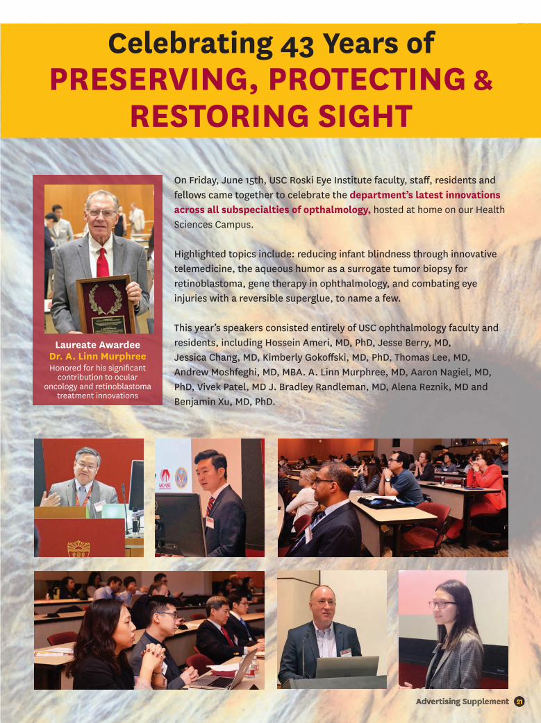
21Advertising Supplement 21Advertising Supplement
Celebrating 43 Years of PRESERVING, PROTECTING &
RESTORING SIGHT
On Friday, June 15th, USC Roski Eye Institute faculty, staff , residents and fellows came together to celebrate the department’s latest innovations across all subspecialties of opthalmology, hosted at home on our Health Sciences Campus.
Highlighted topics include: reducing infant blindness through innovative telemedicine, the aqueous humor as a surrogate tumor biopsy for retinoblastoma, gene therapy in ophthalmology, and combating eye injuries with a reversible superglue, to name a few.
This year’s speakers consisted entirely of USC ophthalmology faculty and residents, including Hossein Ameri, MD, PhD, Jesse Berry, MD, Jessica Chang, MD, Kimberly Gokoff ski, MD, PhD, Thomas Lee, MD, Andrew Moshfeghi, MD, MBA. A. Linn Murphree, MD, Aaron Nagiel, MD, PhD, Vivek Patel, MD J. Bradley Randleman, MD, Alena Reznik, MD and Benjamin Xu, MD, PhD.
Laureate AwardeeDr. A. Linn MurphreeHonored for his signifi cant
contribution to ocular oncology and retinoblastoma
treatment innovations
22 eye.keckmedicine.org
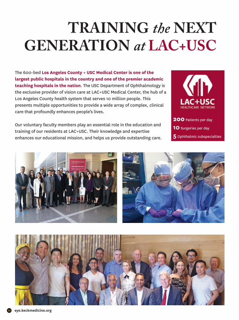
TRAINING the NEXT GENERATION at LAC+USC
The 600-bed Los Angeles County + USC Medical Center is one of the largest public hospitals in the country and one of the premier academic teaching hospitals in the nation. The USC Department of Ophthalmology is the exclusive provider of vision care at LAC+USC Medical Center, the hub of a Los Angeles County health system that serves 10 million people. This presents multiple opportunities to provide a wide array of complex, clinical care that profoundly enhances people’s lives.
Our voluntary faculty members play an essential role in the education and training of our residents at LAC+USC. Their knowledge and expertiseenhances our educational mission, and helps us provide outstanding care.
200 Patients per day
10 Surgeries per day
5 Ophthalmic subspecialties
23Advertising Supplement
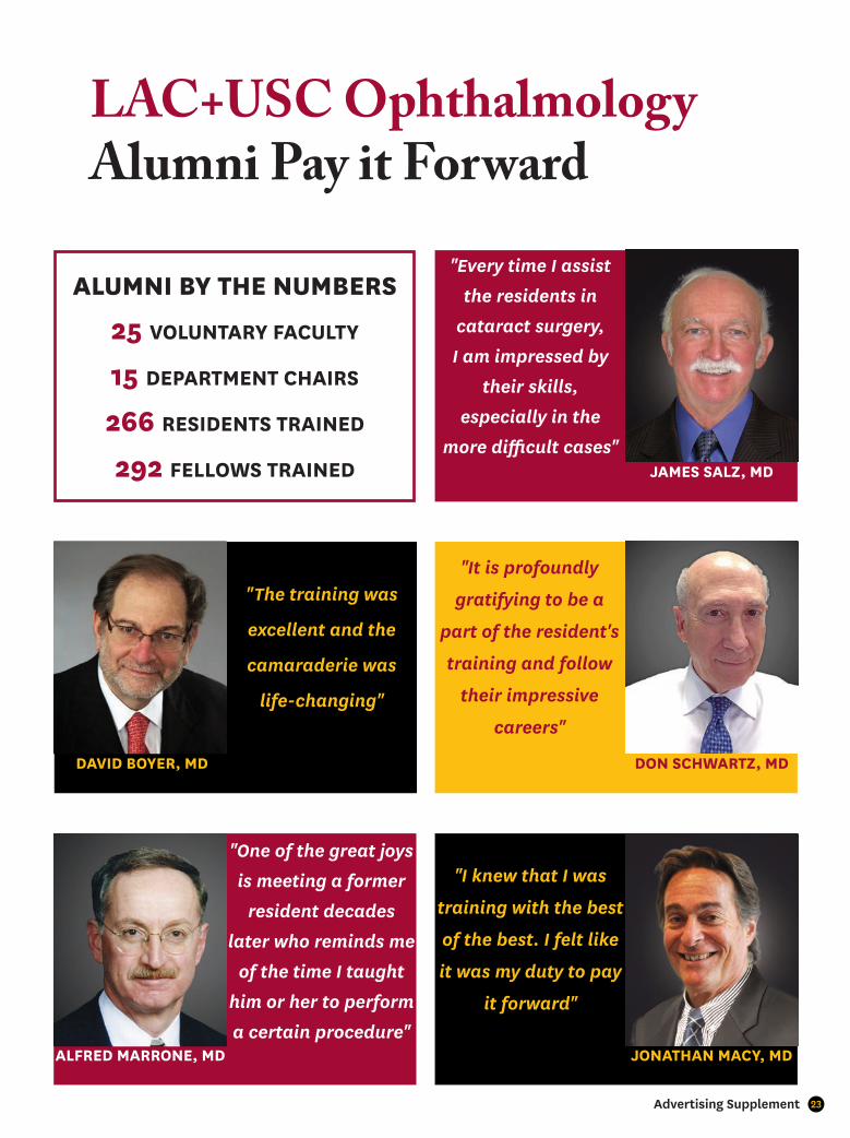
LAC+USC Ophthalmology Alumni Pay it Forward
"Every time I assist
the residents in
cataract surgery,
I am impressed by
their skills,
especially in the
more difficult cases"
"One of the great joys
is meeting a former
resident decades
later who reminds me
of the time I taught
him or her to perform
a certain procedure"
"The training was
excellent and the
camaraderie was
life-changing"
"I knew that I was
training with the best
of the best. I felt like
it was my duty to pay
it forward"
ALUMNI BY THE NUMBERS
25 VOLUNTARY FACULTY
15 DEPARTMENT CHAIRS
266 RESIDENTS TRAINED
292 FELLOWS TRAINED
DAVID BOYER, MD
JAMES SALZ, MD
ALFRED MARRONE, MD JONATHAN MACY, MD
DON SCHWARTZ, MD
"It is profoundly
gratifying to be a
part of the resident's
training and follow
their impressive
careers"
24 eye.keckmedicine.org
Keck School of Medicine of USCDepartment of OphthalmologyUSC Roski Eye Institute1450 San Pablo Street, 4th FloorLos Angeles, CA 90033(323) 442-6335
USC Roski Eye Institute - Arcadia65 N. First Avenue, Suite 101Arcadia, CA 91006(626) 446 -2122
USC Roski Eye Institute - Beverly Hills9033 Wilshire Boulevard, Suite 360Beverly Hills, CA 90211(310) 601-3366
USC Roski Eye Institute - Pasadena625 S. Fair Oaks Avenue, Suite 400Pasadena, CA 91105(323) 442-6335
USC Roski Eye Institute - USC Village835 W. Jeff erson Boulevard, Suite 1720Los Angeles, CA 90089(833) USC-EYES
Children’s Hospital Los AngelesThe Vision Center4650 Sunset BoulevardLos Angeles, CA 90027(323) 660-2450
FACEBOOK.COM/USCROSKIEYE
INSTAGRAM.COM/USCEYE
TWITTER.COM/USCEYE
LINKEDIN.COM/COMPANY/USC-EYE-INSTITUTE
YOUTUBE.COM/USCEYEINSTITUTE
eye.keckmedicine.org#usceye
BE VISIONARYGive the gift of sight today
eye.keckmedicine.org/giving/
For more information, please contact Rebecca Melville, executive director of development, at (323) 442–5396 or [email protected]
The USC Roski Eye Institute sees patients at the following locations:
USC Department of Ophthalmology
#1IN RESEARCH FUNDING
2017by the
National Institutes of Health*Source: Blue Ridge Institute for Medical Research
$54MACTIVE
RESEARCHGRANT
FUNDING*As of December 2018
211PEER-REVIEWEDPUBLICATIONS
Ranked #8LAC+USC OPHTHALMOLOGY
RESIDENCY PROGRAMby Doximity
25 CONSECUTIVE
YEARS