Embed Size (px)
Citation preview

Listen to this manuscript’s
audio summary by
JACC Editor-in-Chief
Dr. Valentin Fuster.
J O U R N A L O F T H E A M E R I C A N C O L L E G E O F C A R D I O L O G Y V O L . 6 7 , N O . 1 4 , 2 0 1 6
ª 2 0 1 6 B Y T H E A M E R I C A N C O L L E G E O F C A R D I O L O G Y F O U N D A T I O N I S S N 0 7 3 5 - 1 0 9 7 / $ 3 6 . 0 0
P U B L I S H E D B Y E L S E V I E R h t t p : / / d x . d o i . o r g / 1 0 . 1 0 1 6 / j . j a c c . 2 0 1 5 . 1 2 . 0 7 3
REVIEW TOPIC OF THE WEEK
Kawasaki Disease
Jane W. Newburger, MD, MPH,a,b Masato Takahashi, MD,c Jane C. Burns, MDdABSTRACT
Fro
Me
Sch
Sa
Bu
rel
Ma
Kawasaki disease is an acute, self-limited vasculitis of unknown etiology that occurs predominantly in infants and
children. If not treated early with high-dose intravenous immunoglobulin, 1 in 5 children develop coronary artery
aneurysms; this risk is reduced 5-fold if intravenous immunoglobulin is administered within 10 days of fever onset.
Coronary artery aneurysms evolve dynamically over time, usually reaching a peak dimension by 6 weeks after illness
onset. Almost all the morbidity and mortality occur in patients with giant aneurysms. Risk of myocardial infarction
from coronary artery thrombosis is greatest in the first 2 years after illness onset. However, stenosis and occlusion
progress over years. Indeed, Kawasaki disease is no longer a rare cause of acute coronary syndrome presenting in
young adults. Both coronary artery bypass surgery and percutaneous intervention have been used to treat Kawasaki
disease patients who develop myocardial ischemia as a consequence of coronary artery aneurysms and stenosis.
(J Am Coll Cardiol 2016;67:1738–49) © 2016 by the American College of Cardiology Foundation.
K awasaki disease (KD) is an acute, self-limited vasculitis of unknown etiologythat occurs predominantly in infants and
young children. Manifested initially by high fever,mucocutaneous inflammation, and cervical lymph-adenopathy, KD targets the coronary arteries andother cardiovascular structures (1). Approximately1 in 5 children who are not treated with intrave-nous immunoglobulin (IVIG) in the acute phase ofillness develop coronary artery aneurysms (CAA).Indeed, KD has replaced rheumatic fever as theleading cause of acquired cardiac disease in chil-dren in the developed world. This review describesour current understanding, as well as knowledgegaps, of the pathophysiology of KD and generalprinciples guiding the care and management ofthese patients in the absence of evidence-basedguidelines (Central Illustration). Diagnostic criteriafor complete and incomplete KD are detailed inthe 2004 American Heart Association/AmericanAcademy of Pediatrics guidelines (2).
m the aDepartment of Cardiology, Boston Children’s Hospital, Boston,
dical School, Boston, Massachusetts; cDepartment of Pediatrics, Seattle
ool of Medicine, Seattle, Washington; and the dDepartment of Pediatrics
n Diego, School of Medicine, La Jolla, California. Dr. Newburger was supp
rns was supported by a grant from the Gordon and Marilyn Macklin Fou
ationships relevant to the contents of this paper to disclose.
nuscript received September 21, 2015; revised manuscript received Decem
EPIDEMIOLOGY
The epidemiology of KD may yield important clues tothe etiology of this mysterious disease. First, KDstrikes predominantly infants and young children;80% of patients are younger than 5 years of age,although the disease can occur even in adolescence.The young age of onset suggests that susceptibilitymay be linked to maturation of the immune system.Second, although KD has been recognized on everycontinent and in all racial groups (3), the incidence ofdisease varies widely among different populations. InJapan, the country of highest incidence, the numbersof cases are steadily increasing and the most recentsurvey reported an incidence of 265 cases per 100,000children <5 years of age (4). In the United States,passive surveillance and analysis of administrativedatabases suggest a national incidence in children<5 years of age of 19 per 100,000, with a higherrate of 24.7 per 100,000 reported for California (5).An important genetic contribution to disease
Massachusetts; bDepartment of Pediatrics, Harvard
Children’s Hospital and University of Washington
, Rady Children’s Hospital, University of California–
orted by a grant from the McCance Family Fund. Dr.
ndation. Dr. Takahashi has reported that he has no
ber 3, 2015, accepted December 22, 2015.
AB BR E V I A T I O N S
AND ACRONYM S
CAA = coronary artery
aneurysm
CABG = coronary artery bypass
graft
CMR = cardiac magnetic
resonance
CTA = computer tomographic
angiography
IL = interleukin
IVIG = intravenous
immunoglobulin
KD = Kawasaki disease
LMP = luminal myofibroblastic
proliferation
MI = myocardial infarction
PCI = percutaneous coronary
intervention
TGFb = transforming growth
factor beta
T helper
J A C C V O L . 6 7 , N O . 1 4 , 2 0 1 6 Newburger et al.A P R I L 1 2 , 2 0 1 6 : 1 7 3 8 – 4 9 Kawasaki Disease
1739
susceptibility is suggested by the higher incidenceamong U.S. children of Asian/Pacific Islanderdescent in Hawaii and California (210 and 50.4 per100,000, respectively) (6,7). A distinct seasonality iscoherent across the Northern Hemisphere (8).
The current KD paradigm is that an immunologicreaction is elicited in genetically susceptible hostsupon exposure to the KD trigger(s), thought to bewidely dispersed in the environment and to enterthrough the upper respiratory tract (9). A subset ofgenetically susceptible children (w25%) will sufferirreversible damage to the coronary arterial wall.There is no evidence to support person-to-personspread, although temporal and spatial clustering ofcases is well-documented (10). Two potential para-digms postulate that the trigger is either of thefollowing: 1) an infectious agent that replicates in thesuperficial epithelial cells of the upper airway (11); or2) an antigen widely dispersed in the environment.Recent intriguing data support the hypothesis thatthe trigger for KD is carried by large-scale tropo-spheric winds and that provinces in northeasternChina serve as a source region for the seasonal clus-tering and annual epidemic of KD cases in Japan,Hawaii, and southern California (12).
PATHOPHYSIOLOGY
The complex picture that is emerging of the immuneresponse in acute KD includes activation of both theinnate and adaptive immune systems. Neutrophilsare among the first responders to invade the arterialwall and are followed by CD8þ T cells, dendritic cells,and monocyte/macrophages (13,14). Strong supportfor activation of the interleukin (IL)-1 pathwayincludes increased transcript abundance for IL-1-related genes by microarray and quantitative real-time polymerase chain reaction, and by increasedlevels of IL-1 pathway proteins in plasma from acuteKD patients (15). In KD, as in giant cell arteritis (16), 2dominant cytokine clusters are recognized: the IL-6/Thelper (Th)-17 axis and the IL-12/interferon gammaaxis. IL-6, in combination with transforming growthfactor beta (TGFb), polarizes naïve T cells toward aTh-17 phenotype, resulting in these cells invadingthe vessel wall and elaborating a proinflammatorycytokine profile (17,18). Evidence is accumulatingthat, in a subset of patients who develop CAA, theIL-12/interferon gamma axis may be activatedand proinflammatory cytokines may play a role ininducing activation of Th-1 cells in the vessel wall(19–22).
Mouse models of coronary arteritis mimicking KDhave been created by intraperitoneal injection of cell
wall extracts of both Lactobacillus casei andCandida albicans into certain genetic strains(23). These models have been used to inter-rogate responses to different therapies andprovide support for the use of IVIG, blockadeof tumor necrosis factor alpha and IL-1, and,potentially, the use of atorvastatin to modu-late the acute phase of vascular wall inflam-mation (24–26).
The complex genetics of KD is beginning toyield to efforts by multinational collaborativegroups to determine the genetic influences ondisease susceptibility and outcome (27).Roughly 65% of the genetic risk for KD sus-ceptibility is accounted for by polymorphismsin calcium signaling pathways, the TGFbpathway, and human leukocyte antigens(28-31).
PATHOLOGY
The pathological changes in KD affect
medium-sized, extra-parenchymal muscular arteries,most commonly the coronary arteries. A recentcomprehensive review of 32 KD autopsies and 8explanted hearts described 3 linked vasculopathicprocesses in the arterial wall: necrotizing arteritis,subacute/chronic vasculitis, and luminal myofibro-blastic proliferation (LMP) (32). The acute arteritis ischaracterized by a neutrophilic infiltrate originatingfrom the lumen of the vessel and can be associatedwith extensive necrosis of all layers of the vessel wallin both the coronaries and other medium-size arteries(13). Neutrophil elastases may also play a role indestruction of the internal and external elasticlaminae that provide recoil for the vessel wall andwhose destruction contributes to aneurysm forma-tion. Neutrophil elastase inhibitors have been used inJapan to block this pathway, but no randomizedclinical trial data are available (33).Subacute vasculitis begins weeks after the onset offever, can still be detected months to years later, andis closely associated with the third process, LMP (32).The inflammatory infiltrate is predominantly lym-phocytic and originates in the adventitia. CD8þ
cytotoxic T lymphocytes have been documented inthe media (14), suggesting that anti-T-cell therapies,such as the calcineurin inhibitors cyclosporine andtacrolimus, might be effective (34,35). LMP, withmyofibroblasts possibly derived from medial smoothmuscle cells, is a pathological process that is likelymediated by TGFb (36,37). Polymorphisms in the TGFbpathway are associated with increased susceptibilityto aneurysm formation in KD patients (30). LMP can
Th =

CENTRAL ILLUSTRATION Management of Kawasaki Disease
Anti-inflammatory therapy with intravenous immunoglobulin (IVIG) and ASA (acetylsalicylic acid or aspirin).Echocardiogram to assess coronary arteries and determine Z score.
Defervescence with no coronary dilation (Z score <2) Persistent/recrudescent fever 36 hrs after IVIG and/or Z score ≥2.5
Additional anti-inflammatory therapy
Diagnosis of Kawasaki disease according to American Heart Association criteria*
Risk level
1 Normal coronary arteries
2 Transient dilation
3 Small aneurysm
4 Medium aneurysm
5 Large or giant aneurysm
Management
Antiplatelet therapy 4–6 weeks and cardiac counseling.No invasive cardiac tests are warranted.
Same as risk level 1
Low-dose aspirin beyond first 6 weeks
Z score
<2
2 to <2.5
≥2.5 to <5
≥5 to <10
≥10 or absolutedimension ≥8mm
Long-term antiplatelet therapy
Long-term antiplatelet plusanticoagulation therapy, +/– beta blockers
Coronary risk stratification
Life-long surveillance tailoredto coronary artery status and age:• Echocardiography (serial)• Angiography (CT, CMR, invasive)• Stress-testing for inducible myocardial ischemia• Coronary intervention
- Catheter- Surgical
• Counseling- Exercise recommendations- Reproductive counseling- CV risk management
Repeat echocardiograms until dimensions stabilize
Newburger, J.W. et al. J Am Coll Cardiol. 2016;67(14):1738–49.
*Fever for at least 5 days plus rash, bilateral nonexudative conjunctivitis, oral changes (red, cracked lips, strawberry tongue, erythema of the oropharynx);
extremity changes (swollen hands and feet, erythema of the palms and soles, and/or subungual desquamation at 2 weeks); cervical lymphadenopathy
($1.5 cm, usually unilateral) or fever plus fewer criteria with coronary aneurysms. ASA ¼ acetylsalicylic acid; CMR ¼ cardiac magnetic resonance;
CT ¼ computed tomography; CV ¼ cardiovascular; IVIG ¼ intravenous immunoglobulin.
Newburger et al. J A C C V O L . 6 7 , N O . 1 4 , 2 0 1 6
Kawasaki Disease A P R I L 1 2 , 2 0 1 6 : 1 7 3 8 – 4 9
1740
result in luminal narrowing and consequent myo-cardial ischemia. A prominent feature of the late his-tology of aneurysms is the almost universal findingof layered thrombus in the aneurysms associatedwith calcification that can be detected by computedtomography (CT) calcium score (38).
It is important to note that only the most severeforms of vascular and myocardial pathology havebeen characterized, as the histological description ofKD is largely on the basis of autopsies of individualswho died of complications of their vasculitis.
Coronary artery histology in individuals with a his-tory of KD who died of unrelated causes has not beensystematically studied. These data are needed toallow accurate counseling of patients about potentialfuture cardiovascular complications.
EVALUATION IN ACUTE PHASE OF ILLNESS
In the absence of a pathognomonic laboratory test,the diagnosis of KD depends on recognition of clinicalcriteria (2). The most important elements in timely
J A C C V O L . 6 7 , N O . 1 4 , 2 0 1 6 Newburger et al.A P R I L 1 2 , 2 0 1 6 : 1 7 3 8 – 4 9 Kawasaki Disease
1741
diagnosis and treatment are meticulous history-taking and thorough physical examination. Theclinical signs may evolve over the first week of theillness or may be evanescent, particularly in infants<6 months of age. An unexplained high fever andmarked irritability or lethargy may be the only initialfindings in very young infants. Enumeration of diag-nostic criteria must incorporate parents’ or othercaregivers’ descriptions, as well as medical providers’direct observation. Clinical laboratory investigationmay be used to support the clinician’s suspicion ofKD, to assist in the differential diagnosis, and togauge the intensity of inflammation.
The evolution of clinical laboratory values over theacute and subacute phases of the illness has beensummarized (39). Leukocytosis with a predominanceof immature and mature granulocytes, normochro-mic, normocytic anemia, and elevated acute phaseproteins are characteristic of the acute phase of KD.Thrombocytopenia may occur in the setting of con-sumption with intravascular clot formation anddegradation, as evidenced by a markedly elevatedD-dimer level. Thrombocytosis is a characteristicfeature of the subacute phase of KD. Mild to moderateelevations in serum transaminase or gamma-glutamyltranspeptidase activity occur in w35% of patientsand mild hyperbilirubinemia in about 10%. Hypo-albuminemia is associated with more severe acutedisease. Urinalysis may show sterile pyuria in up to80% of children.
The echocardiogram is the mainstay of cardiacimaging during the acute phase. Japanese criteria(40,41) define the size of an aneurysm according tointernal lumen dimensions: small, #4 mm; medium,>4 to #8 mm; and giant, >8 mm. For children age$5 years, aneurysm size can also be classified by theratio of the internal diameter compared to an adja-cent segment: small, 1.5�; medium, 1.5� to 4�; andgiant, >4�. In North America, echocardiographicmeasurements of the internal dimension of theproximal coronary segments are normalized on thebasis of body surface area and expressed in standarddeviation units from the mean (42). CAA are consid-ered small if z-scores are $2.5 to <5, medium if z-scores are $5 to <10, and large or giant if z-scores areeither $10 or >8 mm in diameter (43).
When abnormal, echocardiography is a usefuladjunct to diagnosis. However, a normal echocardio-gram does not exclude the diagnosis of KD. Moreover,a normal baseline echocardiogram in the first week ofillness does not exclude the possibility of laterdevelopment of coronary aneurysms; thus, echocar-diography should be repeated at 1 to 2 weeks and 4 to6 weeks after treatment. Those with coronary z-scores
>2 at baseline or with high-risk clinical features (e.g.,persistent fever, IVIG resistance) should be studiedmore frequently.
Two-dimensional and M-mode echocardiographymay also show transient left ventricular dilation,systolic dysfunction, pericardial effusion, or valvular,especially mitral, regurgitation (44). Systolicdysfunction on baseline echocardiography is a riskfactor for coronary aneurysms (44). Rarely, patientsmay present with KD shock syndrome, generallywarm shock with decreased peripheral vascularresistance (45), which can be confused with toxicshock syndrome or sepsis.
TREATMENT IN THE ACUTE STAGE
The goal of treatment is to abrogate systemic andtissue-level inflammation as rapidly as possible andto prevent thrombosis in developing aneurysms(Table 1). Clinical trials in the 1980s established that ahigh dose of IVIG plus aspirin administered within thefirst 10 days after fever onset could reduce the rate ofCAA from 25% to 5% (46,47). In vitro studies withpatient peripheral blood mononuclear cells haveestablished 2 mechanisms of action of IVIG. The firstis stimulation of a myeloid dendritic cell populationby the constant region of the immunoglobulin mole-cule Fc to secrete IL-10 and influence T cell differ-entiation toward a regulatory phenotype (48). Thesecond mechanism is the presentation of processedFc peptides to a subset of regulatory T cells thatexpand and produce IL-10 (49). Peptide mappingstudies have identified specific Fc regions thatmediate this expansion (50). Because the effects ofIVIG on cessation of fever and mucocutaneous signsare very rapid, other mechanisms, such as provisionof anticytokine and anti-idiotype antibodies, mayalso be important, although specific data are lacking.The majority of patients experience rapid clinicalimprovement and cessation of fever following a sin-gle infusion of IVIG, but approximately 10% to 20% ofpatients will experience recrudescent fever andrequire additional anti-inflammatory treatment.These IVIG-resistant patients have a higher risk ofdeveloping CAA and require additional therapy tocontrol inflammation (51). In Japan, clinical scoreshave been successfully used to identify these in-dividuals. A recent randomized clinical trial showed alower rate of CAA in high-risk patients selected withsuch a score who were treated with 3 to 5 weeks oforal steroids in addition to conventional IVIG (52).Unfortunately, these scoring systems do not performwell in mixed ethnic populations in the UnitedStates (53).
TABLE 1 Principles in Acute Management of KD
1. The goal of therapy is to reduce systemic and tissue-level inflammation as rapidly as possible. For this reason, patients should be treated as soon asdiagnosis can be confidently established.
2. All patients within the first 10 days of fever onset should be treated with IVIG. Patients diagnosed after 10 days should receive IVIG treatment ifthey are still febrile, have markedly elevated inflammatory parameters, or have coronary artery dilation.
3. Recrudescent fever at least 36 h after the end of IVIG infusion without other explanation is a marker for persistent inflammation and should promptimmediate and aggressive anti-inflammatory therapy
a. Antibody-mediated hemolysis has become common in KD patients who have received IVIG retreatment and have type A or B blood; rescuetherapies other than IVIG (e.g., infliximab, corticosteroids) should be considered.
4. Patients with coronary artery dilation (z-score >2.0) should be followed with a repeat echocardiogram at least twice a week until dimensionsstabilize; additional anti-inflammatory therapy should be considered.
5. Patients with giant aneurysms should have frequent echocardiograms in the first 3 months of illness for thrombus surveillance, even afterdimensions stabilize.
6. Infants under 6 months of age are at extremely high risk of aneurysm formation, even with timely therapy. They require echocardiograms every fewdays until dimensions have stabilized.
7. Patients with giant CAA (z-score $10) are at highest risk for thrombosis during the first 3 months after fever onset
a. Systemic anticoagulation together with an antiplatelet agent should be administered until coronary dimensions improve.
b. Low-molecular-weight heparin is easier to regulate than warfarin in infants, as well as in patients of any age, during the acute phase of illness oruntil hsCRP normalizes.
CAA ¼ coronary artery aneurysm; hsCRP ¼ high-sensitivity C-reactive protein; IVIG ¼ intravenous immunoglobulin; KD ¼ Kawasaki disease.
Newburger et al. J A C C V O L . 6 7 , N O . 1 4 , 2 0 1 6
Kawasaki Disease A P R I L 1 2 , 2 0 1 6 : 1 7 3 8 – 4 9
1742
The progression of aneurysm formation in somepatients, despite timely diagnosis and treatment, hasfueled the search for more effective therapies.Rational selection of the optimal rescue therapy forIVIG-resistant patients would benefit from improvedunderstanding of the mechanisms of resistance (54).One study has suggested differences in host proteinsialylation as a potential mechanism (55). Geneticstudies are being conducted to shed light on themechanisms of IVIG-resistance (56). Treatment al-ternatives for this group of patients currently in-clude a second infusion of IVIG, either alone orin combination with steroids and infliximab. In a2-center, retrospective study of either second IVIGinfusion or infliximab as the first retreatment, pa-tients with IVIG resistance who were treated withinfliximab had more rapid resolution of fever andinflammatory markers, fewer days in hospital, andlower costs of care (57). However, coronary arteryoutcomes were similar between groups. The efficacyof cyclosporine has been suggested by case series andsupported by basic research suggesting that therapydirected at the calcium signaling pathway may pre-vent T-cell destruction of the coronary arterial wall(34,35). The cytotoxic agent cyclophosphamide,often in conjunction with corticosteroids, is used onrare occasions in the patient with severe progressiveaneurysms refractory to other treatments (58).Enrollment of patients with evolving aneurysms iscurrently ongoing in a trial of the IL-1 receptorantagonist anakinra (Anakinra in Infants and Chil-dren With Coronary Artery Abnormalities in AcuteKawasaki Disease; NCT02179853), and a phase I/IIatrial is exploring whether statin therapy (Pharmaco-kinetics [PK]/Safety Study of Atorvastatin in
Children With Kawasaki Disease and Coronary ArteryAbnormalities; NCT01431105) can inhibit endothelialto mesenchymal transition and promote T-cellregulation. Finally, clinical trials are needed to refinethe approach to KD treatment in resource-limitedsettings (59).
In the setting of severe inflammation, patientsdeveloping giant aneurysms have a high risk of coro-nary artery thrombosis; improved outcomes have beenassociated with aggressive systemic anticoagulationand antiplatelet therapy (60,61). One single-centerobservational series suggested that arterial remodel-ing was superior with low-molecular-weight heparincompared with warfarin (62). Levels of antithrombinIII may be low, necessitating antithrombin supple-mentation to facilitate efficacy of heparin or use ofdirect thrombin inhibitors (63). Administration ofdual antiplatelet therapy is reasonable in childrenwith moderate-sized aneurysms (z-scores of 5to <10 mm).
NATURAL HISTORY
Risk factors for development of CAA include bothdemographic variables and those related to theseverity of vasculitis on presentation (2,64,65). Evenadjusting for their higher incidence of KD, boysare more likely than girls to develop aneurysms. In-fants younger than 6 months of age have the highestincidence of CAA and are most likely to present withincomplete criteria; those older than 8 years of agealso have high risk. Laboratory tests indicating worsesystemic inflammation (e.g., higher C-reactive pro-tein, worse anemia, lower platelet count, higher per-centage of neutrophils, and lower serum sodium) are
J A C C V O L . 6 7 , N O . 1 4 , 2 0 1 6 Newburger et al.A P R I L 1 2 , 2 0 1 6 : 1 7 3 8 – 4 9 Kawasaki Disease
1743
also predictors of CAA. Patients whose coronary di-mensions are small at the time of presentation areless likely to develop CAA (66). Finally, resistance toIVIG portends much higher risk of aneurysm devel-opment (65).
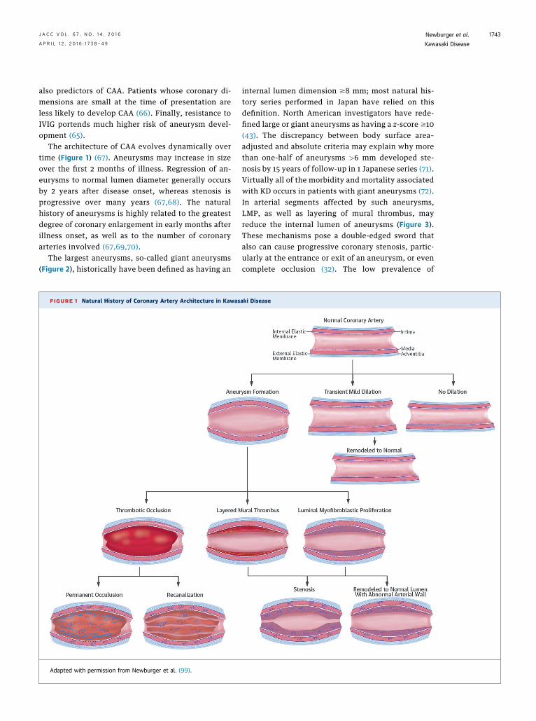
The architecture of CAA evolves dynamically overtime (Figure 1) (67). Aneurysms may increase in sizeover the first 2 months of illness. Regression of an-eurysms to normal lumen diameter generally occursby 2 years after disease onset, whereas stenosis isprogressive over many years (67,68). The naturalhistory of aneurysms is highly related to the greatestdegree of coronary enlargement in early months afterillness onset, as well as to the number of coronaryarteries involved (67,69,70).
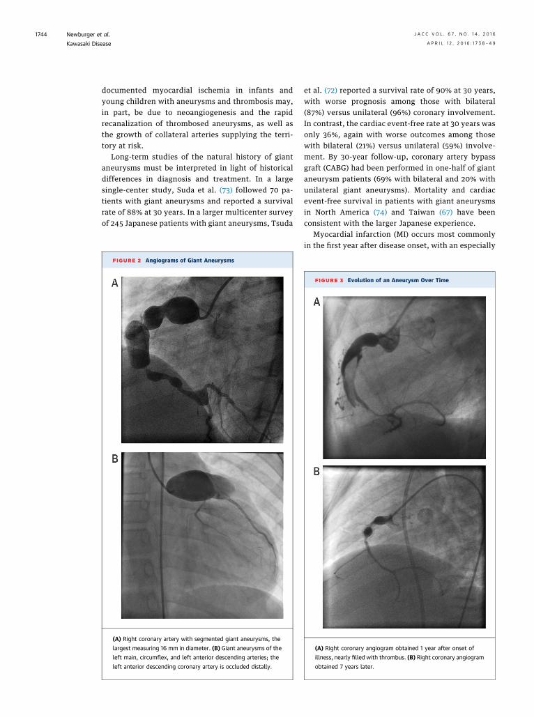
The largest aneurysms, so-called giant aneurysms(Figure 2), historically have been defined as having an
FIGURE 1 Natural History of Coronary Artery Architecture in Kawas
Adapted with permission from Newburger et al. (99).
internal lumen dimension $8 mm; most natural his-tory series performed in Japan have relied on thisdefinition. North American investigators have rede-fined large or giant aneurysms as having a z-score $10(43). The discrepancy between body surface area–adjusted and absolute criteria may explain why morethan one-half of aneurysms >6 mm developed ste-nosis by 15 years of follow-up in 1 Japanese series (71).Virtually all of the morbidity and mortality associatedwith KD occurs in patients with giant aneurysms (72).In arterial segments affected by such aneurysms,LMP, as well as layering of mural thrombus, mayreduce the internal lumen of aneurysms (Figure 3).These mechanisms pose a double-edged sword thatalso can cause progressive coronary stenosis, partic-ularly at the entrance or exit of an aneurysm, or evencomplete occlusion (32). The low prevalence of
aki Disease
Newburger et al. J A C C V O L . 6 7 , N O . 1 4 , 2 0 1 6
Kawasaki Disease A P R I L 1 2 , 2 0 1 6 : 1 7 3 8 – 4 9
1744
documented myocardial ischemia in infants andyoung children with aneurysms and thrombosis may,in part, be due to neoangiogenesis and the rapidrecanalization of thrombosed aneurysms, as well asthe growth of collateral arteries supplying the terri-tory at risk.
Long-term studies of the natural history of giantaneurysms must be interpreted in light of historicaldifferences in diagnosis and treatment. In a largesingle-center study, Suda et al. (73) followed 70 pa-tients with giant aneurysms and reported a survivalrate of 88% at 30 years. In a larger multicenter surveyof 245 Japanese patients with giant aneurysms, Tsuda
FIGURE 2 Angiograms of Giant Aneurysms
(A) Right coronary artery with segmented giant aneurysms, the
largest measuring 16 mm in diameter. (B) Giant aneurysms of the
left main, circumflex, and left anterior descending arteries; the
left anterior descending coronary artery is occluded distally.
et al. (72) reported a survival rate of 90% at 30 years,with worse prognosis among those with bilateral(87%) versus unilateral (96%) coronary involvement.In contrast, the cardiac event-free rate at 30 years wasonly 36%, again with worse outcomes among thosewith bilateral (21%) versus unilateral (59%) involve-ment. By 30-year follow-up, coronary artery bypassgraft (CABG) had been performed in one-half of giantaneurysm patients (69% with bilateral and 20% withunilateral giant aneurysms). Mortality and cardiacevent-free survival in patients with giant aneurysmsin North America (74) and Taiwan (67) have beenconsistent with the larger Japanese experience.
Myocardial infarction (MI) occurs most commonlyin the first year after disease onset, with an especially
FIGURE 3 Evolution of an Aneurysm Over Time
(A) Right coronary angiogram obtained 1 year after onset of
illness, nearly filled with thrombus. (B) Right coronary angiogram
obtained 7 years later.

TABLE 2 Principles in the Long-Term Management of Patients With KD
1. On the basis of available data, patients with no demonstrated coronary arterydilation by echocardiogram with excellent visualization of all arterial segmentsduring the first weeks of illness appear to have normal cardiovascular status inearly adulthood.
2. Remodeling (so-called regression) of aneurysms, especially if moderate or large,to normal internal lumen diameter is often accompanied by luminalmyofibroblastic proliferation and abnormal vascular reactivity.
3. Patients with persistent CAA are at lifelong risk of progressive coronary arterystenosis or occlusion and worsening ischemia.
4. Patients with CAA documented at any stage require lifelong cardiovascularsurveillance tailored to disease severity and age.
5. Testing should minimize exposure to ionizing radiation whenever possible.
6. Sedentary life-style should be avoided.
7. Women with coronary aneurysms can carry pregnancy successfully, but shouldhave reproductive counseling.
8. Monitoring and counseling regarding traditional CV risk factors is appropriate toreduce the likelihood of later atherosclerosis.
CV ¼ cardiovascular; other abbreviations as in Table 1.
J A C C V O L . 6 7 , N O . 1 4 , 2 0 1 6 Newburger et al.A P R I L 1 2 , 2 0 1 6 : 1 7 3 8 – 4 9 Kawasaki Disease
1745
high risk in the period from 15 to 45 days after diseaseonset (75). Intravascular clot formation and degrada-tion is a consequence of severe vascular inflammationduring the early illness and altered hemodynamics inthe aneurysm (76). Although the risk of MI appears tofall after 1 to 2 years, cases continue to occur, andadults can present with MI several decades after KDonset (77,78). Indeed, in a recent study in San DiegoCounty, 5% of patients younger than age 40 years ofage who presented with acute coronary syndromeshad aneurysms secondary to KD in childhood (79). Ina multicenter survey of Japanese patients with giantaneurysms, by 30 years, 26% had suffered at least 1acute MI (unilateral aneurysms 15%, bilateral 3%)(72). Among 60 subjects with MI, survival was 79% at20 years, and 50% of survivors had ventriculartachycardia (80).
Regression to normal internal lumen diameter iscommon in children with moderate or small aneu-rysms, but has been associated with late myointimalthickening of the coronary arterial wall on intravas-cular ultrasound (81) and optical coherence tomog-raphy (82). In such arterial segments, coronaryvascular reactivity is impaired (83), and myocardialblood flow and coronary flow reserve may bedecreased (84).
In the current era, the majority of children treatedwith IVIG in the acute phase of illness never developcoronary artery abnormalities. Some studies haveraised concerns, even in this “always normal” group.Myocardial blood flow and coronary flow reserve withadenosine were diminished in 1 study (85), and somestudies performed in Asia have suggested impairedbrachial artery reactivity (86) and arterial stiffness(87). However, peripheral vascular studies in NorthAmerica have not detected long-term changes in pe-ripheral vascular function (88,89). Moreover, reas-surance can be derived from the absence of latecoronary artery calcification (38), clinical manifesta-tions, or increased mortality in those with always-normal coronary arteries with more than 30 years offollow-up in Japan (90).
LONG-TERM MANAGEMENT
All patients with a history of CAA require lifelongsurveillance. Goals of long-term management are toprevent coronary thrombosis and treat myocardialischemia and associated complications (Table 2).There are few evidence-based studies to guide theoptimal frequency and types of cardiovascular testingafter KD. Thus, long-term management is based uponfirst principles and evidence in adults with athero-sclerotic coronary artery disease. Because the arterial
wall may be abnormal in remodeled coronaries, evenwhen the internal lumen diameter measures in thenormal range, both the current status of aneurysmsand the worst-ever coronary artery dimension orz-score, must be considered in devising a plan forlong-term management. Types and intervals fortesting should be tailored to the severity of coronaryinvolvement (2).
With respect to cardiovascular testing during long-term follow-up, echocardiographic measurements ofthe coronary artery lumen become progressively lessreliable as children grow and the chest wall thickens.Echocardiography is also less reliable for detectionof vascular stenosis or thrombosis than for dilation.For these reasons, advanced imaging techniques,including computerized tomographic angiography(CTA) and magnetic resonance angiography, are usedwith increasing frequency (91,92). As currently prac-ticed at most centers, CTA provides greater detail ofvascular structures (Figure 4), whereas cardiac mag-netic resonance (CMR)/magnetic resonance angiog-raphy is superior for cardiovascular function andassessment of wall motion and myocardial fibrosis.The role of CT calcium scoring in the context of KD isstill being defined, but preliminary data suggest thatthis low-radiation technique can be used to screenpatients with a history of KD and an unclear history ofechocardiographic abnormalities, as only youngadults with aneurysms had a positive score (38).
Tests for inducible ischemia are chosen accordingto the patient’s age and institutional practice andare helpful in determining the need for coronary in-terventions. Wherever possible, the mode of stresstesting should minimize risks of anesthesia andionizing radiation. Children too young to exercise

FIGURE 4 Layering Thrombus Is Common in Giant Aneurysms
With Late Follow-Up
This computed tomographic angiography shows nonobstructive
mural thrombus in the left anterior descending coronary artery
21 years after disease onset.
Newburger et al. J A C C V O L . 6 7 , N O . 1 4 , 2 0 1 6
Kawasaki Disease A P R I L 1 2 , 2 0 1 6 : 1 7 3 8 – 4 9
1746
undergo pharmacological stress testing (e.g., dobut-amine stress echocardiography or CMR, adenosine-stress CMR). For example, dobutamine stress echo-cardiography was shown to be an independent riskfactor for major adverse cardiac events during follow-up of patients with KD and aneurysms (93). For olderchildren, exercise stress testing with myocardial im-aging using echocardiography, nuclear imaging, orpositron emission tomography scan is preferable.Among children with MI, including those incident-ally noted during CMR, annual Holter monitoringshould be performed for surveillance of ventriculartachycardia.
With respect to medications, beta-blocker therapyis often used in the highest-risk patients with giantaneurysms (2), and some experts believe that statinsmay be beneficial for their pleotropic anti-inflammatory effects. For patients with giant aneu-rysms, a combination of antiplatelet therapy andanticoagulation is used to prevent coronary throm-bosis (60,61). Indeed, patients with giant aneurysmsand a recent history of coronary thrombosis aresometimes treated with anticoagulation and dualantiplatelet therapy (63).
Recommendations for participation in competitivesports are on the basis of coronary status, results ofstress testing, and antithrombotic treatment (94). AllKD patients should be encouraged to avoid a seden-tary life-style. For patients with persistent giant CAA,multiple or complex aneurysms without obstruction,or coronary artery obstruction who do not havesymptoms, or exercise-induced ischemia or ar-rhythmia on annual stress testing, and whose leftventricular ejection fraction is normal, it is reason-able to allow participation in noncontact low- tomoderate-intensity static and dynamic competitivesports. Some experts do not restrict such patientsfrom any noncontact sport. Patients who have had MIor coronary revascularization should follow exerciserecommendations for adults with atheroscleroticcoronary artery disease (94). Patients with small- tomedium-sized solitary coronary aneurysms in whomno exercise-induced ischemia or arrhythmia is pres-ent on stress testing every 1 to 2 years do not requireexercise restrictions. Finally, those without a historyof coronary aneurysms require neither stress testingnor exercise restriction.
Most coronary thrombosis is nonocclusive anddetected in asymptomatic children during frequentechocardiography surveillance. If a new thrombusappears in the first few months after disease onset,when the risk of rapid progression to occlusion andMI is greatest, treatment with thrombolytic therapy,generally with tissue plasminogen activator, is
indicated. Treatment with anticoagulation and dualantiplatelet therapy is generally administered forsome months following thrombolytic therapy. Afterthe first year of illness, the nonocclusive coronarythrombus noted incidentally by echocardiographymay be followed with close surveillance duringtreatment with anticoagulation and aspirin.
Coronary revascularization (surgical or percuta-neous) is performed for symptoms of angina or evi-dence of a significant territory of inducible ischemiaon stress testing. No randomized trials of CABGversus percutaneous coronary intervention (PCI)have been performed in KD patients. CABG has beenperformed in young children, but graft longevity isbetter after 12 years of age; internal mammary arterygrafts appear to grow with somatic growth (95). Inthe largest single-center series from Japan, cardiacevent-free survival after bypass was 67% at 20 years(95). Graft failure is more common when CABG isperformed in children without inducible ischemia.
Japanese guidelines for catheter interventionrecommend PCI for patients with ischemic symptoms,inducible ischemia, or >75% stenosis in the leftanterior descending coronary artery; the presence ofcomplex aneurysms with multiple, ostial, or long-segment stenosis are considered a contraindicationfor PCI (96). In a survey of outcomes after PCI versusCABG, the primary composite endpoint of mortalityor Q-wave MI were similar, but repeat target vesselrevascularization was significantly more commonamong those treated with PCI (97). When consideringstent placement, adult cardiologists should be awarethat the walls of a stenotic artery may be composed ofconsiderable thrombus; intravascular ultrasound maybe helpful to assess the true lumen diameter. Finally,
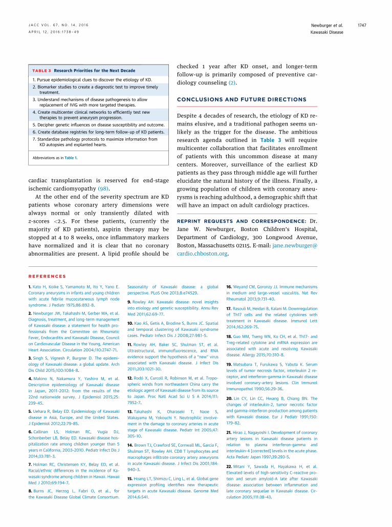
TABLE 3 Research Priorities for the Next Decade
1. Pursue epidemiological clues to discover the etiology of KD.
2. Biomarker studies to create a diagnostic test to improve timelytreatment.
3. Understand mechanisms of disease pathogenesis to allowreplacement of IVIG with more targeted therapies.
4. Create multicenter clinical networks to efficiently test newtherapies to prevent aneurysm progression.
5. Decipher genetic influences on disease susceptibility and outcome.
6. Create database registries for long-term follow-up of KD patients.
7. Standardize pathology protocols to maximize information fromKD autopsies and explanted hearts.
Abbreviations as in Table 1.
J A C C V O L . 6 7 , N O . 1 4 , 2 0 1 6 Newburger et al.A P R I L 1 2 , 2 0 1 6 : 1 7 3 8 – 4 9 Kawasaki Disease
1747
cardiac transplantation is reserved for end-stageischemic cardiomyopathy (98).
At the other end of the severity spectrum are KDpatients whose coronary artery dimensions werealways normal or only transiently dilated withz-scores <2.5. For these patients, (currently themajority of KD patients), aspirin therapy may bestopped at 4 to 8 weeks, once inflammatory markershave normalized and it is clear that no coronaryabnormalities are present. A lipid profile should be
checked 1 year after KD onset, and longer-termfollow-up is primarily composed of preventive car-diology counseling (2).
CONCLUSIONS AND FUTURE DIRECTIONS
Despite 4 decades of research, the etiology of KD re-mains elusive, and a traditional pathogen seems un-likely as the trigger for the disease. The ambitiousresearch agenda outlined in Table 3 will requiremulticenter collaboration that facilitates enrollmentof patients with this uncommon disease at manycenters. Moreover, surveillance of the earliest KDpatients as they pass through middle age will furtherelucidate the natural history of the illness. Finally, agrowing population of children with coronary aneu-rysms is reaching adulthood, a demographic shift thatwill have an impact on adult cardiology practices.
REPRINT REQUESTS AND CORRESPONDENCE: Dr.Jane W. Newburger, Boston Children’s Hospital,Department of Cardiology, 300 Longwood Avenue,Boston, Massachusetts 02115. E-mail: [email protected].
RE F E RENCE S
1. Kato H, Koike S, Yamamoto M, Ito Y, Yano E.Coronary aneurysms in infants and young childrenwith acute febrile mucocutaneous lymph nodesyndrome. J Pediatr 1975;86:892–8.
2. Newburger JW, Takahashi M, Gerber MA, et al.Diagnosis, treatment, and long-term managementof Kawasaki disease: a statement for health pro-fessionals from the Committee on RheumaticFever, Endocarditis and Kawasaki Disease, Councilon Cardiovascular Disease in the Young, AmericanHeart Association. Circulation 2004;110:2747–71.
3. Singh S, Vignesh P, Burgner D. The epidemi-ology of Kawasaki disease: a global update. ArchDis Child 2015;100:1084–8.
4. Makino N, Nakamura Y, Yashiro M, et al.Descriptive epidemiology of Kawasaki diseasein Japan, 2011–2012: from the results of the22nd nationwide survey. J Epidemiol 2015;25:239–45.
5. Uehara R, Belay ED. Epidemiology of Kawasakidisease in Asia, Europe, and the United States.J Epidemiol 2012;22:79–85.
6. Callinan LS, Holman RC, Vugia DJ,Schonberber LB, Belay ED. Kawasaki disease hos-pitalization rate among children younger than 5years in California, 2003–2010. Pediatr Infect Dis J2014;33:781–3.
7. Holman RC, Christensen KY, Belay ED, et al.Racial/ethnic differences in the incidence of Ka-wasaki syndrome among children in Hawaii. HawaiiMed J 2010;69:194–7.
8. Burns JC, Herzog L, Fabri O, et al., forthe Kawasaki Disease Global Climate Consortium.
Seasonality of Kawasaki disease: a globalperspective. PLoS One 2013;8:e74529.
9. Rowley AH. Kawasaki disease: novel insightsinto etiology and genetic susceptibility. Annu RevMed 2011;62:69–77.
10. Kao AS, Getis A, Brodine S, Burns JC. Spatialand temporal clustering of Kawasaki syndromecases. Pediatr Infect Dis J 2008;27:981–5.
11. Rowley AH, Baker SC, Shulman ST, et al.Ultrastructural, immunofluorescence, and RNAevidence support the hypothesis of a “new” virusassociated with Kawasaki disease. J Infect Dis2011;203:1021–30.
12. Rodó X, Curcoll R, Robinson M, et al. Tropo-spheric winds from northeastern China carry theetiologic agent of Kawasaki disease from its sourceto Japan. Proc Natl Acad Sci U S A 2014;111:7952–7.
13. Takahashi K, Oharaseki T, Naoe S,Wakayama M, Yokouchi Y. Neutrophilic involve-ment in the damage to coronary arteries in acutestage of Kawasaki disease. Pediatr Int 2005;47:305–10.
14. Brown TJ, Crawford SE, Cornwall ML, Garcia F,Shulman ST, Rowley AH. CD8 T lymphocytes andmacrophages infiltrate coronary artery aneurysmsin acute Kawasaki disease. J Infect Dis 2001;184:940–3.
15. Hoang LT, Shimizu C, Ling L, et al. Global geneexpression profiling identifies new therapeutictargets in acute Kawasaki disease. Genome Med2014;6:541.
16. Weyand CM, Goronzy JJ. Immune mechanismsin medium and large-vessel vasculitis. Nat RevRheumatol 2013;9:731–40.
17. Rasouli M, Heidari B, Kalani M. Downregulationof Th17 cells and the related cytokines withtreatment in Kawasaki disease. Immunol Lett2014;162:269–75.
18. Guo MM, Tseng WN, Ko CH, et al. Th17- andTreg-related cytokine and mRNA expression areassociated with acute and resolving Kawasakidisease. Allergy 2015;70:310–8.
19. Matsubara T, Furukawa S, Yabuta K. Serumlevels of tumor necrosis factor, interleukin 2 re-ceptor, and interferon-gamma in Kawasaki diseaseinvolved coronary-artery lesions. Clin ImmunolImmunopathol 1990;56:29–36.
20. Lin CY, Lin CC, Hwang B, Chiang BN. Thechanges of interleukin-2, tumor necrotic factorand gamma-interferon production among patientswith Kawasaki disease. Eur J Pediatr 1991;150:179–82.
21. Hirao J, Nagayoshi I. Development of coronaryartery lesions in Kawasaki disease patients inrelation to plasma interferon-gamma andinterleukin-4 [corrected] levels in the acute phase.Acta Pediatr Japan 1997;39:293–5.
22. Mitani Y, Sawada H, Hayakawa H, et al.Elevated levels of high-sensitivity C-reactive pro-tein and serum amyloid-A late after Kawasakidisease: association between inflammation andlate coronary sequelae in Kawasaki disease. Cir-culation 2005;111:38–43.
Newburger et al. J A C C V O L . 6 7 , N O . 1 4 , 2 0 1 6
Kawasaki Disease A P R I L 1 2 , 2 0 1 6 : 1 7 3 8 – 4 9
1748
23. Schulte DJ, Yilmaz A, Shimada K, et al.Involvement of innate and adaptive immunity in amurine model of coronary arteritis mimicking Ka-wasaki disease. J Immunol 2009;183:5311–8.
24. Lee Y, Schulte DJ, Shimada K, et al. Inter-leukin-1b is crucial for the induction of coronaryartery inflammation in a mouse model of Kawasakidisease. Circulation 2012;125:1542–50.
25. Blankier S, McCrindle BW, Ito S, Yeung RS. Therole of atorvastatin in regulating the immuneresponse leading to vascular damage in a model ofKawasaki disease. Clin Exp Immunol 2011;164:193–201.
26. Oharaseki T, Yokouchi Y, Yamada H, et al. Therole of TNF-a in a murine model of Kawasaki dis-ease arteritis induced with a Candida albicans cellwall polysaccharide. Mod Rheumatol 2014;24:120–8.
27. Onouchi Y. Genetics of Kawasaki disease: whatwe know and don’t know. Circ J 2012;76:1581–6.
28. Onouchi Y, Gunji T, Burns JC, et al. ITPKCfunctional polymorphism associated with Kawa-saki disease susceptibility and formation of coro-nary artery aneurysms. Nat Genet 2008;40:35–42.
29. Onouchi Y, Ozaki K, Burns JC, et al., for theU.S. Kawasaki Disease Genetics Consortium.A genome-wide association study identifies threenew risk loci for Kawasaki disease. Nat Genet2012;44:517–21.
30. Shimizu C, Jain S, Davila S, et al. Transforminggrowth factor-b signaling pathway in patients withKawasaki disease. Circ Cardiovasc Genet 2011;4:16–25.
31. Shrestha S, Wiener HW, Aissani B, Shendre A,Tang J, Portman MA. Imputation of class I and IIHLA loci using high-density SNPs from Immu-noChip and their associations with Kawasaki dis-ease in family-based study. Int J Immunogenet2015;42:140–6.
32. Orenstein JM, Shulman ST, Fox LM, et al.Three linked vasculopathic processes characterizeKawasaki disease: a light and transmission elec-tron microscopic study. PLoS One 2012;7:e38998.
33. Kanai T, Ishiwata T, Kobayashi T, et al. Uli-nastatin, a urinary trypsin inhibitor, for the initialtreatment of patients with Kawasaki disease: aretrospective study. Circulation 2011;124:2822–8.
34. Suzuki H, Terai M, Hamada H, et al. Cyclo-sporin A treatment for Kawasaki disease refractoryto initial and additional intravenous immunoglob-ulin. Pediatr Infect Dis J 2011;30:871–6.
35. Tremoulet AH, Pancoast P, Franco A, et al.Calcineurin inhibitor treatment of intravenousimmunoglobulin-resistant Kawasaki disease.J Pediatr 2012;161:506–12.e1.
36. Shimizu C, Oharaseki T, Takahashi K, Kottek A,Franco A, Burns JC. The role of TGF-b and myofi-broblasts in the arteritis of Kawasaki disease. HumPathol 2013;44:189–98.
37. Lee AM, Shimizu C, Oharaseki T, et al. Role ofTGF-b signaling in remodeling of noncoronary ar-tery aneurysms in Kawasaki disease. Pediatr DevPathol 2015;18:310–7.
38. Kahn AM, Budoff MJ, Daniels LB, et al. Calciumscoring in patients with a history of Kawasakidisease. J Am Coll Cardiol Img 2012;5:264–72.
39. Tremoulet AH, Jain S, Chandrasekar D, Sun X,Sato Y, Burns JC. Evolution of laboratory values inpatients with Kawasaki disease. Pediatr Infect Dis J2011;30:1022–6.
40. Research Committee on Kawasaki Disease.Report of Subcommittee on Standardization ofDiagnostic Criteria and Reporting of Coronary Ar-tery Lesions in Kawasaki Disease. Tokyo, Japan:Ministry of Health and Welfare, 1984.
41. JCS Joint Working Group. Guidelines for diag-nosis and management of cardiovascular sequelaein Kawasaki disease (JCS 2008)—digest version.Circ J 2010;74:1989–2020.
42. Parameter(z). Pediatric and fetal echoZ-scores. Available at: http://parameterz.blogspot.com. Accessed February 1, 2016.
43. Manlhiot C, Millar K, Golding F, McCrindle BW.Improved classification of coronary artery abnor-malities based only on coronary artery z-scoresafter Kawasaki disease. Pediatr Cardiol 2010;31:242–9.
44. Printz BF, Sleeper LA, Newburger JW, et al.,for the Pediatric Heart Network Investigators.Noncoronary cardiac abnormalities are associatedwith coronary artery dilation and with laboratoryinflammatory markers in acute Kawasaki disease.J Am Coll Cardiol 2011;57:86–92.
45. Kanegaye JT, Wilder MS, Molkara D, et al.Recognition of a Kawasaki disease shock syn-drome. Pediatrics 2009;123:e783–9.
46. Furusho K, Kamiya T, Nakano H, et al. High-dose intravenous gammaglobulin for Kawasakidisease. Lancet 1984;2:1055–8.
47. Newburger JW, Takahashi M, Beiser AS, et al.A single intravenous infusion of gamma globulinas compared with four infusions in the treatmentof acute Kawasaki syndrome. N Engl J Med 1991;324:1633–9.
48. Burns JC, Song Y, Bujold M, et al. Immune-monitoring in Kawasaki disease patients treatedwith infliximab and intravenous immunoglobulin.Clin Exp Immunol 2013;174:337–44.
49. Franco A, Touma R, Song Y, et al. Specificity ofregulatory T cells that modulate vascular inflam-mation. Autoimmunity 2014;47:95–104.
50. Burns JC, Touma R, Song Y, et al. Fine speci-ficities of natural regulatory T cells after IVIGtherapy in patients with Kawasaki disease. Auto-immunity 2015;48:181–8.
51. Hwang JY, Lee KY, Rhim JW, et al. Assessmentof intravenous immunoglobulin non-respondersin Kawasaki disease. Arch Dis Child 2011;96:1088–90.
52. Kobayashi T, Saji T, Otani T, et al., for theRAISE Study Group Investigators. Efficacy ofimmunoglobulin plus prednisolone for preventionof coronary artery abnormalities in severe Kawa-saki disease (RAISE study): a randomised, open-label, blinded-endpoints trial. Lancet 2012;379:1613–20.
53. Sleeper LA, Minich LL, McCrindle BM, et al., forthe Pediatric Heart Network Investigators. Evalu-ation of Kawasaki disease risk-scoring systems forintravenous immunoglobulin resistance. J Pediatr2011;158:831–5.e3.
54. Burns JC, Franco A. The immunomodulatoryeffects of intravenous immunoglobulin therapy inKawasaki disease. Expert Rev Clin Immunol 2015;11:819–25.
55. Ogata S, Shimizu C, Franco A, et al. Treatmentresponse in Kawasaki disease is associated withsialylation levels of endogenous but not thera-peutic intravenous immunoglobulin G. PLoS One2013;8:e81448.
56. Shendre A, Wiener HW, Zhi D, Vazquez AI,Portman MA, Shrestha S. High-density genotypingof immune loci in Kawasaki disease and IVIGtreatment response in European-American case-parent trio study. Genes Immun 2014;15:534–42.
57. Son MB, Gauvreau K, Burns JC, et al. Infliximabfor intravenous immunoglobulin resistance in Ka-wasaki disease: a retrospective study. J Pediatr2011;158:644–9.e1.
58. Wallace CA, French JW, Kahn SJ, Sherry DD.Initial intravenous gammaglobulin treatment fail-ure in Kawasaki disease. Pediatrics 2000;105:E78.
59. Singh S, Newburger JW, Kuijpers T, Burgner D.Management of Kawasaki disease in resource-limited settings. Pediatr Infect Dis J 2015;34:94–6.
60. Su D, Wang K, Qin S, Pnag Y. Safety and ef-ficacy of warfarin plus aspirin combination therapyfor giant coronary artery aneurysm secondary toKawasaki disease: a meta-analysis. Cardiology2014;129:55–64.
61. Suda K, Kudo Y, Higaki T, et al. Multicenter andretrospective case study of warfarin and aspirincombination therapy in patients with giant coro-nary aneurysms caused by Kawasaki disease. Circ J2009;73:1319–23.
62. Manlhiot C, Brandão LR, Somji Z, et al. Long-term anticoagulation in Kawasaki disease: initialuse of low molecular weight heparin is a viableoption for patients with severe coronary arteryabnormalities. Pediatr Cardiol 2010;31:834–42.
63. Giglia TM, Massicotte MP, Tweddell JS, et al.,for the American Heart Association CongenitalHeart Defects Committee of the Council on Car-diovascular Disease in the Young, Council on Car-diovascular and Stroke Nursing, Council onEpidemiology and Prevention, and Stroke Council.Prevention and treatment of thrombosis in pedi-atric and congenital heart disease: a scientificstatement from the American Heart Association.Circulation 2013;128:2622–703.
64. Kim T, Choi W, Woo CW, et al. Predictive riskfactors for coronary artery abnormalities in Ka-wasaki disease. Eur J Pediatr 2007;166:421–5.
65. Uehara R, Belay ED, Maddox RA, et al. Analysisof potential risk factors associated with nonre-sponse to initial intravenous immunoglobulintreatment among Kawasaki disease patients inJapan. Pediatr Infect Dis J 2008;27:155–60.
66. McCrindle BW, Li JS, Minich LL, et al., for thePediatric Heart Network Investigators. Coronaryartery involvement in children with Kawasaki dis-ease: risk factors from analysis of serial normalizedmeasurements. Circulation 2007;116:174–9.
67. Lin MT, Sun LC, Wu ET, Wang JK, Lue HC,Wu MH. Acute and late coronary outcomes in 1073patients with Kawasaki disease with and without
J A C C V O L . 6 7 , N O . 1 4 , 2 0 1 6 Newburger et al.A P R I L 1 2 , 2 0 1 6 : 1 7 3 8 – 4 9 Kawasaki Disease
1749
intravenous g-immunoglobulin therapy. Arch DisChild 2015;100:542–7.
68. Kato H, Sugimura T, Akagi T, et al. Long-termconsequences of Kawasaki disease: a 10- to 21-year follow-up study of 594 patients. Circulation1996;94:1379–85.
69. Nakano H, Ueda K, Saito A, Nojima K.Repeated quantitative angiograms in coronaryarterial aneurysm in Kawasaki disease. Am J Car-diol 1985;56:846–51.
70. Suzuki A, Kamiya T, Ono Y, Kohata T, Kimura K,Takamiya M. Follow-up study of coronary arterylesions due to Kawasaki disease by serial selectivecoronary arteriography in 200 patients. HeartVessels 1987;3:159–65.
71. Tsuda E, Miyazaki S, Takamuro M, Fuse S,Tsuji Y, Echigo S. Strategy for localized stenosiscaused by Kawasaki disease: midterm results ofpercutaneous transluminal coronary balloon an-gioplasty in two infants. Pediatr Cardiol 2006;27:272–5.
72. Tsuda E, Hamaoka K, Suzuki H, et al. A surveyof the 3-decade outcome for patients with giantaneurysms caused by Kawasaki disease. Am HeartJ 2014;167:249–58.
73. Suda K, Iemura M, Nishiono H, et al. Long-termprognosis of patients with Kawasaki diseasecomplicated by giant coronary aneurysms: asingle-institution experience. Circulation 2011;123:1836–42.
74. McNeal-Davidson A, Fournier A, Scuccimarri R,et al. The fate and observed management of giantcoronary artery aneurysms secondary to Kawasakidisease in the Province of Quebec: the completeseries since 1976. Pediatr Cardiol 2013;34:170–8.
75. Kato H, Ichinose E, Kawasaki T. Myocardialinfarction in Kawasaki disease: clinical analyses in195 cases. J Pediatr 1986;108:923–7.
76. Sengupta D, Kahn AM, Kung E, et al. Throm-botic risk stratification using computationalmodeling in patients with coronary artery aneu-rysms following Kawasaki disease. Biomech ModelMechanobiol 2014;13:1261–76.
77. Tsuda E, Matsuo M, Naito H, Noguchi T,Nonogi H, Echigo S. Clinical features in adults withcoronary arterial lesions caused by presumed Ka-wasaki disease. Cardiol Young 2007;17:84–9.
78. Rizk SR, El Said G, Daniels LB, et al. Acutemyocardial ischemia in adults secondary to missedKawasaki disease in childhood. Am J Cardiol 2015;115:423–7.
79. Daniels LB, Tjajadi MS, Walford HH, et al.Prevalence of Kawasaki disease in young adults
with suspected myocardial ischemia. Circulation2012;125:2447–53.
80. Tsuda E, Hirata T, Matsuo O, Abe T,Sugiyama H, Yamada O. The 30-year outcome forpatients after myocardial infarction due to coro-nary artery lesions caused by Kawasaki disease.Pediatr Cardiol 2011;32:176–82.
81. Tsuda E, Kamiya T, Kimura K, Ono Y, Echigo S.Coronary artery dilatation exceeding 4.0 mmduring acute Kawasaki disease predicts a highprobability of subsequent late intima-medialthickening. Pediatr Cardiol 2002;23:9–14.
82. Dionne A, Ibrahim R, Gebhard C, et al. Coro-nary wall structural changes in patients with Ka-wasaki disease: new insights from opticalcoherence tomography (OCT). J Am Heart Assoc2015;4:e001939.
83. Yamakawa R, Ishii M, Sugimura T, et al.Coronary endothelial dysfunction after Kawasakidisease: evaluation by intracoronary injectionof acetylcholine. J Am Coll Cardiol 1998;31:1074–80.
84. Furuyama H, Odagawa Y, Katoh C, et al.Altered myocardial flow reserve and endothelialfunction late after Kawasaki disease. J Pediatr2003;142:149–54.
85. Muzik O, Paridon SM, Singh TP, Morrow WR,Dayanikli F, Di Carli MF. Quantification ofmyocardial blood flow and flow reserve in childrenwith a history of Kawasaki disease and normalcoronary arteries using positron emission tomog-raphy. J Am Coll Cardiol 1996;28:757–62.
86. Dhillon R, Clarkson P, Donald AE, et al.Endothelial dysfunction late after Kawasaki dis-ease. Circulation 1996;94:2103–6.
87. Cheung YF, Yung TC, Tam SC, Ho MH,Chau AK. Novel and traditional cardiovascular riskfactors in children after Kawasaki disease: impli-cations for premature atherosclerosis. J Am CollCardiol 2004;43:120–4.
88. Selamet Tierney ES, Gal D, Gauvreau K, et al.Vascular health in Kawasaki disease. J Am CollCardiol 2013;62:1114–21.
89. McCrindle BW, McIntyre S, Kim C, Lin T,Adeli K. Are patients after Kawasaki disease atincreased risk for accelerated atherosclerosis?J Pediatr 2007;151:244–8. 248.e1.
90. Nakamura Y, Aso E, Yashiro M, et al. Mortalityamong persons with a history of Kawasaki diseasein Japan: mortality among males with cardiacsequelae is significantly higher than that of thegeneral population. Circ J 2008;72:134–8.
91. Tacke CE, Romeih S, Kuipers IM,Spijkerboer AM, Groenink M, Kuipers TW. Evalua-tion of cardiac function by magnetic resonanceimaging during the follow-up of patients withKawasaki disease. Circ Cardiovasc Imaging 2013;6:67–73.
92. Kanamaru H, Sato Y, Takayama T, et al.Assessment of coronary artery abnormalities bymultislice spiral computed tomography in adoles-cents and young adults with Kawasaki disease. AmJ Cardiol 2005;95:522–5.
93. Noto N, Kamiyama H, Karasawa K, et al.Long-term prognostic impact of dobutaminestress echocardiography in patients with Kawa-saki disease and coronary artery lesions: a15-year follow-up study. J Am Coll Cardiol 2014;63:337–44.
94. Thompson PD, Myerburg RJ, Levine BD,Udelson JE, Kovacs RJ. Eligibility and disqualifi-cation recommendations for competitive athleteswith cardiovascular abnormalities: Task Force 8:Coronary Artery Disease: A Scientific Statementfrom the American Heart Association and Amer-ican College of Cardiology. J Am Coll Cardiol 2015;66:2406–11.
95. Kitamura S, Tsuda E, Kobayashi J, et al.Twenty-five-year outcome of pediatric coronaryartery bypass surgery for Kawasaki disease 104.Circulation 2009;120:60–8.
96. Ishii M, Ueno T, Akagi T, et al., forthe Research Committee of Ministry of Health.Labour and Welfare—“Study of Treatment andLong-Term Management in Kawasaki Disease.”Guidelines for catheter intervention in coronaryartery lesion in Kawasaki disease. Pediatr Int2001;43:558–62.
97. Muta H, Ishii M. Percutaneous coronary inter-vention versus coronary artery bypass grafting forstenotic lesions after Kawasaki disease. J Pediatr2010;157:120–6.
98. Checchia PA, Pahl E, Shaddy RE, Shulman ST.Cardiac transplantation for Kawasaki disease.Pediatrics 1997;100:695–9.
99. Newburger JW, Kato H. Kawasaki disease. In:Willerson JT, Holmes DR, editors. Coronary ArteryDisease. London, UK: Springer-Verlag, 2015:581–96.
KEY WORDS aneurysm, coronaryaneurysm, coronary thrombosis,immunoglobulin, intravenous,mucocutaneous lymph node syndrome,myocardial infarction