Embed Size (px)
Citation preview
Genitourin Med 1997;73:571-574
Kaposi's sarcoma in retrospect
Comelus J G Sanders
A historical overview of Kaposi's sarcoma is provided, scrutinising in particular past clinical andhistological studies of the disease and the conclusions on aetiology and pathogenesis that werereached.(Genitourin Med 1997;73:571-574)
Keywords: Kaposi's sarcoma; historical overview
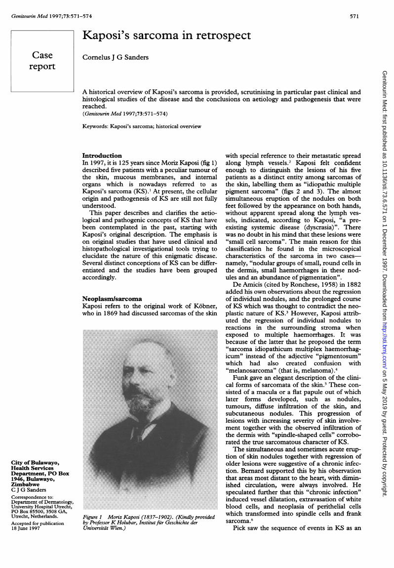
IntroductionIn 1997, it is 125 years since Moriz Kaposi (fig 1)described five patients with a peculiar tumour ofthe skin, mucous membranes, and internalorgans which is nowadays referred to asKaposi's sarcoma (KS).' At present, the cellularorigin and pathogenesis of KS are still not fullyunderstood.
This paper describes and clarifies the aetio-logical and pathogenic concepts of KS that havebeen contemplated in the past, starting withKaposi's original description. The emphasis ison original studies that have used clinical andhistopathological investigational tools trying toelucidate the nature of this enigmatic disease.Several distinct conceptions ofKS can be differ-entiated and the studies have been groupedaccordingly.
NeoplasmisarcomaKaposi refers to the original work of K6bner,who in 1869 had discussed sarcomas of the skin
City ofBulawayo,Health ServicesDepartment, PO Box1946, Bulawayo,ZimbabweC J G SandersCorrespondence to:Department of Dermatology,University Hospital Utrecht,PO Box 85500, 3508 GA,Utrecht, Netherlands.
Accepted for publication18 June 1997
Figure 1 Moriz Kaposi (1837-1902). (Kindly providedby ProfessorK Holubar, Institutfir Geschichte derUniversitat Wien.)
with special reference to their metastatic spreadalong lymph vessels.2 Kaposi felt confidentenough to distinguish the lesions of his fivepatients as a distinct entity among sarcomas ofthe skin, labelling them as "idiopathic multiplepigment sarcoma" (figs 2 and 3). The almostsimultaneous eruption of the nodules on bothfeet followed by the appearance on both hands,without apparent spread along the lymph ves-
sels, indicated, according to Kaposi, "a pre-existing systemic disease (dyscrasia)". Therewas no doubt in his mind that these lesions were"small cell sarcoma". The main reason for thisclassification he found in the microscopicalcharacteristics of the sarcoma in two cases-
namely, "nodular groups of small, round cells inthe dermis, small haemorrhages in these nod-ules and an abundance of pigmentation".De Amicis (cited by Ronchese, 1958) in 1882
added his own observations about the regressionof individual nodules, and the prolonged courseof KS which was thought to contradict the neo-
plastic nature of KS.3 However, Kaposi attrib-uted the regression of individual nodules toreactions in the surrounding stroma whenexposed to multiple haemorrhages. It was
because of the latter that he proposed the term"sarcoma idiopathicum multiplex haemorrhag-icum" instead of the adjective "pigmentosum"which had also created confusion with"melanosarcoma" (that is, melanoma).4Funk gave an elegant description of the clini-
cal forms of sarcomata of the skin.5 These con-
sisted of a macula or a flat papule out of whichlater forms developed, such as nodules,tumours, diffuse infiltration of the skin, andsubcutaneous nodules. This progression oflesions with increasing severity of skin involve-ment together with the observed infiltration ofthe dermis with "spindle-shaped cells" corrobo-rated the true sarcomatous character of KS.The simultaneous and sometimes acute erup-
tion of skin nodules together with regression ofolder lesions were suggestive of a chronic infec-tion. Bernard supported this by his observationthat areas most distant to the heart, with dimin-ished circulation, were always involved. Hespeculated further that this "chronic infection"induced vessel dilatation, extravasation of whiteblood cells, and neoplasia of perithelial cellswhich transformed into spindle cells and franksarcoma.6
Pick saw the sequence of events in KS as an
Casereport
571 on 5 M
ay 2019 by guest. Protected by copyright.
http://sti.bmj.com
/G
enitourin Med: first published as 10.1136/sti.73.6.571 on 1 D
ecember 1997. D
ownloaded from

Sanders
Figure 2 Sarcoma melanodes. (From Hebra F, Elfinger A, Heitzmann K. Atlas derHautkrankheiten. Vienna, K-KHofund Staatsdruckerei, 1856-1876.) Reprinted bypermission. Watercolour painting.
initial dilatation of lymphatic vessels and a sub-sequent accumulation of plasma cells (that is,lymphocytic elements).7 Accordingly, hegrouped KS with other lymphocytic malignan-cies such as mycosis fungoides and leukaemia.The plasma cells in this context were believed tobe multipotential cells, able to transform intoendothelial-like elements and eventually intofibrotic particles (spindle cells), thus accountingfor all the cellular components of KS.A more recent proponent of the malignant
nature of KS argued that it was a neoplasticfibroblast tumour.8 The tumour cells (spindlecells) of KS were supposed to derive from der-mal fibroblasts because of their structural simi-larities and the mature collagenous fibrils foundamong them. Since "no cell other than thefibroblast is known to produce collagenous fib-rils . . ., it follows as a logical inference that thespindle cells in question are fibroblasts."
Infectious hyperplasiaSteiner (1896) mentioned a male predominancein patients with KS, who were usually over 40
Figure 3 Sarcoma melanodes. (From Hebra F, Elfinger A, Heitzmann K Adas derHautkrankheiten. Vienna, K-KHofund Staatsdruckerei, 1856-1876.) Reprinted bypermission. Watercolour painting.
years of age and otherwise remained healthy.9He stated that "the clinical course of the diseasegives the impression of a chronic infectious dis-ease." This paradigm soon gained support fromother clinical and histological observations.An example of the latter were the observed
leucocytes and other inflammatory cells nearareas of capillary proliferation. Philippsonattributed the multifocal expression of the dis-ease to multiple entries into the skin orhaematogenous spread of the causative organ-ism rather than metastasis of tumour cells.'0
In an extensive study Dalla Favera denoted ageographical predisposition for KS from easternEurope and Italy, rather than a racial predomi-nance and to him this also confirmed an infec-tious origin.'1Among those who favoured an infectious
cause for KS there was still debate about thecellular origin and exact nature of KS. Steinercharacterised KS by "a dilatation and neoplasiaof lymph vessels", whereas Philippson regardedthe spindle cells as the actual tumour cells. Hefigured that they must be "connective tissuecells", because the axis of these cells "runs paral-lel to connective tissue fibres in which they areimbedded".'0 A similar argument was used bySymmers when he described the spindle cells asfibroblasts.8
Steiner regarded KS as a benign processwhile others considered it to have malignantpotential as well because of observed metastasisalong lymphatic vessels."'
In their histological study of KS, Dillard andWeidman found nodular accumulations of lym-phocytes and stated that "a specific micro-organism is the irritant, [although]demonstration by current laboratory examina-tions of all kinds ... has failed". 12 This was con-troversial since Justus had claimed that hisinoculation experiments with KS tissue had ledto tumours in white mice at the inoculated areaand also in internal organs." Becker andThatcher repeated inoculation experimentswhereby KS tissue was "injected into a patientwith general paresis with only slight inflamma-tory reaction ... A portion of a 6 day [KS] cul-ture growth ... was implanted under the skin ofthe patient... [and] at the site of the implanta-tion...a bright red infiltrated plaque appeared,[which] showed features typical of very earlyKS".'4 These findings, however, were not con-firmed by other investigators and the infectiousorigin ofKS remained elusive.
Reticuloendothelial diseaseThe reticuloendothelial system was a conceptthat was proposed by Aschoff in 1924 andencompassed the phagocytic macrophages andspecialised endothelia lining sinusoids in theliver, spleen, and bone marrow that were able totake up small dye particles in his experiments.This notion was soon applied to other tissuesand seemed to provide a suitable origin for KS.From the onset the reticuloendothelial systemwas perceived as a flexible system in which acontinuous dedifferentiation between differentcells took place. Thus, vascular cells couldchange into reticulum cells and vice versa and
572 on 5 M
ay 2019 by guest. Protected by copyright.
http://sti.bmj.com
/G
enitourin Med: first published as 10.1136/sti.73.6.571 on 1 D
ecember 1997. D
ownloaded from
Kaposi's sarcoma in retrospect
lymphocyte-like cells could form new vesselsand were capable of phagocytosis.'5 ConsideringKS as a reticuloendothelial disease seemed toresolve the variable histology and clinical coursewhich were otherwise difficult to interpret.
This viewpoint is accurately depicted by thehistological account in Bluefarb and Webster'sreport.'6 "[T]lhere is evidence of hemorrhage;newly formed blood vessels, including prolifera-tion of endothelium and adventitial connectivetissue; spindle cells, and cellular infiltration,including early lymphoid elements and reticu-lum. What tissue other than that of reticuloen-dothelial system presents such a diversity ofstructures?"
In 1932 Dorffel published a much quotedstudy in which he proposed that "the hemor-rhage is the initial pathologic feature of this dis-ease", caused by vascular changes for examplevaricose veins, trauma, cold or arteriosclerosis.'7The observed cellular infiltrate which appearedat the same time was thought to be composed ofcells arising from the reticuloendothelial system.Because these cells "are distributed almostentirely about the blood vessels; ... they form areticulum ... [and] ... there is a multiplicity ofcell forms with many transitional changes".Tedeschi maintained that the pleomorphicnature of the lesions indicated an origin fromthe reticuloendothelial system.'8
These claims, however, were difficult to testscientifically because of the inbuilt flexibility ofthe reticuloendothelial system.
Systemic vascular diseaseObviously the clinical characteristics of KStogether with the microscopic vessel dilatationand proliferation suggested a relation with thevascular system. In this regard early 20th cen-tury investigators often focused on the spindlecells which were considered the true tumourcells of KS.
Steinberg implied that they were actuallysmooth muscle cells because of the "cellularand ... nuclear form", the characteristics of theGieson stain ("yellow ... protoplasm") and thesubmucosal localisation in the ileum where thespindle cells could be compared with surround-ing smooth muscle cells and appeared strikinglysimilar.'9 Others attributed the origin of thespindle cells with the hyperaemic vascular slitsto perivascular embryonic mesenchyme,20 orinterpreted them as transforming and regressionsteps in the vessel proliferation.2' Sachs et aldoubted whether the spindle cells were a spe-cific cell type, stating that "many different typesof cells, even epithelial cells, may have spindleshapes".22Few authors regarded KS as a simple vascu-
lar malformation with no malignant poten-tial,'9 21 whereas most proponents of thesystemic vascular disease concept pointed outthat malignant degeneration of KS could anddid occur.McCarthy and Pack reported on the typical
histopathological evolution of KS, whereby theearly "inflammation-like macule" is followed by a"granulomatous process", which ultimately pro-gresses into the sarcoma stage.2' They suggested
that "some systemic carcinogen acting upon thevascular tissues" might provoke KS. Thisnotion was extended to the recently describedlymphangiosarcoma in female patients withpostmastectomy elephantiasis.24 They impliedthat these lymphangiosarcomas "are truly identi-cal with KS". In fact one ofMcCarthy's patientswith KS was included in the original seriesof postmastectomy lymphangiosarcoma. Sub-sequently Stewart contradicted these findingsbecause the vascular spaces in these angiosarco-mas were often lined by atypical endothelialcells unlike the vascular slits in KS.20Cox and Helwig noticed enlargement of the
nuclei and mitotic figures in spindle cells andalso malignant KS tumour cells in pulmonaryvessels.20 They could hardly escape the conclu-sion that KS is a "neoplastic disease of the vas-cular system with multiple foci of origin", inwhich malignant transformation and metastasiscan occur.
Understanding KS as a disease of the vascu-lar system remained tempting but controversyreigned regarding the constituting elements andthe precise nature of the lesions.25
Neurovascular diseaseSemenow in 1897 was one of the first authors tosuggest that KS was linked to the nervous sys-tem when he found changes in trophic nervesleading to vessel dilatation and proliferation ofthe connective tissue.26The glomus body was first described by
Masson in tips of fingers and toes whereafter itwas soon found that an increase in size pro-voked radiating pain sensation. Neurologicalsymptoms such as pain and cramps heralded theonset of hypodermic nodules of KS in a patientthat was reported by Pautrier and Diss.27 Onhistological examination they observed a prolif-eration of vessels with a neuromuscular sheathand a network of nerve fibrils, proliferation ofSchwann's cells, and formation of touch corpus-cles of Wagner-Meissner type. In their view thiscomplex architecture of KS was akin to a neu-romyoarterial glomus body.
Hudelo and Cailliau on the other hand postu-lated that "an inflammatory event (maybe infec-tious) leads to proliferation of nerve cells[sympathetic fibres (of Remak)] of the mediaand adventitia of normal vessels. ...28 HenceKS and the spindle cells were seen as hyperplas-tic nerve cell lesions. Yet most authors failed toconfirm these findings.'4 17
Endemic (African) KSIn the second part of this century it becameclear that apart from the "classic" form of KSwhich had been studied so far, a similar tumour,endemic KS, was prevalent in Africa and wasfound to be much more common. According toThijs, KS accounted for 9% of the histologicallyconfirmed malignant tumours in a centralAfrican region.29 There was, however, markedvariation with a lower prevalence being reportedfrom western and southern Africa.30 This geo-graphical clustering of cases indicated eithergenetic susceptibility or environmental influ-
573 on 5 M
ay 2019 by guest. Protected by copyright.
http://sti.bmj.com
/G
enitourin Med: first published as 10.1136/sti.73.6.571 on 1 D
ecember 1997. D
ownloaded from
Sanders
ences as aetiological factors in KS. So far therehas been no evidence for either of these factorsto influence endemic KS.The male to female ratio of endemic KS,
although showing marked regional differencesagain,3' was similar to that reported for classicKS.The clinical presentation of endemic KS
showed several distinct features. It occurredmore often in young children who presentedwith lymph node involvement, and was associ-ated with poor outcome.'4 Also in adultsendemic KS could behave more aggressivelywith bone involvement and ulcerating skinlesions.35
Relatively more female children presentedwith lymphadenopathic KS and the sex ratio of3:1 was different from the adults. Sex hormoneswere suggested to provide some protection topost-pubertal females. When oestrogen treat-ment yielded no beneficial results and KS wasdescribed in pregnancy,35 36 this explanationseemed less convincing. Recent evidence thathuman chorionic gonadotrophin induces theregression of AIDS related KS, however, indi-cates some beneficial effects of female hor-mones.37
Notwithstanding these clinical differences,endemic and classic KS appeared strikingly sim-ilar under the microscope."'5'8 Several authorsdescribed distinct cellular patterns in endemicKS such as mixed group, spindle cell predomi-nant or monocellular, and the anaplasticgroup.'5'9 They conceded, however, that theseprobably indicated subsequent stages in the evo-lution of KS.39Most studies of endemic KS mentioned the
spindle cell as the actual tumour cell of KS butopinions about its origin varied. Among the cellsthat were considered as likely candidates werereticulum cells, endothelial cells, mesenchymalcells (that is, "pericytes"), and Schwanncells.30'8'9 Doubt was expressed as to whetherKS was a true sarcoma. Although the associa-tion of endemic KS and lymphoreticular malig-nancies was less commonly found than in othercontinents,'0'9 several authors indicated that thereticuloendothelial system may be closelyrelated to KS.30 31 33The era of relying solely on clinical and con-
ventional histological examination is slowly com-ing to an end and numerous studies haveappeared on the histochemical, electron micro-scopical, and molecular biological characteristicsof KS. The relation between KS, immunodefi-cient states, and the recently described KS asso-ciated herpes virus have provided arguments foran infectious origin of KS which was first sug-gested more than 100 years ago.
This historical overview shows how pastinvestigators with their modest investigationaltools of astute clinical and histopathologicalobservation have paved the way for our presentinterpretation of the nature and aetiology of KS.
1 Kaposi M. Idiopatisches multiples Pigmentsarkom der Haut.Arch Dermatol Syphilis 1872;4:265-73.
2 Kobner H. Zur Kenntniss der allgemeinen Sarcomatose undder Hautsarcome im Besonderen. Arch Dermatol Syphilis1869;1:369-81.
3 Ronchese F. Kaposi's sarcoma. An overlooked essay of 1882.
Arch Dermatol 1958;77:542-5.4 Kaposi M. Zur Nomenclatur des idiopathischen
Pigmentsarcoms Kaposi. Arch Dermatol Syphilis 1894;29:164.
5 Funk I. Clinical studies on sarcomata of the skin. Br JDermatol 1889;1:143-56.
6 Bernard R. Sarcomata idiopathica multiplicia pigmentosacutis (Kaposi). Arch Dermatol Syphilis 1899;49:207-26.
7 Pick W. Zur Kenntnis des Kaposi'schen Pigmentsarkoms.Arch Dermatol Syphilis 1907;87:267-86.
8 Symmers D. Kaposi's disease. Arch Pathol Lab Med 1941;32:764-86.
9 Steiner V. Zwei Falle von Pigmentsarkom der Haut. DtscheMed Wochenschr 1896;33:531-3.
10 Philippson L. Ueber das Sarcoma idiopathicum cutis Kaposi.Ein Beitrag zur Sarcomlehre. Arch Pathol Anat Physiol klinMed Virchow 1902;167:58-81.
11 Dalla Favera GB. Uber das sog. Sarcoma idiop. multiplexhaemorrhagicum (Kaposi). Kilnische und histologischeBeitrage. Arch Dermatol Syphilis 191 1;J09:387-440.
12 Dullard GJ, Weidman FD. Multiple hemorrhagic sarcoma ofKaposi. Histologic studies of two cases, one disclosingintestinal lesions at necropsy. Arch Dermatol Syphilol1925;11:203-31.
13 Justus. Uber Ubertragung von Sarcoma idiopathicumhaemorrhagicum Kaposi auf Tiere. Arch Dermatol Syphilis1910;99:446.
14 Becker SW, Thatcher HW. Multiple idiopathic hemorrhagicsarcoma of Kaposi. Historical review, nomenclature; andtheories relative to the nature of the disease, with experi-mental studies of two cases. J Invest Dermatol 1938;1:379-98.
15 Puhr L. Uber das idiopathische multiple Pigmentsarkom derHaut (Kaposi). Arch Dermatol Syphilis 1931;164:167-80.
16 Bluefarb SM, Webster JR. Kaposi's sarcoma associated withlymphosarcoma. Arch Intern Med 1953;91:97-105.
17 Dorifel J. Histogenesis of multiple idiopathic hemorrhagicsarcoma of Kaposi. Arch Dermatol Syphilol 1932;26:608-34.
18 Tedeschi CG. Some considerations concerning the nature ofthe so-called sarcoma of Kaposi. Arch Pathol 1958;66:656-84.
19 Steinberg C. Uber das Sarcoma multiplex haemorrhagicum(Kaposi). Arch Dermatol Syphilis 1912;111:331-40.
20 Cox FH, Helwig EB. Kaposi's sarcoma. Cancer 1959;12:289-98.
21 Lang FJ, Haslhofer L. Uber die Auffassung der KaposischenKrankheit als systematisierte Angiomatosis. ZKrebsforschung 1935;42:68-75.
22 Sachs W, Azulay RD, Convit J. Multiple idiopathic hemor-rhagic sarcoma of Kaposi. Histopathologic study. J7 InvestDennatol 1947;8:317-26.
23 McCarthy WD, Pack GT. Malignant blood vessel tumors. Areport of 56 cases of angiosarcoma and Kaposi's sarcoma.Surg Gynec Obstet 1950;91:465-82.
24 Stewart FW, Treves N. Lymphangiosarcoma in postmastec-tomy lymphedema; report of 6 cases in elephantiasischirurgica. Cancer 1948;1:64-81.
25 Reynolds WA, Winkelnann RK, Soule EH. Kaposi's sar-coma: a clinicopathologic study with particular reference toits relationship to the reticuloendothelial system. Medicine1965;44:419-43.
26 Semenow ThV. Zehn Falle des Sarcoma idiopathicum pig-mentosum multiplex cutis. Monats prakt Dermnatol 1897;25:539-45.
27 Pautrier LM, Diss A. Kaposi's idiopathic sarcoma is not agenuine sarcoma but a neurovascular dysgenesis. Br J7Dermatol Syph 1929;41:93-105.
28 Hudelo L, Cailliau F. La sarcomatose idiopathique pigmen-taire multiple de Kaposi et ses interpretations histo-genetiques et pathogeniques. Ann Dermatol Syphiligraph1931;2:417-45.
29 Thijs A. L'Angiosarcomatose de Kaposi au Congo belge etau Ruanda-Urundi. Ann Soc Belge Med Trop 1957;37:295-307.
30 Gomes D'Oliveira JJ, Oliveira Torres F. Kaposi's sarcoma inthe Bantu ofMozambique. Cancer 1972;30:553-61.
31 Taylor JF, Smith PG, Bull D, Pike MC. Kaposi's sarcoma inUganda: geographic and ethnic distribution. Br J Cancer1972;26:483-97.
32 Melbye M, Kestens L, Biggar RJ, Schreuder GMT, GigasePL. HLA studies of endemic African Kaposi's sarcomapatients and matched controls: no association with HLA-DR5. IntJtCancer 1987;39:182-4.
33 Slavin G, Cameron HMcD, Singh H. Kaposi's sarcoma inmainland Tanzania: a report of 117 cases. Br J Cancer1969;23:349-57.
34 Slavin G, Cameron HMcD, Forbes C, Morton Mitchell R.Kaposi's sarcoma in East African children: a report of 51cases. 7 Pathol 1970;100:187-99.
35 Templeton AC. Studies in Kaposi's sarcoma. Postmortemfindings and disease patterns in women. Cancer 1972;30:854-67.
36 Taylor JF, Templeton AC, Kyalwazi SK, Lubega A. Kaposi'ssarcoma inpregnancy. Two case reports. BrJY Surg 1971;58:577-9.
37 Gill PS, Lunardi-Iskandar Y, Louie S, Tulpule A, Zheng T,Espina BM, et al. The effects of preparations of humanchorionic gonadotropin on AIDS-related Kaposi's sar-coma. NEngl3'Med 1996;335:1261-9.
38 Murray JF, Lothe F. The histopathology of Kaposi's sar-coma. Acta Unio Int Contra Cancrum 1962;18:413-28.
39 O'Connell KM. Kaposi's sarcoma: histopathological study of159 cases from Malawi. J Clin Pathol 1977;30:687-95.
574
on 5 May 2019 by guest. P
rotected by copyright.http://sti.bm
j.com/
Genitourin M
ed: first published as 10.1136/sti.73.6.571 on 1 Decem
ber 1997. Dow
nloaded from