Embed Size (px)
Citation preview

Neurobiology of Disease
Kalirin-7 Mediates Cocaine-Induced AMPA Receptor andSpine Plasticity, Enabling Incentive Sensitization
Xiaoting Wang,1 Michael E. Cahill,3 Craig T. Werner,1 Daniel J. Christoffel,4 Sam A. Golden,4 Zhong Xie,3
Jessica A. Loweth,1 Michela Marinelli,2 Scott J. Russo,4 Peter Penzes,3 and Marina E. Wolf1
1Department of Neuroscience and 2Department of Cellular and Molecular Pharmacology, The Chicago Medical School at Rosalind Franklin University ofMedicine and Science, North Chicago, Illinois 60064, 3Department of Physiology, Northwestern University Feinberg School of Medicine, Chicago, Illinois60611, and 4Fishberg Department of Neuroscience, Mount Sinai School of Medicine, New York, New York 10029
It is well established that behavioral sensitization to cocaine is accompanied by increased spine density and AMPA receptor (AMPAR)transmission in the nucleus accumbens (NAc), but two major questions remain unanswered. Are these adaptations mechanisticallycoupled? And, given that they can be dissociated from locomotor sensitization, what is their functional significance? We tested thehypothesis that the guanine-nucleotide exchange factor Kalirin-7 (Kal-7) couples cocaine-induced AMPAR and spine upregulation andthat these adaptations underlie sensitization of cocaine’s incentive-motivational properties—the properties that make it “wanted.” Ratsreceived eight daily injections of saline or cocaine. On withdrawal day 14, we found that Kal-7 levels and activation of its downstreameffectors Rac-1 and PAK were increased in the NAc of cocaine-sensitized rats. Furthermore, AMPAR surface expression and spine densitywere increased, as expected. To determine whether these changes require Kal-7, a lentiviral vector expressing Kal-7 shRNA was injectedinto the NAc core before cocaine exposure. Knocking down Kal-7 abolished the AMPAR and spine upregulation normally seen duringcocaine withdrawal. Despite the absence of these adaptations, rats with reduced Kal-7 levels developed locomotor sensitization. However,incentive sensitization, which was assessed by how rapidly rats learned to self-administer a threshold dose of cocaine, was severelyimpaired. These results identify a signaling pathway coordinating AMPAR and spine upregulation during cocaine withdrawal, demon-strate that locomotor and incentive sensitization involve divergent mechanisms, and link enhanced excitatory transmission in the NAc toincentive sensitization.
IntroductionRepeated cocaine exposure leads to a progressive and persistentincrease in behavioral responses to cocaine termed behavioralsensitization. Most studies measure sensitization of cocaine’spsychomotor-activating effects, reflected as enhanced locomo-tion and/or stereotyped behavior (referred to hereafter as loco-motor sensitization for simplicity and because we measuredlocomotion in the present study). However, sensitization also
occurs to the incentive-motivational properties of cocaine, theproperties that make cocaine “wanted.” Incentive sensitizationmay contribute to compulsive drug-seeking in addiction (Robin-son and Berridge, 1993, 2008; Vezina, 2004), but little is knownabout its underlying mechanism.
Many neuroadaptations occur in the nucleus accumbens(NAc) during withdrawal from noncontingent cocaine regimens,leading to locomotor sensitization. Here we focus on two adap-tations directly related to excitatory synaptic transmission. First,AMPA receptor (AMPAR) transmission in the NAc is enhanced.Thus, AMPAR levels are increased on the cell surface (Boudreauand Wolf, 2005; Boudreau et al., 2007, 2009; Ferrario et al., 2010;Schierberl et al., 2011) and in synaptic membrane fractions(Ghasemzadeh et al., 2009; Schumann and Yaka, 2009) andAMPAR-mediated synaptic transmission is strengthened (Kour-rich et al., 2007). This begins during the first week of withdrawal,persists on withdrawal day (WD) 21 (Wolf and Ferrario, 2010),but dissipates by WD41 (McCutcheon et al., 2011). Second, suchregimens increase dendritic spine density in the NAc of adultrodents during the first month of withdrawal (Robinson andKolb, 1999; Norrholm et al., 2003; Li et al., 2004; Pulipparacha-ruvil et al., 2008; Russo et al., 2009; Kiraly et al., 2010; LaPlant etal., 2010; Maze et al., 2010; Ren et al., 2010; Brown et al., 2011;Dobi et al., 2011; Martin et al., 2011; Wissman et al., 2011; Dietzet al., 2012; Golden and Russo, 2012; Li et al., 2012; Marie et al.,2012; Zhang et al., 2012).
Received Feb. 19, 2013; revised May 22, 2013; accepted May 22, 2013.Author contributions: X.W. and M.E.W. designed research; X.W., M.E.C., C.T.W., and J.A.L. performed research;
Z.X., S.J.R., and P.P. contributed unpublished reagents/analytic tools; X.W., M.E.C., C.T.W., D.J.C., S.A.G., and M.M.analyzed data; X.W., M.M., and M.E.W. wrote the paper.
This work was supported by the National Institute on Drug Abuse and the National Institute of Mental Health ofthe National Institutes of Health (Grant #R01DA009621, Grant #R37DA015835, and Grant #K05DA029099 to M.E.W.,Grant #MH078064 to P.P., and postdoctoral National Research Service Award Grant #F32DA030844 to J.A.L.). Thecontent is solely the responsibility of the authors and does not necessarily represent the official views of the NationalInstitutes of Health. We thank Dr. A.W. Lasek for preparing the lentiviral vectors, Drs. D.D. Kiraly and B.A. Eipper forproviding Kalirin immunoprecipitation protocols, P.A. Loomis and the Werner Straus Live Cell Imaging Facility fortraining in confocal microscopy, Dr. W.C. Wong for help with experimental design and statistical analysis, Dr. C.R.Ferrario for comments on the manuscript, and K.A. Ford for help with animal surgery.
M.E.W. owns shares in Grace Laboratories, LLC, and CIS Biotech, Inc., but these companies are unrelated to thepresent work. The remaining authors declare no competing financial interests.
Correspondence should be addressed to Marina E. Wolf, Department of Neuroscience, Rosalind FranklinUniversity of Medicine and Science, 3333 Green Bay Road, North Chicago, IL 60064-3095. E-mail:[email protected].
X. Wang’s present address: Department of Neurobiology, Duke University, Durham, NC 27710.DOI:10.1523/JNEUROSCI.1097-13.2013
Copyright © 2013 the authors 0270-6474/13/3311012-11$15.00/0
11012 • The Journal of Neuroscience, July 3, 2013 • 33(27):11012–11022
The increases in AMPAR levels and spine density describedabove presumably potentiate excitatory synaptic transmission inthe NAc. Because such transmission is critical for motivated be-haviors, including drug-seeking (Kalivas and Volkow, 2005),these adaptations are thought to be critical for addiction. However,nothing is known about whether AMPAR and spine plasticity aremechanistically linked in cocaine-sensitized rats. Furthermore, al-though it is apparent that AMPAR and spine plasticity can be disso-ciated from locomotor sensitization (Singer et al., 2009; for review,see Russo et al., 2010; Wolf and Ferrario, 2010), their relationship toincentive sensitization is unexplored.
Kalirin-7 (Kal-7), a brain-specific guanine-nucleotide ex-change factor (GEF), is a key regulator of spine plasticity andexcitatory transmission (Penzes and Jones, 2008; Ma, 2010). Inhippocampal neurons, Kal-7 is required for activity-dependentspine enlargement and increased GluA1 content in spines (Xie etal., 2007), suggesting Kal-7 as a mechanistic link between struc-tural and functional plasticity. Kal-7 has also been implicated incocaine-induced spine plasticity (Kiraly et al., 2010). The goal ofthis study was to determine whether activation of Kal-7 signalingmediates cocaine-induced AMPAR and spine plasticity in theNAc and to test the hypothesis that these adaptations are requiredfor the development of incentive sensitization to cocaine.
Materials and MethodsSubjects. Adult male Sprague Dawley rats (275–300 g on arrival; HarlanLaboratories) were housed 2–3/cage on a 12 h light:dark cycle, and accli-mated to the colony for �1 week before use. Experiments were approvedby the Institutional Animal Care and Use Committee of Rosalind Frank-lin University of Medicine and Science.
Locomotor sensitization. Animals were injected intraperitoneally withsaline or cocaine (15 mg/kg; Sigma-Aldrich) on 8 consecutive days inphotobeam cages (San Diego Instruments). To assess locomotor activity,horizontal beam breaks were recorded throughout a 40 min habituationphase and for 90 min after each injection. In some experiments, a chal-lenge injection of cocaine (15 mg/kg) was administered on WD14 andactivity was recorded.
LP1 preparation. The LP1 fraction was prepared from the NAc of in-dividual rats after 14 d of withdrawal from the regimen of repeated salineor cocaine injections described above. The NAc dissection consisted pri-marily of core, although lateral shell was also present. The subcellularfractionation protocol was based on a previous study (Goel et al., 2006).Briefly, each NAc sample was first homogenized in sucrose homogeniza-tion buffer consisting of the following: 10 mM HEPES, 0.32 M sucrose, 1mM Na3VO4, 5 mM NaF, 2 mM EDTA, and 1:100 protease inhibitormixture set I (Calbiochem). The homogenate was centrifuged (800 � g,10 min, 4°C). The resulting supernatant (S1) was centrifuged (10,000 �g, 15 min, 4°C) to yield the crude membrane pellet (P2). The P2 pellet wasresuspended in sucrose homogenization buffer and then centrifuged(10,000 � g, 15 min, 4°C) to yield the washed crude membrane pellet(P2�). Last, P2� was lysed with hypo-osmotic shock in cold 4 mM HEPESand centrifuged (25,000 � g, 20 min, 4°C) to yield the final synaptosomalmembrane fraction (LP1).
Immunoblotting. Protein samples were heated (95°C, 5 min) in Laem-mli sample treatment buffer with 5% (v/v) �-mercaptoethanol. LP1 sam-ples were loaded 5 �g per lane and NAc homogenates were loaded 10 �gper lane. Samples were electrophoresed on 4 –15% Bis-Tris gradient gels(catalog #161-1158; Bio-Rad) for analysis of GluA1 and PAKs, and on4 –20% Bis-Tris gradient gels (catalog #161-1159; Bio-Rad) for analysisof Kalirin isoforms. Rac1 immunoprecipitated samples (30 �g) wereelectrophoresed on 4 –20% Bis-Tris gradient gels. For analysis of BS 3
crosslinked proteins, samples were loaded 20 �g per lane on 4 –15%Bis-Tris gradient gels. Immunoblotting was performed as described pre-viously (Ferrario et al., 2010; Boudreau et al., 2012) using the followingprimary antibodies: Kal-spectrin (1:500; catalog #07-122; Millipore), Kal-7(1:500; catalog #18-202-335512; Genway), PAK1/2/3 (1:1000; catalog #2604;
Cell Signaling Technology), p-PAK1(Thr423)/PAK2(Thr402) (1:1000; cat-alog #2601S; Cell Signaling Technology), phosphotyrosine (1:500; catalog#1400-01; Southern Biotech), phosphothreonine (1:500; catalog #13-9200;Invitrogen), phosphoserine (1:500; catalog #61-8100; Invitrogen), GluA1(1:1000; catalog #PA1-37776; Thermo Scientific), GluA2 (1:200; catalog#75-002; University of California–Davis/National Institutes of Health Neu-roMab Facility), GluA3 (1:1000; catalog #3437S; Cell Signaling Technology),and GluA2/3 (1:2000; catalog #B1506; Millipore). After immunoblotting,the immunoreactivity of each band was quantified using Quantity One anal-ysis software (Bio-Rad) and normalized to either total protein in the lane(determined by staining with Ponceau S; catalog #P7170-1L; Sigma-Aldrich)or a loading control (GADPH; catalog #CB1001; Calbiochem).
Analysis of Kal-7 signaling. For biochemistry, rats were killed on WD14without a challenge injection. GluA1 and Kal-7 were measured in an LP1fraction of NAc prepared as described above. PAKs and Rac1 were as-sessed in NAc homogenates. A Rac1 activation assay kit (catalog #80501;New East Biosciences) was used to pull down active Rac1, which was thendetected by immunoblotting with Rac1 antibody included in the kit (1:1000). To measure Kal-7 phosphorylation, Kal-7 was immunoprecipi-tated as described previously (Kiraly et al., 2011b). The starting materialwas 0.4 – 0.5 mg of total NAc protein from each rat on WD14 after thecocaine sensitization regimen. To solubilize Kal-7, lysates were preparedusing boiling SDS, followed by addition of NP-40 to achieve an NP-40/SDS weight ratio of 7.5:1. Kal-7 protein was immunoprecipitated usingantibody specific for the C terminus of Kal-7 (catalog #18-202-335512;Genway). Each sample of lysate was mixed with 6 �l of antibody and 30�l of Pierce protein A/G agarose beads (catalog #20421; Thermo Scien-tific). The final yield of bound fraction from each sample was �60 �l.Fifty microliters of each bound fraction was loaded on 4 –20% Bis-Trisgradient gels (catalog #161-1159; Bio-Rad) and further processed forimmunoblotting.
Immunohistochemistry. A previously described Kal shRNA (Xie et al.,2007) was cloned into a pLL3.7 lentiviral vector that expresses GFP(Lasek et al., 2007). Rats received intra-NAc core infusions (2 �l/side) ofthis construct (termed shKal) or pLL3.7 vector (termed GFP). Knock-down (�50%) was confirmed with immunohistochemistry. Two and 4weeks after viral injection, coronal NAc sections (12 �m) were mountedon gelatin-coated slides. Sections were blocked in PBS containing 5%(v/v) goat serum for 1 h at room temperature. Sections were then incu-bated with Kal-spectrin antibody (1:200; catalog #07–122; Millipore) at4°C for 72 h. A biotinylated goat anti-rabbit antibody (1:250; catalog#BA-1000; Vector Laboratories) was incubated with the sections for 90min after washing off primary antibody. Last, sections were treated withAlexa Fluor 594-conjugated streptavidin (1:200; catalog #S32356; Invit-rogen) for 2 h, followed by washing with PBS and ddH2O. Kal stainingintensity was measured from images taken with a confocal microscope(Fluoview 300; Olympus).
Stereotaxic surgery for virus injection. Before surgery, rats were anesthe-tized with a ketamine-xylazine mixture (80 and 10 mg/kg, respectively)and placed in a stereotaxic frame. Either GFP or shKal viruses (2 �l perside) were bilaterally microinjected into NAc core (0.2 �l per min) usinga Hamilton syringe. The coordinates for NAc core were as follows: AP:�1.2 mm; L: �2.6 mm (6° angle) and DV: �7.0 mm (Conrad et al.,2008). Each 2 �l microinjection was made over 10 min and the injectorswere left in place for 5 min after the injection. We targeted the corebecause robust AMPAR and spine plasticity are observed in this region(Robinson and Kolb, 2004; Ferrario et al., 2005). The placement of eachinjection was verified at the end of behavioral testing by preparing brainslices and determining the location of GFP expression using a fluores-cence microscope (Eclipse C600; Nikon). Only rats that exhibited GFP-expressing cells within the NAc core were included in data analysis.
AMPAR and spine analysis after Kal-7 knock-down. Two weeks aftervirus injection, rats began the regimen of 8 daily saline or cocaine injec-tions described above. They were decapitated on WD14. For each rat,hemispheres were separated so that one could be used for AMPAR anal-ysis and the other for spine analysis. The hemisphere destined forAMPAR analysis was placed in a brain matrix (ASI Instruments). Toimmobilize it, the space normally occupied by the other hemisphere wasfilled with a piece of mezze penne (al dente). A 2 mm coronal section
Wang et al. • Kalirin-7 Signaling Mediates Incentive Sensitization J. Neurosci., July 3, 2013 • 33(27):11012–11022 • 11013

containing the NAc was then obtained. NAc tissue (primarily core) waspunched, minced, and incubated (30 min, 4°C) with the membrane im-permeant protein crosslinking agent BS 3 (catalog #21585; Thermo Sci-entific). BS 3 selectively modifies cell surface proteins, increasing theirapparent molecular weight and enabling them to be separated from un-modified intracellular proteins by SDS-PAGE/immunoblotting (Boud-reau et al., 2012). The other hemisphere was used for spine analysis asdescribed previously (Russo et al., 2009; Christoffel et al., 2011). It wasfixed in 2.5% (w/v) paraformaldehyde (EMS) overnight, stored in PBScontaining 30% (w/v) sucrose, and sectioned (100 �m) on a Vibratome(Ted Pella). Sections were immunostained using antibody against GFP tobetter discriminate spines in transfected neurons (1:5000; catalog #290;Abcam). Distal dendrites, which show robust cocaine-induced plasticity(Robinson and Kolb, 2004; Ferrario et al., 2005), were randomly selectedby an experimenter blind to experimental groups. Dendritic segmentswere imaged on a confocal microscope (LSM 510; Carl Zeiss) using a100� oil lens (numerical aperture 1.4) and a zoom of 2.5. Images weretaken with a resolution of 1024� �300 �m ( y dimension was adjustedto the particular dendritic segment), x–y scaling at 0.02 �m and a z-stepat 0.2 �m. NeuronStudio software (Rodriguez et al., 2008; Dumitriu etal., 2011; http://www.mssm.edu/cnic/downloads/ns0.9.92.zip) was usedfor quantitative analysis of spine number and shape (3–5 segments/neu-ron, 5–7 neurons/rat, 5–7 rats per group).
Test for acquisition of cocaine self-administration. Sensitization of therewarding effects of cocaine can be measured as the ability of cocainepreexposure to increase the rate at which rats learn to self-administer alow dose of cocaine (Horger et al., 1990; Schenk and Partridge, 2000).Although other approaches exist (Mendrek et al., 1998; Lorrain et al.,2000; Wyvell and Berridge, 2001), we adopted this measure of incentivesensitization because it captures the effect of cocaine preexposure onvoluntary drug taking and minimizes exposure of our control group(saline-preexposed rats) to cocaine during the self-administration phase.Such exposure could compromise preexisting group differences. Despitethese advantages, the rate of acquisition of cocaine self-administration isnot a “pure” measure of incentive sensitization and additional studiesusing more precise approaches (Wyvell and Berridge, 2001) should beperformed in the future. For our acquisition experiments, rats receivedintra-NAc core injections of shKal or GFP lentivirus followed by repeatedintraperitoneal injections of cocaine or saline as described above. Oneweek after the last injection (WD7), all rats were implanted with intrave-nous catheters (Conrad et al., 2008). Allowing 1 week of recovery fromsurgery, the test for acquisition of cocaine self-administration began onWD14. All rats were given access to cocaine for 2 h/d on 10 consecutivedays in operant chambers (Med Associates). Nose poking in the activehole delivered an infusion of cocaine (0.15 mg/kg per infusion over 3 s,fixed ratio 1) paired with a 5 s light cue inside the nose-poke hole, fol-lowed by a 5 s time-out period. Nose poking in the inactive hole had noconsequences. After the last training session, the patency of the catheterwas tested by infusing 10 mg/kg sodium brevital (Henry Schein). Ratsthat failed to be anesthetized within seconds were excluded from dataanalysis.
Test for acquisition of sucrose self-administration. Two weeks after in-jection of shKal or GFP viruses into the NAc core, a separate group ofanimals was tested in the same operant chambers used for cocaine self-administration, but the drug-delivery apparatus was replaced by a su-crose pellet dispenser. All rats were given access to sucrose for 2 h/d on 9consecutive days. Nose poking in the active hole delivered a sucrose pelletpaired with a 5 s light cue inside the nose-poke hole, followed by a 10 stime-out period. Nose poking in the inactive hole had no consequences.The number of uneaten pellets at the end of each session was subtractedfrom total pellets delivered before data analysis.
Data analysis. Data from Western blots, Kal immunostaining, andspine analysis were analyzed with unpaired t tests (two-tailed unless oth-erwise stated). Locomotor activity data were evaluated using repeated-measures ANOVA with drug pretreatment as between-subject factorsand days as the repeated measure. Post hoc analyses were done withDuncan’s tests. In experiments on acquisition of cocaine or sucrose self-administration, both locomotor activity and self-administration datawere analyzed using repeated-measures ANOVA with drug pretreatment
as between-subject factors and days as the repeated measure. Post hocanalyses were done with Duncan’s tests. In the figures, n indicates rats/group unless otherwise indicated, error bars show SEM, and n.s. indi-cates not significantly different.
ResultsCocaine increases Kal-7 levels and activates Rac1 and PAKTo examine the role of Kal-7 in cocaine-induced behavioral sen-sitization, we compared adult rats injected with cocaine (Coc; 15mg/kg, i.p.) or saline (Sal) for 8 consecutive days in photobeamcages. Measurement of locomotor activity on days 1 and 8 in bothgroups verified that cocaine treatment produced a sensitized lo-comotor response whereas saline treatment did not [main effectof pretreatment: F(1,22) � 110, p � 0.001; main effect of days:F(1,22) � 19.8, p � 0.001; interaction between pretreatment anddays: F(1,22) � 25.7, p � 0.001; therefore, locomotor activity washigher on injection day 8 than day 1 for Coc rats (p � 0.001), butnot for Sal rats (n.s.); Coc n � 12, Sal n � 12; data not shown].Rats were killed on WD14, a moderate withdrawal time at whichincreased AMPAR surface expression and spine density havebeen observed using the same cocaine regimen (Li et al., 2004;Ferrario et al., 2010).
Kal-7 is localized almost exclusively to the postsynaptic den-sity (Penzes et al., 2000; Mains et al., 2011). Therefore, we pre-pared a synaptosomal membrane fraction (LP1) to compareGluA1 and Kal-7 levels in the NAc of cocaine- and saline-treatedrats. As expected (Wolf and Ferrario, 2010), GluA1 was increasedin the cocaine group (Fig. 1A, t(20) � 2.0, *p � 0.05, one-tailed ttest), although the magnitude of the increase was smaller than wehave found previously using a BS 3 protein crosslinking assay
Figure 1. Biochemical analysis of Kal-7 and signaling proteins in the NAc after 14 d of with-drawal from repeated cocaine injections. Data are expressed as average percent change fromsaline controls. A, B, Significant increases in GluA1 and Kal-7 protein levels were detected in anLP1 fraction from the NAc of cocaine-sensitized rats, but no significant change was found forKal-9 or Kal-12. GluA1 and Kal isoforms were analyzed in the same LP1 fractions (Sal, n � 11rats; Coc, n � 11 rats). C, Increased Rac1-GTP was detected in NAc homogenates from cocaine-sensitized rats (Sal, n � 8; Coc, n � 8). D, E, Protein levels of PAK2 and PAK2 phosphorylation[p-PAK2 (Thr402)] were increased in NAc homogenates from cocaine-sensitized rats (Sal, n �10; Coc, n � 11). Representative bands are shown at the bottom of each panel. Kal-7 wasdetected by Kal-spectrin antibody recognizing a common sequence among Kalirin isoforms.*p � 0.05, **p � 0.01, t tests.
11014 • J. Neurosci., July 3, 2013 • 33(27):11012–11022 Wang et al. • Kalirin-7 Signaling Mediates Incentive Sensitization
(Ferrario et al., 2010; see also Fig. 5). This is probably because theLP1 fraction contains some GluA1 that is membrane associatedbut not externalized (e.g., GluA1 in trafficking endosomes)whereas the BS 3 assay selectively detects surface AMPARs. Kal-7was the most abundant Kal isoform in the adult rat NAc (Fig. 1B),as reported previously in mice (Mains et al., 2011). Only Kal-7,not the other two isoforms (Kal-9 and Kal-12), increased in thecocaine group on WD14 (Fig. 1B, Kal-7: t(20) � 2.9, **p � 0.01;Kal-9: t(20) � 0.3, n.s.; Kal-12: t(20) � 0.9, n.s.). Levels of the splicevariant �Kal-7 were too low for reliable quantification. Theseresults on WD14 extend prior studies showing that Kal-7 expres-sion is increased 24 h after cocaine exposure (Kiraly et al., 2010;Mains et al., 2011; see Discussion).
Kal-7 regulates actin cytoskeletal dynamics by activating thesmall GTPase Rac1 (Penzes et al., 2000). By measuring its activeGTP-bound form (Rac1-GTP), we found that Rac1 activity wasmarkedly increased in NAc homogenates from cocaine-sensitized rats (Fig. 1C, t(14) � 2.31, *p � 0.05). PAKs (p21-activated kinases) are major substrates of Rac1. Binding ofactivated Rac1 causes PAK autophosphorylation and activation(Hayashi et al., 2002). We therefore investigated whether chroniccocaine treatment engages the Rac1-PAK pathway by measuringexpression and phosphorylation of all three PAK isoforms in NAchomogenates. Although no group difference was observed forPAK1/3 (Fig. 1D; these isoforms could not be resolved), bothPAK2 protein levels and PAK2 phosphorylation were increasedin cocaine-sensitized animals (Fig. 1E, PAK2: t(19) � 2.4, *p �0.05; P-PAK2: t(19) � 2.3, *p � 0.05). The significance of selectivePAK2 activation is unclear because little is known about PAK2’sspecific role in the brain (Kreis and Barnier, 2009). These resultsdemonstrate increased protein levels of Kal-7 and activation of itsdownstream effectors Rac1 and PAK2 in the NAc of cocaine-sensitized rats on WD14.
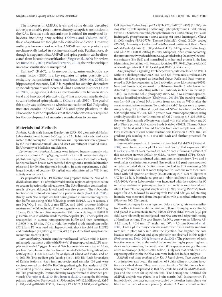
Kal-7 phosphorylation after cocaine or saline treatmentAlthough Kal-7 contains multiple phosphorylation sites (Kiralyet al., 2011b), the functional significance of phosphorylation forKal-7’s GEF activity is unclear (Xie et al., 2007; Kiraly et al.,2011b). To determine whether repeated cocaine injections alterKal-7 phosphorylation, we immunoprecipitated Kal-7 from theNAc of individual cocaine-treated and saline-treated animals us-ing Kal-7 antibody as described previously (Kiraly et al., 2011b).The majority of Kal-7 protein was successfully pulled down(Fig. 2A). However, in both groups, no phosphorylation signalor only a trace signal was detected on immunoblots by anti-bodies recognizing phosphorylated tyrosine, serine, or threo-nine residues (Fig. 2B). It is possible that the phosphorylationlevel of Kal-7 is below our detection limit and/or that cocaineproduces a transient change in Kal-7 phosphorylation notcaptured on WD14.
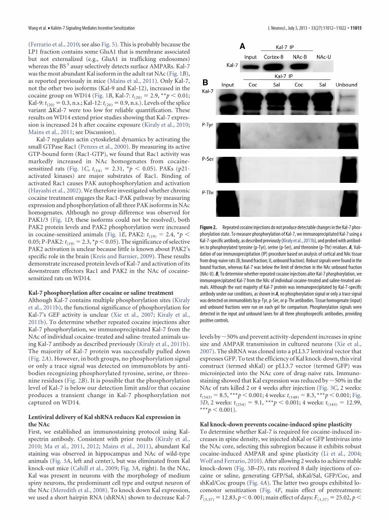
Lentiviral delivery of Kal shRNA reduces Kal expression inthe NAcFirst, we established an immunostaining protocol using Kal-spectrin antibody. Consistent with prior results (Kiraly et al.,2010; Ma et al., 2011, 2012; Mains et al., 2011), abundant Kalstaining was observed in hippocampus and NAc of wild-typeanimals (Fig. 3A, left and center), but was eliminated from Kalknock-out mice (Cahill et al., 2009; Fig. 3A, right). In the NAc,Kal was present in neurons with the morphology of mediumspiny neurons, the predominant cell type and output neuron ofthe NAc (Meredith et al., 2008). To knock down Kal expression,we used a short hairpin RNA (shRNA) shown to decrease Kal-7
levels by �50% and prevent activity-dependent increases in spinesize and AMPAR transmission in cultured neurons (Xie et al.,2007). The shRNA was cloned into a pLL3.7 lentiviral vector thatexpresses GFP. To test the efficiency of Kal knock-down, this viralconstruct (termed shKal) or pLL3.7 vector (termed GFP) wasmicroinjected into the NAc core of drug-naive rats. Immuno-staining showed that Kal expression was reduced by �50% in theNAc of rats killed 2 or 4 weeks after injection (Fig. 3C, 2 weeks:t(343) � 8.5, ***p � 0.001; 4 weeks: t(148) � 8.3, ***p � 0.001; Fig.3D, 2 weeks: t(254) � 9.1, ***p � 0.001; 4 weeks: t(145) � 12.99,***p � 0.001).
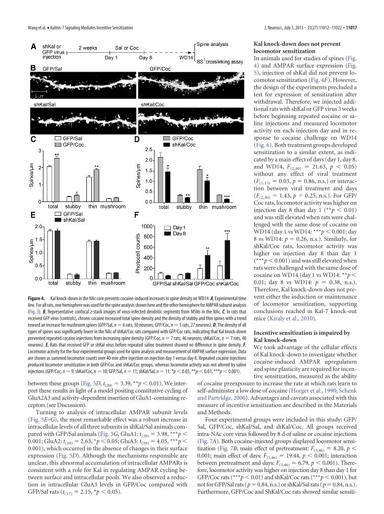
Kal knock-down prevents cocaine-induced spine plasticityTo determine whether Kal-7 is required for cocaine-induced in-creases in spine density, we injected shKal or GFP lentivirus intothe NAc core, selecting this subregion because it exhibits robustcocaine-induced AMPAR and spine plasticity (Li et al., 2004;Wolf and Ferrario, 2010). After allowing 2 weeks to achieve stableknock-down (Fig. 3B–D), rats received 8 daily injections of co-caine or saline, generating GFP/Sal, shKal/Sal, GFP/Coc, andshKal/Coc groups (Fig. 4A). The latter two groups exhibited lo-comotor sensitization (Fig. 4F, main effect of pretreatment:F(3,37) � 12.83, p � 0. 001; main effect of days: F(1,37) � 25.02, p �
Figure 2. Repeated cocaine injections do not produce detectable changes in the Kal-7 phos-phorylation state. To measure phosphorylation of Kal-7, we immunoprecipitated Kal-7 using aKal-7-specific antibody, as described previously (Kiraly et al., 2011b), and probed with antibod-ies to phosphorylated tyrosine (p-Tyr), serine (p-Ser), and threonine (p-Thr) residues. A, Vali-dation of our immunoprecipitation (IP) procedure based on analysis of cortical and NAc tissuefrom drug-naive rats (B, bound fraction; U, unbound fraction). Robust signals were found in thebound fraction, whereas Kal-7 was below the limit of detection in the NAc unbound fraction(NAc-U). B, To determine whether repeated cocaine injections alter Kal-7 phosphorylation, weimmunoprecipitated Kal-7 from the NAc of individual cocaine-treated and saline-treated ani-mals. Although the vast majority of Kal-7 protein was immunoprecipitated by Kal-7-specificantibody under our conditions, as shown in A, no phosphorylation signal or only a trace signalwas detected on immunoblots by p-Tyr, p-Ser, or p-Thr antibodies. Tissue homogenate (input)and unbound fractions were run on each gel for comparison. Phosphorylation signals weredetected in the input and unbound lanes for all three phosphospecific antibodies, providingpositive controls.
Wang et al. • Kalirin-7 Signaling Mediates Incentive Sensitization J. Neurosci., July 3, 2013 • 33(27):11012–11022 • 11015
0.001; interaction between pretreatmentand days: F(3,37) � 11.87, p � 0.001).Therefore, locomotor activity was higheron injection day 8 than day 1 for GFP/Cocrats (**p � 0.01) and shKal/Coc rats(***p � 0.001), but not for GFP/Sal rats(p � 0.51, n.s.) or shKal/Sal rats (p �0.72, n.s.). Furthermore, GFP/Coc andShKal/Coc rats showed similar sensitiza-tion (main effect of viral treatment: F(1,17)
� 1.9, n.s.; main effect of days: F(1,17) �23.29, p � 0.001; interaction between viraltreatment and days: F(1,17) � 2.37, n.s.).Rats were killed on WD14. Brains wererapidly removed and divided into rightand left hemispheres. One hemispherewas used for spine analysis. The other wasused for AMPAR analysis (see next sec-tion). Because we could not simultane-ously process all four groups, threeexperiments using separate cohorts of an-imals were conducted to compare twogroups at a time: (1) GFP/Sal versus GFP/Coc, (2) GFP/Coc versus shKal/Coc, and(3) GFP/Sal versus shKal/Sal.
By comparing rats that received con-trol virus followed by repeated saline orcocaine injections (GFP/Sal and GFP/Cocgroups, respectively), we replicated priorfindings of increased total spine density inthe NAc of cocaine-sensitized animals(Fig. 4B,C, t(9) � 4.2, **p � 0.01). Fur-thermore, by classifying spines using Neu-ronStudio, we demonstrated that cocaineincreased the density of thin and stubbyspines with a trend toward increased densityof mushroom spines (Fig. 4C, Stubby: t(9) �2.24, *p�0.05; Thin: t(9) �3.52, **p�0.01;Mushroom: t(9) �1.83, p�0.10, n.s.). Con-versely, the shKal/Coc group showed mark-edly lower spine density compared with theGFP/Coc group, indicating that Kal knock-down prevented repeated cocaine injectionsfrom increasing spine density (Fig. 4B,D,Total spines: t(12) � 2.44, *p � 0.05; Stubby:t(12) � 3.84, **p � 0.01; Thin: t(12) � 2.39, *p � 0.05; Mushroom:t(12) � 6.60, ***p � 0.001). No significant differences were observedbetween GFP/Sal and shKal/Sal groups (Fig. 4B,E). Therefore,Kal knock-down prevented the cocaine-induced increase in spinedensity while having no effect in saline-treated animals.
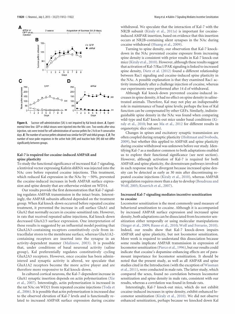
Kal knock-down preventscocaine-induced AMPAR upregulationBased on a role for Kal-7 in AMPAR synaptic delivery in other celltypes (Xie et al., 2007; Cahill et al., 2009), we hypothesized thatKal-7 was involved in the cocaine-induced increase in AMPARsurface expression. We tested this by using a protein crosslinkingassay (Boudreau et al., 2012) to compare AMPAR subunit surfaceand intracellular levels in the same experimental groups gener-ated for the spine studies (see previous section).
Focusing first on AMPAR surface expression (Fig. 5B–D),GFP/Coc rats exhibited increased surface GluA1 and GluA2 and atrend toward increased GluA2/3 compared with GFP/Sal rats
(Fig. 5B, GluA1: t(17) � 2.32, *p � 0.05; GluA2: t(17) � 2.50, *p �0.05). This is consistent with prior work showing increased sur-face or synaptic expression of GluA1A2-containing AMPARs inthe NAc of cocaine-sensitized rats (Boudreau and Wolf, 2005;Boudreau et al., 2007, 2009; Kourrich et al., 2007; Ghasemzadehet al., 2009; Schumann and Yaka, 2009; Ferrario et al., 2010; Schi-erberl et al., 2011). However, GluA1 surface expression wasmarkedly reduced in shKal/Coc animals compared with GFP/Coc animals, indicating that Kal knock-down in the NAc pre-vented the cocaine-induced increase in surface GluA1 (Fig. 5C,t(15) � 2.75, *p � 0.05). In addition, the shKal/Coc group showedtrends toward reductions in surface GluA2, GluA3, and GluA2A3(Fig. 5C), consistent with prevention of the upregulation ofGluA1-containing AMPARs that also contained GluA2 or GluA3.However, when shKal animals were treated with repeated salineinjections, it was GluA2/3 (detected with antibody recognizingboth subunits) that showed a decrease in surface expression com-pared with GFP/Sal animals, whereas surface GluA1 did not differ
Figure 3. Lentivirus expressing Kal shRNA (shKal) reduces Kal immunostaining in the NAc by �50%. A, We detected robust Kalimmunostaining in the hippocampus of naive rats (left), moderate staining in the NAc of naive rats (center), and loss of signal in theNAc of Kal knock-out mice (right). Immunostaining was performed using a Kal-spectrin antibody. B, To knock down Kal expression,a previously characterized shRNA (Xie et al., 2007), which targets the Kal sequence GCAGTACAATCCTGGCCATGT (beginning atposition 1229), was cloned into a pLL3.7 lentiviral vector that expresses GFP. To determine the efficiency of Kal knock-down, thisviral construct (termed shKal) or the pLL3.7 vector (termed GFP) was injected into the NAc core. Rats were killed either 2 or 4 weeksafter virus injection. GFP (left) and Kal staining (center) were visualized (right panel shows merged images). C, Quantification ofthese results revealed a reduction of �50% when comparing transfected (GFP-positive) NAc neurons in shKal rats (yellow arrow)with nontransfected neurons in the same brain section (white arrow; 2 weeks: n � 345 cells; 4 weeks: n � 150 cells; ***p �0.001). D, A similar reduction in Kal staining was found when comparing transfected (GFP-positive) NAc neurons from shKal ratswith transfected neurons from GFP rats (2 weeks: n � 256 cells; 4 weeks: n � 147 cells; ***p � 0.001).
11016 • J. Neurosci., July 3, 2013 • 33(27):11012–11022 Wang et al. • Kalirin-7 Signaling Mediates Incentive Sensitization
between these groups (Fig. 5D, t(20) � 3.39, **p � 0.01). We inter-pret these results in light of a model positing constitutive cycling ofGluA2A3 and activity-dependent insertion of GluA1-containing re-ceptors (see Discussion).
Turning to analysis of intracellular AMPAR subunit levels(Fig. 5E–G), the most remarkable effect was a robust increase inintracellular levels of all three subunits in shKal/Sal animals com-pared with GFP/Sal animals (Fig. 5G, GluA1: t(20) � 3.98, ***p �0.001; GluA2: t(20) � 2.63, *p � 0.05; GluA3: t(20) � 4.05, ***p �0.001), which occurred in the absence of changes in their surfaceexpression (Fig. 5D). Although the mechanisms responsible areunclear, this abnormal accumulation of intracellular AMPARs isconsistent with a role for Kal in regulating AMPAR cycling be-tween surface and intracellular pools. We also observed a reduc-tion in intracellular GluA3 levels in GFP/Coc compared withGFP/Sal rats (t(17) � 2.15, *p � 0.05).
Kal knock-down does not preventlocomotor sensitizationIn animals used for studies of spines (Fig.4) and AMPAR surface expression (Fig.5), injection of shKal did not prevent lo-comotor sensitization (Fig. 4F). However,the design of the experiments precluded atest for expression of sensitization afterwithdrawal. Therefore, we injected addi-tional rats with shKal or GFP virus 3 weeksbefore beginning repeated cocaine or sa-line injections and measured locomotoractivity on each injection day and in re-sponse to cocaine challenge on WD14(Fig. 6). Both treatment groups developedsensitization to a similar extent, as indi-cated by a main effect of days (day 1, day 8,and WD14, F(2,30) � 21.63, p � 0.05)without any effect of viral treatment(F(1,15) � 0.03, p � 0.86, n.s.) or interac-tion between viral treatment and days(F(2,30) � 1.43, p � 0.25, n.s.). For GFP/Coc rats, locomotor activity was higher oninjection day 8 than day 1 (**p � 0.01)and was still elevated when rats were chal-lenged with the same dose of cocaine onWD14 (day 1 vs WD14: ***p � 0.001; day8 vs WD14: p � 0.26, n.s.). Similarly, forshKal/Coc rats, locomotor activity washigher on injection day 8 than day 1(***p � 0.001) and was still elevated whenrats were challenged with the same dose ofcocaine on WD14 (day 1 vs WD14: **p �0.01; day 8 vs WD14: p � 0.38, n.s.).Therefore, Kal knock-down does not pre-vent either the induction or maintenanceof locomotor sensitization, supportingconclusions reached in Kal-7 knock-outmice (Kiraly et al., 2010).
Incentive sensitization is impaired byKal knock-downWe took advantage of the cellular effectsof Kal knock-down to investigate whethercocaine-induced AMPAR upregulationand spine plasticity are required for incen-tive sensitization, measured as the ability
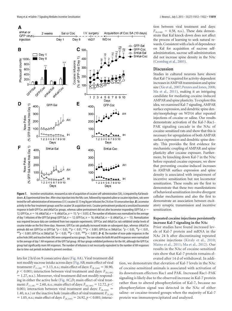
of cocaine preexposure to increase the rate at which rats learn toself-administer a low dose of cocaine (Horger et al., 1990; Schenkand Partridge, 2000). Advantages and caveats associated with thismeasure of incentive sensitization are described in the Materialsand Methods.
Four experimental groups were included in this study: GFP/Sal, GFP/Coc, shKal/Sal, and shKal/Coc. All groups receivedintra-NAc core virus followed by 8 d of saline or cocaine injections(Fig. 7A). Both cocaine-injected groups displayed locomotor sensi-tization (Fig. 7B; main effect of pretreatment: F(3,46) � 8.20, p �0.001; main effect of days: F(1,46) � 19.44, p � 0.001; interactionbetween pretreatment and days: F(3,46) � 6.79, p � 0.001). There-fore, locomotor activity was higher on injection day 8 than day 1 forGFP/Coc rats (***p � 0.01) and shKal/Coc rats (***p � 0.001), butnot for GFP/Sal rats (p � 0.84, n.s.) or shKal/Sal rats (p � 0.84, n.s.).Furthermore, GFP/Coc and ShKal/Coc rats showed similar sensiti-
Figure 4. Kal knock-down in the NAc core prevents cocaine-induced increases in spine density on WD14. A, Experimental timeline. For all rats, one hemisphere was used for the spine analysis shown here and the other hemisphere for AMPAR subunit analysis(Fig. 5). B, Representative confocal z-stack images of virus-infected dendritic segments from MSNs in the NAc. C, In rats thatreceived GFP virus (controls), chronic cocaine increased total spine density and the density of stubby and thin spines with a trendtoward an increase for mushroom spines (GFP/Sal, n � 6 rats, 30 neurons; GFP/Coc, n � 5 rats, 27 neurons). D, The density of alltypes of spines was significantly lower in the NAc of shKal/Coc rats compared with GFP/Coc rats, indicating that Kal knock-downprevented repeated cocaine injections from increasing spine density (GFP/Coc, n � 7 rats, 46 neurons; shKal/Coc, n � 7 rats, 40neurons). E, Rats that received GFP or shKal virus before repeated saline treatment showed no difference in spine density. F,Locomotor activity for the four experimental groups used for spine analysis and measurement of AMPAR surface expression. Dataare shown as summed locomotor counts over 40 min after injection on injection day 1 versus day 8. Repeated cocaine injectionsproduced locomotor sensitization in both GFP/Coc and shKal/Coc groups, whereas locomotor activity was not altered by salineinjections (GFP/Coc, n � 9; shKal/Coc, n � 10; GFP/Sal, n � 11; shKal/Sal, n � 11; *p � 0.05, **p � 0.01, ***p � 0.001).
Wang et al. • Kalirin-7 Signaling Mediates Incentive Sensitization J. Neurosci., July 3, 2013 • 33(27):11012–11022 • 11017

zation (main effect of viral treatment: F(1,27) � 0.24, n.s.; main effectof days: F(1,27) �28.13, p�0.001; viral treatment�days interaction:F(1,27) � 0.45, n.s.). On WD7, jugular catheters were implanted toenable cocaine self-administration. On WD14, when locomotorsensitization was maintained in shKal/Coc rats but neither AMPARnor spine upregulation had occurred (Figs. 4, 5, 6), we began cocaineself-administration training (2 h/d for 10 d). A low dose of cocaine(0.15 mg/kg) was selected based on preliminary studies (data notshown). This dose was sufficient for cocaine preexposed animals toreadily acquire cocaine self-administration because cocaine alreadyhas an elevated incentive value for these animals, whereas it wassubthreshold for saline-pretreated or drug-naive animals, whotherefore took longer to acquire stable self-administration behavior.
We found that pretreatment (saline vs cocaine injections) mod-ified cocaine intake across days (Fig. 7C; main effect of pretreatment:F(3,47) � 7.66, p � 0.001; main effect of days: F(9,423) � 8.30, p �0.001; interaction between days and pretreatment: F(27,423) � 1.71,p � 0.05). Specifically, GFP/Coc rats did not differ significantly fromother groups on days 1–3, but they gradually increased their intakeon subsequent days (GFP/Coc vs GFP/Sal: *p � 0.05, **p � 0.01,***p � 0.001; GFP/Coc vs ShKal/Coc: #p � 0.05, ##p � 0.01, ###p �0.001; GFP/Coc vs ShKal/Sal: &p � 0.05, &&p � 0.01, &&&p �
0.001), whereas the intake of shKal/Coc animals plateaued at thesame level reached by the GFP/Sal group. A comparison was alsomade for active versus inactive nose-poke responses (Fig. 7D–G).Pretreatment modified responding in the active hole, with GFP/Cocrats consistently showing higher responding than all other groups[main effect of pretreatment: F(3,47) � 5.60, p � 0.01; main effect ofdays: F(9,423) � 3.37, p � 0.001; interaction between days and pre-treatment: F(27,423) � 0.71, p � 0.86, n.s.; GFP/Coc vs GFP/Sal: p �0.01; GFP/Coc vs ShKal/Sal: p � 0.05; GFP/Coc vs ShKal/Coc: p �0.01]. Pretreatment did not modify responding in the inactive hole(main effect of pretreatment: F(3,47) �0.30, p�0.83, n.s.; main effectof days: F(9,423) � 7.39, p � 0.001; interaction between days andpretreatment: F(27,423) � 1.03, p � 0.43, n.s.). Therefore, with respectto both infusions and nose pokes, shKal/Coc rats resembled GFP/Coc rats during the initial training days, but came to resemble GFP/Sal and shKal/Sal rats over subsequent days. We interpret these datato indicate that, based on their prior intraperitoneal cocaine injec-tion experience, the shKal/Coc rats “recognized” cocaine and rapidlylearned the operant response. However, compared with the GFP/Coc rats, the shKal/Coc rats took fewer infusions over the remainingdays of training, reflecting lower motivation for cocaine.
After completion of the self-administration experiment, wemeasured Kal immunostaining in the NAc. Kal expression intransfected cells from shKal/Coc rats was reduced by 43% com-pared with nontransfected cells, verifying that shKal virus wasstably expressed throughout our experiment (data not shown).
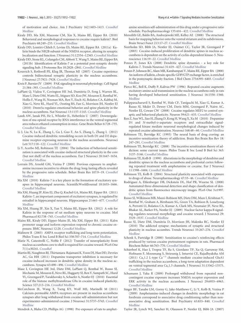
Sucrose self-administration is not impaired by Kalknock-downTo rule out the possibility that Kal knock-down caused a generalimpairment of reward-seeking behavior, we determined the ef-fect of shKal on self-administration of a natural reward, sucrose.Two weeks after injection of GFP or shKal virus into theNAc core, rats were tested for self-administration of sucrose pel-
Figure 5. Kal knock-down in the NAc core prevents cocaine-induced increases in AMPARsubunit surface expression on WD14. A, Experimental time line. For all rats, one hemisphere wasused for spine analysis (Fig. 4) and the other hemisphere was used for the AMPAR subunitanalysis shown here. B–G, Cell surface levels (left panels) and intracellular levels (right panels)of AMPAR subunits measured using a BS 3 protein crosslinking assay and immunoblotting. B,Surface-expressed GluA1 and GluA2 increased after repeated cocaine injections (GFP/Sal, n �10; GFP/Coc, n � 9). C, Compared with GFP/Coc rats, surface GluA1 was significantly lower inrats that received shKal before repeated cocaine injections (GFP/Coc, n � 8; shKal/Coc, n � 9).There were also trends toward relatively lower surface expression of other subunits in ShKal/Cocrats. D, Knock-down of Kal before repeated saline injections decreased surface GluA2/3 com-pared with GFP/Sal rats (GFP/Sal, n � 11; shKal/Sal, n � 11). E, After cocaine treatment, therewas a reduction in intracellular GluA3 levels in GFP/Coc compared with GFP/Sal rats. F, Nodifferences in intracellular AMPAR subunit levels were observed between GFP/Coc and shKal/Coc rats. G, Knock-down of Kal before repeated saline injections produced a marked intracellularaccumulation of GluA1–3 (*p � 0.05, **p � 0.01, ***p � 0.001).
Figure 6. Kal knock-down does not prevent the development or maintenance of locomotorsensitization. A, Experimental time line. B–E, Locomotor sensitization was assessed by com-paring photocell counts on injection days 1 and 8 and after a challenge injection on WD14. Dataare presented as mean number of beam breaks per 5 min interval during the first 40 min aftercocaine injection (B,C) and total beam breaks summed over 20 min after injection (D,E). In bothGFP/Coc and shKal/Coc rats, the locomotor response to cocaine was markedly greater on day 8than day 1 and sensitization was expressed at approximately the same level on day 8 and WD14.The cocaine dose was 15 mg/kg on all days (GFP/Coc, n � 8; ShKal/Coc, n � 9; *p � 0.05,**p � 0.01, ***p � 0.001 vs day 1).
11018 • J. Neurosci., July 3, 2013 • 33(27):11012–11022 Wang et al. • Kalirin-7 Signaling Mediates Incentive Sensitization
lets for 2 h/d on 9 consecutive days (Fig. 8A). Viral treatment didnot modify sucrose intake across days (Fig. 8B; main effect of viraltreatment: F(1,18) � 3.13, n.s.; main effect of days: F(8,144) � 38.90,p � 0.001; interaction between viral treatment and days: F(8,144)
� 1.27, n.s.). Moreover, viral treatment did not modify respond-ing in either the active hole (Fig. 8C,D; main effect of viral treat-ment: F(1,18) � 2.60, n.s.; main effect of days: F(8,144) � 12.72, p �0.001; interaction between viral treatment and days: F(8,144) �1.40, n.s.) or the inactive hole (main effect of viral treatment: F(1,18)
� 1.05, n.s.; main effect of days: F(8,144) � 24.92, p � 0.001; interac-
tion between viral treatment and days:F(8,144) � 0.58, n.s.). These data demon-strate that Kal knock-down does not affectthe process of learning to seek natural re-wards. Consistent with a lack of dependenceon Kal for acquisition of sucrose self-administration, sucrose self-administrationdid not increase spine density in the NAc(Crombag et al., 2005).
DiscussionStudies in cultured neurons have shownthat Kal-7 is required for activity-dependentincreases in AMPAR transmission and spinesize (Xie et al., 2007; Penzes and Jones, 2008;Ma et al., 2011), making it an intriguingcandidate for mediating cocaine-inducedAMPAR and spine plasticity. To explore thisidea, we examined Kal-7 signaling, AMPARsurface expression, and dendritic spine den-sity/morphology on WD14 after repeatedinjections of cocaine or saline. Our resultsdemonstrate activation of the Kal-7-Rac1-PAK signaling cascade in the NAc ofcocaine-sensitized rats and show that this isnecessary for upregulation of both AMPARsurface expression and dendritic spine den-sity. This provides the first evidence formechanistic coupling of AMPAR and spineplasticity after cocaine exposure. Further-more, by knocking down Kal-7 in the NAcbefore repeated cocaine exposure, we showthat preventing cocaine-induced increasesin AMPAR surface expression and spinedensity is associated with impairment ofincentive sensitization but not locomotorsensitization. These results are the first todemonstrate that these two manifestationsof behavioral sensitization involve divergentcellular mechanisms and also the first todemonstrate an association between excit-atory synaptic transmission and incentivesensitization.
Repeated cocaine injections persistentlyincrease Kal-7 signaling in the NAcPrior studies have found increased lev-els of Kal-7 protein and mRNA in theNAc 24 h after discontinuing repeatedcocaine injections (Kiraly et al., 2010;Mains et al., 2011; Ma et al., 2012). Ourresults in the NAc of cocaine-sensitizedrats show that Kal-7 protein remains el-evated after 14 d of withdrawal. In addi-
tion, we demonstrate that elevation of Kal-7 levels in the NAcof cocaine-sensitized animals is associated with activation ofits downstream effectors Rac1 and PAK. Increased Rac1-PAKsignaling is likely due to the observed increase in Kal-7 proteinrather than to altered phosphorylation of Kal-7, because nophosphorylation signal was detected in the NAc of eithersaline- or cocaine-treated groups when the majority of Kal-7protein was immunoprecipitated and analyzed.
Figure 7. Incentive sensitization, measured as rate of acquisition of cocaine self-administration (SA), is impaired by Kal knock-down. A, Experimental time line. After virus injection into the NAc core, followed by repeated saline or cocaine injections, rats weretested for self-administration of intravenous (I.V.) cocaine (0.15 mg/kg per infusion) for 2 h/d on 10 consecutive days. B, Locomotoractivity in the four treatment groups used for cocaine SA acquisition tests. Cocaine pretreatment produced a sensitized locomotorresponse in both GFP/Coc and shKal/Coc groups, whereas saline pretreatment did not alter locomotor responding (GFP/Sal, n �12; GFP/Coc, n � 14; shKal/Sal, n � 9; shKal/Coc, n � 15; *p � 0.05). C, The number of infusions was normalized to the averageof day 1 infusions of the GFP/Sal group (GFP/Sal, n � 12; GFP/Coc, n � 16; shKal/Sal, n � 8; shKal/Coc, n � 15). Normalizationwas required because data are combined from two separate experiments. GFP/Coc and shKal/Coc rats exhibited similar levels ofcocaine intake on the first three days. However, GFP/Coc rats gradually increased intake on subsequent days, whereas shKal/Cocanimals did not (GFP/Coc vs GFP/Sal: *p � 0.05, **p � 0.01, ***p � 0.001; GFP/Coc vs ShKal/Coc: #p � 0.05, ##p � 0.01,###p � 0.001; GFP/Coc vs ShKal/Sal: &p � 0.05, &&p � 0.01, &&&p � 0.001). D–G, The number of nose-poke responses in theactive hole (AH) and inactive hole (IH) were compared across groups. The raw values for both AH and IH responses were normalizedto the average of day 1 AH responses of the GFP/Sal group. All four groups exhibited preference for the AH, although the GFP/Cocgroup had significantly more AH responses. The number of infusions is not necessarily equivalent to the number of AH responsesdue to time-out periods included to prevent overdose.
Wang et al. • Kalirin-7 Signaling Mediates Incentive Sensitization J. Neurosci., July 3, 2013 • 33(27):11012–11022 • 11019
Kal-7 is required for cocaine-induced AMPAR andspine plasticityTo study the functional significance of increased Kal-7 signaling,a lentiviral vector expressing Kalirin shRNA was injected into theNAc core before repeated cocaine injections. This treatment,which reduced Kal expression in the NAc by �50%, preventedthe cocaine-induced increases in both AMPAR surface expres-sion and spine density that are otherwise evident on WD14.
Our results provide the first demonstration that Kal-7 signal-ing regulates AMPAR transmission in the intact brain. Interest-ingly, the AMPAR subunits affected depended on the treatmentgroup. When Kal knock-down occurred before repeated cocainetreatment, it prevented the increase in cell surface GluA1 andGluA2 that normally occurs in cocaine-sensitized rats. However,in rats that received repeated saline injections, Kal knock-downdecreased GluA2/3 surface expression. One interpretation ofthese results is suggested by an influential model positing thatGluA2A3-containing receptors constitutively cycle from in-tracellular stores to the membrane surface, whereas GluA1A2-containing receptors are inserted into the synapse in anactivity-dependent manner (Malinow, 2003). It is possiblethat, under conditions of basal neuronal activity (salinegroup), Kal preferentially regulates constitutively cyclingGluA2A3 receptors. However, once cocaine has been admin-istered and synaptic activity is altered, we speculate thatGluA1A2 receptors become the more active player and aretherefore more responsive to Kal knock-down.
In cultured cortical neurons, the Kal-7-dependent increase inGluA1 synaptic insertion depends on actin polymerization (Xieet al., 2007). Interestingly, actin polymerization is increased inthe rat NAc on WD21 from repeated cocaine injections (Toda etal., 2006). It is possible that actin polymerization is increased dueto the observed elevation of Kal-7 levels and is functionally re-lated to increased AMPAR surface expression during cocaine
withdrawal. We speculate that the interaction of Kal-7 with theNR2B subunit (Kiraly et al., 2011a) is important for cocaine-induced AMPAR insertion, based on evidence that this insertionoccurs at NR2B-containing silent synapses in the NAc duringcocaine withdrawal (Huang et al., 2009).
Turning to spine density, our observation that Kal-7 knock-down in the NAc prevented cocaine exposure from increasingspine density is consistent with prior results in Kal-7 knock-outmice (Kiraly et al., 2010). However, although these results suggestthat activation of Kal-7/Rac1/PAK signaling is linked to increasedspine density, Dietz et al. (2012) found a different relationshipbetween Rac1 signaling and cocaine-induced spine plasticity inthe NAc. A possible explanation is that they examined Rac1 ac-tivity immediately after a challenge injection of cocaine, whereasour experiments were performed after 14 d of withdrawal.
Although Kal knock-down prevented cocaine-induced in-creases in spine density, it had no effect on spine density in saline-treated animals. Therefore, Kal may not play an indispensablerole in maintenance of basal spine levels; perhaps the loss of Kalfunction can be compensated by other GEFs. Similarly, indistin-guishable spine density in the NAc was found when comparingwild-type and Kal7 knock-out mice under basal conditions (Ki-raly et al., 2010; but see Ma et al., 2012 for different results inorganotypic slice cultures).
Changes in spines and excitatory synaptic transmission areoften coupled during synaptic plasticity (Holtmaat and Svoboda,2009), but whether this applied to AMPAR and spine plasticityduring cocaine withdrawal was unknown before our study. Iden-tifying Kal-7 as a mediator common to both adaptations enabledus to explore their functional significance (see next section).However, although activation of Kal-7 is required for bothAMPAR and spine plasticity, the downstream pathways involvedin each response may be divergent because increased spine den-sity can be detected as early as 30 min after discontinuing re-peated cocaine injections (Kiraly et al., 2010), whereas AMPARupregulation requires more than a day to develop (Boudreau andWolf, 2005; Kourrich et al., 2007).
Increased Kal-7 signaling mediates incentive sensitizationto cocaineLocomotor sensitization is the most commonly used measure ofbehavioral sensitization to cocaine. Although it is accompaniedby increased AMPAR surface expression and increased spinedensity, both adaptations can be dissociated from locomotor sen-sitization either temporally or using molecular manipulations(Singer et al., 2009; Russo et al., 2010; Wolf and Ferrario, 2010).Indeed, our results show that Kal-7 knock-down impairsAMPAR and spine plasticity, but not locomotor sensitization.More work is required to understand this dissociation becausesome results implicate AMPAR transmission in expression oflocomotor sensitization (Pierce et al., 1996), but our results couldindicate that cocaine’s dopamine-enhancing effects are of para-mount importance for locomotor sensitization. It should benoted that the present study, as well as all AMPAR and spinestudies cited in the Introduction (with the exception of Wissmanet al., 2011), were conducted in male rats. The latter study, whichcompared the sexes, found no correlation between locomotorsensitization and spine density in male rats, consistent with ourresults, whereas a correlation was found in female rats.
Interestingly, Kal-7 knock-out mice, which do not exhibitcocaine-induced increases in spine density, exhibit enhanced lo-comotor sensitization (Kiraly et al., 2010). We did not observeenhanced sensitization, perhaps because we knocked down Kal
Figure 8. Sucrose self-administration (SA) is not impaired by Kal knock-down. A, Experi-mental time line. GFP or shKal viruses were injected into the NAc core. Two weeks after virusinjection, rats were tested for self-administration of sucrose pellets for 2 h/d on 9 consecutivedays. B, The number of sucrose pellets obtained was similar for GFP and shKal groups. C, D, Thenumber of nose-poke responses in the active hole (AH) and inactive hole (IH) did not differsignificantly between groups.
11020 • J. Neurosci., July 3, 2013 • 33(27):11012–11022 Wang et al. • Kalirin-7 Signaling Mediates Incentive Sensitization

specifically in the NAc during adulthood. In the prior study (Ki-raly et al., 2010), there was the potential for developmental adap-tations or changes in Kal-7 signaling in other brain regions due toits constitutive deletion.
Psychostimulant pretreatment enhances learning about drugsand drug-related cues and willingness to work for them (Vezina,2004). These are potential manifestations of incentive sensitiza-tion. With the exception of results in genetically modified ani-mals, all studies support a positive correlation between the levelof excitatory transmission in the NAc and the motivation to seekcocaine (Wolf and Ferrario, 2010). Therefore, we hypothesizedthat the cocaine-induced, Kal-7-dependent increases in surfaceAMPARs and spines strengthen synaptic connections onto NAcneurons and thereby elicit incentive sensitization. Indeed, ourresults clearly demonstrate that knocking down Kal-7 signalingin the NAc core reduces incentive sensitization, measured as theability of cocaine preexposure to facilitate self-administration ofa low dose of cocaine. Additional studies will be required to de-termine whether incentive sensitization requires increases inboth surface AMPAR levels and spine density or if one is morecritical.
Other studies have found a relationship between increasedNAc spine density and associative drug conditioning. Therefore,Kal-7 knock-out mice, which do not exhibit cocaine-inducedincreases in spine density, show impaired conditioned place pref-erence (Kiraly et al., 2010). Conversely, Marie et al. (2012) founda correlation between the magnitude of conditioned place pref-erence and the increase in NAc spine density. Finally, resonatingclosely with our results, Singer et al. (2009) found that repeatedamphetamine exposure increases spine density in the NAc only ifthe amphetamine regimen leads to conditioned hyperactivity,not if the regimen leads to locomotor sensitization in the absenceof conditioned hyperactivity. One interpretation is that increasedspine density in these studies reflects acquisition of incentiveproperties by the drug context (a form of incentive learning).
The original formulation of the incentive sensitization hy-pothesis of addiction suggested that common neuroadaptations,in the mesocorticolimbic dopamine system and others, enablesensitization of motor activating and incentive motivationaleffects (Robinson and Berridge, 1993). Indeed, there is strongevidence for overlapping mechanisms (Norrholm et al., 2003;Vezina, 2004; Taylor et al., 2007). Our results are not in oppo-sition to these findings; rather, they indicate that additionalKal-7-dependent adaptations must occur to enable incentivesensitization.
In conclusion, these results identify Kal-7 as an essential linkbetween the mechanisms underlying cocaine-induced structuraland functional plasticity. Furthermore, they demonstrate thatthese Kal-7-dependent adaptations are required for sensitizationof the incentive-motivational properties of cocaine but not forsensitization of its locomotor-activating properties.
ReferencesBoudreau AC, Wolf ME (2005) Behavioral sensitization to cocaine is asso-
ciated with increased AMPA receptor surface expression in the nucleusaccumbens. J Neurosci 25:9144 –9151. CrossRef Medline
Boudreau AC, Reimers JM, Milovanovic M, Wolf ME (2007) Cell surfaceAMPA receptors in the rat nucleus accumbens increase during cocainewithdrawal but internalize after cocaine challenge in association withaltered activation of mitogen-activated protein kinases. J Neurosci 27:10621–10635. CrossRef Medline
Boudreau AC, Ferrario CR, Glucksman MJ, Wolf ME (2009) Signalingpathway adaptations and novel protein kinase A substrates related tobehavioral sensitization to cocaine. J Neurochem 110:363–377. CrossRefMedline
Boudreau AC, Milovanovic M, Conrad KL, Nelson C, Ferrario CR, Wolf ME(2012) A protein cross-linking assay for measuring cell surface expres-sion of glutamate receptor subunits in the rodent brain after in vivotreatments. Curr Protocols Neurosci, Unit 5.30.1–19. CrossRef Medline
Brown TE, Lee BR, Mu P, Ferguson D, Dietz D, Ohnishi YN, Lin Y, Suska A,Ishikawa M, Huang YH, Shen H, Kalivas PW, Sorg BA, Zukin RS, NestlerEJ, Dong Y, Schluter OM (2011) A silent synapse-based mechanism forcocaine-induced locomotor sensitization. J Neurosci 31:8163– 8174.CrossRef Medline
Cahill ME, Xie Z, Day M, Photowala H, Barbolina MV, Miller CA, Weiss C,Radulovic J, Sweatt JD, Disterhoft JF, Surmeier DJ, Penzes P (2009) Ka-lirin regulates cortical spine morphogenesis and disease-related behav-ioral phenotypes. Proc Natl Acad Sci U S A 106:13058 –13063. CrossRefMedline
Christoffel DJ, Golden SA, Dumitriu D, Robison AJ, Janssen WG, Ahn HF,Krishnan V, Reyes CM, Han MH, Ables JL, Eisch AJ, Dietz DM, FergusonD, Neve RL, Greengard P, Kim Y, Morrison JH, Russo SJ (2011) I�Bkinase regulates social defeat stress-induced synaptic and behavioral plas-ticity. J Neurosci 31:314 –321. CrossRef Medline
Conrad KL, Tseng KY, Uejima JL, Reimers JM, Heng LJ, Shaham Y, MarinelliM, Wolf ME (2008) Formation of accumbens GluR2-lacking AMPA re-ceptors mediates incubation of cocaine craving. Nature 454:118 –121.CrossRef Medline
Crombag HS, Gorny G, Li Y, Kolb B, Robinson TE (2005) Opposite effectsof amphetamine self-administration experience on dendritic spines in themedial and orbital prefrontal cortex. Cereb Cortex 15:341–348. CrossRefMedline
Dietz DM, Sun H, Lobo MK, Cahill ME, Chadwick B, Gao V, Koo JW, Mazei-Robison MS, Dias C, Maze I, Damez-Werno D, Dietz KC, Scobie KN,Ferguson D, Christoffel D, Ohnishi Y, Hodes GE, Zheng Y, Neve RL,Hahn KM, Russo SJ, Nestler EJ (2012) Rac1 is essential in cocaine-induced structural plasticity of nucleus accumbens neurons. Nat Neuro-sci 15:891– 896. CrossRef Medline
Dobi A, Seabold GK, Christensen CH, Bock R, Alvarez VA (2011) Cocaine-induced plasticity in the nucleus accumbens is cell specific and developswithout prolonged withdrawal. J Neurosci 31:1895–1904. CrossRefMedline
Dumitriu D, Rodriguez A, Morrison JH (2011) High-throughput, detailed,cell-specific neuroanatomy of dendritic spines using microinjection andconfocal microscopy. Nat Protoc 6:1391–1411. CrossRef Medline
Ferrario CR, Gorny G, Crombag HS, Li Y, Kolb B, Robinson TE (2005)Neural and behavioral plasticity associated with the transition from con-trolled to escalated cocaine use. Biol Psychiatry 58:751–759. CrossRefMedline
Ferrario CR, Li X, Wang X, Reimers JM, Uejima JL, Wolf ME (2010) Therole of glutamate receptor redistribution in locomotor sensitization tococaine. Neuropsychopharmacol 35:818 – 833. CrossRef Medline
Ghasemzadeh MB, Mueller C, Vasudevan P (2009) Behavioral sensitizationto cocaine is associated with increased glutamate receptor trafficking tothe postsynaptic density after extended withdrawal period. Neurosci 159:414 – 426. CrossRef Medline
Goel A, Jiang B, Xu LW, Song L, Kirkwood A, Lee HK (2006) Cross-modalregulation of synaptic AMPA receptors in primary sensory cortices byvisual experience. Nat Neurosci 9:1001–1003. CrossRef Medline
Golden SA, Russo SJ (2012) Mechanisms of psychostimulant-inducedstructural plasticity. Cold Spring Harb Perspect Med 2:pii:a011957.CrossRef Medline
Hayashi K, Ohshima T, Mikoshiba K (2002) Pak1 is involved in dendriteinitiation as a downstream effector of Rac1 in cortical neurons. Mol CellNeurosci 20:579 –594. CrossRef Medline
Holtmaat A, Svoboda K (2009) Experience-dependent structural synapticplasticity in the mammalian brain. Nat Rev Neurosci 10:647– 658.CrossRef Medline
Horger BA, Shelton K, Schenk S (1990) Pre-exposure sensitizes rats to therewarding effects of cocaine. Pharmacol Biochem Behav 37:707–711.CrossRef Medline
Huang YH, Lin Y, Mu P, Lee BR, Brown TE, Wayman G, Marie H, Liu W, YanZ, Sorg BA, Schluter OM, Zukin RS, Dong Y (2009) In vivo cocaineexperience generates silent synapses. Neuron 63:40 – 47. CrossRefMedline
Kalivas PW, Volkow ND (2005) The neural basis of addiction: a pathology
Wang et al. • Kalirin-7 Signaling Mediates Incentive Sensitization J. Neurosci., July 3, 2013 • 33(27):11012–11022 • 11021
of motivation and choice. Am J Psychiatry 162:1403–1413. CrossRefMedline
Kiraly DD, Ma XM, Mazzone CM, Xin X, Mains RE, Eipper BA (2010)Behavioral and morphological responses to cocaine require kalirin7. BiolPsychiatry 68:249 –255. CrossRef Medline
Kiraly DD, Lemtiri-Chlieh F, Levine ES, Mains RE, Eipper BA (2011a) Ka-lirin binds the NR2B subunit of the NMDA receptor, altering its synapticlocalization and function. J Neurosci 31:12554 –12565. CrossRef Medline
Kiraly DD, Stone KL, Colangelo CM, Abbott T, Wang Y, Mains RE, Eipper BA(2011b) Identification of Kalirin-7 as a potential post-synaptic densitysignaling hub. J Proteome Res 10:2828 –2841. CrossRef Medline
Kourrich S, Rothwell PE, Klug JR, Thomas MJ (2007) Cocaine experiencecontrols bidirectional synaptic plasticity in the nucleus accumbens.J Neurosci 27:7921–7928. CrossRef Medline
Kreis P, Barnier JV (2009) PAK signaling in neuronal physiology. Cell Signal21:384 –393. CrossRef Medline
LaPlant Q, Vialou V, Covington HE 3rd, Dumitriu D, Feng J, Warren BL,Maze I, Dietz DM, Watts EL, Iniguez SD, Koo JW, Mouzon E, Renthal W,Hollis F, Wang H, Noonan MA, Ren Y, Eisch AJ, Bolanos CA, Kabbaj M,Xiao G, Neve RL, Hurd YL, Oosting RS, Fan G, Morrison JH, Nestler EJ(2010) Dnmt3a regulates emotional behavior and spine plasticity in thenucleus accumbens. Nat Neurosci 13:1137–1143. CrossRef Medline
Lasek AW, Janak PH, He L, Whistler JL, Heberlein U (2007) Downregula-tion of mu opioid receptor by RNA interference in the ventral tegmentalarea reduces ethanol consumption in mice. Gene Brain Behav 6:728 –735.CrossRef Medline
Li J, Liu N, Lu K, Zhang L, Gu J, Guo F, An S, Zhang L, Zhang L (2012)Cocaine-induced dendritic remodeling occurs in both D1 and D2 dopa-mine receptor-expressing neurons in the nucleus accumbens. NeurosciLett 517:118 –122. CrossRef Medline
Li Y, Acerbo MJ, Robinson TE (2004) The induction of behavioral sensiti-zation is associated with cocaine-induced structural plasticity in the core(but not shell) of the nucleus accumbens. Eur J Neurosci 20:1647–1654.CrossRef Medline
Lorrain DS, Arnold GM, Vezina P (2000) Previous exposure to amphet-amine increases incentive to obtain the drug: long-lasting effects revealedby the progressive ratio schedule. Behav Brain Res 107:9 –19. CrossRefMedline
Ma XM (2010) Kalirin-7 is a key player in the formation of excitatory syn-apses in hippocampal neurons. ScientificWorldJournal 10:1655–1666.CrossRef Medline
Ma XM, Huang JP, Kim EJ, Zhu Q, Kuchel GA, Mains RE, Eipper BA (2011)Kalirin-7, an important component of excitatory synapses, is regulated byestradiol in hippocampal neurons. Hippocampus 21:661– 677. CrossRefMedline
Ma XM, Huang JP, Xin X, Yan Y, Mains RE, Eipper BA (2012) A role forKalirin in the response of rat medium spiny neurons to cocaine. MolPharmacol 82:738 –745. CrossRef Medline
Mains RE, Kiraly DD, Eipper-Mains JE, Ma XM, Eipper BA (2011) Kalrnpromoter usage and isoform expression respond to chronic cocaine ex-posure. BMC Neurosci 12:20. CrossRef Medline
Malinow R (2003) AMPA receptor trafficking and long-term potentiation.Philos Trans R Soc Lond B Biol Sci 358:707–714. CrossRef Medline
Marie N, Canestrelli C, Noble F (2012) Transfer of neuroplasticity fromnucleus accumbens core to shell is required for cocaine reward. PLoS One7(1):e30241. CrossRef
Martin BJ, Naughton BJ, Thirtamara-Rajamani K, Yoon DJ, Han DD, DevriesAC, Gu HH (2011) Dopamine transporter inhibition is necessary forcocaine-induced increases in dendritic spine density in the nucleus ac-cumbens. Synapse 65:490 – 496. CrossRef Medline
Maze I, Covington HE 3rd, Dietz DM, LaPlant Q, Renthal W, Russo SJ,Mechanic M, Mouzon E, Neve RL, Haggarty SJ, Ren Y, Sampath SC, HurdYL, Greengard P, Tarakhovsky A, Schaefer A, Nestler EJ (2010) Essentialrole of the histone methyltransferase G9a in cocaine-induced plasticity.Science 327:213–216. CrossRef Medline
McCutcheon JE, Wang X, Tseng KY, Wolf ME, Marinelli M (2011)Calcium-permeable AMPA receptors are present in nucleus accumbenssynapses after long withdrawal from cocaine self-administration but notexperimenter-administered cocaine. J Neurosci 31:5737–5743. CrossRefMedline
Mendrek A, Blaha CD, Phillips AG (1998) Pre-exposure of rats to amphet-
amine sensitizes self-administration of this drug under a progressive ratioschedule. Psychopharmacology 135:416 – 422. CrossRef Medline
Meredith GE, Baldo BA, Andrezjewski ME, Kelley AE (2008) The structuralbasis for mapping behavior onto the ventral striatum and its subdivisions.Brain Struct Funct 213:17–27. CrossRef Medline
Norrholm SD, Bibb JA, Nestler EJ, Ouimet CC, Taylor JR, Greengard P(2003) Cocaine-induced proliferation of dendritic spines in nucleus ac-cumbens is dependent on the activity of cyclin-dependent kinase-5. Neu-roscience 116:19 –22. CrossRef Medline
Penzes P, Jones KA (2008) Dendritic spine dynamics - a key role forKalirin-7. Trends Neurosci 31:419 – 427. CrossRef Medline
Penzes P, Johnson RC, Alam MR, Kambampati V, Mains RE, Eipper BA (2000)An isoform of kalirin, a brain-specific GDP/GTP exchange factor, is enrichedin the postsynaptic density fraction. J Biol Chem 275:6395–6403. CrossRefMedline
Pierce RC, Bell K, Duffy P, Kalivas PW (1996) Repeated cocaine augmentsexcitatory amino acid transmission in the nucleus accumbens only in ratshaving developed behavioral sensitization. J Neurosci 16:1550 –1560.Medline
Pulipparacharuvil S, Renthal W, Hale CF, Taniguchi M, Xiao G, Kumar A,Russo SJ, Sikder D, Dewey CM, Davis MM, Greengard P, Nairn AC,Nestler EJ, Cowan CW (2008) Cocaine regulates MEF2 to control syn-aptic and behavioral plasticity. Neuron 59:621– 633. CrossRef Medline
Ren Z, Sun WL, Jiao H, Zhang D, Kong H, Wang X, Xu M (2010) DopamineD1 and N-methyl-D-aspartate receptors and extracellular signal-regulated kinase mediate neuronal morphological changes induced byrepeated cocaine administration. Neurosci 168:48 – 60. CrossRef Medline
Robinson TE, Berridge KC (1993) The neural basis of drug craving: anincentive-sensitization theory of addiction. Brain Res Brain Res Rev 18:247–291. CrossRef Medline
Robinson TE, Berridge KC (2008) The incentive sensitization theory of ad-diction: some current issues. Philos Trans R Soc Lond B Biol Sci 363:3137–3146. CrossRef Medline
Robinson TE, Kolb B (1999) Alterations in the morphology of dendrites anddendritic spines in the nucleus accumbens and prefrontal cortex follow-ing repeated treatment with amphetamine or cocaine. Eur J Neurosci11:1598 –1604. CrossRef Medline
Robinson TE, Kolb B (2004) Structural plasticity associated with exposureto drugs of abuse. Neuropharmacology 47:33– 46. CrossRef Medline
Rodriguez A, Ehlenberger DB, Dickstein DL, Hof PR, Wearne SL (2008)Automated three-dimensional detection and shape classification of den-dritic spines from fluorescence microscopy images. PLoS One 3:e1997.CrossRef Medline
Russo SJ, Wilkinson MB, Mazei-Robison MS, Dietz DM, Maze I, Krishnan V,Renthal W, Graham A, Birnbaum SG, Green TA, Robison B, LesselyongA, Perrotti LI, Bolanos CA, Kumar A, Clark MS, Neumaier JF, Neve RL,Bhakar AL, Barker PA, Nestler EJ (2009) Nuclear factor kappa B signal-ing regulates neuronal morphology and cocaine reward. J Neurosci 29:3529 –3537. CrossRef Medline
Russo SJ, Dietz DM, Dumitriu D, Morrison JH, Malenka RC, Nestler EJ(2010) The addicted synapse: mechanisms of synaptic and structuralplasticity in nucleus accumbes. Trends Neurosci 33:267–276. CrossRefMedline
Schenk S, Partridge B (2000) Sensitization to cocaine’s reinforcing effectsproduced by various cocaine pretreatment regimens in rats. PharmacolBiochem Behav 66:765–770. CrossRef Medline
Schierberl K, Hao J, Tropea TF, Ra S, Giordano TP, Xu Q, Garraway SM,Hofmann F, Moosmang S, Striessnig J, Inturrisi CE, Rajadhyaksha AM(2011) Cav1.2 L-type Ca �2 channels mediate cocaine-induced GluA1trafficking in the nucleus accumbens, a long-term adaptation dependenton ventral tegmental area Cav1.3 channels. J Neurosci 31:13562–13575.CrossRef Medline
Schumann J, Yaka R (2009) Prolonged withdrawal from repeated non-contingent cocaine exposure increases NMDA receptor expression andERK activity in the nucleus accumbens. J Neurosci 29:6955– 6963.CrossRef Medline
Singer BF, Tanabe LM, Gorny G, Jake-Matthews C, Li Y, Kolb B, Vezina P(2009) Amphetamine-induced changes in dendritic morphology in ratforebrain correspond to associative drug conditioning rather than non-associative drug sensitization. Biol Psychiatry 65:835– 840. CrossRefMedline
Taylor JR, Lynch WJ, Sanchez H, Olausson P, Nestler EJ, Bibb JA (2007)
11022 • J. Neurosci., July 3, 2013 • 33(27):11012–11022 Wang et al. • Kalirin-7 Signaling Mediates Incentive Sensitization

Inhibition of CDK5 in the nucleus accumbens enhances the locomotor-activating and incentive-motivational effects of cocaine. Proc Natl AcadSci U S A 104:4147– 4152. CrossRef Medline
Toda S, Shen HW, Peters J, Cagle S, Kalivas PW (2006) Cocaine increasesactin cycling: effects in the reinstatement model of drug seeking. J Neu-rosci 26:1579 –1587. CrossRef Medline
Vezina P (2004) Sensitization of midbrain dopamine neuron reactivity andthe self-administration of psychomotor stimulant drugs. Neurosci Biobe-hav Rev 27:827– 839. CrossRef Medline
Wissman AM, McCollum AF, Huang GZ, Nikrodhanond AA, Woolley CS(2011) Sex differences and effects of cocaine on excitatory synapses in thenucleus accumbens. Neuropharmacology 61:217–227. CrossRef Medline
Wolf ME, Ferrario CR (2010) AMPA receptor plasticity in the nucleus ac-
cumbens after repeated exposure to cocaine. Neurosci Biobehav Rev 35:185–211. CrossRef Medline
Wyvell CL, Berridge KC (2001) Incentive sensitization by previous amphet-amine exposure: Increased cue-triggered “wanting” for sucrose reward.J Neurosci 21:7831–7840. Medline
Xie Z, Srivastava DP, Photowala H, Kai L, Cahill ME, Woolfrey KM, Shum CY,Surmeier DJ, Penzes P (2007) Kalirin-7 controls activity-dependent struc-tural and functional plasticity of dendritic spines. Neuron 56:640–656.CrossRef Medline
Zhang L, Li J, Liu N, Wang B, Gu J, Zhang M, Zhou Z, Jiang Y, Zhang L, ZhangL (2012) Signaling via dopamine D1 and D3 receptors oppositely regu-lates cocaine-induced structural remodeling of dendrites and spines.Neurosignals 20:15–34. CrossRef Medline
Wang et al. • Kalirin-7 Signaling Mediates Incentive Sensitization J. Neurosci., July 3, 2013 • 33(27):11012–11022 • 11022a