Embed Size (px)
Citation preview

IRMER Procedure Justification of Exposures Oxford University Hospitals NHS Trust Radiology Department
File: justification-guidelines.doc Version No: 5 Issue Date: March 2011 Page No:1 of 20 Author’s Initials: DS Authorised By: MC Review Date: March 2012
Justification of exposure including referral criteria and exposure
protocols guidelines
GENERAL RADIOGRAPHY
Under the Ionising Radiation (Medical Exposures) Regulations 2000 no medical exposure to radiation can take place without prior
justification of the exposure by a practitioner.
General radiographic exposures can be authorised by the operator if the referral complies with the enclosed guidelines and criteria
which have been approved by the entitled practitioner.
Referrers should provide sufficient medical data relevant to the medical exposure requested to enable the operator who is authorising, or the practitioner, to decide whether there is a
sufficient net benefit.
Radiographers, acting as operator authorising the exposure, should be satisfied that the information provided by the referrer
conforms to the approved referral criteria.
Any referral not meeting the criteria should be referred to an entitled practitioner who will make a decision on the justification of
the exposure.
The person authorising or justifying the exposure should be recorded on the referral and the RIS according to the IRMER
Pathways charts.
Practitioner for General Radiography
DR. S. ANTHONY ……………………….
Practitioner for Trauma, Musculoskeletal, Emergency Department and Orthopaedic Referrals
DR. S. OSTLERE ………………………
IRMER Procedure Justification of Exposures Oxford University Hospitals NHS Trust Radiology Department
File: justification-guidelines.doc Version No: 5 Issue Date: March 2011 Page No:2 of 20 Author’s Initials: DS Authorised By: MC Review Date: March 2012
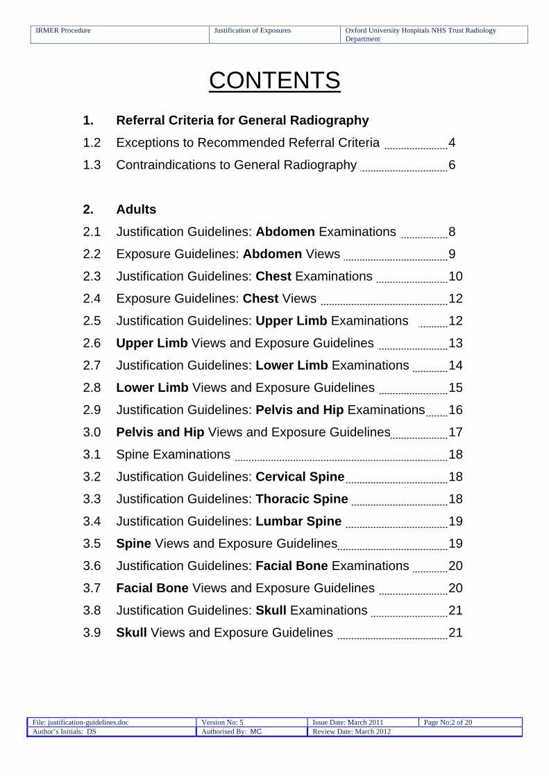
CONTENTS
1. Referral Criteria for General Radiography
1.2 Exceptions to Recommended Referral Criteria 4
1.3 Contraindications to General Radiography 6
2. Adults
2.1 Justification Guidelines: Abdomen Examinations 8
2.2 Exposure Guidelines: Abdomen Views 9
2.3 Justification Guidelines: Chest Examinations 10
2.4 Exposure Guidelines: Chest Views 12
2.5 Justification Guidelines: Upper Limb Examinations 12
2.6 Upper Limb Views and Exposure Guidelines 13
2.7 Justification Guidelines: Lower Limb Examinations 14
2.8 Lower Limb Views and Exposure Guidelines 15
2.9 Justification Guidelines: Pelvis and Hip Examinations 16
3.0 Pelvis and Hip Views and Exposure Guidelines 17
3.1 Spine Examinations 18
3.2 Justification Guidelines: Cervical Spine 18
3.3 Justification Guidelines: Thoracic Spine 18
3.4 Justification Guidelines: Lumbar Spine 19
3.5 Spine Views and Exposure Guidelines 19
3.6 Justification Guidelines: Facial Bone Examinations 20
3.7 Facial Bone Views and Exposure Guidelines 20
3.8 Justification Guidelines: Skull Examinations 21
3.9 Skull Views and Exposure Guidelines 21
IRMER Procedure Justification of Exposures Oxford University Hospitals NHS Trust Radiology Department
File: justification-guidelines.doc Version No: 5 Issue Date: March 2011 Page No:3 of 20 Author’s Initials: DS Authorised By: MC Review Date: March 2012
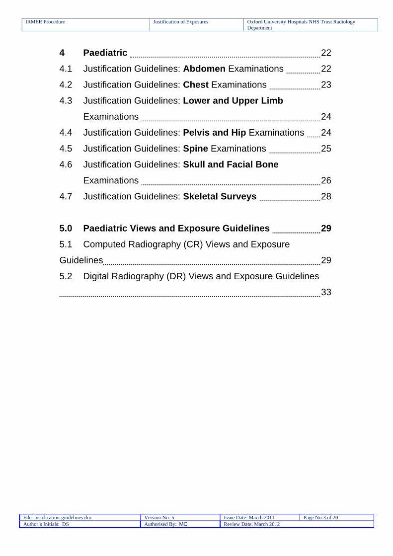
4 Paediatric 22
4.1 Justification Guidelines: Abdomen Examinations 22
4.2 Justification Guidelines: Chest Examinations 23
4.3 Justification Guidelines: Lower and Upper Limb
Examinations 24
4.4 Justification Guidelines: Pelvis and Hip Examinations 24
4.5 Justification Guidelines: Spine Examinations 25
4.6 Justification Guidelines: Skull and Facial Bone
Examinations 26
4.7 Justification Guidelines: Skeletal Surveys 28
5.0 Paediatric Views and Exposure Guidelines 29
5.1 Computed Radiography (CR) Views and Exposure
Guidelines 29
5.2 Digital Radiography (DR) Views and Exposure Guidelines
33

IRMER Procedure Justification of Exposures Oxford University Hospitals NHS Trust Radiology Department
File: justification-guidelines.doc Version No: 5 Issue Date: March 2011 Page No:4 of 20 Author’s Initials: DS Authorised By: MC Review Date: March 2012
1. Referral Criteria for General Radiography
Referral Criteria
Referral criteria will be based on the current version of Royal
College of Radiologists (RCR) booklet entitled “Making the best use of clinical radiology services” (Version 6.03, 2007), MBUR 6th
Edition.
These RCR recommendations are available on the Trust’s intranet on the ‘Radiology and PACS’ site.
1.2 Exceptions to recommended referral criteria
OUH referral criteria which deviates from the RCR Guidelines (version 6).
Referral Action Suggested
Examination Cardio-vascular / Thoracic System
Air entry decrease
Added to guidelines
CXR PA or AP
Anaphylactic reaction if pulmonary oedema suspected
Added to guidelines
CXR PA or AP
Aspiration Added to guidelines
CXR PA or AP
Chronic Cough Added to guidelines
CXR PA or AP
Cardiomegaly Added to guidelines
CXR PA or AP PA preferred to see enlargement of heart
Respiratory Tract Infection
Added to guidelines
CXR PA or AP
Tuberculosis Added to guidelines
CXR PA or AP
IRMER Procedure Justification of Exposures Oxford University Hospitals NHS Trust Radiology Department
File: justification-guidelines.doc Version No: 5 Issue Date: March 2011 Page No:5 of 20 Author’s Initials: DS Authorised By: MC Review Date: March 2012
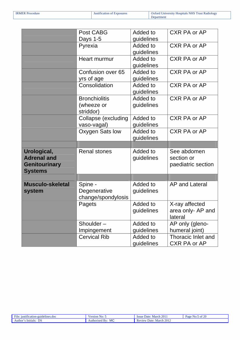
Post CABG Days 1-5
Added to guidelines
CXR PA or AP
Pyrexia Added to guidelines
CXR PA or AP
Heart murmur Added to guidelines
CXR PA or AP
Confusion over 65 yrs of age
Added to guidelines
CXR PA or AP
Consolidation Added to guidelines
CXR PA or AP
Bronchiolitis (wheeze or striddor)
Added to guidelines
CXR PA or AP
Collapse (excluding vaso-vagal)
Added to guidelines
CXR PA or AP
Oxygen Sats low Added to guidelines
CXR PA or AP
Urological, Adrenal and Genitourinary Systems
Renal stones Added to guidelines
See abdomen section or paediatric section
Musculo-skeletal system
Spine - Degenerative change/spondylosis
Added to guidelines
AP and Lateral
Pagets Added to guidelines
X-ray affected area only- AP and lateral
Shoulder – Impingement
Added to guidelines
AP only (gleno-humeral joint)
Cervical Rib Added to guidelines
Thoracic Inlet and CXR PA or AP

IRMER Procedure Justification of Exposures Oxford University Hospitals NHS Trust Radiology Department
File: justification-guidelines.doc Version No: 5 Issue Date: March 2011 Page No:6 of 20 Author’s Initials: DS Authorised By: MC Review Date: March 2012
1.3 Contraindications to General Radiography The following cannot be justified for general X-ray Clinical Problem Suggested Investigation
Musculo-Skeletal Heel pain: Suspected plantar fasciitis
NM, US, MRI
Chronic Back Pain: Unless osteoporotic collapse
MRI
Bony Metastases NM Soft tissue mass MRI Radiolucent Foreign Body US Rotator cuff shoulder US Severs Disease (heel pain with no history of trauma)
None. Clinical management only
Sternoclavicular joints CT Trauma 2nd to 5th toes: undisplaced fracture
None. Clinical management only
Coccyx # None. Clinical management onlyNasal Bones None. Clinical management onlyFractured Ribs None. Clinical management onlyC-spine injury over 65 years of age
CT
Gastrointestinal System Abdominal Aortic Aneurysm US, CT, MRI GI Bleed CTA Dysphagia/ Difficulty in Swallowing
Ba Swallow
Heartburn/ Hiatus Hernia Ba Swallow/Meal
IRMER Procedure Justification of Exposures Oxford University Hospitals NHS Trust Radiology Department
File: justification-guidelines.doc Version No: 5 Issue Date: March 2011 Page No:7 of 20 Author’s Initials: DS Authorised By: MC Review Date: March 2012
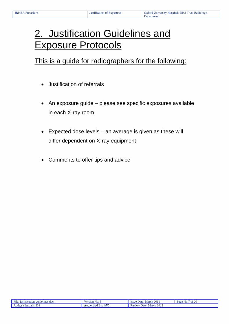
2. Justification Guidelines and Exposure Protocols
This is a guide for radiographers for the following:
Justification of referrals
An exposure guide – please see specific exposures available
in each X-ray room
Expected dose levels – an average is given as these will
differ dependent on X-ray equipment
Comments to offer tips and advice
IRMER Procedure Justification of Exposures Oxford University Hospitals NHS Trust Radiology Department
File: justification-guidelines.doc Version No: 5 Issue Date: March 2011 Page No:8 of 20 Author’s Initials: DS Authorised By: MC Review Date: March 2012
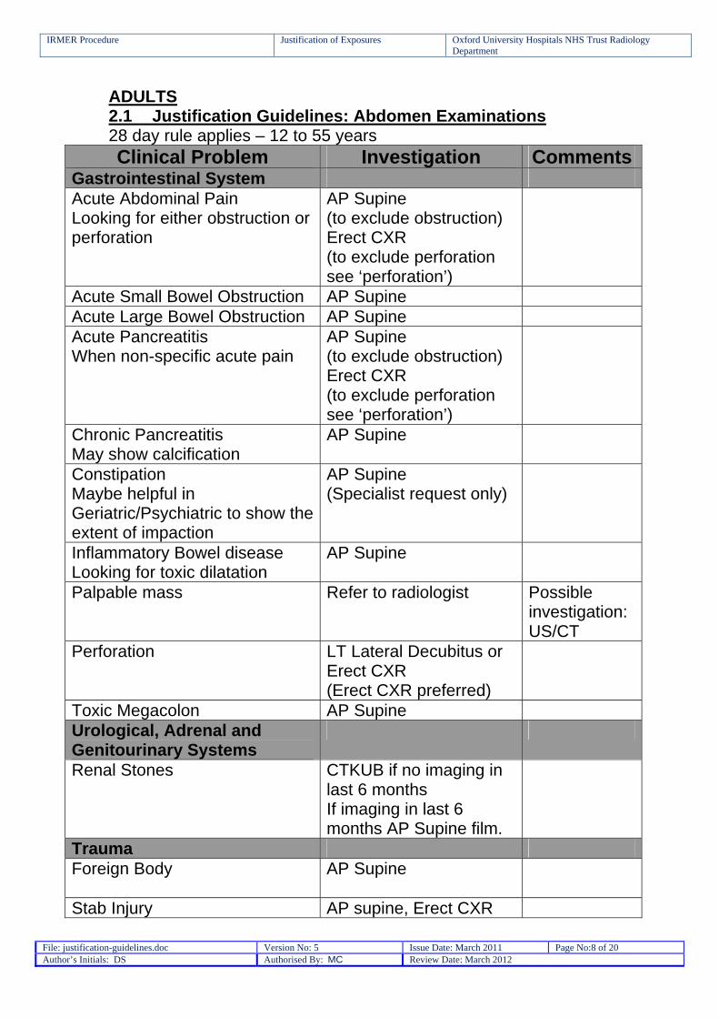
ADULTS 2.1 Justification Guidelines: Abdomen Examinations 28 day rule applies – 12 to 55 years Clinical Problem Investigation Comments
Gastrointestinal System Acute Abdominal Pain Looking for either obstruction or perforation
AP Supine (to exclude obstruction) Erect CXR (to exclude perforation see ‘perforation’)
Acute Small Bowel Obstruction AP Supine Acute Large Bowel Obstruction AP Supine Acute Pancreatitis When non-specific acute pain
AP Supine (to exclude obstruction) Erect CXR (to exclude perforation see ‘perforation’)
Chronic Pancreatitis May show calcification
AP Supine
Constipation Maybe helpful in Geriatric/Psychiatric to show the extent of impaction
AP Supine (Specialist request only)
Inflammatory Bowel disease Looking for toxic dilatation
AP Supine
Palpable mass Refer to radiologist Possible investigation:US/CT
Perforation LT Lateral Decubitus or Erect CXR (Erect CXR preferred)
Toxic Megacolon AP Supine Urological, Adrenal and Genitourinary Systems
Renal Stones CTKUB if no imaging in last 6 months If imaging in last 6 months AP Supine film.
Trauma Foreign Body AP Supine
Stab Injury AP supine, Erect CXR
IRMER Procedure Justification of Exposures Oxford University Hospitals NHS Trust Radiology Department
File: justification-guidelines.doc Version No: 5 Issue Date: March 2011 Page No:9 of 20 Author’s Initials: DS Authorised By: MC Review Date: March 2012
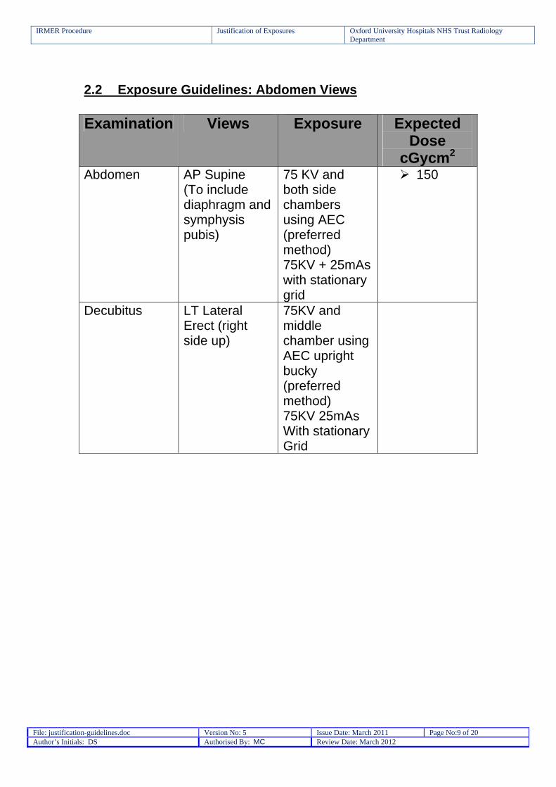
2.2 Exposure Guidelines: Abdomen Views Examination Views Exposure Expected
Dose cGycm2
Abdomen AP Supine (To include diaphragm and symphysis pubis)
75 KV and both side chambers using AEC (preferred method) 75KV + 25mAs with stationary grid
150
Decubitus LT Lateral Erect (right side up)
75KV and middle chamber using AEC upright bucky (preferred method) 75KV 25mAs With stationary Grid
IRMER Procedure Justification of Exposures Oxford University Hospitals NHS Trust Radiology Department
File: justification-guidelines.doc Version No: 5 Issue Date: March 2011 Page No:10 of 20 Author’s Initials: DS Authorised By: MC Review Date: March 2012
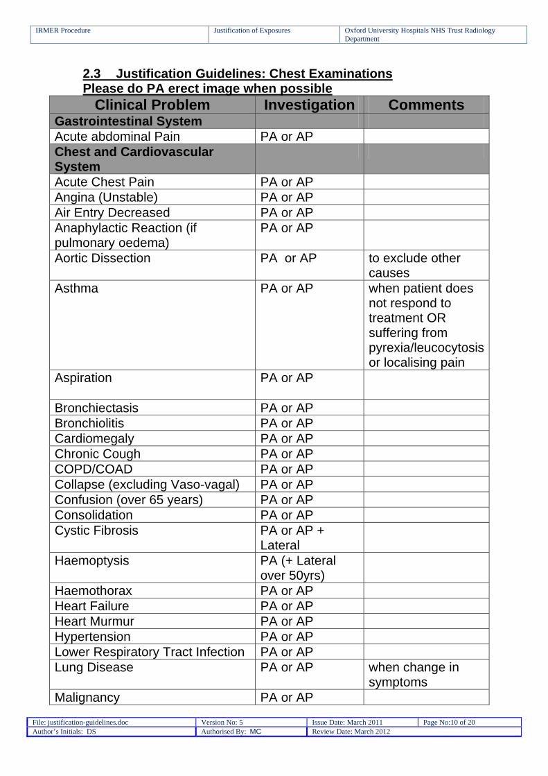
2.3 Justification Guidelines: Chest Examinations Please do PA erect image when possible
Clinical Problem Investigation Comments Gastrointestinal System Acute abdominal Pain PA or AP Chest and Cardiovascular System
Acute Chest Pain PA or AP Angina (Unstable) PA or AP Air Entry Decreased PA or AP Anaphylactic Reaction (if pulmonary oedema)
PA or AP
Aortic Dissection PA or AP
to exclude other causes
Asthma PA or AP
when patient does not respond to treatment OR suffering from pyrexia/leucocytosis or localising pain
Aspiration
PA or AP
Bronchiectasis PA or AP Bronchiolitis PA or AP Cardiomegaly PA or AP Chronic Cough PA or AP COPD/COAD PA or AP Collapse (excluding Vaso-vagal) PA or AP Confusion (over 65 years) PA or AP Consolidation PA or AP Cystic Fibrosis PA or AP +
Lateral
Haemoptysis PA (+ Lateral over 50yrs)
Haemothorax PA or AP Heart Failure PA or AP Heart Murmur PA or AP Hypertension PA or AP Lower Respiratory Tract Infection PA or AP Lung Disease PA or AP
when change in symptoms
Malignancy PA or AP
IRMER Procedure Justification of Exposures Oxford University Hospitals NHS Trust Radiology Department
File: justification-guidelines.doc Version No: 5 Issue Date: March 2011 Page No:11 of 20 Author’s Initials: DS Authorised By: MC Review Date: March 2012
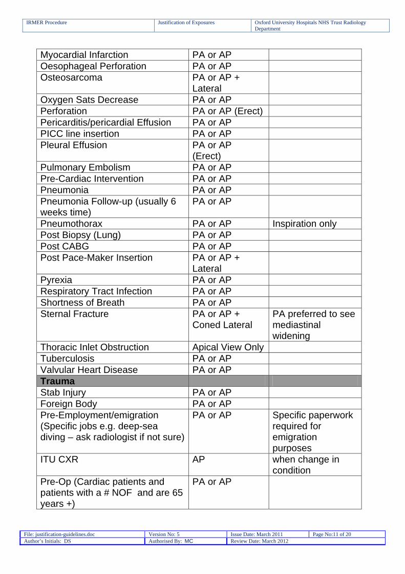
Myocardial Infarction PA or AP Oesophageal Perforation PA or AP Osteosarcoma PA or AP +
Lateral
Oxygen Sats Decrease PA or AP Perforation PA or AP (Erect) Pericarditis/pericardial Effusion PA or AP PICC line insertion PA or AP Pleural Effusion PA or AP
(Erect)
Pulmonary Embolism PA or AP Pre-Cardiac Intervention PA or AP Pneumonia PA or AP Pneumonia Follow-up (usually 6 weeks time)
PA or AP
Pneumothorax PA or AP Inspiration only Post Biopsy (Lung) PA or AP Post CABG PA or AP Post Pace-Maker Insertion PA or AP +
Lateral
Pyrexia PA or AP Respiratory Tract Infection PA or AP Shortness of Breath PA or AP Sternal Fracture PA or AP +
Coned Lateral PA preferred to see mediastinal widening
Thoracic Inlet Obstruction Apical View Only Tuberculosis PA or AP Valvular Heart Disease PA or AP Trauma Stab Injury PA or AP Foreign Body PA or AP Pre-Employment/emigration (Specific jobs e.g. deep-sea diving – ask radiologist if not sure)
PA or AP Specific paperwork required for emigration purposes
ITU CXR AP
when change in condition
Pre-Op (Cardiac patients and patients with a # NOF and are 65 years +)
PA or AP

IRMER Procedure Justification of Exposures Oxford University Hospitals NHS Trust Radiology Department
File: justification-guidelines.doc Version No: 5 Issue Date: March 2011 Page No:12 of 20 Author’s Initials: DS Authorised By: MC Review Date: March 2012
2.4 Exposure Guidelines: Chest Views Please refer to specific room settings Examination Views Exposure Expected
Dose cGycm2
Chest PA FFD = 150cm 150kV + 2.5mAs (use Airgap)
< 5
Chest AP FFD = 100cm 85kV + 2.5mAs
< 10
Chest Lateral FFD = 120cm 150kV + 10mAs (use airgap)
< 20
2.5 Justification Guidelines: Upper Limb Examinations Refer to Views and Exposure Guidelines for Specific Investigation Clinical Problem Investigation Comments
Musculo-skeletal System Arthropathy AP (affected area only) Bony Mass/Primary Bone Tumour
AP + Lateral
for all cases of unresolved bone pain
Bone Pain AP + Lateral Diabetes – Hands Only DP Osteomalacia AP + Lateral Osteomyelitis AP + Lateral Painful Prosthesis AP + Lateral Pagets AP + Lateral (affected
area only)
Trauma Trauma AP + Lateral Trauma Follow-up (e.g. post manipulation/reduction)
AP + Lateral
Stress Fracture AP + Lateral Subluxation AP + Lateral Dislocation AP + Lateral Foreign Body (Radio-opaque only)
AP and Lateral and tangential view of affected area.
Use marker to indicate site/wound

IRMER Procedure Justification of Exposures Oxford University Hospitals NHS Trust Radiology Department
File: justification-guidelines.doc Version No: 5 Issue Date: March 2011 Page No:13 of 20 Author’s Initials: DS Authorised By: MC Review Date: March 2012
Remove dressings
Foreign Body ? bony involvement (Radio-opaque only)
AP and Lateral view of Object
2.6 Upper Limb Views and Exposure Guidelines Please refer to specific room settings
Examination Views Exposure Expected Dose
cGycm2 Fingers DP, Lateral
(45° Oblique for MCPJ)
50-52kV + 1.4-1.6mAs
< 2
Hand DP + Oblique (lateral if #’d MC)
52-55kV + 1.6mAs (60Kv + 2mAs for lateral)
< 3
Thumb AP + Lateral 50-52kV + 1.4-1.6mAs
< 2
Scaphoid DP, Lateral, Oblique, 25° Axial
52-55kV + 1.5mAs – 2mAs
< 3
Wrist DP + Lateral DP = 55kV + 2mAs Lateral = 56kV + 2mAs
< 4
Forearm AP + Lateral 55kV + 2.5mAs < 5 (for both views)
Elbow AP + Lateral 60kV + 2mAs < 2 Humerus AP + Lateral 65kV + 3.2mAs < 10 (for both
views) Shoulder (Trauma) AP+
Axial/modified axial (Lateral for proximal humerus)
AP: 64.5kV + 4mAs Lateral/axial: 75kV + 3.2mAs
< 4
< 8
Shoulder Joint (Trauma) (post manipulation and follow-up)
AP Oblique (45° to view gleno-humeral joint) + Axial/modified axial –see
AP: As above Axial: 75kV + 3.2mAs
< 6
IRMER Procedure Justification of Exposures Oxford University Hospitals NHS Trust Radiology Department
File: justification-guidelines.doc Version No: 5 Issue Date: March 2011 Page No:14 of 20 Author’s Initials: DS Authorised By: MC Review Date: March 2012
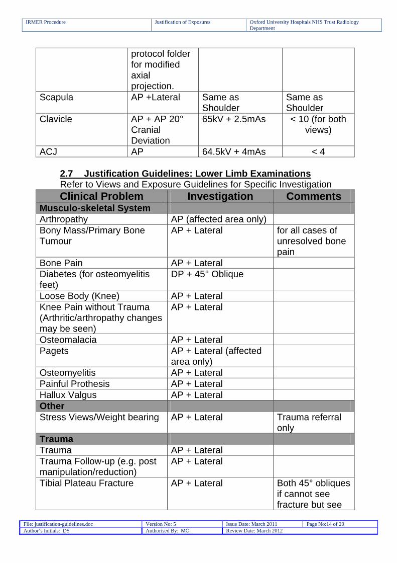
protocol folder for modified axial projection.
Scapula AP +Lateral Same as Shoulder
Same as Shoulder
Clavicle AP + AP 20° Cranial Deviation
65kV + 2.5mAs < 10 (for both views)
ACJ AP 64.5kV + 4mAs < 4 2.7 Justification Guidelines: Lower Limb Examinations Refer to Views and Exposure Guidelines for Specific Investigation Clinical Problem Investigation Comments
Musculo-skeletal System Arthropathy AP (affected area only) Bony Mass/Primary Bone Tumour
AP + Lateral
for all cases of unresolved bone pain
Bone Pain AP + Lateral Diabetes (for osteomyelitis feet)
DP + 45° Oblique
Loose Body (Knee) AP + Lateral Knee Pain without Trauma (Arthritic/arthropathy changes may be seen)
AP + Lateral
Osteomalacia AP + Lateral Pagets AP + Lateral (affected
area only)
Osteomyelitis AP + Lateral Painful Prothesis AP + Lateral Hallux Valgus AP + Lateral Other Stress Views/Weight bearing AP + Lateral Trauma referral
only Trauma Trauma AP + Lateral Trauma Follow-up (e.g. post manipulation/reduction)
AP + Lateral
Tibial Plateau Fracture AP + Lateral
Both 45° obliques if cannot see fracture but see
IRMER Procedure Justification of Exposures Oxford University Hospitals NHS Trust Radiology Department
File: justification-guidelines.doc Version No: 5 Issue Date: March 2011 Page No:15 of 20 Author’s Initials: DS Authorised By: MC Review Date: March 2012
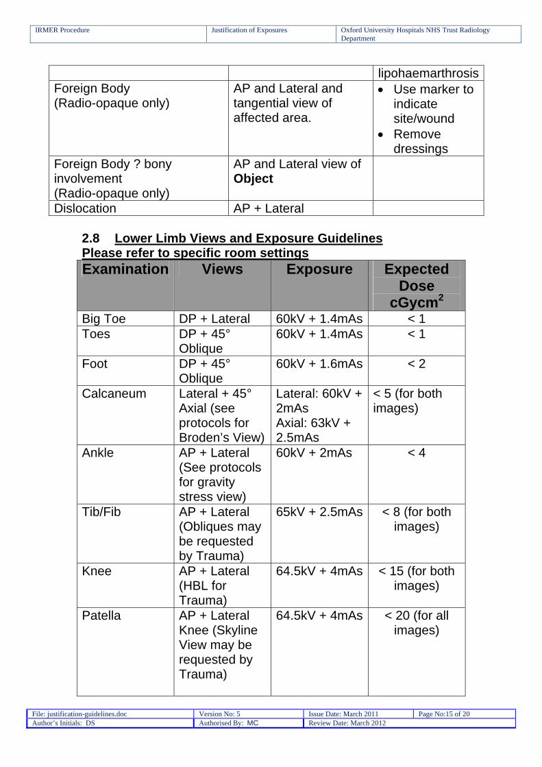
lipohaemarthrosisForeign Body (Radio-opaque only)
AP and Lateral and tangential view of affected area.
Use marker to indicate site/wound
Remove dressings
Foreign Body ? bony involvement (Radio-opaque only)
AP and Lateral view of Object
Dislocation AP + Lateral 2.8 Lower Limb Views and Exposure Guidelines Please refer to specific room settings Examination Views Exposure Expected
Dose cGycm2
Big Toe DP + Lateral 60kV + 1.4mAs < 1 Toes DP + 45°
Oblique 60kV + 1.4mAs < 1
Foot DP + 45° Oblique
60kV + 1.6mAs < 2
Calcaneum Lateral + 45° Axial (see protocols for Broden’s View)
Lateral: 60kV + 2mAs Axial: 63kV + 2.5mAs
< 5 (for both images)
Ankle AP + Lateral (See protocols for gravity stress view)
60kV + 2mAs < 4
Tib/Fib AP + Lateral (Obliques may be requested by Trauma)
65kV + 2.5mAs < 8 (for both images)
Knee AP + Lateral (HBL for Trauma)
64.5kV + 4mAs < 15 (for both images)
Patella AP + Lateral Knee (Skyline View may be requested by Trauma)
64.5kV + 4mAs < 20 (for all images)
IRMER Procedure Justification of Exposures Oxford University Hospitals NHS Trust Radiology Department
File: justification-guidelines.doc Version No: 5 Issue Date: March 2011 Page No:16 of 20 Author’s Initials: DS Authorised By: MC Review Date: March 2012
Femur AP + Lateral 70kV + AEC centre chamber (16mAs with Grid)
< 15
2.9 Justification Guidelines: Pelvis and Hip Examinations 28 day rule applies – 12 to 55 years Clinical Problem Investigation Comments
Musculo-skeletal System Arthropathy AP Pelvis Avascular Necrosis AP Pelvis Bone Pain AP Pelvis and Lateral Hip Pain AP Pelvis + Lateral Osteomyelitis AP + Lateral Osteomalacia AP + Lateral Painful Prosthesis AP + Lateral Post op – THR, ETS (All prosthesis must be included; DHS patients should have had X-rays in theatre)
AP Pelvis (Top of cassette at ASIS for hips) + HBL Lateral
Primary Bone Tumour AP + Lateral Sacroiliac Pain AP Pelvis Pagets AP Pelvis(affected area
only)
Trauma Trauma AP Pelvis + (HBL
Lateral for Hip, Judet views for acetabular)
Trauma Follow-up (Post reduction)
AP + Lateral
Acetabular Fixation/Fracture Judet Views Fall AP Pelvis + HBL Lateral Injury to pelvic ring Inlet and Outlet Trauma referral
only
IRMER Procedure Justification of Exposures Oxford University Hospitals NHS Trust Radiology Department
File: justification-guidelines.doc Version No: 5 Issue Date: March 2011 Page No:17 of 20 Author’s Initials: DS Authorised By: MC Review Date: March 2012
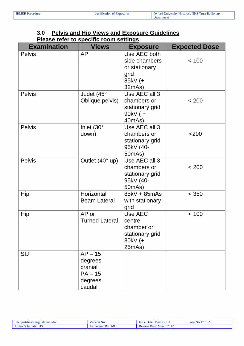
3.0 Pelvis and Hip Views and Exposure Guidelines Please refer to specific room settings
Examination Views Exposure Expected Dose Pelvis AP Use AEC both
side chambers or stationary grid 85kV (+ 32mAs)
< 100
Pelvis Judet (45° Oblique pelvis)
Use AEC all 3 chambers or stationary grid 90kV ( + 40mAs)
< 200
Pelvis Inlet (30° down)
Use AEC all 3 chambers or stationary grid 95kV (40-50mAs)
<200
Pelvis Outlet (40° up) Use AEC all 3 chambers or stationary grid 95kV (40-50mAs)
< 200
Hip Horizontal Beam Lateral
85kV + 85mAs with stationary grid
< 350
Hip AP or Turned Lateral
Use AEC centre chamber or stationary grid 80kV (+ 25mAs)
< 100
SIJ AP – 15 degrees cranial PA – 15 degrees caudal

IRMER Procedure Justification of Exposures Oxford University Hospitals NHS Trust Radiology Department
File: justification-guidelines.doc Version No: 5 Issue Date: March 2011 Page No:18 of 20 Author’s Initials: DS Authorised By: MC Review Date: March 2012
3.1 Spine Examinations 3.2 Justification Guidelines: Cervical Spine Clinical Problem Investigation Comments
Musculo-Skeletal System Atlanto-Axial Subluxation (To identify congenital or structural abnormalities)
Lateral
Atlanto-occipital Subluxation Lateral Brachialgia Refer to radiologist MRI Degenerative change/spondylosis
AP + Lateral
Nerve Compression Refer to radiologist MRI Trauma Suspected Ligamentous Injury
Flexion + Extension (movement undertaken by referrer)
Trauma referral only
Trauma AP, Peg, Lateral – swimmers if C7/T1 is not visualised
Unconscious Trauma Refer to radiologist CT Foreign Body Lateral or tangential
Views (dependent on location)
Neck Pain/Injury with Neurological Deficit
AP, Peg, Lateral -swimmers if C7/T1 is not visualised
CT If patient over 65 years of age
3.3 Justification Guidelines: Thoracic Spine Clinical Problem Investigation Comments
Musculo-Skeletal System Degenerative change/spondylosis
AP + Lateral
Osteoporotic Collapse Lateral Spondyloarthropathies AP + Lateral Trauma Trauma AP + Lateral Trauma with neurological deficit
AP + Lateral
IRMER Procedure Justification of Exposures Oxford University Hospitals NHS Trust Radiology Department
File: justification-guidelines.doc Version No: 5 Issue Date: March 2011 Page No:19 of 20 Author’s Initials: DS Authorised By: MC Review Date: March 2012
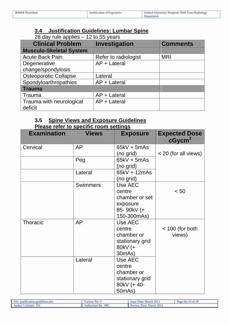
3.4 Justification Guidelines: Lumbar Spine 28 day rule applies – 12 to 55 years Clinical Problem Investigation Comments
Musculo-Skeletal System Acute Back Pain Refer to radiologist MRI Degenerative change/spondylosis
AP + Lateral
Osteoporotic Collapse Lateral Spondyloarthropathies AP + Lateral Trauma Trauma AP + Lateral Trauma with neurological deficit
AP + Lateral
3.5 Spine Views and Exposure Guidelines Please refer to specific room settings
Examination Views Exposure Expected Dose cGycm2
Cervical AP 65kV + 5mAs (no grid)
Peg 65kV + 5mAs (no grid)
Lateral 65kV + 12mAs (no grid)
< 20 (for all views)
Swimmers Use AEC centre chamber or set exposure 85- 90kV (+ 150-300mAs)
< 50
Thoracic AP Use AEC centre chamber or stationary grid 80kV (+ 30mAs)
Lateral Use AEC centre chamber or stationary grid 80kV (+ 40-50mAs)
< 100 (for both
views)

IRMER Procedure Justification of Exposures Oxford University Hospitals NHS Trust Radiology Department
File: justification-guidelines.doc Version No: 5 Issue Date: March 2011 Page No:20 of 20 Author’s Initials: DS Authorised By: MC Review Date: March 2012
Lumbar AP Use AEC centre chamber or stationary grid 90kV (+ 40 mAs)
Lateral Use AEC centre chamber or stationary grid 95kV(+50mAs)
< 300 (for both views)
3.6 Justification Guidelines: Facial Bone Examinations Clinical Problem Investigation Comments
Trauma Blunt Injury OM +OM 30 Middle Third of Face OM +OM 30 Mandibular Trauma OPG + PA Mandible Dislocation OPG + PA Mandible Subluxation of TMJ OPG Foreign Body Tangential Views Orbits Orbit Views
ENT/Head and Neck Abscess OPG Dental Reasons OPG Impacted 8’S OPG Other Pre-Op valve replacement ? tooth decay
OPG
3.7 Facial Bone Views and Exposure Guidelines Please refer to specific room settings Examination Views Exposure Expected
Dose cGycm2
Facial Bones OM Use AEC centre chamber or skull unit 85kV (+ 12mAs)
< 20
IRMER Procedure Justification of Exposures Oxford University Hospitals NHS Trust Radiology Department
File: justification-guidelines.doc Version No: 5 Issue Date: March 2011 Page No:21 of 20 Author’s Initials: DS Authorised By: MC Review Date: March 2012
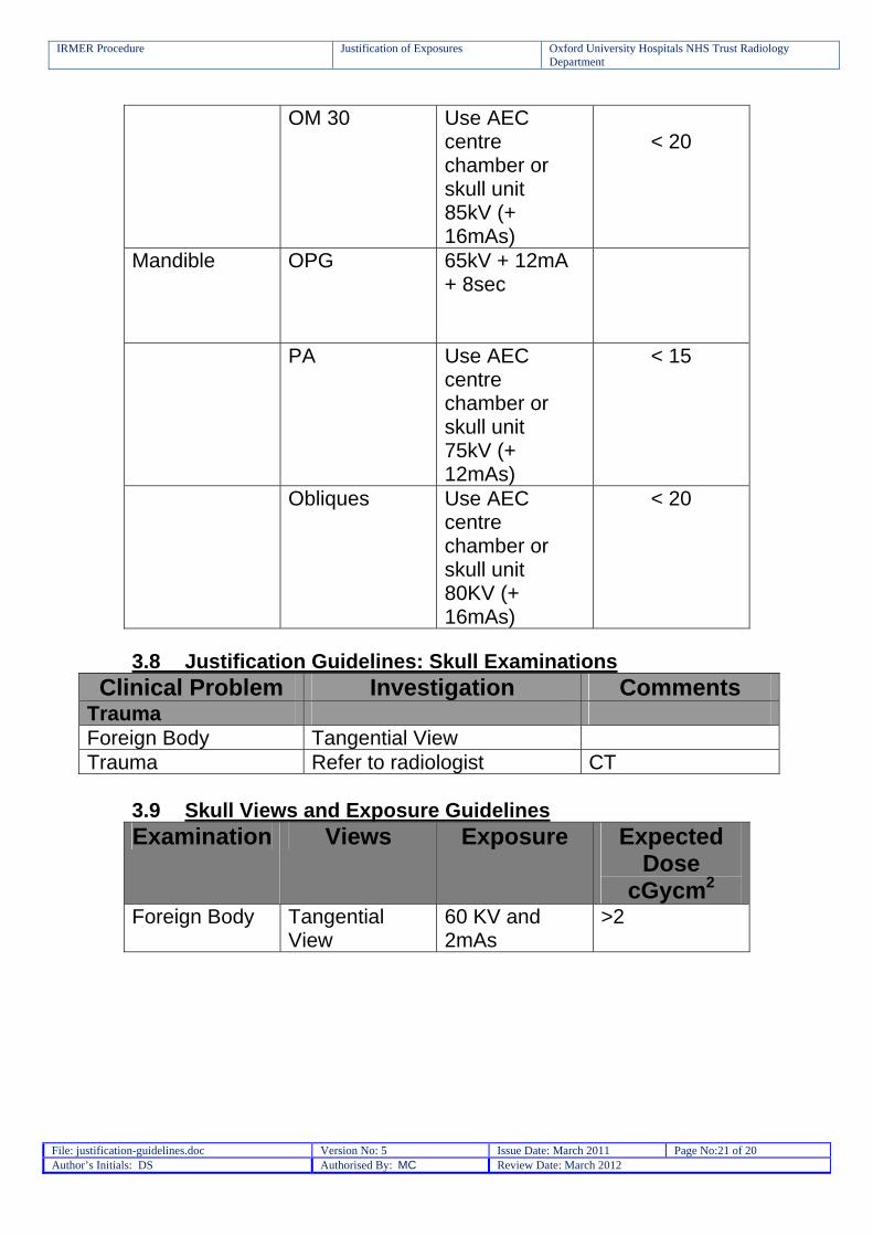
OM 30 Use AEC centre chamber or skull unit 85kV (+ 16mAs)
< 20
Mandible OPG 65kV + 12mA + 8sec
PA Use AEC centre chamber or skull unit 75kV (+ 12mAs)
< 15
Obliques Use AEC centre chamber or skull unit 80KV (+ 16mAs)
< 20
3.8 Justification Guidelines: Skull Examinations
Clinical Problem Investigation Comments Trauma Foreign Body Tangential View Trauma Refer to radiologist CT
3.9 Skull Views and Exposure Guidelines Examination Views Exposure Expected
Dose cGycm2
Foreign Body Tangential View
60 KV and 2mAs
>2
IRMER Procedure Justification of Exposures Oxford University Hospitals NHS Trust Radiology Department
File: justification-guidelines.doc Version No: 5 Issue Date: March 2011 Page No:22 of 20 Author’s Initials: DS Authorised By: MC Review Date: March 2012
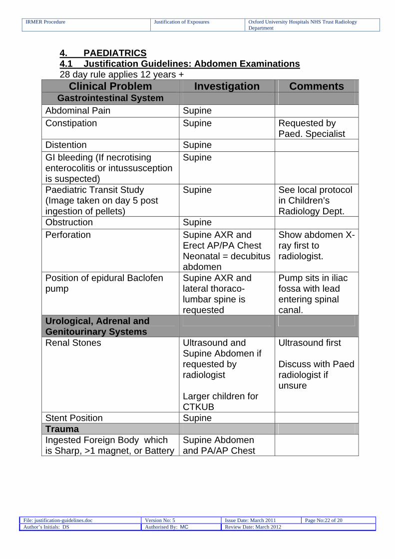
4. PAEDIATRICS 4.1 Justification Guidelines: Abdomen Examinations 28 day rule applies 12 years +
Clinical Problem Investigation Comments Gastrointestinal System
Abdominal Pain Supine Constipation Supine Requested by
Paed. Specialist Distention Supine GI bleeding (If necrotising enterocolitis or intussusception is suspected)
Supine
Paediatric Transit Study (Image taken on day 5 post ingestion of pellets)
Supine See local protocol in Children’s Radiology Dept.
Obstruction Supine Perforation Supine AXR and
Erect AP/PA Chest Neonatal = decubitus abdomen
Show abdomen X-ray first to radiologist.
Position of epidural Baclofen pump
Supine AXR and lateral thoraco-lumbar spine is requested
Pump sits in iliac fossa with lead entering spinal canal.
Urological, Adrenal and Genitourinary Systems
Renal Stones Ultrasound and Supine Abdomen if requested by radiologist Larger children for CTKUB
Ultrasound first Discuss with Paed radiologist if unsure
Stent Position Supine Trauma Ingested Foreign Body which is Sharp, >1 magnet, or Battery
Supine Abdomen and PA/AP Chest
IRMER Procedure Justification of Exposures Oxford University Hospitals NHS Trust Radiology Department
File: justification-guidelines.doc Version No: 5 Issue Date: March 2011 Page No:23 of 20 Author’s Initials: DS Authorised By: MC Review Date: March 2012
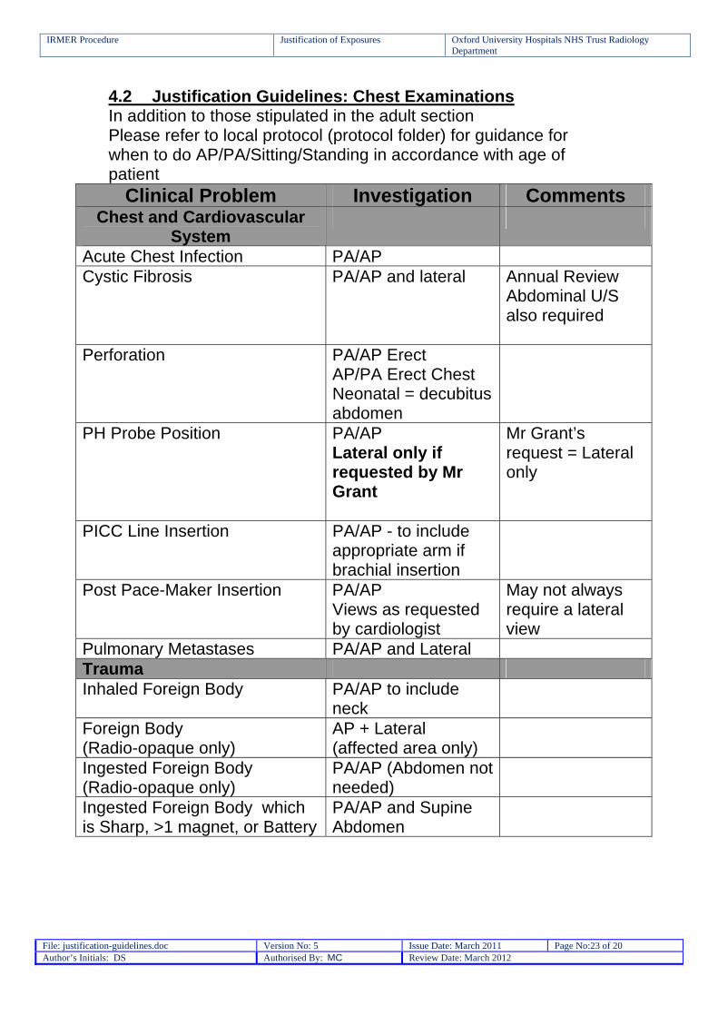
4.2 Justification Guidelines: Chest Examinations In addition to those stipulated in the adult section Please refer to local protocol (protocol folder) for guidance for when to do AP/PA/Sitting/Standing in accordance with age of patient
Clinical Problem Investigation Comments Chest and Cardiovascular
System
Acute Chest Infection PA/AP Cystic Fibrosis PA/AP and lateral Annual Review
Abdominal U/S also required
Perforation PA/AP Erect AP/PA Erect Chest Neonatal = decubitus abdomen
PH Probe Position PA/AP Lateral only if requested by Mr Grant
Mr Grant’s request = Lateral only
PICC Line Insertion PA/AP - to include appropriate arm if brachial insertion
Post Pace-Maker Insertion PA/AP Views as requested by cardiologist
May not always require a lateral view
Pulmonary Metastases PA/AP and Lateral Trauma Inhaled Foreign Body PA/AP to include
neck
Foreign Body (Radio-opaque only)
AP + Lateral (affected area only)
Ingested Foreign Body (Radio-opaque only)
PA/AP (Abdomen not needed)
Ingested Foreign Body which is Sharp, >1 magnet, or Battery
PA/AP and Supine Abdomen
IRMER Procedure Justification of Exposures Oxford University Hospitals NHS Trust Radiology Department
File: justification-guidelines.doc Version No: 5 Issue Date: March 2011 Page No:24 of 20 Author’s Initials: DS Authorised By: MC Review Date: March 2012
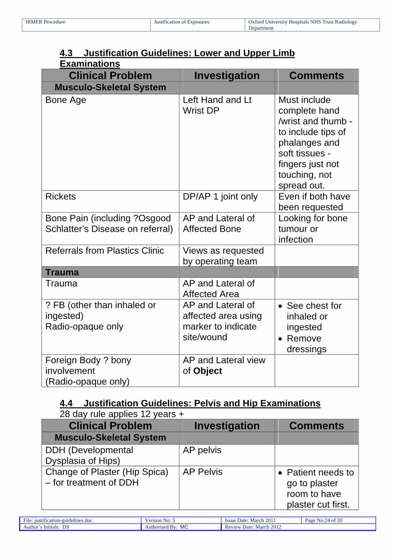
4.3 Justification Guidelines: Lower and Upper Limb Examinations
Clinical Problem Investigation Comments Musculo-Skeletal System
Bone Age Left Hand and Lt Wrist DP
Must include complete hand /wrist and thumb - to include tips of phalanges and soft tissues -fingers just not touching, not spread out.
Rickets DP/AP 1 joint only Even if both have been requested
Bone Pain (including ?Osgood Schlatter’s Disease on referral)
AP and Lateral of Affected Bone
Looking for bone tumour or infection
Referrals from Plastics Clinic Views as requested by operating team
Trauma Trauma AP and Lateral of
Affected Area
? FB (other than inhaled or ingested) Radio-opaque only
AP and Lateral of affected area using marker to indicate site/wound
See chest for inhaled or ingested
Remove dressings
Foreign Body ? bony involvement (Radio-opaque only)
AP and Lateral view of Object
4.4 Justification Guidelines: Pelvis and Hip Examinations 28 day rule applies 12 years +
Clinical Problem Investigation Comments Musculo-Skeletal System
DDH (Developmental Dysplasia of Hips)
AP pelvis
Change of Plaster (Hip Spica) – for treatment of DDH
AP Pelvis Patient needs to go to plaster room to have plaster cut first.
IRMER Procedure Justification of Exposures Oxford University Hospitals NHS Trust Radiology Department
File: justification-guidelines.doc Version No: 5 Issue Date: March 2011 Page No:25 of 20 Author’s Initials: DS Authorised By: MC Review Date: March 2012
Remove top section and X-ray child whilst still in posterior section of cast.
Child needs to be immobilised in cast for X-ray.
Replace anterior section and bandage in place for transfer back to ward.
Limping Child-request to X-ray whole leg
AP Pelvis and AP and lateral limb bones as directed by clinical team
Gonad protection not to be used on 1st image but should be used on subsequent imaging.
Limping Child ?Irritable Hip AP pelvis if requested by radiologist
U/S first
Perthes/Avascular necrosis Frog Legs Lateral only
SUFE (Slipped Upper Femoral Epiphysis) – Approx. age 10-16 yrs
Frog Legs Lateral only
Trauma Trauma AP pelvis and HBL
lateral
4.5 Justification Guidelines: Spine Examinations 28 day rule applies for L-Spine 12 years +
Clinical Problem Investigation Comments Musculo-Skeletal System
Post Scoliosis Repair
AP and Lateral Thoracic and Lumbar Spine Standing AP and Lateral views may be requested
Images must overlap and include whole T and L Spine Images may be requested whilst
IRMER Procedure Justification of Exposures Oxford University Hospitals NHS Trust Radiology Department
File: justification-guidelines.doc Version No: 5 Issue Date: March 2011 Page No:26 of 20 Author’s Initials: DS Authorised By: MC Review Date: March 2012
patient is sitting in their own wheel chair
Spinal vertebral Anomalies AP and Lateral Lumbar/Sacral Spine
Constipation with suspected underlying spinal cause
AP Lumbar/scaral Spine Review with Pead. Radiologist as a lateral may also be required
Vertebral anomaly may affect nerve supply to bowel hence causing constipation. Often can’t see on AP due to constipation but this is view of choice.
Chronic Back Pain Refer to Radiologist Spondylolisthesis Lateral
Lumbar/Sacral Spine and review with Paed. Radiologist
Often presents in sporty children
C-Spine Instability/Subluxation As requested May need Flexion and Extension Views. A Lateral may suffice
Must be performed in presence of referring clinician
Trauma Trauma AP and Lateral of
Effected Area Peg view for C-spine injury
4.6 Justification Guidelines: Skull and Facial Bone Examinations
Clinical Problem Investigation Comments Musculo-Skeletal System
Craniosynostosis (premature fusing of sutures)
AP, Townes and Lateral
With copper ruler on edge of image
Post Cranio-Facial Surgery. Frontal Advancement
Views as requested by cranio-facial team.
May ask for both laterals
ENT/Head and Neck Cochlear Implants Coned AP Centre through
EAMs
IRMER Procedure Justification of Exposures Oxford University Hospitals NHS Trust Radiology Department
File: justification-guidelines.doc Version No: 5 Issue Date: March 2011 Page No:27 of 20 Author’s Initials: DS Authorised By: MC Review Date: March 2012
Only need to see position of leads in cochlea
No need to include the external component attached to head
Most patients have bilateral
Post Nasal Space for enlarged adenoids
Lateral Face Ideally with “Sniffing In” Collimate to avoid eyes
Shunt Insertion – to show position of Ventroperitoneal (VP)Shunt
VP Shunt Series: Lateral Skull to include neck PA/AP chest to include lower neck Supine Abdomen to include lung bases to symphysis pubis
VP shunt drains from ventricles in the brain into the peritoneum Treatment for hydrocephalus Important to get overlap of the images to ensure that there are no breaks in the shunt
Trauma Facial Trauma OM and OM30° If unsure speak to
Consultant Paediatric
Head Trauma (18mths and under)
AP/PA and Lateral, (even if CT requested)
If unsure speak to Consultant Paediatric Radiologist
IRMER Procedure Justification of Exposures Oxford University Hospitals NHS Trust Radiology Department
File: justification-guidelines.doc Version No: 5 Issue Date: March 2011 Page No:28 of 20 Author’s Initials: DS Authorised By: MC Review Date: March 2012
4.7 Justification Guidelines: Skeletal Surveys
NAI Must be discussed with Paediatric Consultant Radiologist
Abdomen to include Pelvis
Chest to include all ribs
Oblique Ribs to include all ribs
Lateral C-spine Lateral thoraco-
lumbar Spine Skull AP and
Lateral – lateral to include mandible
Separate AP views on both:
Feet Femurs Tib/Fib Humeri Rad/Ulna Hands
Additional views as directed by Consultant Paediatric Radiologist
Please see specific folder in Paediatric Hospital or Level One For live children
arrange a mutually convenient time with the patient’s nurse and the radiologist
Make sure the patient has had a good feed and/or sleep and comes to the dept with a dummy if they have one
2 people will be required to immobilise the patient. Ensure that neither are pregnant before they come to the dept
If parent is assisting please ensure they know why the examination is being carried out before arrival to the X-ray Dept
General As directed by radiologist Protocol for each
individual patient

IRMER Procedure Justification of Exposures Oxford University Hospitals NHS Trust Radiology Department
File: justification-guidelines.doc Version No: 5 Issue Date: March 2011 Page No:29 of 20 Author’s Initials: DS Authorised By: MC Review Date: March 2012
5.0 Paediatric Views and Exposure Guidelines 5.1 Computed Radiography (CR) Views and Exposure
Guidelines Please refer to specific room settings
Examinations Views Exposure Expected Dose
cGycm2 Based on CR method
Chest Chest 0 - 6 months
Supine 60 – 63kV + 1 - 2mAs 180 FFD
1
Chest 6 months – 5 years
AP Sitting 65kV + 1.6 – 3.2mAs 180cm FFD
1-2
Chest 5 years + AP/PA Standing
65-77KV + 2 – 3.2 180cm FFD
1-5
Lateral Chest < 5 years
Lateral Sitting or Standing
70KV + 3.2mAs 180cm FFD
3
Lateral Chest > 5 years
Lateral Sitting or Standing
73KV + 4-5mAs 180cm FFD
5-8
Abdomen/Pelvis Abdomen or Pelvis Baby
Supine 60KV + 1-2 mAs 100cm FFD
1 -2
Abdomen or Pelvis 1- 10 years
Supine No Grid 65kV -75KV + 2-10 mAs 100cm FFD
2-11
Abdomen or Pelvis 10 + years
Supine Use AEC, both side chambers 75kV (+16-25mAs with stationary grid) 100cm FFD
<150
IRMER Procedure Justification of Exposures Oxford University Hospitals NHS Trust Radiology Department
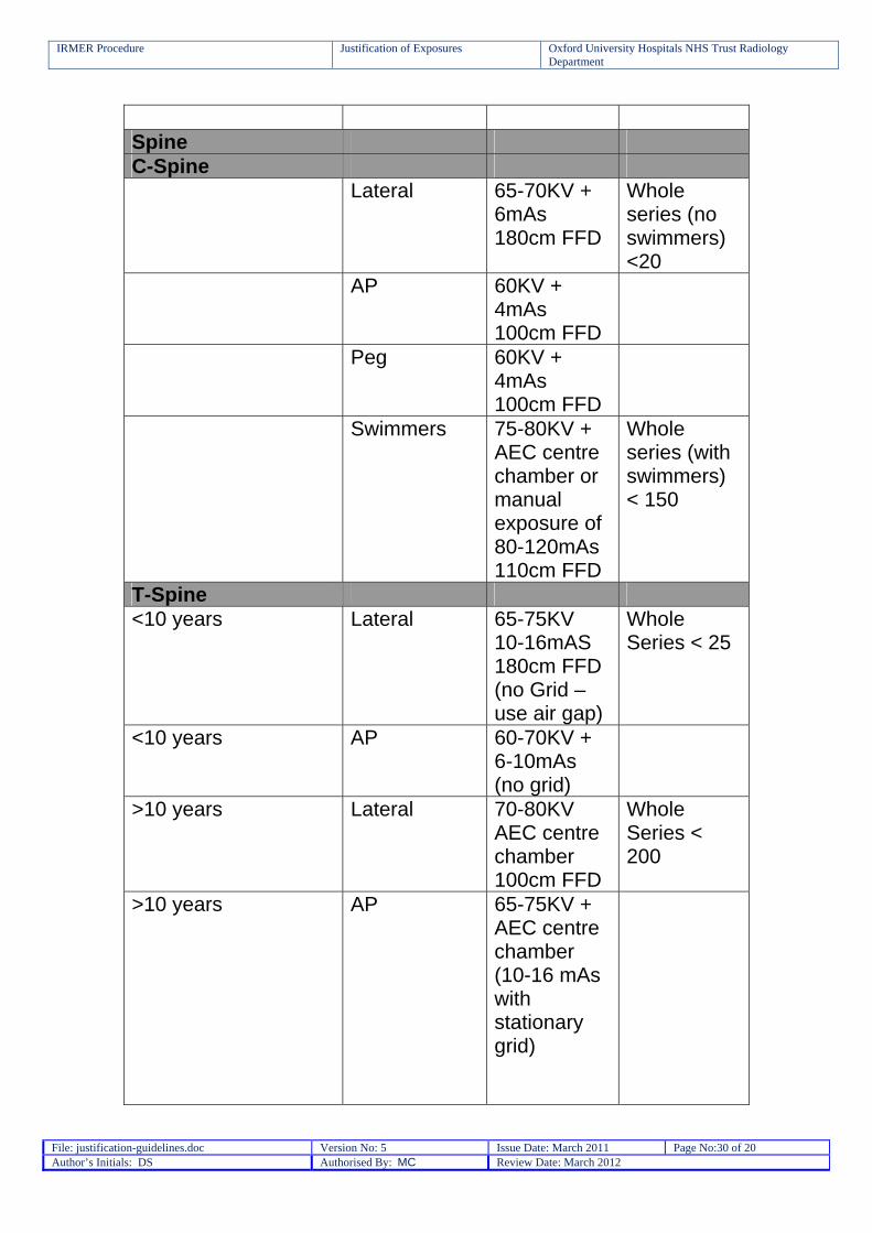
File: justification-guidelines.doc Version No: 5 Issue Date: March 2011 Page No:30 of 20 Author’s Initials: DS Authorised By: MC Review Date: March 2012
Spine C-Spine Lateral 65-70KV +
6mAs 180cm FFD
Whole series (no swimmers) <20
AP 60KV + 4mAs 100cm FFD
Peg 60KV + 4mAs 100cm FFD
Swimmers 75-80KV + AEC centre chamber or manual exposure of 80-120mAs 110cm FFD
Whole series (with swimmers) < 150
T-Spine <10 years Lateral 65-75KV
10-16mAS 180cm FFD (no Grid – use air gap)
Whole Series < 25
<10 years AP 60-70KV + 6-10mAs (no grid)
>10 years Lateral 70-80KV AEC centre chamber 100cm FFD
Whole Series < 200
>10 years AP 65-75KV + AEC centre chamber (10-16 mAs with stationary grid)
IRMER Procedure Justification of Exposures Oxford University Hospitals NHS Trust Radiology Department
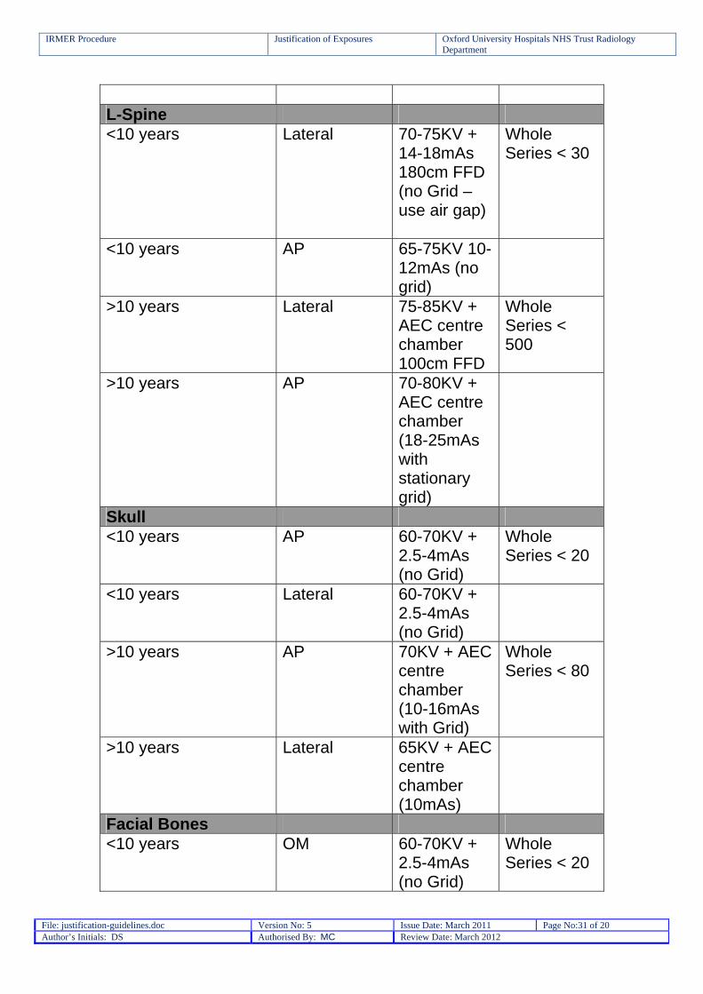
File: justification-guidelines.doc Version No: 5 Issue Date: March 2011 Page No:31 of 20 Author’s Initials: DS Authorised By: MC Review Date: March 2012
L-Spine <10 years Lateral 70-75KV +
14-18mAs 180cm FFD (no Grid – use air gap)
Whole Series < 30
<10 years AP 65-75KV 10-12mAs (no grid)
>10 years Lateral 75-85KV + AEC centre chamber 100cm FFD
Whole Series < 500
>10 years AP 70-80KV + AEC centre chamber (18-25mAs with stationary grid)
Skull <10 years AP 60-70KV +
2.5-4mAs (no Grid)
Whole Series < 20
<10 years Lateral 60-70KV + 2.5-4mAs (no Grid)
>10 years AP 70KV + AEC centre chamber (10-16mAs with Grid)
Whole Series < 80
>10 years Lateral 65KV + AEC centre chamber (10mAs)
Facial Bones <10 years OM 60-70KV +
2.5-4mAs (no Grid)
Whole Series < 20

IRMER Procedure Justification of Exposures Oxford University Hospitals NHS Trust Radiology Department
File: justification-guidelines.doc Version No: 5 Issue Date: March 2011 Page No:32 of 20 Author’s Initials: DS Authorised By: MC Review Date: March 2012
<10 years OM 30° 60-70KV +
2.5-4mAs (no Grid)
>10 years OM 70KV + AEC centre chamber (10-16mAs with Grid)
Whole Series < 80
>10 years OM 30° 70KV + AEC centre chamber (10-16mAs with Grid)
Upper and Lower Limbs
Hands DP + Oblique (lateral if #’d MC)
50-55KV + 1mAs
<2
Fingers DP, Lateral (45° Oblique for MCPJ)
50-55KV + 1mAs
<2
Thumb AP + Lateral 50-55KV + 1mAs
<2
Feet DP + Oblique
50-55KV + 1.6mAs
<2
Toes DP + Oblique
50-55KV + 1mAs
<2
Long Bones + Joints (Humeri, tib/fib, radius/ulna, femora, elbow,)
AP + Lateral (Scaphoid does not appear till about 10 years of age)
55-60KV + 2mAs
<5
Shoulder (Trauma) AP and review Axial/modified axial (Lateral for proximal humerus)
55-60KV + 2mAs
<5
Scapula (Trauma) AP + Lateral 55-60KV + 2mAs
<5
IRMER Procedure Justification of Exposures Oxford University Hospitals NHS Trust Radiology Department
File: justification-guidelines.doc Version No: 5 Issue Date: March 2011 Page No:33 of 20 Author’s Initials: DS Authorised By: MC Review Date: March 2012
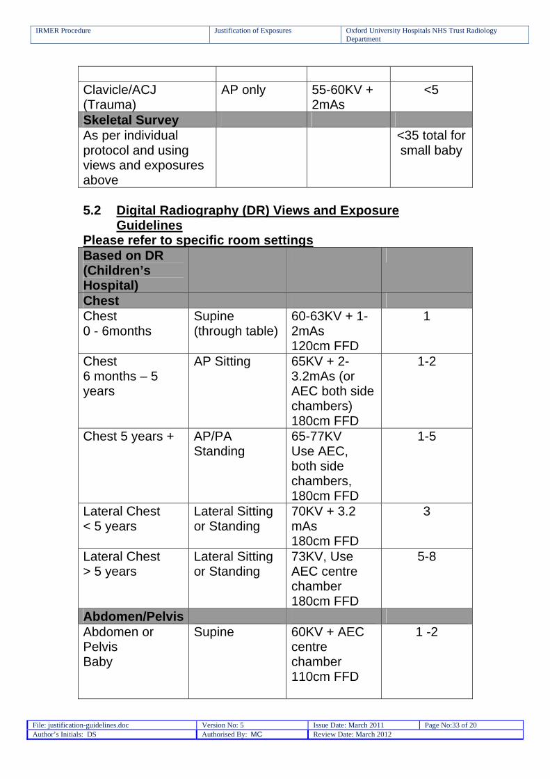
Clavicle/ACJ (Trauma)
AP only 55-60KV + 2mAs
<5
Skeletal Survey As per individual protocol and using views and exposures above
<35 total for small baby
5.2 Digital Radiography (DR) Views and Exposure
Guidelines Please refer to specific room settings Based on DR (Children’s Hospital)
Chest Chest 0 - 6months
Supine (through table)
60-63KV + 1-2mAs 120cm FFD
1
Chest 6 months – 5 years
AP Sitting 65KV + 2-3.2mAs (or AEC both side chambers) 180cm FFD
1-2
Chest 5 years + AP/PA Standing
65-77KV Use AEC, both side chambers, 180cm FFD
1-5
Lateral Chest < 5 years
Lateral Sitting or Standing
70KV + 3.2 mAs 180cm FFD
3
Lateral Chest > 5 years
Lateral Sitting or Standing
73KV, Use AEC centre chamber 180cm FFD
5-8
Abdomen/Pelvis Abdomen or Pelvis Baby
Supine 60KV + AEC centre chamber 110cm FFD
1 -2
IRMER Procedure Justification of Exposures Oxford University Hospitals NHS Trust Radiology Department
File: justification-guidelines.doc Version No: 5 Issue Date: March 2011 Page No:34 of 20 Author’s Initials: DS Authorised By: MC Review Date: March 2012
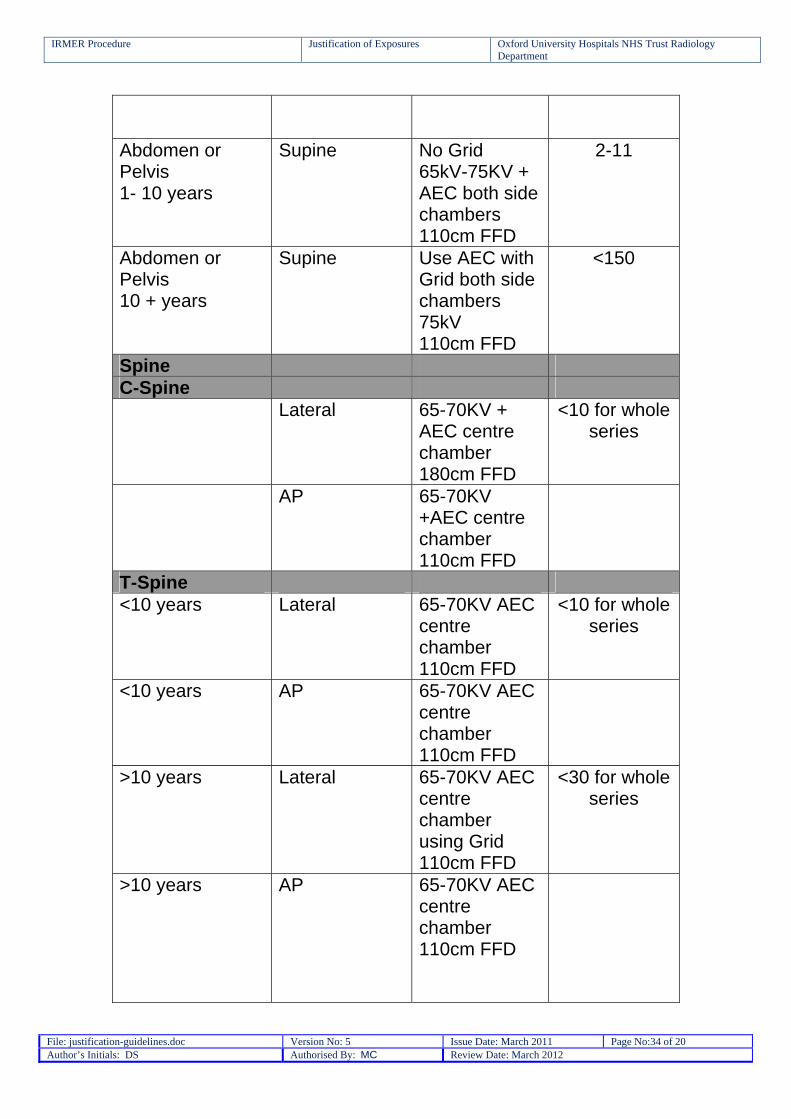
Abdomen or Pelvis 1- 10 years
Supine No Grid 65kV-75KV + AEC both side chambers 110cm FFD
2-11
Abdomen or Pelvis 10 + years
Supine Use AEC with Grid both side chambers 75kV 110cm FFD
<150
Spine C-Spine Lateral 65-70KV +
AEC centre chamber 180cm FFD
<10 for whole series
AP 65-70KV +AEC centre chamber 110cm FFD
T-Spine <10 years Lateral 65-70KV AEC
centre chamber 110cm FFD
<10 for whole series
<10 years AP 65-70KV AEC centre chamber 110cm FFD
>10 years Lateral 65-70KV AEC centre chamber using Grid 110cm FFD
<30 for whole series
>10 years AP 65-70KV AEC centre chamber 110cm FFD
IRMER Procedure Justification of Exposures Oxford University Hospitals NHS Trust Radiology Department
File: justification-guidelines.doc Version No: 5 Issue Date: March 2011 Page No:35 of 20 Author’s Initials: DS Authorised By: MC Review Date: March 2012
L-Spine <10 years Lateral 65KV -75KV
AEC centre chamber 110cm FFD
<20 for whole series
<10 years AP 65-75KV AEC centre chamber 110FFD
>10 years Lateral 65KV -75KV AEC centre chamber with grid 110cm FFD
<150 for whole series
>10 years AP 65-75KV AEC centre chamber 110FFD
Skull <10 years AP/PA 63-75KV AEC
Centre chamber 110cm FFD
<15
<10 years Lateral 63-75KV AEC Centre chamber 110cm FFD
<15
>10 years AP/PA 63-75KV AEC Centre chamber with Grid 110cm FFD
<15
>10 years Lateral 63-75KV AEC Centre chamber with Grid 110cm FFD
<15
Facial Bones <10 years OM 63-75KV
AEC Centre chamber
<15
IRMER Procedure Justification of Exposures Oxford University Hospitals NHS Trust Radiology Department
File: justification-guidelines.doc Version No: 5 Issue Date: March 2011 Page No:36 of 20 Author’s Initials: DS Authorised By: MC Review Date: March 2012
110cm FFD <10 years OM 30° 63-75KV
AEC Centre chamber 110cm FFD
<15
>10 years OM 63-75KV AEC Centre chamber with Grid 110cm FFD
<15
>10 years OM 30° 63-75KV AEC Centre chamber with Grid 110cm FFD
<15
Post nasal space Lateral 63-75KV AEC Centre chamber 110cm FFD
<10
Upper and Lower Limbs
Hands DP + Oblique (lateral if #’d MC)
60KV + 1.25mAs directly onto detector 110cm FFD
<2
Fingers DP, Lateral (45° Oblique for MCPJ)
60KV + 1.25mAs directly onto detector 110cm FFD
<2
Thumb AP + Lateral 60KV + 1.25mAs directly onto detector 110cm FFD
<2
Feet DP + Oblique
60KV + 1.25mAs directly onto detector 110cm FFD
<2
IRMER Procedure Justification of Exposures Oxford University Hospitals NHS Trust Radiology Department
File: justification-guidelines.doc Version No: 5 Issue Date: March 2011 Page No:37 of 20 Author’s Initials: DS Authorised By: MC Review Date: March 2012
Toes DP + Oblique
60KV + 1.25mAs directly onto detector 110cm FFD
<2
Long Bones + Joints (Humeri, tib/fib, radius/ulna, femora, elbow,)
AP + Lateral (Scaphoid does not appear till about 10 years of age)
60KV + AEC Centre Chamber 110cm FFD
<5
Shoulder (Trauma)
AP and review Axial/modified axial (Lateral for proximal humerus)
60KV + AEC Centre Chamber 110cm FFD
<5
Scapula (Trauma)
AP + Lateral 60KV + AEC Centre Chamber 110cm FFD
<5
Clavicle/ACJ (Trauma)
AP only 60KV + AEC Centre Chamber 110cm FFD
<5
Skeletal Survey As per individual protocol and using views and exposures above
Directly onto detector where possible
<35 total for small baby