Embed Size (px)
Citation preview

- June 2009
Istanbul, Turkey

2
TABLE OF CONTENTS
Organisation 3
Introduction 4
Conference Program 6
Oral Presentations
Friday 13
Saturday 24
Sunday 32
Monday 43
Poster Presentations 54
List of Participants 93
Maps, Pictures, etc. 102

3
Organization
International Committee Oddur Ingolfsson, Iceland, Chair
Karina Morgenstern, Germany, Vice-Chair
Michael Allan, Switzerland
Flemming Besenbacher, Denmark
Petr CARSKY, Czech Republic
Stephan Denifil, Austria
Jacques Delwiche, Belgium
Gerald Dujardin, France
Karl-Heinz Ernst, Switzerland
David Field, Denmark
Thomas Field, United Kingdom
Mieczyslaw Forys, Poland
Gustavo Garcia, Spain
Armin Golzhauser, Germany
Cornelis W. Hagen
Werner Hofer, United Kingdom
Jiri Hoacek Czech Republic
Marie-Jeanne Hubin-Franskin, Belgium
Anne Lafosse, France
Paulo Limao-Vieira, Portugal
Bratislav Marinkovic, Serbia
Stefan Matejcik, Slovakia
Nigel Mason, United Kingdom
Sveinn Olafsson, Iceland
Ahmet Oral, Turkey
Paulo Ribeiro, Portugal
Paul Scheier, Austria
Jan Skalny, Slovakia
Petra Swiderek, Germany
Harold Zandvliet, Netherlands
Mariusz Zubek, Poland
Scientific Committee Werner Hofer
Oddur Ingolfsson
Anne Lafosse
Paulo Limao-Vieira
Nigel Mason
Karina Morgenstern
Petra Swiderek
Local Committee Ahmet Oral, Chair
Selin Manukyan, Local Secretary
Derya Gemici
Administrative Support Beverley Harker Secretary
Dr. Nykola Jones, Web master
Host Institution Faculty of Engineering & Natural
Sciences
Sabanci University
www.sabanciuniv.edu
Tuzla, 34956 Istanbul, TURKEY
TEL : + 90 216 483 9522
Conference Venue
Golden Age 1 Hotel
http://www.grandoztanik.com/gol
den1/indexen.html
Topçu Cad. No: 22
Taksim / Istanbul Turkey
Tel: +90 212 254 4906
Principal Organization
http://www.cost.esf.org

4
Introduction
Dear Colleagues,
Welcome to Istanbul and ECCL 2009, the second annual meeting of the COST Action
CM0601 on ‘Electron Controlled Chemical Lithography’.
The COST Action CM0601, which we more commonly refer to as ECCL was approved by
the COST Committee for Chemistry and Molecular Science and Technology in 2006. The
Action which runs for four years was initially ratified by 13 countries and officially
launched in May 2007. Presently 16 European countries formally joined and the Action
has grown to be a valuable forum for cutting edge science in the field of electron
controlled chemical processes.
The Action consists of three independent working groups:
WG01 focuses on gas phase dissociative electron attachment and measurements of
absolute, differential and integral cross sections for electron scattering. These experiments
and the quantities acquired are fundamental to the Action underpinning its science base.
WG02 focuses on fundamental studies on electron transport and dissociative electron
attachment in the condensed phase, on the investigation of electron-induced reactions in
clusters, mixed ices and model biomolecular films and on applications of electron-induced
reactions in nanofabrication.
WG03 focuses on the investigation of the process of inelastic electron tunnelling both
theoretically and experimentally, and on the prospective of molecular manipulation with
the tip of the scanning tunnelling microscope (STM). The mastering of such manipulations
being the key to electron controlled chemical lithography at the molecular level.
The concerted goal of the Action is to build up an interdisciplinary European programme
to combine state-of-the-art in electron induced chemistry and surface science with the
recent and very exciting advances emerging from the field of scanning tunnel microscopy.
The first annual meeting of the Action; ECCL 2008, which was given in Lisbon in March
2008 showed clearly that this goal is achievable.
The Lisbon meeting was a great success and provided, for the first time, a privileged forum
where the leading European groups in the field of electron interactions with molecules in
the gas phase, clusters and condensates met with the leading European groups in the field
of scanning tunnelling microscopy to share and develop the scientific expertise on
molecular manipulation with electrons.

5
ECCL 2009, which we are proud to host here in Istanbul, has the capacity to be no less
exciting and stimulating than the Lisbon meeting in 2008. The programme offers 36 oral
presentations and about 40 posters are presented. In this forum most of Europe’s leading
groups in the field of electron/molecule interaction and STM research are represented. In
addition to the representatives from the European research groups we also have the
honour to welcome a number of very distinct speakers from overseas. Among those are
Prof. John Polanyi from Toronto Canada and Prof. Wilson Ho from Irvine, California. Prof.
Polanyi received the Nobel Price in Chemistry 1986 for his “contributions concerning the
dynamics of chemical elementary processes”. Prof. Ho is one of the pioneering researchers
in the field of scanning tunnelling microscopy.
We are also proud of the stimulation and encouragement this Action is to young
researchers, a fact which is well documented by the large number of progress reports and
posters given by students and young scientist at this meeting.
Finally we thank the local conference secretary Ms Selin Manukyan and Ms Derya Gemici
for their valuable assistance with local arrangements we also thank Ms Beverley Harker
from the Open University and our web master Dr. Nykola Jones from the University of
Aarhus who’s hard work made this meeting possible
We hope you will experience a stimulating scientific programme, enjoy the conference
ambience and its social experience.
Istanbul, June 2009
Oddur Ingólfsson; Chair of COST Action CM0601
Ahmet Oral; Local Organizer of ECCL 2009

6
Conference Program
Thursday 4th June 2009
12:00 - Arrival & Registration
Friday 5th June 2009
08:50 Opening Remarks/ Session 1, Chair:: Prof. Oddur Ingólfsson / Prof. Ahmet Oral
09:00 - 09:45 Prof. John C. Polanyi, Nobel Laureate (CA). Electron-Controlled Adsorbate Motion and Reaction at a Silicon Surface.
09:45 - 10:25 Prof. Michael Allan (CH). On the relation between gas phase electron scattering and processes at the STM tip
10:25 - 11:10 Coffee break
Session 2, Chair: Dr. Roman Curik
11:10 - 11:35 Dr..Isabella Baccarelli (IT). Electron-scattering on biosystems: a step forward a high-throughput approach
11:35 - 12:00 Prof. Stefan Matecjik (SK). Electron attachment and electron impact ionisation of selected derivatives of silane
12:00 - 12:25 Dr. Janina Kopyra (PL). Low energy electron interactions with biologically relevant molecules
12:25 - 14:10 Lunch Break
Session 3 and 4, Chair: Prof. Armin Gölzhäuser
14:10 - 14:50 Dr. Hubertus Marbach (DE). Lithographic fabrication of clean nanostructures via focused electron-beam induced processing in UHV.
14:50 - 15:15 Dr. Bianca Hermann (DE). Predicting Molecular Pattern Diversity: Elementary Geometrical Features Encoding Molecular Ordering
15:15 - 15:40 Dr. Roberto Otero (ES). Charge transfer-driven molecular self-assembly at organic/metal surfaces.
15:40 - 16:25 Coffee break
16:25 - 16:50 Mr. Filipe Ferreira da Silva (AT). Magic L-serine clusters in cold helium nanodroplets
16:50 - 17:15 Ms. Helga Dögg Flosadóttir (IS). Predictive simulations for metastable dissociation of negative ions.
18:15 - Welcome reception

7
Saturday 6th June 2009
Session 5 Chair: Dr. Isabella Baccarelli
09:00 - 09:40
Prof. Leon Sanche (UK)/(CA).
Low energy electrons in Nanolithography.
09:40 - 10:05
Dr. Roman Curik (CZ).
Cold electron collisions: Theory and experiment.
10:05 - 10:30
Dr. Andreas Mauracher (AT).
Comparative study of delayed fragmentation resulting from electron attachment to nitro-aromatic compounds
10:30 - 11:15
Coffee break
Session 6 Chair: Prof. Anne Lafosse
11:15 - 11:55
Prof. Nigel Mason (UK).
Electron induced chemistry on surfaces – Some unanswered questions
11:55 - 12:20
Dr. Hassan Abdoul-Carime (FR).
Dissociative Electron Attachment to Amino-Acid: the case of Leucine.
12:20 - 12:45 Prof. Bratislav Marinkovic (YU).
Measurements of electron interactions with metal vapour atoms
12:45 - 13:10
Prof. Eugen Illenberger (DE).
Formation of CN– in Electron Attachment to Organic Molecules
12:45 - 14:30
Lunch Break
14:30 - Conference Excursion
20:00 - MC Meeting

8
Sunday 7th June 2009
Session 5 Chair: Prof. .Karina Morgenstern
09:00 - 09:45 Prof. Wilson Ho (USA). Atomic Scale Inelastic Electron Tunneling Phenomena.
09:45 - 10:10 Dr. Willem van Dorp (NL). A surface science whodunnit: the cross section for electron induced dissociation of the precursor Me3PtMeCp
10:10 - 10:35 Prof. Richard E. Palmer (UK). Non-local molecular manipulation by STM charge injection into selected surface electronic states.
10:35 - 11:15 Coffee break
Session 8 Chair: Prof. Nigel Mason
11:15 - 11:55 Dr. Anne Lafosse (FR). Low-energy electron (0-20 eV) induced degradation of 11-Mercapto-undecanoic acid SAMs
11:55 - 12:20 Dr. Paulo Gomez (PT). Impedance Spectroscopy - A Tool for DNA Irradiation Studies.
12:20 - 12:45 Dr. Richard Balog (DK). Graphene modification by atomic hydrogen.
Lunch Break
Session 9 Chair: Richard Palmer
14:30 - 15:10 Prof. Karina Morgenstern (DE). Switching molecules by electrons: From isomerisation to chirality flip.
15:10 - 15:35 Dr. Marie-Laure Bocquet (FR). Probing the proton location in a water bilayer on Pd(111) by inelastic spectroscopy simulations.
15:35 - 16:00 Dr. Manfred Parschau (CH). Surface dynamics and chemistry of propene on Cu(211) induced by inelastic electron tunneling.
16:15 - 18:00 Poster Session
18:00 - 19:30 Dinner Break
19:30 - Poster Session

9
Monday 8th June 2009
Session 10 Chair: Prof. Prof. Armin Gölzhäuser
09:00 - 09:40 Prof. Adam Foster (FI). Adsorption, diffusion and manipulation of molecules on insulating surfaces
09:40 - 10:05 Prof. Sveinn Ólafsson (IS). The outlook for growing larger nanostructures with STM using chemical amplified feedback and other energy assisted processes.
10:05 - 10:30 Dr. Meike Stöhr (CH). Reactions on surfaces for the creation of supramolecular polymers.
10:30 - 11:10 Coffee break
Session 11 Chair: Gerald.Dujardin
11:10 - 11:50 Prof. E. Krishnakumar (UK)/(IN). Momentum imaging of negative ions and dissociative electron attachment
11:50 - 12:15 Ms. Martina Fuss (ES). Single electron track simulation in methane.
12:15 - 12:40 Mr. Olivier May (CH). Isotope effect in dissociative electron attachment cross sections in acetylene.
12:40 - 14:00 Lunch Break
Session 12 Chair: Klaus Edinger
14:00 - 14:40 Prof. Armin Gölzhäuser (DE). 1 nm thin carbon nanosheets with tunable conductivity and stiffness
14:40 - 15:05 Mr. Thorben Hamann (DE). Low-energy electron-induced reaction between ammonia and ethylene driven by ionization
15:05 - 15:30 Dr. Harun Solak (CH). Nanolithography in the extreme ultraviolet range
Closing remarks
18:00 - Departure for conference dinner

10
Poster Session
Sunday 7th June, 2009
# Presenting Author Title
P01 Prof. Michael Allan (CH). Electron collisions with cyclopropane
P02 Prof. Michael Allan (CH). Electron collisions with Pt(PF3)4
P03 Mr. Bogdan C. Ibănescu (CH). Rules governing the dissociation of Feshbach resonances in oxygen containing compounds
P04 Mr. Ivo Annen (AT). Delayed fragmentations of nitro aromatic explosives resulting from electron attachment
P05 Mr. Elias H. Bjarnason (IS). Construction of an instrument to identify suitable molecules for ECCL – work in progress
P06 Dr. Genevieve Comtet (FR). Nanometer-scale electron lithography on the hydrogenated diamond surface with a conducting AFM
P07 Mr. Marcin Dampc (PL). Threshold excitation of diethyl ether and tetrahydrofuran by electron impact
P08 Mr. Marcin Dampc (PL). High resolution threshold photoionization studies of isoxazole
P09 Dr. Gérald Dujardin (FR). Electronic control of single molecules on insulating layers
P10 Dr. Juraj Fedor (CZ).
Dissociative electron attachment cross sections for HCl, HBr and their deuterated analogs – comparison of experiment and theory
P11 Dr. Juraj Fedor (CZ).
Anisotropy in electron distributions from fullerenes excited by femtosecond laser pulses.
P12 Dr. Thomas Field (UK).
Autodetachment Rates of SF6-* Anions: Some Agreement Between Theory And Experiment
P13 Prof. Gustavo Garcia (ES).
Total electron scattering and ionization cross sections in THF
P14 Mr. Paulo Gomes (PT). Interactions of DNA with Ionic Phospholipids in Langmuir Monolayers at the Air−Water Interface
P15 Ms. Michaela Hager (AT). Characterization of Titanium and Silicon Nanoparticle Films by STM and XPS techniques
P16 Mr. Thorben Hamann (DE). Low-energy electron-induced reactions in NH3-containing molecular films
P17 Mr. Sean Haughey (UK) Dissociative Electron Attachment to 2-Nitro-m-xylene

11
P18 Prof. Oddur Ingolfsson (IS).
Theoretical approach to predict metastable dissociation of deprotonated anions in gas phase
P19 Prof Ismet Kaya (TK) Dual-probe scanning tunneling microscope for study of nanoscale metal-semiconductor interfaces
P20 Prof. Paulo Limao-Vieira (PT). The electronic state spectroscopy of isoprene [CH2CHC(CH3)CH2] studied by electron and photon impact
P21 Mr. Olivier May (CH). Isotope effect in dissociative electron attachment cross sections in acetylene
P22 Dr. Bernd Nestmann (DE). Locally restricted resonances in electron-molecule scattering
P23 Prof. Yuri Nunes (PT).
Understanding the VUV spectroscopy of CF3COOCH3 as a route to environmental friendly hydrofluorinated ethers (HFEs)
P24 Mr. Benedikt Ómarsson (IS). The simulated effects of fringing fields and orthogonal velocity components on peak shape and electron energy resolution in the Trochoidal Electorn Monochromator
P25 Mr. Benedikt Ómarsson (IS). HF formation as a driving force for selective bond cleavage
P26 Prof. Ahmet Oral (TK). Extreme nanotribology: Atomistic level lateral force measurement using novel home-made combined nc-AFM/STM
P27 Dr. Chiara Panosetti (IT). Modelling dissociative fragmentation of aminoacids after resonant electron attachment: A quantum picture
P28 Dr. Peter Papp (SK). Studies of low energy electron interaction with furanose-structured alcohols
P29 Mr. Branko Petrusevski (YU). Measurement of laser-induced fluorescence of molecules using a time-resolved spectrometar
P30 Mr. Ivan Pshenichnyuk (CZ). Vibrational Dynamics of Tunneling Molecular Junctions
P31 Prof. Paulo Ribeiro (PT). Dynamics of Birefringence Creation and Relaxation in Azobenzene Containing Surfaces
P32 Prof. Paulo Ribeiro (PT). Creation of Polarized light by Orientation of Electroluminescent organic molecules Induced by Photorientation of Azobenzene Molecules
P33 Mr Paulo Gomes (PT). Characterization of DNA Intercalation with 2,2’-Bipyridyl on Solid Surfaces
P34 Prof. Leon Sanche (UK)/(CA). Low energy electron-induced processes in lithographic materials: electron stimulated desorption from methyl methacrylate (MMA) films
P35 Prof. Leon Sanche (UK)/(CA). Radiation-induced processes on gold nanoparticles: Raman SERS analysis of adsorbed amino acids and peptides.

12
P36 Prof. Iwona Szamrej-Forys (PL). Interaction of thermal electrons with chloro- and bromo- containing hydrocarbons
P37 Dr. Ivo Utke et al. (CH). Characterization of focused electron beam induced carbon deposits from organic precursors
P38 Prof. Mariusz Zubek (PL). Fragmentation of diethyl ether by electron impact

13
ORAL
PRESENTATIONS
ABSTRACTS
FRIDAY, 5th
of June

14
Friday, 9:00
Electron-Controlled Adsorbate Motion and Reaction at a Silicon Surface.
John C. Polanyi
Department of Chemistry, University of Toronto, Toronto, Canada, M5S 3H6
Electron impact from the tip of a Scanning Tunneling Microscope (STM) can result in motion of physisorbed adsorbate, leading to configuration-change or to chemical reaction. Examples will be given for single molecules, pairs of molecules and self-assembled lines of molecules on Si(111) and Si(100). Recent results show that in the case of some adsorbates on Si(100) reaction is ‘cooperative’, i.e. a single electron invariably triggers the reaction of two adjacent molecules, rather than one. The underlying dynamics for this novel type of reaction will be described. The experiments were performed at the University of Toronto, the theory at Liverpool University (Professor Werner Hofer’s group) and at McGill University (Professor Hong Guo’s group).
References 1. Krishnan R. Harikumar, John C. Polanyi, Peter A. Sloan, Serge Ayissi, and Werner A. Hofer, Electronic Switching of Single Silicon Atoms by Molecular Field-Effects, J. Am. Chem. Soc. 128, 16791 (2006). (See also Nature Nanotech Research Highlights, Online January 5, 2007).
2. S. Dobrin, K.R. Harikumar, I.R. McNab. J.C. Polanyi, Z. Waqar, J. (S.Y.) Yang, Molecular Dynamics of Haloalkane Corral Formation and Surface Halogenation at Si(111)-7×7, J. Chem. Phys. 125, 133407 (2006).
3. S. Dobrin, K.R. Harikumar, T.B. Lim, L. Leung, I.R. McNab. J.C. Polanyi, P.A. Sloan, Z. Waqar, J. (S.Y.) Yang, S. Ayissi and W.A. Hofer, Maskless Nanopatterning and Formation of Nanocorrals and Switches, for Haloalkanes at Si(111)-7×7, Nanotechnology 18, 044012 (2007).
4. K.R. Harikumar, Tingbin Lim, Iain R. McNab, John C. Polanyi, Linda Zotti, Serge Ayissi and Werner A. Hofer, Dipole-Directed Assembly of Lines of 1,5-Dichloropentane on Silicon Substrates by Displacement of Surface Charge, Nature Nanotech 3, 222 (2008). (News article: Stacey F. Bent, Silicon Falls into Line, Nature Nanotech 3, 185 (2008)).
5. Hong Guo, Wei Ji, John C. Polanyi and Jody (S.Y.) Yang, Molecular Dynamics of Localized Reaction, Experiment and Theory: Methyl Bromide on Si(111)-7×7, ACS Nano 2, 699 (2008).
6. K.R. Harikumar, Lydie Leung, Iain R. McNab, John C. Polanyi, Haiping Lin and Werner A. Hofer, Cooperative Molecular Dynamics in Surface-Reaction. Nature Chemistry, Submitted (2009).

15
Friday, 9:45
On the relation between gas phase electron scattering and processes at the STM tip
Michael Allan
Department of Chemistry, University of Fribourg, chemin du Musée 9, 1700 Fribourg, Switzerland
There are many conceptual similarities between electron collisions in an electron beam
experiment and the processes at the tip of a scanning tunneling electron microscope (STM):
• Electrons are inserted into the molecule and may be temporarily or permanently
captured.
• The electrons are inserted into normally unoccupied orbitals [1].
• The insertion of the electron may cause vibrational [2] or electronic [3] excitation,
dissociation or other chemical change [4] of the target.
• The electron energy may be varied in both experiments and spectroscopic information
on both the final excited states of the target and on the intermediate states with
temporary LUMO occupation may be gained.
• There is a relation between the properties of the temporarily occupied orbital and the
selectivity of the vibrational excitation, including symmetry selection rules, both in the
STM [5,6,7] and the gas phase [8,9] experiments.
The talk will highlight the analogies and the differences between the two experiments.
References
[1] J. Repp and G. Meyer, Physik in Unserer Zeit 37, 266 (2006).
[2] J. I. Pascual, Eur. Phys. J. D 35, 327 (2005).
[3] J. W. Alldredge et al., Nature Physics 4, 319 (2008).
[4] K. Morgenstern, Acc. Chem. Res 42, 213 (2009).
[5] M. Ohara et al., Phys. Rev. Lett. 100, 136104 (2008).
[6] N. Lorente et al., Phys. Rev. Lett. 86, 2593 (2001).
[7] N. Gawronski, M. Melhorn and K. Morgenstern, Science 319, 930 (2008).
[8] I. C. Walker, A. Stamatović and S. F. Wong, J. Chem. Phys. 69, 5532 (1978).
[9] T. Skalický, C. Chollet, N. Pasquier and M. Allan, Phys. Chem. Chem. Phys. 4, 3583 (2002).

16
Friday, 11:10
Electron-scattering on biosystems: a step forward a high-throughput approach
I. Baccarelli1, F. Sebastianelli2, M.D. Gonzalez-Sanchez3, C. Panosetti 2, F. A. Gianturco2, G. Morelli1, Nico Sanna1
1 CASPUR Supercomputing Consortium, via dei Tizii 6, 00185 Rome, Italy 2 Department of Chemistry and CNISM, University of Rome 'La Sapienza', Ple A. Moro 5, 00185 Rome,
Italy 3Departamento de Quimica Fisica, Facultad de Farmacia, Campus Miguel deUnamuno, 37007
Salamanca, Spain
Recent years have witnessed a remarkable growth of both experimental and theoretical
investigations on the capabilities of low-energy electrons to selectively break molecular bonds.
Such a remarkable property shed a new light on the general understanding of complex
phenomena, such as the damage to a living tissue under exposure to an ionizing radiation, and
it opened the way to the `engineering' use of electrons in the chemical control of processes.
In the last few years we devoted a great amount of effort in the study of electrons as probes of
the various DNA building blocks in the gas-phase [1]. The quantum mechanical analysis of such
processes is a challenging task, due to the size and the generally low symmetry of the system
under study and in the perspective of extending the analysis from individual building blocks to
larger biological macromolecules. Very recently we released updated versions of our
electron/positron-molecule scattering packages [2,3] in order to cope with more massive
calculations both for the number of systems which can be analysed in a fast, automated way
and for the size of the compounds. Our scope is in fact to develop a computational tool which
allows from one hand to efficiently study a great variety of molecules (conformers, chemically-
substituted systems for use in tumor imaging and radiotherapy, etc.) and, ultimately, to extend
the calculations to small fragments of, e.g., DNA.
We will present here an overall picture of our last findings and discuss our preliminary results on
large-scale calculations. References [1] I. Baccarelli, F.A. Gianturco, A. Grandi, R.R. Lucchese and N. Sanna, “Electron-driven molecular processes induced in biological systems by electromagnetic and other ionizing sources”, Adv. Quantum Chem. 52, 189(2007).
[2] N. Sanna, I. Baccarelli, G. Morelli, “SCELib3.0: the new revision of SCELib, the parallel computational library of molecular properties in the Single Center Approach”, Comp. Phys. Comm., accepted (2009).
[3] N. Sanna, I. Baccarelli, G. Morelli, “The VOLSCAT package for electron and positron scattering off molecular targets: a new high throughput approach to cross-section and resonances computation”, Comp. Phys. Comm., accepted (2009).

17
Friday, 11:35
Electron attachment and electron impact ionisation of selected derivatives of silane
Jaroslav Kočíšek, František Krčma2, Štefan Matejčík 1Department of Experimental Physics, Comenius University, Mlynska dolina F2, 84248 Bratislava Slovak
republic 2University of Technology, Purkyňova 118, 61200 Brno, Czech republic
The derivatives of silane are widely used in the industry to prepare thin films. Various types of
plasma techniques are used to prepare such thin layers [1, 2, 3]. The processes in the
technological plasma are in large extend drived by the electron induced reactions with
molecules. Except of elestic and inelastic scattering the ionisation reactions such as Electron
Impact Ionisation (EII) and Electron Attachment (EA) are of high importance [4].
In present experimental study we present new data concerning EII and EA to two derivatives of
silan, tetravinylsilan (TVS) and dimethyl-phenyl-silane (DMPS). The study was carried out using
crossed electron/molecular beam apparatus at Comenius Univeristy in Bratislava.
We have measured positive and negative mass spectra, apearance energies and partial cross
sections for EII and EA reactions to these molecules.
References
[1] O. Salyk, P. Broya, N. Dokoupil, R. Herrmann, I. Kurytka, J. Prycek, M. Weiter, Surface & Coatings Technology 200 (2005) 486 [2] V. Cech, J. Studynka, F. Janos, V. Perina, Plasma Process. Polym. 4 (2007) S776 [3] Sung-Gyu Park and Shi-Woo Rheea, J. Vac. Sci. Technol. A 24 (2006) 291 [4] J. Ch. Choe, International Journal of Mass Spectrometry 242 (2005) 5

18
Friday, 12:00
Low energy electron interactions with biologically relevant molecules
Janina Kopyraa,b, Constanze König-Lehmannb, Iwona Szamreja and Eugen Illenbergerb
a Chemistry Department, University of Podlasie, 3 Maja 54, 08-110 Siedlce, Poland
b Institut fur Chemie und Biochemie - Physikalische und Theoretische Chemie, Freie Universitat Berlin,
Takustrasse 3, D-14195 Berlin, Germany
In the past electron attachment studies in the gas phase were focused on halogenated
compounds such as halocarbons. The high interest in these investigations is due to many
technological applications since they are widely used as gaseous dielectrics or in plasma
etching industry. Within the last decade, increasing interest in the study of biologically relevant
molecules could be observed. Among them the organic acids are of particular importance. They
are considered as the simplest building blocks of biomolecules and can hence serve as model
systems for the properties of large and more complex amino acids, or proteins with respect to
their behaviour during exposure to high energy radiation. Its also believed that molecules with
carboxylic group could be of great importance in functionalization of semi-conductor material
due to its ability to enhance a stickiness of the compounds on the substrate surface.
Here we present results on electron capture by the halo-substituted organic acids,
chlorodifluoroacetic acid and trichloroacetic acid, derived by means of a crossed electron-
molecular beam apparatus. We find that these molecules are very ready to attach low energy
electrons which results in the formation of low energy resonances. These resonances arise
either from simple bond cleavage or from more complicated reaction pathways that go along
with multiple bond cleavage and rearrangement in the precursor ion resulting in the formation of
new molecules. For both molecules the most intensive reaction channel is the formation of two
complementary ions (M-Cl- and Cl-) due to the cleavage of the C-Cl bond with the excess
charge localized on either of the fragment. In the case of CClF2COOH we observe interesting
and unusual feature in a sense that the parent anion M- is formed at an energy 0.75 eV while
very efficient dissociative electron attachment reactions already occur at energy close to zero
eV.
Additionally, we report results on thermal electron attachment rate constant for CClF2COOH
obtained using a pulsed Townsend technique.
We acknowledgment: Work supported by the COST action CM0601 (Electron Controlled
Chemical Lithography, ECCL), by the Polish Ministry of Science and Higher Education, the
Deutsche Forschungsgemeinschaft (DFG), the Fonds der Chemischen Industrie and the Freie
Universität Berlin.

Friday, 14:10
Lithographic fabrication of clean nanostructures via focused electron-beam induced processing in UHV
Lehrstuhl für Physikalische Chemie II and Interdisciplinary Center for Molecular Materials (ICMM), Universität Erlangen
The fabrication of chemically and structurally well
at the same time important for a large number of envisioned technological applications. We
explore the technique of electron
such nanostructures: By exploiting a highly focused electron
microscope (SEM) we directly write nanostructures by locally dissociating adsorbed pr
molecules [1,2]. In contrast to previous studies our novel approach is to work in an ultra high
vacuum (UHV) environment. This allows us to overcome the hitherto existing limitation
concerning the rather poor cleanliness of the deposits [3
In this work, the successful generation of clean metallic (Fe) and oxidic nanostructures (TiOx)
with lithographically controlled shapes and lateral dimensions partially smaller than 10 nm on
different substrates is presented. In our “surface science approach” to
role of the chemical and structural properties of the substrate and the observation of catalytic
effects already at room temperature
promising perspectives and applications of this nanostructuring technique will be presented.
This work is supported by the DFG under grant MA 4246/1
Figure 1. SEM images of different EBID structurTemplates for the deposits were either the seal structure shown in C (upper row) or two vertical lines (lower row). Comparing G and H (generated at the indicated sample temperatures) it is generated from Fe(CO)5 differ depending on the sample temperature during the EBID process (all SEM images shown are acquired at room temperature, the scale bars represent 200 nm).
[1] van Dorp W. and Hagen C. W., J. Appl. Phys. 104 (2008) 081301.[2] Utke I., Hoffmann P. and Melngailis J[3] Lukasczyk T., Schirmer M., Steinrück H.
Lithographic fabrication of clean nanostructures via focused beam induced processing in UHV
Hubertus Marbach
Lehrstuhl für Physikalische Chemie II and Interdisciplinary Center for Molecular Materials (ICMM), Erlangen-Nürnberg, Egerlandstr. 3, D-91058 Erlangen
The fabrication of chemically and structurally well-defined nanostructures is still a challenge and
at the same time important for a large number of envisioned technological applications. We
echnique of electron-beam induced deposition (EBID) to realize the engineering of
such nanostructures: By exploiting a highly focused electron-beam of a scanning electron
microscope (SEM) we directly write nanostructures by locally dissociating adsorbed pr
. In contrast to previous studies our novel approach is to work in an ultra high
vacuum (UHV) environment. This allows us to overcome the hitherto existing limitation
r cleanliness of the deposits [3].
s work, the successful generation of clean metallic (Fe) and oxidic nanostructures (TiOx)
with lithographically controlled shapes and lateral dimensions partially smaller than 10 nm on
different substrates is presented. In our “surface science approach” to EBID we also discuss the
role of the chemical and structural properties of the substrate and the observation of catalytic
already at room temperature. The concept of EBID, basic physical principles and the
promising perspectives and applications of this nanostructuring technique will be presented.
supported by the DFG under grant MA 4246/1-1.
SEM images of different EBID structures generated with ethene (A, E) and iron pentacarbonyl (B, G, H). Templates for the deposits were either the seal structure shown in C (upper row) or two vertical lines (lower row).
(generated at the indicated sample temperatures) it is obvious that the morphology of the deposits differ depending on the sample temperature during the EBID process (all SEM images shown
are acquired at room temperature, the scale bars represent 200 nm).
, J. Appl. Phys. 104 (2008) 081301. Utke I., Hoffmann P. and Melngailis J, J. Vac. Sci. Technol. B 26(4) (2008) 1197. Lukasczyk T., Schirmer M., Steinrück H.-P. and Marbach H., Small 4(6) (2008) 841.
Lithographic fabrication of clean nanostructures via focused
Lehrstuhl für Physikalische Chemie II and Interdisciplinary Center for Molecular Materials (ICMM),
defined nanostructures is still a challenge and
at the same time important for a large number of envisioned technological applications. We
beam induced deposition (EBID) to realize the engineering of
beam of a scanning electron
microscope (SEM) we directly write nanostructures by locally dissociating adsorbed precursor
. In contrast to previous studies our novel approach is to work in an ultra high
vacuum (UHV) environment. This allows us to overcome the hitherto existing limitation
s work, the successful generation of clean metallic (Fe) and oxidic nanostructures (TiOx)
with lithographically controlled shapes and lateral dimensions partially smaller than 10 nm on
EBID we also discuss the
role of the chemical and structural properties of the substrate and the observation of catalytic
The concept of EBID, basic physical principles and the
promising perspectives and applications of this nanostructuring technique will be presented.
and iron pentacarbonyl (B, G, H). Templates for the deposits were either the seal structure shown in C (upper row) or two vertical lines (lower row).
obvious that the morphology of the deposits differ depending on the sample temperature during the EBID process (all SEM images shown

20
Friday, 14:50
Predicting Molecular Pattern Diversity: Elementary Geometrical Features Encoding Molecular Ordering
B. A. Hermann1, C. L. Rohr1, K. Gruber1, J. Büttner1, M. Balbás Gambra2, E. C. Constable3, E. Frey2, T. Franosch2,
1Department of Physics/Center for Nano Science (CeNS) LMU Munich and Walther-Meissner-Institute of Low Temperature Research of the Bavarian Academy of Science and Humanities, Walther-Meissner-Str.
8, 85748 Garching, Germany
²Arnold Sommerfeld Center for Theo. Phys. (ASC)/Center for Nano Science (CeNS), Department of Physics, LMU München, Theresienstr. 37, 80333 München, Germany
³Department of Chemistry, University of Basel, Spitalstr. 51, 4056 Basel, Switzerland
Self-organization of supramolecular monolayers constitutes a major challenge for applications
e.g. of locally controlled chemical lithography. One main difficulty lies in making reliable
predictions of and controlling the complex molecular ordering on the nanometer scale. We
investigate a versatile model system consisting of Fréchet dendrons for molecular self-
organization on graphite surfaces by scanning tunneling microscopy (STM). By engineering the
interactions of this complex molecule, we find a surprisingly rich diversity of structural phases
even for a single type of molecule. Based on energy-minimized molecular mechanics (MM)
simulations atomic positions can be provided as input to derive an electronic image employing
density functional theory (DFT). A slice of the integrated local density of states (LDOS) of a free
single molecule can so be compared with the STM images. In order to understand and predict
the ground state pattern with Monte Carlo (MC) simulations, we created a conceived coarse-
grained model condensing the molecular architecture into a backbone and ten interaction sites.
The pattern diversity is successfully reproduced confirming that geometry as well as few salient
weak interactions encode the structural motifs.

21
Friday, 15:15
Charge Transfer-Driven Molecular Self-Assembly at Organic/Metal Interfaces
Tzu-Chun Tseng1, Christian Urban2, Wang Yang3, Roberto Otero2,4,* Steven L. Tait1, Manuel Alcamí³, David Écija2, Marta Trelka2, José María Gallego5, María Ángeles Herranz6, Fernando Martín3, Nazario
Martín6, Klaus Kern1, and Rodolfo Miranda2,4 1Max-Planck-Institut für Festkörperforschung, D-70569 Stuttgart, Germany
2Departamento de Física de la Materia Condensada, Universidad Autónoma de Madrid, Cantoblanco, 28049 - Madrid. Spain
3Departamento de Química, Universidad Autónoma de Madrid, 28049 - Madrid. Spain 4Instituto Madrileño de Estudios Avanzados en Nanociencia (IMDEA-Nanociencia), Cantoblanco, 28049 -
Madrid, Spain 5Instituto de Ciencia de Materiales de Madrid - CSIC, Cantoblanco, 28049 - Madrid. Spain
6Departamento de Química Orgánica, Universidad Complutense de Madrid, 28040 - Madrid. Spain
Organic heterostructures based on blends of molecules with electron-accepting (large electron-
affinity) and electron-donating (small ionization potential) character display interesting electrical
and optical properties with promising technological applications. For example, they show
electroluminiscence for Organic Light Emission Diodes (OLEDs), photovoltaic response for solar
cell devices and one-dimensional conduction for low molecular-weight metallic films, while
strong acceptors or donors are the basis for metalorganic magnets. These blends of molecules
are deposited onto or contacted with metallic layers and their performance depends crucially on
the alignment of energy levels, the molecular nanostructure and crystalline perfection. Interfaces
between organic species with either donor or acceptor character and metal surfaces are, thus,
of paramount importance for the performance of the devices described above. This observation
has motivated a large effort aimed at understanding the electronic structure of organic/metal
interfaces and, in particular, the alignment of the energy levels at the interface related to the
charge transfer between the organic donor or acceptor species and the metallic surface. Charge
transfer, however, not only leads to modifications in the alignment of energy levels; usually, it is
also related to structural transformations in both donating and accepting species. Unfortunately,
too often it is assumed that the substrate is just an inert spectator, playing no active role in the
supramolecular organization. We describe here experiments and theoretical simulations that
unequivocally demonstrate that for strong charge transfer systems, both the molecules and the
substrate suffer strong structural rearrangements that may even control the resulting molecular
ordering. Such charge transfer-induced structural rearrangements at both sides of the
organic/metal interface might have significant effects on the subsequent growth and structure of
the organic film and, thereby, on device performance.

22
Friday, 16:25
Magic L-serine clusters in cold helium nanodroplets
F. Ferreira da Silva1, P. Bartl1, S. Denifl1, A. Ellis2, T. D. Märk1 and P. Scheier1
1 Institut für Ionenphysik und Angewandte Physik, Universität Innsbruck, Technikerstr. 25, A-6020 Innsbruck, Austria
2 Department of Chemistry, University of Leicester, University Road, Leicester LE1 7RH, UK
The emergence of homochirality in living systems is not fully explained. Why is it that L-amino acids and D-sugars predominate in living organisms? How did this homochirality begin?
Cooks et al. and Hodyss et al. have reported a strong chirality preference in clusters of serine. In their studies, they propose that serine could be the original chiral progenitor. The initial chiral transmission may have occurred in serine clusters where the chiral preference of a molecular assembly is transferred to the other amino acids or sugars through the entantioselective substitution reactions.
Recently our group has performed the first measurements with L-serine in cold He nanodroplets. The nanodroplets are produced by supersonic expansion and then doped with L-serine monomers generated by heating solid L-serine. The serine-doped helium droplets are then probed by electron impact ionization mass spectrometry. The dominant ionization process of the doped nanodroplets proceeds through the initial helium ionization followed by charge hopping between the helium atoms towards final localization on the L-serine cluster in the center of the droplet. We have studied clustering of serine in the nanodroplets for different expansion parameters which defines the mean He droplet size. We observe with decreasing droplet size a profound change in the shape of the serine cluster distribution. For this case we observe a distinct magic peak for Ser8H+ and also a magic peak for the protonated dimer Ser2H+.
1 Possible explanations for such a pronounced cluster distribution will be discussed.
Acknowledgements: This work has been supported by the FWF, Wien, Austria and the European Commission, Brussels. A.E. is grateful for the support of his visit to Innsbruck by ECCL COST Action CM0601. S.D. gratefully acknowledges an APART-fellowship from the Austrian Academy of Sciences.
1 F. Ferreira da Silva et al, submited to ChemPhysChem 2009

23
Friday, 16:50
Predictive simulations for metastable dissociation of deprotonated anions
H. D. Flosadóttir, H Jónsson, O Ingólfsson
Science Institute, University of Iceland, Dunhagi 3, 107 Reykjavík, Iceland
The predictive power of theoretical approaches has been improving enormously the last
decades or even years, and many very complicated problems can now be reliably modelled. In
a recent work we simulated the metastable fragmentation of [L-Valine-H]- formed upon
dehydrogenation via dissociative electron attachment (DEA) and deprotonation in matrix
assisted laser desorption ionisation (MALDI). These simulations gave very promising results
and compared excellently with our experimental results1.
We have now extended our work to the more complicated nucleosides. The nucleosides have
many different possible deprotonation sites. On the sugar moiety the 2’, 3’ and 5’-OH hydroxyl
protons are present and on the nucleobases there is at least one acidic proton. In the case of
nucleobases, the site of hydrogen abstraction following electron attachment is clearly
dependent upon the incoming electron energy2, and further fragmentation pathways are
dependent upon the site of hydrogen abstraction.
We have measured the metastable decay of deprotonated uridine, guanosine and 2’-
deoxyguanosine using matrix assisted laser desorption ionisation time of flight mass
spectrometry (MALDI TOF MS). To explain the different fragmentation patterns we have carried
out density functional theory (DFT) calculations with the PW91 functional and the VASP code3, 4
on uridine and 2’-deoxyguanosine. First a geometric energy minimization of the different
deprotonated parent anions was carried out. These structures were heated to 298 K to account
for the room temperature. Ten geometric snapshots were taken for each ion and to account for
the additional energy from the laser desorption an internal energy of 8 eV was added to the
system by scaling the atomic velocities. Constant energy trajectories were then calculated for
500 fs.
The results obtained from the simulations show an excellent agreement with the measurments
of both uridine and guanosine. We have modified the nucleosides uridine and guanosine in
order to block selectively different deprotonation sites, and will compare the mass spectrum with
the simulations. So far we have shown that each metastable fragmentation pathway results
selectively from only one specific deprotonation site, and the method we have used to calculate
the metastable fragmentation can be used to predict the mass spectra for the nucleoside and
clarify the reaction pathway of the dissociation.
References
1. H. D. Flosadottir, S. Denifl, F. Zappa, N. Wendt, A. Mauracher, A. Bacher, H. Jonsson, T. D. Mark, P. Scheier and O. Ingolfsson, Angew. Chem.-Int. Edit. 46 (42), 8057-8059 (2007).
2. S. Ptasinska, S. Denifl, P. Scheier, E. Illenberger and T. Märk, Angewandte Chemie-International Edition 44, 6941-6943 (2005).
3. G. Kresse and J. Furthmuller, Physical Review B 54 (16), 11169-11186 (1996). 4. G. Kresse and J. Hafner, Physical Review B 49 (20), 14251-14269 (1994).

24
ORAL
PRESENTATIONS
ABSTRACTS
SATURDAY, 6th
of June

25
Saturday, 9:00
Low Energy Electrons in Nanolithography
Léon Sanche
Groupe en Sciences des Radiations, Faculté de Médecine, Université de Sherbrooke, Sherbrooke, QC, Canada J1H 5N4 and Department of Physics and Astronomy, Walton Hall, The Open University, Milton
Keynes, UK.
Low energy electrons (LEE) play an important role in almost all lithographic processes. They
can directly induce specific reactions from molecules condensed on semi-conductor or
conductor substrates via dissociative electron attachment (DEA) or electron-exciton complex
formation. These processes will be discussed during the talk as well as the possible application
of LEE to the direct writing of a specific reaction on a nanometer scale.
LEE are also involved as secondary particles in high energy electron beam and extreme
ultraviolet (EUV) lithographic procedures. At the conference, I will address the mechanisms by
which EUV photons (92 eV) break chemical bonds and activate EUV resists. Secondary
electrons are created in large numbers when the primary EUV-generated photoelectron excite
and ionize the absorbing material. It will be shown that (1) the energy distribution of electrons
emitted from resist-coated semi-conductor samples under 13.5 nm synchrotron irradiation
consists essentially of electrons with energies lower than 15 eV; (2) these low energy secondary
electrons can effectively induce dissociation of organic molecules, particularly in PMMA
(Polymethyl methacrylate) resists, due to the high dissociation cross sections involved; (3) LEE
can be the main source of resist activation; (4) DEA is especially effective for inducing reactions
with molecules containing fluorine atoms, including the photoacid generators (PAG), which are
used to sensitize resist materials. Results with be presented for PMMA having 0% PAG, 7.5%
PAG, and 15% PAG deposited on a Si wafer and other surfaces coated with MMA molecules
(Methyl methacrylate, C5H8O2).

26
Saturday, 9:40
Cold Electron Collisions: Theory and Experiment
Roman Čurík1, Miroslav Šulc1, David Field2
1 J.Heyrovský Institute of Physical Chemistry, Prague, Czech Republic
2 Department of Physics and Astronomy, University of Aarhus, Denmark
Cold electrons are electrons whose matter wavelengths are large compared with the
dimensions of the target molecules. Cold electrons have the remarkable property that they can
interact very strongly with molecular targets, resulting in elastic scattering, ro-vibrationally
inelastic scattering and in some cases chemical dissociation with cross-sections of hundreds or
thousands of A2.
In this report we will describe how to find quantum defects with respect to dipole waves [1] using
experimental data, obtained on ASTRID [2] in Denmark. These short-range parameters are then
used to determine absolute rotationally inelastic cross-sections and elastic cross-sections for
polar molecules. The resulting data have applications in the modeling of all plasmas, both
industrial and naturally occurring in the atmosphere of the Earth and in the interstellar medium.
The results will be presented for several polar targets including SO2 and CH3Cl molecules.
References [1] Čurik R, Ziesel JP, Jones NC, Field TA, Field D, Phys. Rev. Lett. 97 123202 (2006) [2] Field D, Lunt SL, Ziesel JP, Acc. Chem. Res. 34, 291 (2001)

27
Saturday,10:05
Comparative study of delayed fragmentation resulting from electron attachment to nitro-aromatic compounds
A. Mauracher1, K. Graupner2, E. Alizadeh1, S. Haugehy2, S. Denifl1, T.A. Field2, M. Probst1, T.D. Märk1, P. Scheier1
1Institut für Ionenphysik und Angewandte Physik, Leopold-Franzens-Universität Innsbruck, Technikerstraße 25, A-6020 Innsbruck, Austria.
2Centre for Plasma Physics, Department of Physics and Astronomy, Queen’s University Belfast, Belfast BT7 1NN (UK)
Summary: We report electron attachment processes to isomers of dinitrotoluene and 2-nitro-m-xylene. These conformers belong to the wide group of nitro aromatic explosives and they also have in common a rich fragmentation pattern resulting from low energy electron interactions. In these investigations we concentrate on the delayed fragmentation in the time range up to several tenth of µs. Special interest is given to the loss of a neutral NO from the metastable parent anion.
Experimental setup: The Innsbruck apparatus consists of a double focusing mass spectrometer with a Nier-type ion source. The molecular beam is crossed at 608 with an electron beam of controllable energy (energy resolution 1 eV at low energies). The extracted anions are momentum-analyzed by a magnetic sector field, energy-selected by an electric sector field and detected by a channeltron-type electron multiplier. Metastable dissociation of anions in the field free region between the magnetic and electric sector, and the kinetic energy release distribution (KERD) of the reaction are investigated by the mass analyzed ion kinetic energy (MIKE) technique.
The Belfast apparatus consists of a trochiodal electron monochromator with an electron resolution of > 200 meV followed by a time of flight (TOF) mass spectrometer. Fragment anions formed in dissociative electron attachment are pushed with an ion repeller from the source region into the acceleration region where they are further accelerated before they pass through the drift region and strike the multichannel plate detector.
Theory:
Energetic data obtained from high level ab initio calculations are used to identify possible
fragments as well as reaction channels. Also the electronic structures of the neutral and anionic
compounds have been investigated.

28
1 0 6 0 1 0 4 0 1 0 2 0 1 0 0 0 9 8 0 9 6 0 9 4 0
0 , 0 0 0
0 , 0 0 2
0 , 0 0 4
0 , 0 0 6
0 , 0 0 8
0 , 0 1 0
0 , 0 1 2
0 , 0 1 4
0 , 0 1 6
0 , 0 1 8
1 6 - 1 6 - 1 6
1 6 - 1 6 - 1 8
1 6 - 1 8 - 1 6
1 8 - 1 6 - 1 8
1 8 - 1 8 - 1 6
1 8 - 1 8 - 1 8
Absorbance
W a v e n u m b e r , c m - 1
Saturday, 11:15
Electron induced chemistry on surfaces – Some unanswered questions
N.J. Mason
Department of Physics and Astronomy, The Open University, Walton Hall, Milton Keynes, MK7 6A, United Kingdom
The ECCL action aims to investigate how electrons may be used as a tool for lithography.
Central to this is an understanding of how electrons induce chemistry on surfaces and how this
may be controlled to produce well defined chemical structures.
Our knowledge of such electron induced chemistry has advanced rapidly in recent years with
the process of dissociative electron attachment being shown to be capable of site specific
fragmentation of molecular systems that can then be used to induce specific surface chemistry.
However many questions remain unanswered which may themselves determine how Electron
Controlled Chemical Lithography (ECCL) can be developed as commercial tool. For example
how dependent is such ECCL to the morphology and temperature of the surface? Are the
chemical pathways induced in the gas phase (from which the majority of our data is derived)
relevant in surface chemistry?
In patterning surfaces and developing nanoscale structures using ECCL, what is the most
appropriate methodology for producing the electron sources on suitable nanoscales? Can we
develop nanosized electron beams e.g. using STMs? Or should the electrons be produced
using (EUV) light or high energy ion beams, and if so can the same chemical control be
implemented using such secondary electrons?
Another important criterion for implementing ECCL is quantifying cross sections for electron
induced processes and thence determining rate constants for surface processing. The
methodology for measuring such cross sections and deriving rate constants are in their infancy
and to date there are few experiments that allow those cross sections that have been measured
to be cross checked whilst there are even fewer theoretical calculations and most of these are
not directly applicable to available experimental data.
In this talk I will therefore review the current status of our knowledge of electron induced
chemistry on surfaces and highlight those areas that require the community’s attention, topics
that the ECCL action might highlight and develop in the next two years.
Figure 1: Infrared spectrum of ozone synthesized by electron irradiation of bimolecular film composed of two isomers of molecular oxygen 16O2 and
18O2. Such experiments provide information on the role of surface morphology and chemical reaction mechanisms in ECCL processes

29
Saturday, 11:55
Dissociative Electron Attachment to Amino-Acids:
the case of Leucine
H. Abdoul-Carime a , B. Farizon a, M. Farizon a and E. Illenberger b
a Université de Lyon, F-69003, Lyon, France; Université Lyon 1, Villeurbanne; CNRS/IN2P3, UMR5822, Institut de Physique Nucléaire de Lyon- Gp. IPM
b Institut für Chemie -Physikalische und Theoretische Chemie-, Freie Universität Berlin, Takustrasse 3, D-14195 Berlin, Germany
Chemistry induced by Low Energy Electrons is now established in a large variety of
research fields, among which are nano-scale lithography and radiation science. While
investigating biological molecular systems and their interaction with electrons is in straight line
of radiation science, in the nano-scale field, it arises from the potential use of (bio-) polymers for
processing new type of material such as bio-sensors or for preparation of surface for
subsequent usage (e.g., nano-patterning). (Bio-)polymers (DNA or Proteins) are a rather
complex structure, however, composed by more simpler sub-units linked together via chemical
bonds. In protein, a sub-unit consists of amino-acids. Previous investigation have shown that
damages to bio-polymers by low-energy ( <15 eV) electrons arise by the alteration of their sub-
units 2. Thus, the investigation of the degradation of sub-units becomes a pre-requisit for a better
comprehension of the alteration of bio-polymers, and more particularly their functionalities.
We will present and discuss measurements of Dissociative Electron Attachment to gas phase
Leucine (Leu) amino-acid. We will show why our observation is contrasting with previously
investigated amino-acids 3.
2. Abdoul-Carime, L. Sanche, Int. J. Radiat. Biol. 78 89 (2002); H. Abdoul-Carime, S. Gohlke, E. Illenberger, J. Am. Chem. Soc. 126 12158 (2004).
3 H.Abdoul-Carime, S. Gohlke, E. Illenberger, J. Chem. Phys. Lett. 402 497 (2005), S. Ptasinska et al. Chem. Phys. Lett 403 107 (2005), P.Papp et al., JCP 125
204301 (2006), A.M. Scheer et al., JCP 126 174301 (2007)

30
Saturday, 12:20
MEASUREMENTS OF ELECTRON INTERACTIONS WITH METAL VAPOUR ATOMS
B. P. Marinković1,2,4, S. D. Tošić1, M. S. Rabasović1, D. Šević1,2, V. Pejčev1,3, B. Predojević4 and D. M. Filipović1,5
1Institute of Physics, Belgrade, Serbia 2Electrical Engineering College, Belgrade, Serbia
3Faculty of Science, University of Kragujevac, Serbia 4Faculty of Science, University of Banja Luka, Republic of Srpska, Bosnia and Herzegovina
5Faculty of Physics, University of Belgrade, Serbia
Measurements of electron interactions with metal vapour atoms have been performed in a
systematic study of fundamental interactions of atomic particles [1]. These lead toward deeper
understanding of collisional dynamics and types of interaction potentials that govern the
electron scattering phenomena. Electron impact spectroscopy has been accomplished for
atoms from the IA (Na; Rb - in progress), IB (Ag), IIA (Mg, Ca), IIB (Zn, Cd, Hg), IIIA (In), IIIB
(Yb), IVA (Pb) and VA (Sb, Bi) groups of Periodic Table of Elements. The main observable in
these processes is differential cross section (DCS) that gives the probability of specific
interaction at certain electron energy and scattering angle.
The experimental method used to determine DCS is based on crossed beam technique where
effusive atomic beam is perpendicularly crossed by electron beam. To determine absolute DCS
it is necessary to know absolute atom target density and its spatial distribution, energy and
angular distribution of electron beam and its current density, as well as effective scattering
volume [2] and response function of detection system. Achieving high temperatures necessary
for producing effusive atomic beams of metals makes it a challenge for experimental technique.
The experimental set-up consists of hemispherical monochromator and analyzer, ohmically
heated crucible and a single electron multiplier [3]. A monochromatic electron beam of energies
from 10 to 100 eV was elastically and inelastically scattered by an effusive beam of metal
vapours and angular distributions of scattered electrons are recorded. Absolute DCS are
obtained through the procedure of normalization onto the well defined, both experimentally and
theoretically, optical oscillator strengths [4].
New results for some of above mentioned metal atoms will be presented and discussed in terms
of agreement between experimental findings and calculation predictions.
References [1] B. P. Marinković, A. R. Milosavljević, D. M. Filipović, D. Šević, B. A. Petruševski, D. Pavlović, D. M. Filipović, M. Terzić and V. Pejčev, Acta Physica Polonica A 112 (2007) 1143.
[2] M. S. Rabasović, S. D. Tošić, V. Pejčev, D. Šević, D. M. Filipović and B. P. Marinković, Facta Universitatis, Series Phys. Chem. Technol. 6 (2008) 119.
[3] R. Panajotović, D. Šević, V. Pejčev, D. M. Filipović and B. P. Marinković, Int. J. Mass Spectrom. 233 (2004) 253.
[4] M. S. Rabasović, S. D. Tošić, D. Šević, V. Pejčev, D. M. Filipović and B. P. Marinković, Nucl. Instrum. and Meth. in Phys. B 267 (2009) 279.
4 e-mail: [email protected]

31
Saturday, 12:45
Formation of CN– in Electron Attachment to Organic Molecules
Constanze König-Lehmanna, Janina Kopyrab, Oddur Ingólfssonc and Eugen Illenbergera
a Institut für Chemie und Biochemie - Physikalische und Theoretische Chemie Freie Universität Berlin, Takustrasse 3, D-14195 Berlin, Germany
b Chemistry Department, University of Podlasie, 3 Maja 54, 08-110 Siedlce, Poland cDepartment of Chemistry, University of Iceland, Science Institute, Dunhaga 3, 107 Reykjavík, Iceland
The cyano radical is a well-known pseudo-halogen with an electron affinity of 3.82 eV thus
exceeding even that of the halogen atoms. CN– can readily be observed in dissociative electron
attachment (DEA) to nitriles, where it is formed by a simple cleavage of the corresponding C-CN
bond. The cross section for CN– formation, however, is low compared to X– formation (X:
halogen atom) from halogenated compounds like halocarbons. This can be explained by the
significantly different decomposition mechanism which in the case of halocarbons is direct
electronic dissociation along a repulsive potential energy surface and vibrational predissociation
in the nitriles.
Interestingly, CN– is also observed as a result of electron attachment to biomolecules like the
DNA bases or amino acids. In this case, CN– formation must proceed along a complex reaction
sequence involving multiple bond cleavages and rearrangement within the neutral products.
DEA to biologically relevant molecules is important with respect to the underlying molecular
processes in the course of radiation damage in living cells.
Here we present results on CN– formation from hexafluoroacetone azine ((CF3)2C=N-
N=C(CF3)2) and acetamide (CH3C(O)NH2) including some of its derivatives. In all these
compounds, CN– is formed at appreciable intensity via a low energy shape resonance located in
the range 1-2 eV. Its formation is remarkably selective with respect to the electron energy (no
further fragment ions observed within the energy range of the corresponding low energy
resonance). In each case, the CN– formation represents the excision of the unit from the target
molecule. Energy conservation then dictates that CN– excision must be accompanied by
substantial rearrangement in the neutral counterpart of the system thereby creating new and
stable closed shell compounds like perfluoroethane in the case of hexafluoroacetone azine or
water in the case of acetamide.
Work supported by the ESF COST action CM0601 (Electron Controlled Chemical Lithography,
ECCL), by the Polish Ministry of Science and Higher Education, the Deutsche
Forschungsgemeinschaft (DFG), the Fonds der Chemischen Industrie and the Freie Universität
Berlin.

32
ORAL
PRESENTATIONS
ABSTRACTS
SUNDAY, 7th
of June

33
Sunday, 9:00
Atomic Scale Inelastic Electron Tunneling Phenomena
Wilson Ho
Department of Physics and Chemistry, University of California, Irvine 4129 Frederick Reines Hall, Irvine, CA 92697-4575 U.S.A.
Chemical reactions involve motions of atoms that are coupled to changes in the electron
configurations of the reactants. These changes in the energy, occupation, and spatial
distribution of the electrons can be induced by irradiation with photons and other particles.
Using electrons in a scanning tunneling microscope (STM) allows chemical transformations to
be initiated with atomic scale spatial resolution. The STM makes it possible to inject or remove
an electron from a specific point r0 of the molecule at t=0, i.e. setting up an initial, nonstationary
state Ψ(r0,0), and measure the influence of the chosen r0 on the molecule’s properties. There
are competing relaxation pathways for this initial, excited state that limit the efficiency and
pathway of chemical reactions. The desire to control chemistry with high spatial precision
requires a detailed understanding of the various inelastic decay channels. With the initial
demonstration of inelastic electron tunneling spectroscopy (IETS) and imaging of single
molecule vibrations with a scanning tunneling microscope (STM) 11 years ago in 1998, it is now
possible to investigate with atomic scale resolution a wide range of inelastic tunneling
phenomena. These include both radiative and non-radiative decays such as the excitation of
phonons, vibronic states, optical transitions, and electron spins. The scanning capability of the
STM enables imaging of these excitations that provides a link to the spatial dependence in
chemical reactions.

34
0
1
2
3
4
5
6
1 10 100 1000Incident electron energy (eV)
Cross section ( x 10-16 cm
2 )
a:C-Pt
Clean Au
Sunday, 9:45
A surface science whodunit: the cross section for electron induced dissociation of the precursor Me3PtMeCp
W.F. van Dorp1,2 *, T.E. Madey1, C.W. Hagen2
1 Laboratory of Surface Modification, Department of Physics and Astronomy, Rutgers, The State University of New Jersey, Piscataway, New Jersey 08854-8019
2 Charged Particle Optics Group, Faculty of Applied Sciences, Delft University of Technology, Lorentzweg 1, 2628 CJ Delft, Netherlands
Focused electron beam induced processing (FEBIP) is a direct-write lithography technique that
uses an adsorbed layer of precursor gas molecules to define a pattern on a substrate. In the
typical FEBIP experiment, a precursor gas is introduced in a vacuum system (Pbackground ~ 10-6
Torr) and an electron beam is focused onto the substrate. The adsorbed precursor gas
molecules are dissociated by the electrons and form a deposit on the substrate. The resolution
of FEBIP patterns can be very high; we have used FEBIP to write dots with a diameter as small
as 0.7nm [1].
There are many unanswered questions about FEBIP. What is the dissociation mechanism?
What are the surface reactions? Why are the ligands in the organometallic precursors so difficult
to remove with electrons? Which electrons are more important for the dissociation: primary
electrons (PE’s) or secondary electrons (SE’s)? How is the cross section affected by the
substrate?
To address some of these questions, we studied the electron-induced dissociation of a widely
used Pt-precursor, trimethyl (methylcyclopentadienyl) platinum, MeCpPtMe3 [2]. We used
temperature programmed desorption (TPD) to determine the total cross section for dissociation
as a function of incident electron energy between 3eV and 1keV. Experiments were performed
in ultra-high-vacuum and two different surfaces were used: (1) clean Au (2) many layers of
dissociated precursor material (C/Pt surface, a surface typical for FEBIP conditions). Fig. 1
shows the results. The cross section peaks between 80 and 500eV and is observed to be
consistently higher on the C/Pt surface than on the clean Au surface.
Fig. 1. The total cross section as measured with
TPD for MeCpPtMe3.

35
Fig. 2. Energy spectra of SE’s generated by PE’s with energies of 150, 500 and 1000 eV. These spectra are used as input for the simulation.
The dissociation of the precursor can be initiated by either the PE’s or the SE’s generated by
the interaction of the PE’s with the substrate. It is therefore important to realize that Fig. 1
represents the total cross section for each incident electron energy and not the true cross
section for the given electron energy. To find the true cross section for each electron energy
(and not just the incident electron energy), it is necessary to measure SE yields and energy
distribution. We will present the results from SE yield measurements and simulations and
discuss potential implications of these results in terms of the relative importance of PE’s vs.
SE’s.
References
[1] W.F. van Dorp, C.W. Hagen, P.A. Crozier, P. Kruit, Nanotechnology 19 225305 (2008) [2] JD Wnuk, JM Gorham, S Rosenberg, WF. van Dorp, TE. Madey, CW Hagen DH Fairbrother, J Phys Chem C 113 2487 (2009)

36
Sunday, 10:10
Non-local molecular manipulation by STM charge injection into selected surface electronic states
R. E. Palmer
Nanoscale Physics Research Laboratory, School of Physics and Astronomy, University of Birmingham,
Birmingham B15 2TT, UK
We report the non-local desorption of chlorobenzene molecules from the Si(111)-7x7 surface
via long-range lateral charge transport from the tip of a scanning tunnelling microscope (STM).
Local desorption and dissociation in this system induced with the STM have been identified
previously [1,2]. Non-local desorption is found for the injection of both electrons and holes. The
probability of molecular desorption decays exponentially as a function of radial distance from the
injection site. Typical values for the exponential decay constant are ~ 150-200 Å. Moreover, the desorption probability varies with the precise injection site within the surface unit cell, which
allows us to focus on which surface states transport the charge from tip to target molecule at
both polarities. The desorption probability also depends on the ejection (desorption) site, which
highlights the coupling of these propagating surface states to the localised orbitals of the target
molecule. These results imply new pathways to the control of the dynamics of adsorbed
molecules on the atomic-scale.
References
1. P.A. Sloan, M.F.G. Hedouin, R.E.Palmer, M. Persson, Phys. Rev. Lett. 91 18301 (2003). 2. P.A. Sloan and R.E. Palmer, Nature 434 367 (2005).

37
Sunday, 11:15
Low-energy electron (0-20 eV) induced degradation of 11-Mercapto-undecanoic acid SAMs
I. Martin1, L. Amiaud1, V. Humblot2, R. Azria1, C.-M. Pradier2, A. Lafosse1
1 Laboratoire des Collisions Atomiques et Moléculaires (LCAM) UMR 8625 Université Paris-Sud 11 - CNRS, Bât. 351, 91405 Orsay (France)
2 Laboratoire de Réactivité de Surface (LRS) UMR CNRS 7197 Université Pierre & Marie Curie – 4 place Jussieu, case 178, 75252 Paris Cedex 05 (France)
Substrate nanostructuring using focused electron or ion beams (FEB and FIB) as well as
standard probing techniques (like XPS, NEXAFS, SEM) involve either high-energy electrons,
high-energy ions, or ionizing photons as reactive/probing particles. Among the processes
induced directly by the incoming particles is the release of secondary low-energy electrons (<
20 eV) [1]. The latter act as effective reactive agents, as demonstrated by the efficient and
selective chemistry driven by sub-excitation energy electrons in molecular films [2].
The simplicity of self-assembled monolayer (SAM) elaboration as well as their environmental
friendliness make them excellent candidates to build molecular platforms for molecular
electronic or biomedical applications [3]. 11-Mercapto-undecanoic acid (MUA) SAMs were
chosen, since acid terminal functions can serve as anchoring groups for DNA or protein
immobilization [4]. The study of the degradation of SAMs induced by low-energy electron
irradiation was undertaken as a function of the incident energy (0-20 eV) in order to determine
the mechanisms leading to the surface modifications. MUA samples were prepared and
characterized by Polarisation Modulation Reflection Absorption Infrared Spectroscopy (PM-
RAIRS) and X-ray Photoelectron Spectroscopy (XPS) at the LRS [5], and by High Resolution
Electron Energy Loss Spectroscopy (HREELS) at the LCAM. They are further irradiated using
an electron gun, which supplies, depending on electron energy, a current of 1-20 µA with a
resolution of about 300 meV. Exposures of typically ~2.5·1017 e- (uncertainty estimated to 50%)
were used for processing. The processed layers were then characterized in-situ by HREELS,
and ex-situ by XPS and PM-RAIRS. The irradiation at 1 eV, well below the sample ionization
threshold, damages efficiently the —COOH terminal function and leads to the release of carbon
monoxide CO [6] and water H2O. The analysis of the processed layer as well as the possibility
to take advantage of the selective dissociative electron attachment processes to control the
induced modifications will be discussed.
References [1] e.g. I. Utke et al., J. Vac. Sci. Technol. B 26 (2008) 1197; A. Turchanin et al., Small 3 (2007). [2] I. Bald et al., Int. J. Mass Spectrom. 277 (2008) 4. [3] A. Kudelski, Vibrational Spectroscopy 39 (2005) 200. [4] K. Ushizawa et al., Chem. Phys. Lett. 351 (2002) 105. [5] F. Tielens et al., J. Phys. Chem. C 112 (2008) 182; F. Thery-Merland et al., Sensors and Actuators B 114 (2006) 223.
[6] M.A. Huels et al., J. Chem. Phys. 118 (2003) 11168. [7] R. Magnée et al., J. Phys. Chem. B 107 (2003) 4567.

38
Sunday, 11:55
Impedance Spectroscopy - A Tool for DNA Irradiation Studies
Paulo J. Gomes and Maria Raposo
CEFITEC, Departamento de Física, Faculdade de Ciências e Tecnologia, Universidade Nova de Lisboa, 2829-516 Caparica, Portugal
The DNA molecule, well known from biology for containing the genetic code of all living species,
has recently caught the attention of chemists and physicists, due to its potential use in
nanoelectronic devices, either as a template for assembling nanocircuits or as an element of
such circuits. Without question, a truly conducting form of DNA would have a major impact on
developments in nanotechnology. It has also been suggested that extended electronic states of
DNA play an important role in biology, e.g., through the processes of DNA damage sensing or
repair or through long-range charge transfer. Being DNA a macromolecule consisting of a long
chain of monomers, it is expected that its electrical properties to change when the chain breaks,
feature that can be used for the study of DNA damage by UV radiation. This can be
accomplished using the impedance spectroscopy technique which consists of the measurement
of real and imaginary impedance components of properly electroded DNA thin films samples as
a function of frequency of an applied voltage. In this work, DNA cast films were prepared from
aqueous solutions onto substrates having interdigitated electrodes, exposed to a 254 nm
wavelength radiation for different periods of time, and its impedance spectra, in the 0.01 Hz to 1
MHz range, measured. The obtained results revealed an increase of the dielectric constant and
a decrease of the dielectric loss tangent at given frequencies with the irradiation time. While the
imaginary part of the permittivity curves shows an increase of a relaxation peak at 1 kHz which
can be associated with DNA breaks. In order to relate the damage caused by UV radiation at
254 nm with the electrical properties of DNA molecules, the infrared spectra of the samples
were obtained. The results showed a that the bonds associated with phosphate groups, ether
groups and thymines are the principal bonds being affected. Comparing the results from the
both techniques, it is observed similar time decay behaviour being possible conclude that the
impedance technique can be suitable to characterize the DNA breaks.
0 5 10 15 20 25 30 35 40 45 50 55 60 65 70 75 80 85 90
25250255002575026000262502650026750270002725027500277502800028250285002875029000292502950029750
Imaginary Permitivity at 1E5 Hz (a.u.)
Time (s)
Equation y = A1*exp(-x/t1) + y0
Adj. R-Squ 0,84426
Value Standard Er
C7 y0 25188,712 470,49043
C7 A1 3577,1184 592,40737
C7 t1 17,70288 8,14757
0 5 10 15 20 25 30 35 40 45 50 55 60 65 70 75 803,8
3,9
4,0
4,1
4,2
4,3
4,4
4,5
4,6
4,7
4,8
4,9
5,0
5,1
1097 cm
-1 Area/ 961 cm
-1 Area
Time (s)
Equation y = A1*exp(-x/t1) + y0
Adj. R-Square 0,75019
Value Standard Error
A1097dA961 y0 4,0941 0,09024
A1097dA961 A1 0,93512 0,18346
A1097dA961 t1 12,9182 6,122
Fig.01: a) Imaginary component of dielectric permittivity at 1x10⁵ Hz, of a DNA cast film irradiated at 254 nm radiation at room temperature as a function of irradiation time. b) Decay of phosphate bonds observed on DNA cast films irradiated at 140 nm UV radiation as a function of irradiation time

39
Sunday, 12:20
Graphene modification by atomic hydrogen
Richard Balog, Bjarke Jørgensen, Erik Lægsgaard, Philip
Hofmann#, Silvano Lizzit*, Flemming Besenbacher and
Liv Hornekær.
Dept. Physics and Astronomy and Interdisciplinary Nanoscience Center (iNANO),
Aarhus University, Denmark.
#Institute for Storage Ring Facilities and Interdisciplinary Nanoscience Center (iNANO), Aarhus University,
Denmark
*Sincrotrone Trieste, I-34012 Trieste, Italy
Graphene has potential as the basic constituent for the future high speed integrated circuits [1]
mainly due to its characteristic electronic attributes such as ballistic electron transport, massless
fermions, and anomalous quantum Hall effect. Though graphene sheets of high quality can now
be produced in larger scale [2, 3], controlled manipulating of its electronic properties still
requires considerable research. Graphene is a zero band-gap semiconductor and in order to be
applicable for electronic circuit engineering it is desirable to open up the gap in a tunable way.
Several approaches to achieve this goal has been proposed. One of these is based on the
adsorbtion of hydrogen onto the basal plane of graphene [4-8]. Here we present manipulation of
the electronic properties of graphene by the use of a hot atomic hydrogen beam. The modified
graphene surface is probed by Scanning Tunneling Microscopy (STM) and Angle-Resolved
Ultraviolet Photoelectron Spectroscopy (ARUPS). STM measurements reveal formation of
different hydrogen adsorbate structures after the exposure and show that the underlying
substrate influence the hydrogen-graphene interaction. ARUPS measurements allow the direct
observation of the changes in the band dispersion as a function of H coverage, thus probing
electronic properties of the chemically modified graphene. Finally, comparison with theoretical
predictions are made.
References
[1] A. Geim, and K. Novoselov, Nat Mater, 6, (2007), pp. 183. [2] A. N. Obraztsov, Nat Nanotechnol, 4, (2009), pp212. [3] M. Choucair et al., Nat Nanotechnol, 4, (2009), pp. 30. [4] L. Chernozatonskiĭ et al., Jetp Lett, 85, (2007), pp. 77. [5] Y. Ferro et al., Phys. Rev. B, 78, (2008), p. 8. [6] J. O. Sofo et al., Phys. Rev. B, 75, (2007), p. 153401. [7] S. Ryu et al., Nano Lett, 8, (2008), pp. 4597. [8] D. Elias et al., Science, 323, (2009), pp. 610.

40
Sunday, 14:30
Switching molecules by electrons: From isomerisation to chirality flip
Karina Morgenstern
Devision of Atomic and Molcular Structures (ATMOS), Leibniz Universität Hannover
The development of molecular switches on the single molecule level is a major challenge on the
path towards incorporating molecules as building units into nanoelectronic circuits. With a
scanning tunneling microscope (STM) it is possible to induced chemical reactions on a single
molecule basis by electrons tunneling inelastically from the STM tip into a molecule. The
method is based on high-resolution imaging at low-temperature (5K) that allows us to identify
different groups within the molecule. Chemical reactions are induced by injecting selectively
electrons into specific parts of the molecule. The success of the manipulation is visualized in the
recorded tunneling current during the manipulation and in STM images taken afterwards. We
investigate isomerization of individual molecules adsorbed on metal surfaces. For
chlorobenzene and azobenzene derivatives, the effects of different substitutional groups and
different substrates are explored.

41
Sunday, 15:10
Probing the proton location in a water bilayer on Pd(111) by inelastic spectroscopy simulations
Marie-Laure Bocquet1 and Nicolás Lorente2
1Laboratoire de Chimie, UMR 5182, ENS Lyon, Lyon 69007, France 2Centre d’ Investigació en Nanociència i Nanotecnologia (CIN2-CSIC), Campus de la UAB,Bellaterra E-
08193, Spain and Laboratoire Collisions Agrégats Réactivité, UMR 5589, IRSAMC, Université Paul Sabatier, Toulouse F-31062 cedex, France
First-principles inelastic electron tunneling spectroscopy simulations give distinct signals for
different phases of a water bilayer on a Pd(111) surface. Based on our recent electrochemical
modeling of the water-Pd interface [1], we report on elastic and inelastic scanning tunneling
microscopy simulations on two ice phases. Both phases exhibit only two tunneling-active optical
phonons: the one corresponding to a stretching mode at higher frequency and the
rocking/wagging one at lower frequency. However, the inelastic efficiencies at the phonon
thresholds are very different between phases, enabling the discrimination of ice phases on
metal surfaces.[2]
References
[1] J.S. Filhol and M.-L. Bocquet, Chem. Phys. Lett. 438, 203 (2007). [2] M.-L. Bocquet and N. Lorente, J. Chem. Phys. 130, 124702 (2009)

42
Sunday, 15:35
Surface Dynamics and Chemistry of Propene on Cu(211) Induced by Inelastic Electron Tunneling
Manfred Parschau, Daniele Passerone, Karl-Heinz Rieder, Hans Josef Hug and Karl-Heinz Ernst
Empa - Swiss Federal Laboratories for Materials Testing and Research Nanoscale Materials Science Laboratory, Ueberlandstrasse 129, CH-8600 Dübendorf, Switzerland
Adsorbate motions are the most fundamental steps in surface chemistry. In particular if
collisions with other reaction partners or reactive surface sites are required, they are commonly
the rate determining steps of a heterogeneously catalyzed reaction. Studying the movement of
adsorbates, induced by phonons, photons or electrons, is therefore of paramount interest. The
role of vibrational modes for the process of adsorption has been investigated in much greater
detail than those involved in lateral motion on a surface. An important question is here, how the
vibronic excitation of a molecule couples to the lateral translation across the surface and which
state-resolved excitations are lead preferentially to lateral movement or other actions, like
rotation, dissociation or desorption.
Here we show that the excitation of molecular vibrations of propene (C3H6) on a Cu(211)
surface by means of inelastically scattered electrons from the tip of a scanning tunneling
microscope (STM) can lead to different dynamical processes depending on energy and amount
of tunneling electrons. Action spectroscopy, so the response of vibrationally mediated molecular
motion to applied bias voltage, reveals that the vibrational excitations of the stretching modes
ν(C=C) and νs(CH3) are responsible for the rotation of molecules. The excitation of the three
sp2-C-H stretching vibrations leads to a lateral movement of molecules at low currents, whereas
at higher currents two other processes can be observed: First, a bond cleavage can be induced
creating species not observed by plain adsorption of C3H6 and second, the two distinct
enantiomeric states of adsorbed C3H6 can be interconverted by inelastic electron tunneling
(IET). That is, the prochiral molecule propene first forms a chiral adsorbate on the copper
surface. The absolute handedness of the adsorbate complex is then switched under the STM tip
via IET into the opposite mirror configuration. This process goes beyond the IET-induced
adsorbate motions that have been reported so far.

43
ORAL
PRESENTATIONS
ABSTRACTS
MONDAY, 8th
of June

44
Monday, 9:00
Adsorption, diffusion and manipulation of molecules on insulating surfaces
Adam S. Foster
Department of Physics, Tampere University of Technology, P.O. Box 692, 33101, Finland
Using a combination of first principles simulations and Scanning Probe Microscopy experiments,
we look at two molecule/insulator systems and characterize molecular adsorption, diffusion and
manipulation on the surfaces. In both cases, we demonstrate you must go beyond conventional
experimental or theoretical approaches to reach understanding.
On the TiO2 (110) surface, we use an interplay between simultaneously recorded non-contact
Atomic Force Microscopy (NC-AFM) and Scanning Tunneling Microscopy (STM) images and
simulations based on Density Functional Theory (DFT) [1] to reveal the location of single
hydrogen species in the surface and subsurface layers [2]. Subsurface hydrogen atoms are
found to reside in a stable interstitial site as subsurface OH groups detectable in STM as a
characteristic electronic state, but imperceptible to AFM. Time-resolved images show that the
hydrogen has been manipulated from a surface site into the subsurface by the tip. The
combined AFM/STM and DFT study demonstrates a general scheme to reveal near surface
defects and interstitials in poorly conducting materials.
The adsorption and diffusion of PTCDA molecules on a nanostructured KBr (001) surface were
investigated by combining NC-AFM and first principles calculations. Atomically-resolved
measurements demonstrate trapping of PTCDA molecules in intentionally-created rectangular
monolayer-deep substrate pits and a preferential adsorption at kink sites. In order to understand
the experimental results, we found that it was essential to include a first principles treatment of
the van der Waals interactions. We show that at some sites on the surface, 85% of the
molecular binding is provided by van der Waals interactions, and in general it is always the
dominant contribution to the adsorption energy. It also qualitatively changes molecular diffusion
on the surface. Based on the specificity of the molecular interaction at kink sites, the species of
the imaged ionic sublattice in the NC-AFM measurements could be identified.
References
[1] Georg H. Enevoldsen, Henry Pinto, Adam S. Foster, Mona C. Christensen, Angelika Khunle, Michael Reichling, W.Hofer, Jeppe V. Lauritsen, and Flemming Besenbacher, Phys. Rev. B 78 (2008) 045416 Editors' suggestion [2] Georg H. Enevoldsen, Henry P. Pinto, Adam S. Foster, Mona C. R. Jensen, Werner A. Hofer, Björk Hammer, Jeppe V. Lauritsen, and Flemming Besenbacher, Phys. Rev. Lett. 102 (2009) 136103

45
Monday, 9:40
The outlook for growing larger nanostructures with STM using chemical amplified feedback and other energy assisted
processes
S. Olafsson
Science Institute University of Iceland, Physics Department, Dunhaga 2 107 Rekjavik Iceland
Currently, ECCL with STM is an elegant way of handling controlled movement of single atoms
or molecules on surfaces as well as inducing bond-braking, simple chemical reactions and
vibration excitations in the adsorbed molecule. As such it is extremely good approach for
studying the details of simple electron induced processes. For reaching broader practical
applications STM or SPM has to gain means to bring higher energy input into the processing
region and therefore growing larger nanostructures. A general overview will be given for
different possible technical approaches to this problem. Finally a general research plan is
drafted and discussed in order to advance this field.

46
Monday, 10:05
Reactions on surfaces for the creation of supramolecular polymers
M. Matena1, M. Wahl1, J. Lobo-Checa1, H. Dil2, L.H. Gade3, T.A. Jung4, J. Zegenhagen5, M. Stöhr1
1 Department of Physics, University of Basel, 4056 Basel, Switzerland; 2 Physik-Institut, Universität Zürich, 8057 Zürich, Switzerland;
3 Anorganisch-Chemisches Institut, Universität Heidelberg, 69120 Heidelberg, Germany; 4 Laboratory for Micro- and Nanotechnology, Paul-Scherrer-Institute, 5232 Villigen, Switzerland;
5 ESRF, Bote Postale 220, 38043 Grenoble CEDEX 9, France.
Molecular assemblies on surfaces can be used as templates that allow the study of host guest
interactions and provide in a further step an ideal starting point for the generation of more
complex hierarchic structures. An important prerequisite besides the regularity of such
structures is their stability.
We reported the formation of a molecular honeycomb network generated by thermal
dehydrogenation of a perylene derivative (DPDI) on a Cu(111) surface [1]. By thermal
activation, these molecules become H-bond donors/acceptors and form a highly regular
honeycomb structure which is commensurate to the Cu substrate. This network can be used as
a template for the incorporation of guest molecules in its hexagonal “holes”. For porphyrin
derivatives a thermally activated hindered rotation was found inside the pores [2] while for the
other guest molecule (C60) repositioning experiments were performed resulting in the formation
of a ‘ball bearing’ at the single molecule level by moving a single C60 on top of an individual
porphyrin molecule [3]. Besides utilizing this network for the incorporation of guest molecules in
its hexagonal “holes”, XSW (x-ray standing wave) experiments were carried out to gain more
structural information and by this more information on the molecule substrate interaction. This is
done by determining the vertical height of the molecules above the Cu surface before and after
annealing the sample. Before annealing, the DPDI molecule is chemisorbed. It mainly interacts
via its N atoms with the Cu surface and is in a bridge-like configuration. After annealing, the
height difference between the end groups and the perylene core is lowered what is required to
enable H-bonding between the molecules.
Furthermore, to study the interaction between the electronic Cu surface state and the DPDI
network, scanning tunneling spectroscopy (STS) and angle-resolved photoemission (ARPES)
were used. Each pore of our porous network confines the Cu surface state in what can be
described as a 0D quantum dot. Due to the imperfect confinement observed for all 0D cases
studied so far on surfaces, the quantum dots couple with their neighbors resulting in shallow
dispersive electronic bands. A consequence of this work is the perspective to engineer these
artificially created electronic structures by modification of the dimensions of the molecular
network periodicities together with the appropriate choice of the substrate.A slight modification
of the DPDI molecule results in another perylene derivative, called TAPP (1,3,8,10-
tetraazaperopyrene). Upon annealing submonolayer coverage of this molecule adsorbed on
Cu(111), a metal coordinated porous network is formed for annealing at 150°C and 1D
polymeric chains are formed for annealing at 250°C [4].

47
Fig.1. a) Molecular structure of DPDI and its dehydrogenated form. b) STM image for submonolayer coverage of DPDI deposited on Cu(111) after thermal annealing at 500K (image size: 29x29 nm², -0.1 V, 50 pA). Standing wave patterns in the Cu surface state arise from the scattering of the delocalized electronic states at the border of the adsorbate adlayer.
Reerences [1] M. Stöhr et al., Angew. Chem. Int. Ed. 44 (2005) 7394; [2] M. Wahl et al., Chem. Commun. (2007) 1349; [3] M. Stöhr et al., Small 3 (2007) 1336; [4] M. Matena et al., Angew. Chem. Int. Ed. 47 (2008)2414.

48
Monday, 11:10
Momentum imaging of negative ions and dissociative electron attachment
E. Krishnakumar1,2 1 Tata Institute of Fundamental Research, Homi Bhabha Road, Mumbai 400005, India,
2 The Open University, Walton Hall, Milton Keynes, MK76AA, U. K.
Dissociative electron attachment (DEA) is the dominant process in low energy electron molecule
collisions that produces excited states and radicals, which in turn gives rise to electron induced
chemistry. Thus for all applications involving electron induced chemistry, it is necessary to
understand the dynamics of the DEA process in individual molecules in isolation, in clusters and
on surfaces. The important inputs in understanding the dynamics are the data on the kinetic
energy and angular distribution of the fragment ions arising from DEA. We have recently
developed an ion momentum imaging technique towards this which allows us to obtain the
angular and kinetic energy distribution data with high sensitivity at all angles [1]. Using this set
up we have obtained important information on several basic molecules which has given us a
clearer understanding of the DEA process in these molecules, while raising some new
questions.
In this talk I would like to present the details of the ion momentum imaging technique, some of
the results that we have obtained and the efforts towards building a modified spectrometer for
possible use on molecules in condensed phase.
Reference [1] Dhananjay Nandi, Vaibhav S. Prabhudesai, E. Krishnakumar and A. Chatterjee, Rev. Sci. Instrum. 75 (2005) 053107

49
Monday, 11:50
Single electron track simulation in methane
M. Fuss1, A. Muñoz2, J.C. Oller2, F. Blanco3, M.-J. Hubin-Franskin4, P. Limão-Vieira5 and G. García1
1Instituto de Física Fundamental, Consejo Superior de Investigaciones Científicas (CSIC), Serrano 113-bis, 28006 Madrid, Spain
2Centro de Investigaciones Energéticas, Medioambientales y Tecnológicas (CIEMAT), Avenida Complutense 22, 28040 Madrid, Spain
3Departamento de Física Atómica, Molecular y Nuclear, Universidad Complutense de Madrid, 28040 Madrid, Spain
4Institut de Chimie, Université de Liège, Sarf Tilman, B6c, 4000 Liège 1, Belgium 5Atomic and Molecular Collisions Laboratory, Department of Physics, New University of Lisbon, Campus
de Caparica, 2829-516 Caparica, Portugal
Due to its natural occurrence (in Earth’s atmosphere, other planet’s atmospheres/surfaces,
interstellar space, etc.) as well as its importance as a filling gas in radiation detectors and, more
generally, as a model system for simple hydrocarbons, methane and its interactions with
electrons have long been a subject of interest. However, certain aspects of electron scattering
from methane molecules, such as differential cross sections for medium and high energies
(> 100 eV) and the electron energy loss distributions, have still not been addressed satis-
factorily. Therefore, our recent work focused on establishing a complete set of collisional
interaction data that covers incident electron energies from 0.1eV up to 10keV, in order to
provide a suitable interaction model.
To that end, we combined the critical analysis of published cross sectional data with the results
of our own experiments and calculations. On one hand, using two different experimental setups
corresponding to different energy ranges [1,2] we measured the Electron Energy Loss (EEL)
spectra for energies from 20eV to 3keV. On the other hand, molecular elastic differential cross
sections were calculated from an optical potential method that assumes an independent atom
representation by an addition procedure including screening corrections. Elastic differential
cross sections for lower energies and integral cross sections for total scattering, elastic
scattering, ionization, neutral dissociation, vibrational and rotational excitation and electron
attachment could be found in the literature. After combining selected existing and new data into
a consistent set of interaction parameters, we were able to use this recommended data set as
the input for a Monte Carlo simulation of individual electron tracks through methane gas.
As a first application of the interaction model presented, the mass stopping power of methane
was calculated. Although this quantity is often relied on for dose measurements in biomedical
applications, only scarce data were available up to now [3]. Comparison of our results with the
reference values supplied by the NIST - which are based on the Born-Bethe approximation -
reveals large differences in the overlapping energy region (1-10 keV), suggesting that this
approximation is not applicable until higher energies.
References [1] P. Limão-Vieira, F. Blanco, J.C. Oller, A. Muñoz, J.M. Pérez, M. Vinodkumar, G. García, N. Mason,
Phys Rev. A 71, 032720 (2005) [2] P. Limão-Vieira, A. Giuliani, J. Delwiche, R. Parafita, R. Mota, D. Duflot, J.-P. Flament, E. Drage, P.
Cahillane, N.J. Mason, S.V. Hoffmann, M.-J. Hubin-Franskin, Chem. Phys. 324, 339-349 (2006) [3] E. Waibel, B. Grosswendt, Phys. Med. Biol. 37, 1

50
Monday, 12:15
Isotope effect in dissociative electron attachment cross sections in acetylene
Olivier May, Juraj Fedor and Michael Allan
Department of Chemistry, University of Fribourg, Chemin du Musée 9, 1700 Fribourg, Switzerland
We present absolute partial cross sections for dissociative electron attachment to C2H2 and
C2D2 which have been measured using a new time-of-flight ion spectrometer combined with a
trochoidal electron monochromator.
For C2H2 the new data is in good agreement with the total cross sections measured previously
in our laboratory using the total ion collection principle [1]. Our value of σ(C2H–) = 3.6 pm2 was
in excellent agreement with the theoretical predictions of Chourou and Orel, who obtained 3.44
pm2 [2]. They first used the complex Kohn variational method to derive the complex potential
surface (i.e., its real and imaginary parts) of the π* resonance, as a function of three dimensions (the C-H distance, the CC-H angle and the C-C distance). Then they calculated the temporal
development of the nuclear wave packet on this potential surface.
The sensitivity of our original ion collection instrument was too low to record the C2D–/C2D2
signal. In order to detect lower signal we extended our experimental apparatus by adding a
high-transmission time-of-flight ion mass analyser. The higher sensitivity of the TOF provides
the partial cross sections both for C2H2 and C2D2 and allows a test of the calculated isotope
effect in acetylene.
The present measurement on deuterated acetylene reveals a pronounced isotope effect at the
first dissociative electron attachment band at 3 eV, with the cross section for C2D–/C2D2 being
14.7 times smaller than that for C2H–/C2H2. The dramatic magnitude of the effect is attributed to
slower motion of the D atom as compared to H atom, combined with unfavorable competition of
the dissociation with autodetachment. The recent calculations by Chourou and Orel are in good
agreement with our experimental values for C2H–/C2H2 but underestimate values for C2H2/C2D2,
their ratio σ(C2H–)/σ(C2D
–) being 28.9. The difference could be due, at least in part, to the finite
temperature of the experiment (333K), which leads to a substantial population of bending
vibrations in the target molecule (there are two such modes, πg and πu, each doubly degenerate), together with an expected rapid rise of the DEA cross section with initial vibrational
quantum.
A second band is observed at around 8 eV, corresponding to the light fragments H– or D– which
dominate above the C2– fragment, in contrary to previous assignment. These bands exhibit
much weaker isotope effects, in agreement with their assignment to Feshbach resonances with
narrow autodetachment widths.
References [1] O. May, J. Fedor, B. C. Ibanescu and M. Allan, 2008, Phys. Rev. A, 77, 040701(R). [2] S. T. Chourou and A. E. Orel, 2008, Phys. Rev. A, 77, 042709.

Monday, 14:00
1 nm thin carbon nanosheets with tunable conductivity and
A. Turchanin, A. Beyer, C.T. Nottbohm, X. Zhang, R. StoschA. Sologubenko2, J. Mayer
Universität Bielefeld, Fakultät für Physik, 33615 Bielefeld, Germany1Physikalisch-Technische Bundesanstalt, 38116 Braunschweig, Germany
2RWTH Aachen, Gemeinschaftslabor Elektronenmikroskopie, 52074 Aachen, Germany
We present a route for the fabrication of 1 nm thin carbon films and membranes, whose
electrical behavior can be tuned from insulating to conducting [1]. Self
(SAM) of biphenyls are cross-
surface [2,3] and subsequently pyrolized. Upon annealing at ~1000K, the cross
monolayer forms a mechanically stable graphitic phase consisting of nanosize patches of
graphene. The transition is accompanied by a drop of the sheet resistivity
kΩ/sq (Fig. 1) and a mechanical stiffening of the nanomembranes from ~10 to 50 GPa. The
technical applicability of the nanosheets is demonstrated by incorporating them in a microscopic
pressure sensor.
References
[1] A. Turchanin, A. Beyer, C.T. Nottbohm, X. Zhang, R. Stosch, A. Sologubenko, J. Mayer, P. Hinze, T. Weimann, A. Gölzhäuser, Adv. Mater.[2] W. Eck, A. Küller, M. Grunze, B. Völkel, A. Gölzhäuser, [3] C. T. Nottbohm, A. Beyer, A. S. Sologubenko, I. Ennen, A. Hütten, H. Rösner, W. Eck, J. Mayer, A. Gölzhäuser, Ultramicroscopy 108, 885 (2008).
∗ E-mail: [email protected]
1 nm thin carbon nanosheets with tunable conductivity and stiffness
A. Turchanin, A. Beyer, C.T. Nottbohm, X. Zhang, R. Stosch, J. Mayer2, P. Hinze1, T. Weimann1,
Gölzhäuser∗
Universität Bielefeld, Fakultät für Physik, 33615 Bielefeld, Germany
Technische Bundesanstalt, 38116 Braunschweig, Germany
RWTH Aachen, Gemeinschaftslabor Elektronenmikroskopie, 52074 Aachen, Germany
route for the fabrication of 1 nm thin carbon films and membranes, whose
electrical behavior can be tuned from insulating to conducting [1]. Self-assembled monolayers
-linked by electron-molecule interactions, detached from the
surface [2,3] and subsequently pyrolized. Upon annealing at ~1000K, the cross-linked aromatic
monolayer forms a mechanically stable graphitic phase consisting of nanosize patches of
graphene. The transition is accompanied by a drop of the sheet resistivity from ~10
/sq (Fig. 1) and a mechanical stiffening of the nanomembranes from ~10 to 50 GPa. The
technical applicability of the nanosheets is demonstrated by incorporating them in a microscopic
[1] A. Turchanin, A. Beyer, C.T. Nottbohm, X. Zhang, R. Stosch, A. Sologubenko, J. Mayer, P. Hinze, T. Weimann, A. Gölzhäuser, Adv. Mater. 21, 1233 (2009) [2] W. Eck, A. Küller, M. Grunze, B. Völkel, A. Gölzhäuser, Adv. Mater., 17, 2583 (2005). [3] C. T. Nottbohm, A. Beyer, A. S. Sologubenko, I. Ennen, A. Hütten, H. Rösner, W. Eck, J. Mayer, A.
Ultramicroscopy 108, 885 (2008).
Fig. 1: Room temperature resistivity of carbon nanosheets as a function of annealing temperature.
1 nm thin carbon nanosheets with tunable conductivity and
A. Turchanin, A. Beyer, C.T. Nottbohm, X. Zhang, R. Stosch1, A.
RWTH Aachen, Gemeinschaftslabor Elektronenmikroskopie, 52074 Aachen, Germany
route for the fabrication of 1 nm thin carbon films and membranes, whose
assembled monolayers
molecule interactions, detached from the
linked aromatic
monolayer forms a mechanically stable graphitic phase consisting of nanosize patches of
from ~108 to ~102
/sq (Fig. 1) and a mechanical stiffening of the nanomembranes from ~10 to 50 GPa. The
technical applicability of the nanosheets is demonstrated by incorporating them in a microscopic
[1] A. Turchanin, A. Beyer, C.T. Nottbohm, X. Zhang, R. Stosch, A. Sologubenko, J. Mayer, P.
[3] C. T. Nottbohm, A. Beyer, A. S. Sologubenko, I. Ennen, A. Hütten, H. Rösner, W. Eck, J. Mayer, A.
resistivity of carbon nanosheets as a function

52
Monday, 14:40
Low-energy electron-induced reaction between ammonia and ethylene driven by ionization
T.Hamann, E.Böhler, P.Swiderek
Universität Bremen, Fachbereich 2 (Chemie/Biologie), Institute of Applied and Physical Chemistry, Leobener Straße / NW 2, Postfach 330440, 28334 Bremen, Germany
The functionalization of surfaces by the transformation of structurally simple molecules is one
challenge of chemical research. The introduction specific functional group is an essential step
towards this aim. Amino groups are very valuable because they can be used as linkers to
connect complex molecules to the surface. One important aim of our work is to identify electron-
induced reactions that lead to an attachment of such functional groups to simpler molecules.
Previous studies had suggested that radicals produced under low-energy electron exposure of
molecular films deposited on surfaces undergo further reactions that lead to activation of
adjacent molecules and recombination with remaining radicals. For example, acetonitrile-H
(CH2CN) fragments could be attached to a hydrogen-terminated diamond surface after
activation of the latter by the H radical produced via dissociative electron attachment /1/.
Similarly, electron-induced dissociation of acetaldehyde (CH3CHO) also lead to the production
of the larger homologue propionaldehyde /2/. We thus attempted to trigger a similar reaction
that would introduce amino groups into a hydrocarbon by an electron-induced dissociation of
NH3 yielding NH2 and H fragments in a mixture of NH3 with other molecules like ethane (C2H6)
but also d4-acetaldehyde (CD3CDO) and acetone (CH3COCH3). H was assumed to activate
C2H6 by abstraction of another H so that, subsequently, the NH2 fragment could recombine with
the resulting C2H5 fragment to produce C2H5NH2. Similarly, radicals produced by dissociation of
the other molecules were also expected to recombine with NH2.
In this talk, it will be shown that, instead of initiating the expected reactions, electron exposure of
NH3 as pure film and in mixtures leads to production of N2 and desorption of both the starting
material and the product. If, on the other hand, ethylene is used as a hydrocarbon component,
exposure at energies above the ionisation threshold leads to formation of C2H5NH2 /3/. This
reaction is reminiscent of a hydroamination known from organic chemistry with the difference
that the electron replaces the catalyst used in the organic synthesis. The latter is required to
lower the activation barrier that results from the electrostatic repulsion between neutral C2H4
and NH3. This repulsion can also be eliminated by ionisation of one of the reaction partners.
Therefore, the present finding provides evidence that soft ionisation is an alternative strategy to
control the outcome of electron-induced reactions.
References
/1/ A.Lafosse, M.Bertin, D.Carceres, C.Jäggle, P.Swiderek, D.Pliszka, R.Azria, Eur.Phys.J. D 35, 363 (2005).
/2/ E.Burean, P.Swiderek, J.Phys.Chem. C 112, 19456 (2008). /3/ T.Hamann, E.Böhler, P.Swiderek, Angew.Chem.Int.Ed., in print.

53
Monday, 15:05
Nanolithography in the extreme ultraviolet range
Harun H. Solak 1,2 1 Paul Scherrer Institut, Laboratory for Micro and Nanotechnology, 5232 Villigen, Switzerland
2 EULITHA AG, 5232 Villigen PSI, Switzerland
Fabrication of nanometer-scale patterns is of critical importance in many scientific and
technological applications such as the continuing miniaturization of electronic circuits, sub-
wavelength optics, x-ray optics, templated self-assembly and patterned magnetic media.
Extreme ultraviolet interference lithography (EUV-IL) has recently emerged as a viable tool for
the creation of periodic nano-patterns. Dense line/space patterns with half-pitch as small as 11
nm has been achieved by this method. EUV-IL is also capable of creating other patterns of
interest such as circular structures (e.g. Fresnel Zone Plates) and 2D dot arrays. The
achievement of high resolution in EUV-IL is made possible by the short wavelength and the
practical absence of proximity effect, thanks to the short mean free path of electrons excited by
photons in the EUV energy range. Recent advances in high-resolution photoresists such as
calixarene and HSQ and processing techniques have also been critical in realizing the high-
resolution potential of EUV-IL. With continued progress in EUV-IL tools and photoresist
materials we expect to approach the ultimate resolution available from this technique, which is
equal to half of the wavelength in terms of the pattern period.
The outstanding potential of EUV-IL for resolution well below 10 nm marks, for the first time, the
emergence of a photolithography method that rivals conventional e-beam lithography for the
creation of sub-50 nm patterns. Being a parallel exposure technique EUV-IL has higher
throughput than e-beam lithography and it is insensitive to charging unlike e-beam lithography.
Besides its use in many fields of research for the fabrication of necessary samples, EUV-IL has
been instrumental in the characterization of new photoresist materials for projection EUV
lithography. In this talk I will review the recent advances in EUV-IL and its applications.

54
POSTER
PRESENTATIONS
ABSTRACTS
04.JUNE – 09 JUNE 2009
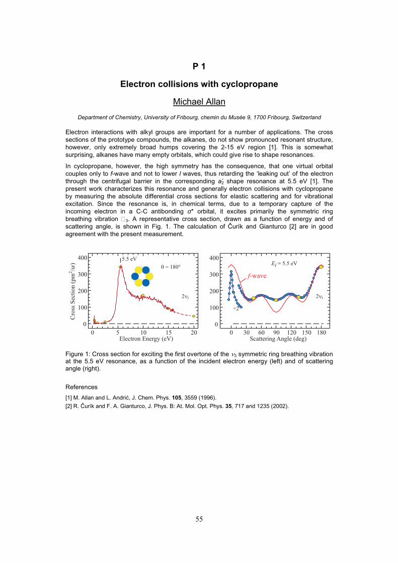
55
P 1
Electron collisions with cyclopropane
Michael Allan
Department of Chemistry, University of Fribourg, chemin du Musée 9, 1700 Fribourg, Switzerland
Electron interactions with alkyl groups are important for a number of applications. The cross sections of the prototype compounds, the alkanes, do not show pronounced resonant structure, however, only extremely broad humps covering the 2-15 eV region [1]. This is somewhat surprising, alkanes have many empty orbitals, which could give rise to shape resonances.
In cyclopropane, however, the high symmetry has the consequence, that one virtual orbital couples only to f-wave and not to lower l waves, thus retarding the ‘leaking out’ of the electron through the centrifugal barrier in the corresponding a2 shape resonance at 5.5 eV [1]. The present work characterizes this resonance and generally electron collisions with cyclopropane by measuring the absolute differential cross sections for elastic scattering and for vibrational excitation. Since the resonance is, in chemical terms, due to a temporary capture of the incoming electron in a C-C antibonding σ* orbital, it excites primarily the symmetric ring breathing vibration 3. A representative cross section, drawn as a function of energy and of scattering angle, is shown in Fig. 1. The calculation of Čurík and Gianturco [2] are in good agreement with the present measurement.
Figure 1: Cross section for exciting the first overtone of the ν3 symmetric ring breathing vibration at the 5.5 eV resonance, as a function of the incident electron energy (left) and of scattering angle (right).
References
[1] M. Allan and L. Andrić, J. Chem. Phys. 105, 3559 (1996).
[2] R. Čurík and F. A. Gianturco, J. Phys. B: At. Mol. Opt. Phys. 35, 717 and 1235 (2002).
0 30 60 90 120 150 180
0
100
200
300
400
Scattering Angle (deg)
÷2
Ei = 5.5 eV
0 5 10 15 20
0
100
200
300
400
Electron Energy (eV)
Cro
ss S
ecti
on
(p
m/s
r)2
2n3
q = 180°
f-wave
2n3
5.5 eV

56
P 2
Electron collisions with Pt(PF3)4
Michael Allan
Department of Chemistry, University of Fribourg, chemin du Musée 9, 1700 Fribourg, Switzerland
The interest in electron interactions with Pt(PF3)4 stems from its use as a platinum precursor in Focused Electron Beam Induced Processing (FEBIP) [1]. It is a volatile platinum compound with a molar mass of 547 amu! For FEBIP it has the advantage of not causing carbon deposits. It is a tetrahedral d10 complex.
Representative vibrational energy-loss spectrum, recorded with about 10 meV resolution at an incident energy of 0.7 eV is shown in Fig. 1, with an assignment based on the vibrational frequencies from Ref. [2]. Cross sections were measured for elastic scattering and for vibrational and electronic excitation, both as a function of electron energy and as a function of scattering angle. The elastic cross sections have deep Ramsauer-Townsend minima and their angular distributions have interesting minima around 40° which drop to lower scattering angles with increasing energy. The cross sections for vibrational excitation, reveal five resonances, probably shape resonances.
Figure 1: Electron energy-loss spectrum of Pt(PF3)4, recorded with an incident electron energy of 0.7 eV at a scattering angle of 45°.
References
[1] I. Utke, P. Hoffmann and J. Melngailis, J. Vac. Sci. Technol. B 26, 1197 (2008).
[2] R. M. Blight-Smith, H. G. M. Edwards and V. Fawcet, Spectrochim Acta 43A, 1069 (1987).
Cro
ss S
ect
ion (
Å/s
r)2
n1 PF strn2 3PF deform
n16 3PF asym deform
Pt(PF )3 4
q = 45°
Ei = 0.70 eV
0.0 0.2 0.4
0
1
2
3
4
Energy Loss (eV)
×1
×4×20
n3 Pt-P str

P 3
Rules governing the dissociation of Feshbach resonances in oxygen containing compounds
Bogdan C. Ibănescu and Michael Allan
Department of Chemistry, University of Fribourg, chemin du Musée 9, 1700 Fribourg, Switzerland
A thorough study of dissociative electron attachment (DEA) in 60 mostly saturated polyatomic molecules revealed a number of regularities and rules governing the dissociation of Feshbach resonances in oxygen and nitrogen containing compounds [1-6]. Some of them are presented here.
DEA bands due to shape resonances are observed below 5 eV in some cases. They are at the dissociation threshold in alcohols (for example the 2.84 eV band in the bottom spectrum of Fig. 1), and slightly above it in ethers (for example the 3.5 eV band in the top spectrum of Fig. 1).
Bands due to dissociation of the 2(n,3s2) Feshbach resonance are found around 6.5 eV when the O-H bond is broken (an example is the bottom spectrum in Fig. 1), but not when the C-O bond is broken (upper three spectra in Fig. 1). This rule has been rationalized by calculations of the potential surfaces on the parent states of the resonances, the 1(n,3s) Rydberg states [5]. Similar rules govern the dissociation of the 2(n-,3s2) Feshbach resonance seen as a shoulder at 7.9 eV in the ethanol spectrum in Fig. 1.
The 9.1-9.2 eV band in the top three spectra in Fig. 1 is at the same energy and has the same width in all three spectra despite the fact that the target molecules are different. A band at the same energy (±0.1 eV) was observed in all cases where a neutral C2H5 group was lost in DEA, for a wide variety of oxygen-containing target molecules [1,6]. Similar observation was made for the loss of other alkyl radicals, except that the band shifts to lower energy with increasing size of the alkyl radical lost, for example to 8 eV for the loss of n-butyl radical [6]. This leads to the conclusion that the excitation is localized on a given alkyl group for the high-lying Feshbach resonances.
References
[1] B.C. Ibanescu, Ph. D. thesis, University of Fribourg (2009).
[3] B.C. Ibanescu, O. May, A. Monney, M. Allan, Phys. Chem. Chem. Phys. 9 (2007) 3163.
[4] B.C. Ibanescu, O. May, M. Allan, Phys. Chem. Chem. Phys. 10 (2008) 1507.
[5] B.C. Ibanescu, M. Allan, Phys. Chem. Chem. Phys. 10 (2008) 5232. [6] B.C. Ibanescu and M. Allan, Phys. Chem. Chem. Phys., DOI:10.1039/B904945B (2009).
Figure 1: DEA spectra. Bottom spectrum: (M-1)- in ethanol; top three spectra: loss of C2H5 from various ethers.

58
P 4
Delayed fragmentations of nitro aromatic explosives resulting from electron attachment
I. Annen1, A. Mauracher1, S. Denifl1, E. Alizadeh1, P. Scheier1, T.D. Märk1, M. Probst1, K. Graupner2, S. Haughey2, T.A. Flield2
1Institut für Ionenphysik und Angewandte Physik, Technikerstraße 25, A-6020 Innsbruck, Austria 2Department of Physics and Astronomy, Queen’s University Belfast, Belfast BT7 1NN, United Kingdom
Introduction: Investigations about explosives and their detections are getting more important motivated by the need to prevent terror attacks with explosives involved. Therefore the group in Innsbruck carried out some experiments on relevant molecules in close collaboration with the group in Belfast.
Experimental Setups: The apparatus in Innsbruck is a double focusing mass spectrometer with a Nier-type ion source. An electron beam with controllable energy (energy resolution ~1 eV at low energies) is crossed with a molecular beam of neutral molecules of interest. Attachment of the electrons leads to the formation of mostly fragment anions. These anions are momentum-analyzed by a magnetic sector field and energy-selected by an electric sensor. Finally, the anions are detected by a channel electron type multiplier. Metastable dissociation of anions in the field free region and the corresponding kinetic energy release distribution (KERD) of the reaction are investigated by the mass analyzed ion kinetic energy (MIKE) technique. The Belfast apparatus consists of a linear Time-Of-Flight (TOF) mass spectrometer with a two-zone extraction region in combination with a pulsed trochoidal electron monochromator (energy resolution ~0.3 eV a low energies). Dinitrotoluene was inserted to the apparatus at room temperature. The ions are extracted ~1 µs after the end of the electron pulse. They pass through an acceleration region before they enter a field free drift region. The TOF is sensitive to metastable dissociations which occur in the interaction and acceleration regions.
Results: The picture on the right side shows a plot of 2,4-dinitrotouluene [1] measured by the TOF in Belfast. The yield of anions shown on a log-scale is plotted vs. the flight time and the electron energy. The narrow vertical bands are due to 2,4-DNT parent ions and prompt fragment ions. The parent anion peaks at about 0.0 eV and the fragment anions peak at about 0.5 eV. Delayed dissociations of anions are responsible for the events between the narrow vertical bands. The most metastable signal is monitored at low electron energies between 152 and 165 Dalton.
Reference
[1] F. Zappa, M. Beikircher, A. Mauracher, S. Denifl, M. Probst, N. Injan, J.Limtrakul, A. Bacher, O. Echt, T. D. Märk, P. Scheier, T. A. Field, K.Graupner, Metastable Dissociation of Anions Formed by Electron Attachment.ChemPhysChem 4/9 (2008) 607-611

59
P 5
Construction of an instrument to identify suitable molecules for ECCL – work in progress
E. H. Bjarnason, B. Ómarsson, Oddur Ingólfsson
University of Iceland, Faculty of science, Department of Chemistry, Science Institute, Dunhaga 3, 107 Reykjavik, Iceland
Electron controlled chemical lithography (ECCL) is based on the ability of low energy electrons to initiate and effectively drive selective bond cleavage processes in molecular systems. The selectivity is being controlled by the kinetic energy of the electron within the range of few electron-volts or by the choice of functional groups of the target molecule. An example of an ECCL process, is where molecules are linked to a substrate by chemical reactions induced by injecting tunneling electrons from a STM tip, resulting in microscopically patterned surfaces with specific chemical or physical properties.
We present a design of an instrument, currently under construction, that serves the purpose of identifying, in an efficient way, suitable molecular targets for use in ECCL. Such candidates need to show high selectivity and high efficiency with regards to bond rupture induced by dissociative electron attachment (DEA), and should maintain that selectivity and efficiency when coupled to an environment in clusters, in condensed phase or adsorbed on surfaces.
The instrument comprises a custom designed differentially pumped high vacuum chamber. It’s design is is based on a typical crossed electron- and molecular beam electron attachment ionization process combined with TOF-MS [1], with electron-beam energy range of 0 to 20 Ev with sub 100 meV resolution. Molecules may be introduced in different environments: isolated or embedded in homogeneous- or heterogeneous molecular clusters and large low vapor pressure molecules with molecular mass up to 100 kDa.
In an external vacuum chamber, either a laser desorption [2] or a laser induced acoustic desorption [3] electron attachment time-of-flight mass spectrometry feature will be included, where the molecules of interest may be desorbed in the presence of a high pressure carrier gas and subsequently the analyte/carrier gas mixture is expanded to form a cold molecular beam via a pulsed valve.
The predominantly neutral beam enters the internal vacuum chamber via a skimmer and may then be ionized by attachment of low energy electrons, formed by a trochoidal electron monochromator [4]. The negatively charged ions, formed upon electron attachment, are mass analyzed with a TOF-MS perpendicular to the molecular beam.
Furthermore, a transferable ion optics system in the TOF-chamber, will measure the lifetime of ions during their drift. The lifetime with regards to autodetachment and the lifetime with regards to dissociation will be measurable and also the part of the electron energy that converts to the molecule-fragment kinetic energy.
References
[1] Wiley W. C. and McLaren I. H., Time-of-Flight Mass Spectrometer with Improved Resolution, Review of Scientific Instruments, 1955, 26, 1150 – 1157 [2] Ingolfsson O. and Wodtke A. M., Laser Desorption Electron Attachment Time-of-Flight Mass Spectrometry: A New Approach to Detection of Involatile Compounds, Journal of American Society for Mass Spectrometry, 2001, 12, 1339 – 1347 [3] Bald I., Dabkowska I. and Illenberger E., Angew. Chem. Int. Ed. 2008, 47, 8518 – 8520

60
P 6
Nanometer-scale electron lithography on the hydrogenated diamond surface with a conducting AFM
E. Boer-Duchemin, G. Comtet, G. Dujardin, A.J. Mayne, E. Tranvouez, T. Wang
Laboratoire de Photophysique Moléculaire, Université de Paris-Sud, 91405, Orsay, France
In recent years, the scanning tunneling microscope (STM) and the atomic force microscope (AFM) have been used to manipulate matter at the atomic-scale. Electronic excitation with the STM or conducting AFM tip appears to be the most versatile method of manipulation as it allows to activate a large variety of processes such as molecular motions, chemical reactions or luminescence1.
Whereas electronic excitation in the electron tunnel regime with the STM is reasonably well understood1, electronic excitation in the contact regime with the conducting AFM has been much less explored.
We report the nanometer-scale electron lithography on the hydrogenated diamond surface with a conducting AFM operating in air. The hydrogenated diamond (C(100)) surface has a p-type doped semiconducting surface conductivity2. When applying a positive surface voltage as low as 1.5 V, electron lithography with the conducting AFM tip enables to locally isolate the hydrogenated diamond surface. This reaction is similar to the previously observed local oxidation of silicon surfaces3. However, here, the local oxidation is produced without any observed change in the AFM topography. This is explained by the removal of the hydrogen layer accompanied by the oxidation of the first carbon layer of diamond.
This electron lithography is used to pattern nanometer-scale isolated areas on the hydrogenated diamond and to investigate electron transport through these patterned structures.
References 1 A.J. Mayne, G. Dujardin, G. Comtet, D. Riedel, Chemical Reviews 106, 4355-4378 (2006) 2 L. Hellner, A.J. Mayne, R. Bernard and G. Dujardin, Diamond and Related Materials 14, 1529 (2005) 3 J.A. Dagata, J. Scheir, H.H. Harary, C.J. Evans, M.T. Postek and J. Bennett, Appl. Phys. Lett. 56, 2001 (1990).

61
P 7
Threshold excitation of diethyl ether and tetrahydrofuran by electron impact
Marcin Dampc, Ireneusz Linert, Tomasz Neumann, Mariusz Zubek
Department of Physics of Electronic Phenomena, Gdańsk University of Technology, Gdańsk 80–233, Poland
Electron impact threshold excitation of the gas-phase diethyl ether (DE), C4H10O and tetrahydrofuran (THF), C4H8O molecules has been investigated. DE molecule has a quasi-linear structure with an oxygen atom binding two ethyl groups. THF molecule, a five-membered hydrocarbon ring also containing an oxygen atom, is considered as a simple model analogue of deoxyribose. Previous studies of the electronic excitation of DE and THF in the gas phase include measurements of the energy loss spectra at 100 eV incident energy [1] and of trapped electron spectra at residual energy of ~0.3 eV [2]. In the present measurements, we have modified electron analyzer of our spectrometer [3] to detect threshold electrons produced in the excitation of the target molecules with residual energies lower than 40 meV. In the modification the technique of penetrating electrostatic field [4,5] has been exploited. The overall energy resolution of the measurements was about 40 meV. Fig. 1 presents the threshold electron spectra of DE and THF obtained at the residual energies of Er = 0.035 eV and 0.02 eV, respectively. Broad bands with maxima at 6.40 and 6.25 eV, respectively appear at lower energies than those of the excitation of the optically allowed Rydberg states [1]. They show presence of two or more triplet states in each of both spectra, excited most likely via negative ion resonances.
5.5 6.0 6.5 7.0 7.5 8.0 8.5 9.0 9.5
THFEr = 0.02 eV
DEEr = 0.035 eV
Threshold electron yield
Electron energy loss (eV)
Fig. 1 Threshold electron energy loss spectra of DE and THF measured at residual energies of Er=0.035 and 0.02 eV, respectively.
References
[1] W-C. Tam, C.E. Brion, J. Electron Spectr. Rel. Phenom. 3, 263 (1974) [2] L. J. Bremner, M.G. Curtis, I.C. Walker, J. Chem. Soc. Faraday Trans. 87, 1049 (1991) [3] I. Linert, M. Zubek, J. Phys. B: At. Mol. Opt. Phys. 39, 4087 (2006) [4] S. Cvejanović, F.H. Read, J. Phys. B: At. Mol. Phys. 7, 1180 (1974) [5] M. Zubek, D.S. Newman, G.C. King, J. Phys. B: At. Mol. Phys. 24, 495 (1991)

P 8
High resolution threshold photoionization studies of isoxazole
Marcin Dampc1, Brygida Mielewska1, M.R.F. Siggel-King2, G.C. King3 , Bhala Sivaraman4, Sylwia Ptasińska4, Nigel Mason4 Mariusz
Zubek1 1 Department of Physics of Electronic Phenomena, Gdańsk University of Technology, 80-233 Gdańsk,
Poland
2 Daresbury Laboratory, Daresbury, Warrington, WA4 4AD, UK 3School of Physics and Astronomy, Manchester University, Manchester M13 9PL, UK
4 Department of Physics and Astronomy, The Open University, Milton Keynes, UK
Isoxazole, C3H3NO is a five-membered heterocycle molecule that is related to furan by replacing the CH group in the furan ring with a N-atom at position 2. It is, with its derivatives, well known for its importance in the synthesis of antibiotic, antitumour and anti-HIV agents [1-3].
In this poster we report the results of threshold photoionization studies of isoxazole using synchrotron radiation at the Daresbury Laboratory. Photoelectron spectra were recorded in the photon energy range 9.9 - 30 eV, such results significantly extend the range of earlier He I photoionization measurements [4-6]. The present studies have been performed using a threshold photoelectron spectrometer employing a penetrating field technique, an apparatus which has been previously described in detail [7]. The overall energy resolution of the present measurements was better than 15meV.
A threshold photoelectron spectrum of isoxazole obtained over the energy range from 9.9 to 10.4eV is shown in figure 1. It covers the region of ionization from the highest occupied molecular orbital 3a”(π3) [4]. The first threshold ionization peak is centered at 9.967eV. The observed vibrational structures have been resolved into four series with vibrational energies of 65meV, 100meV, 136meV and 173meV, tentatively assigned to v18, v16, v9, and v5 modes, respectively. These results are consistent with the most intense vibrational modes reported [6], however allowed to observe much more
vibrational transitions than previously.
References
[1] P. Pevarello et al., Targets Heterocyclic Syst. 3, 301 (1999). [2] K. D. Shin et al., J. Biol. Chem. 280, 41439 (2005). [3] B. L. Deng et al., Bioorg. Med. Chem. 14, 2366 (2006). [4] M. H. Palmer et al., J. Mol. Struct. 40, 191 (1977). [5] T. Kobayashi et al., Bull. Chem. Soc. Jpn. 55, 3915 (1982). [6] I. C. Walker et al., Chem. Phys. 297, 306 (2004). [7] R. I. Hall et al., Meas. Sci. Technol. 3, 316 (1992).
Fig. 1. Threshold photoionization spectrum of isoxazole.
9,9 10,0 10,1 10,2 10,3 10,40
10
20
30
40
50
60
70v16
v5
v9
v18
Photoelectron yield (arb.units)
Photon energy (eV)

63
P 9
Electronic Control of Single Molecules on Insulating Layers
G. Dujardin, A. Bellec, F. Chiaravalloti and D. Riedel
Laboratoire de Photophysique Moléculaire, Orsay, France
In the context of mono-molecular electronics, it is essential to electronically decouple the molecule from the substrate in order to achieve electronic control of the molecular functions (electron transfer, charging, luminescence, etc).
A new class of scanning tunneling microscopy (STM) experiments have been performed by using molecules adsorbed on thin insulating layers grown on metallic substrates [1-2] . However, the use of thin insulating films grown on semiconducting substrates would have a number of advantages as compared to metallic substrates ; absence of leak current within the bulk band gap, fabrication of metallic contact on the semiconductor substrate, etc.
By using a low temperature (5K) STM, we have performed topography, I(V) spectroscopy and manipulation of single molecules on two different insulating layers on semiconductor substrates ; (i) pentacene molecules on the hydrogenated Si(100) surface [3], and (ii) hexaphenyl molecules on a CaF2 thin layer grown on Si(100).
References
[1] X.H. Qiu, G.V. Nazin and W. Ho, Science 299, 542 (2003) [2]J. Repp et al. Phys. Rev. Lett. 94, 026803 (2005) [3] A. Bellec et al. Nano Lett. 9, 144 (2009)

64
P 10
Dissociative electron attachment cross sections for HCl, HBr and their deuterated analogs – comparison of experiment and
theory
J. Fedor1, 2, O. May2, M. Allan2, P. Kolorenč3 , M. Čížek3 ,J. Horáček3 1J. Heyrovský Institute of Physical Chemistry, Dolejškova 3, 18000 Prague, Czech Republic
2Department of chemistry, University of Fribourg Chemin du Musée 9, 1700 Fribourg 3Institute of Theoretical Physics, Charles University, V Holešovičkách 2, 18000 Prague, Czech Republic
We present dissociative electron attachment cross-sections for HX and DX (X= Cl and Br). Experimental results, obtained using a trochoidal electron monochromator with a recently constructed total ion collection collision chamber [1], are compared to the predictions of the nonlocal resonance model [2, 3].
The experimental results for Br from HBr are in excellent quantitative agreement with predictions of the nonlocal resonance model [2], but the experimental cross sections for HCl and DCl are, respectively, 2.2 times and 2.8 times smaller than the theoretical predictions of the HCl model from Ref. [3]. We present a modified model for HCl, with an improved short-range part of the ion potential. The new short-range part has been fitted on recent high quality electron scattering eigenphase sums calculated using the Schwinger Multichannel method [4].
We present an improvement of the model for HCl via modification of the short-range part of ion potential involved in the model. The new short-range part has been fitted on the electron scattering eigenphase sums calculated using the Schwinger Multichannel method [4].
The new model for HCl agrees much better with the experimental data than the model from Ref. [3]. The present model also partly resolves an old controversy in the DEA temperature dependence in HCl – the agreement with experiments of Allan and Wong [5] is better in present model. Additionally, the present model reproduces all qualitative structures observed in the elastic scattering and vibrational excitation cross sections.
References [1] J. Fedor, O. May, M. Allan, Phys. Rev. A 78, 032701, 2008 [2] M. Čížek, J. Horáček, A.-C. Sergenton, D. B. Popović, M. Allan, W. Domcke, T. Leininger, F. X. Gadea, Phys. Rev. A 63, 062710, 2001 [3] M. Čížek, J. Horáček, W. Domcke, Phys. Rev. A 60, 2873, 1999 [4] C. Winstead, V. McKoy, private communication [5] M. Allan, S.F. Wong, J. Chem. Phys. 83, 1687, 1981

65
P 11
Anisotropy in electron distributions from fullerenes excited by femtosecond laser pulses
J. Fedor1, 2, M. Kjellberg3, J. Stenfalk3, O. Johansson3,4, E.E.B. Campbell3, 4, K. Hansen3
1J. Heyrovský Institute of Physical Chemistry, Dolejškova 3, 18000 Prague, Czech Republic 2Department of chemistry, University of Fribourg Chemin du Musée 9, 1700 Fribourg 3Department of Physics, University of Gothenburg, SE-4196 Gothenburg, Sweden
4Department of Chemistry, University of Edinburgh, Scotland
The photoelectron spectra for fullerenes C60 and C70 ionized using 775 nm, 160 fs laser pulses show thermal electron kinetic energy distributions but they also exhibit angular anisotropy with respect to the laser light polarization. Both the electron yield and the electron kinetic energy distribution (related to an apparent electron temperature) increase in the polarization direction. We explain the observed behavior by considering the influence of the electric eld on the energy distribution of electrons emitted from the hot electronic subsystem of the molecule. This influence is unique for statistical emission.

66
Autoionizing States
-0.3 0 0.3 0.6
Electron Energy (eV)
Intensity (arb. units)
150-300
200-400
300-600
SF6-
SF6-theory
SF6neutraltheory
P 12
Autodetachment Rates of SF6-* Anions: Some Agreement
Between Theory And Experiment
Thomas A Field1, Karola Graupner1 1Centre for Plasma Physics, Department of Physics and Astronomy Queen’s University Belfast, Belfast
BT7 1NN, N. Ireland, UK
An experiment to study the autodetachment of metastable SF6-* ions formed in close to zero
energy electron attachment to SF6 has been performed with ERIC [1], which includes a trochoidal electron monochromator and a time-of-flight mass spectrometer. Negative ions are formed in the source region of a time-of-flight mass spectrometer. They are accelerated in two electric field regions before passing through a electric field free drift region. If an anion autodetaches in the drift region then a fast neutral will arrive at the detector rather than a negatively charged ion. Fast neutrals can, like ions, be detected at microchannel plates. In this experiment the microchannel plate voltages were changed to positive ion detection mode with negative ions passing through the mass spectrometer. These conditions repel negative ions from the detector, but fast neutrals are detected.
With this experimental arrangement, autodetachment in the field-free drift region is observed. The SF6
-* ions autodetach in this region 4.2 to 9.4 µs, 3.8 to 8.4 µs and 3.4 to 7.1 µs after electron attachment with the mass spectrometer voltage settings of 150-300, 200-400 and 300-600 indicated in the figure below. Thus, the lifetime of SF6
-* ions with different internal energies was not measured. Instead, the internal energies of SF6
-* ions which dissociate in different time windows is observed. The results show clearly that the earliest time window (300-600 voltage setting) has a slightly higher energy profile, and higher mean SF6
-* internal energy, than the other windows. Furthermore the latest window has the lowest energy profile. These results are consistent with shorted lifetimes at higher internal energies.
Continuous theoretical lines are shown in the figure with experimental data points. The dashed SF6
- line shows a convolution of high resolution SF6
-* formation data [2] with an experimental Gaussian type function. The SF6 neutral theoretical solid line is the same convolution of high resolution data [2] and experimental function, but it is also convoluted with the probability of autodetachment in the 300-600 time
window of 3.4 to 7.1 µs after autodetachment. The autodetachment probabilities were derived from recent theoretical predictions of SF6
-* lifetimes [3]. There is some agreement between the theoretical solid line and the experimental data points in the figure. This may be the first (near) quantitative agreement between experiment and theory on the autodetachment of SF6
-*
References
[1] T.A. Field et al, J. Phys. B: At. Mol. Opt. Phys. 38 (2005) 255–264 [2] M. Braun et al. Eur. Phys. J. D, 35 (2005) 177–191 [3] L.G. Gerchikov and G.F. Gribakin Phys. Rev. A 77 (2008) 042724

67
P 13
Total electron scattering and ionization cross sections in THF
M. Fuss1, A. Muñoz2, F. Blanco3, P. Limão-Vieira4 and G. García1
4Instituto de Física Fundamental, Consejo Superior de Investigaciones Científicas (CSIC), Serrano 113-bis, 28006 Madrid, Spain
2 Centro de Investigaciones Energéticas Medioambientales y Tecnológicas (CIEMAT), Avenida Complutense 22, 28040 Madrid, Spain.
3 Departamento de Física Atómica, Molecular y Nuclear, Universidad Complutense de Madrid, 28040 Madrid, Spain
4Atomic and Molecular Collisions Laboratory, Department of Physics,
New University of Lisbon, Campus de Caparica, 2829-516 Caparica, Portugal
Following the method described recently for water molecules [1], total electron- THF scattering cross sections have been measured from 50 to 5000 eV with experimental errors of about 5%. Integral elastic and inelastic cross sections have been calculated over a broad energy range (1-10000 eV) with an optical potential method assuming an independent atom representation [2]. Improvements based on atomic screening corrections inside the molecule and low scattering angle normalization procedures have been included [3]. Total and partial ionization cross sections have been also derived in this range through the simultaneous electron and ion current measurement complemented by a time of flight analysis of the induced charged fragments. From a detailed evaluation of the present results and their comparison with previous theoretical and experimental data, a set of recommended integral cross sectional data is provided. These data will be used as a complement for modelling radiation damage induced by photons and electrons in biomolecular systems. A typical mass spectrum of charged fragments induced by 500 eV electrons is shown in the figure below.
References [1] Muñoz A, Oller JC, Blanco F, Gorfinkiel JD, Limao-Vieira P, Garcia G. Phys. Rev. A 76, 052707 (2007). [2] Blanco F, Garcia G. Phys. Rev. A 67, 022701 (2003). [3] A. R. Milosavljevic, F. Blanco, D. Sevic and B. P. Marinkovic, Eur. Phys. J. D 40, 107 (2006).
0 20 40 60 80 100Mass (atomic units)
0
20
40
60
80
100
Ion intensity (arbitrary units)
THF ion fragmentation by 500 eV electron impact
C4H8O+
C4X+
C3X+
C2X+
CX+
68
P14
Interactions of DNA with Ionic Phospholipids in Langmuir Monolayers at the Air−Water Interface
P.J. Gomes1#, A.M.P.S. Gonçalves da Silva2, R.S. Romão2 and M. Raposo1
1 Centro de Física e Investigação Tecnológica, Universidade Nova de Lisboa, 2829-516 Caparica, Portugal
2 Centro de Química Estrutural, Complexo I, Instituto Superior Técnico, 1049-001 Lisboa, Portugal
Recently, the research on deoxyrribonucleic acid (DNA)−lipid interactions has been motivated mostly by two medical applications: i) on nonviral gene transfer and DNA complexes with cationic lipids; ii) and on diagnostic applications with detection of specific base pairing (“lab on the chip”), requiring DNA adsorbed on a surface in a suitable way to be exposed to either a complementary chain and/or a DNA polymerase. Another important application is the development of biological sensors, such as the radiation sensor.
A preliminary study of DNA−lipid interactions can be carried out using Langmuir monolayers of phospholipids spread on DNA aqueous solutions. This work addresses the interaction studies of two phospholipids, the anionic 1,2-distearoyl-sn-glycero-3-phospho-(1'-rac-glycerol) (DPPG) and the cationic 1,2-distearoyl-sn-glycero-3-ethylphosphocholine (EDPPC), with DNA.
The surface pressure − area (π−A) isotherm of DPPG progressively deviates to larger areas at low surface pressures with the DNA concentration in the subphase, becomimg nearly invariant at high surface pressures up to the collapse of the monolayer. This means that DNA penetrates in the expanded regime of DPPG monolayer, being excluded from the condensed regime probably due to repulsive electrostatic interactions. Differently, the π−A isotherm of EDPPC is not significantly affected in the expanded regime, while becomes steeper in the condensed regime and the collapse surface pressure increases with the DNA content in the subphase. DNA in the water subphase clearly stabilizes the cationic EDPPC monolayer.
The relaxation curves at constant surface pressure, A−t, confirm the above trend. For the anionic DPPG monolayer, the rate of decay increases with the DNA concentration as a consequence of the repulsive monolayer−subphase interaction. Contrarily, for the cationic EDPPC monolayer, the rate of decay decreases with the DNA concentration, proving that the existence of opposite charges in the subphase, stabilizes the EDPPC monolayer.

69
P 15
Characterization of Titanium and Silicon Nanoparticle Films by STM and XPS techniques
M.Hager1, S. Jaksch1, R. Stijepovic1, N. Endstrasser1, P. Scheier1
1Institut für Ionenphysik und Angewandte Physik, Technikerstraße 25, A-6020 Innsbruck, Austria
Nanoparticle films of titanium and silicon are produced by DC magnetron sputtering onto highly oriented pyrolytic graphite (HOPG). The sputtering is performed with an argon pressure of 3 Pa. The size, shape and electrical properties of the nanoparticles are investigated as a function of the plasma parameters. Without breaking the vacuum the sample is transferred to the STM- or XPS- chamber.
By Scanning Tunneling Microscopy (STM) surface characterization, manipulation of the film and sample stability are investigated. The STM is operated at room temperature working in constant current mode and using a platinum iridium tip. Manipulation is done by varying the bias voltage and the tunneling current.
X-ray Photoelectron Spectroscopy (XPS) measurements are carried out at room temperature and provide information on the chemical composition of the surface layers. In combination with Ar ion beam sputtering depth profiles of the nanoparticle films are obtained. The XPS is operated in constant energy mode and all spectra are collected using monochromatic Al Kα radiation (1486.6 eV). Particular attention is given to the oxidation of the films of nanoparticles. Even long term exposure of the silicon films to ambient air only leads to the oxidation of the surface of the nanoparticles, however with a constant level throughout the whole film thickness. This indicates that molecular oxygen can easily diffuse through the voids between the nanoparticles but is not able to oxidize the interior of the nanoparticles. The figure below shows depth profiles of silicon in various oxidation states.
Charging of the films by electron irradiation and STM imaging at increased bias voltages modifies the properties of the films. We observe increased oxidation at the irradiated regions and with the STM an increase of the local density of states is measured. Apparently such films of nanoparticles provide interesting properties for electron driven reactions that can be controlled locally in the low
nanometer regime.
Acknowledgments: work was supported by the FWF, Wien and the European commission Brussels via the project ITS-LEIF and the cost network CM0601 (ECCL)
0 50 100 150 200 250 300 350 400 450
0
200
400
600
800
1000
1200
As grown fresh SiC [cps.eV]
SiO2 [cps.eV]
Si2O3 [cps.eV]
SiO [cps.eV]
Si2O [cps.eV]
Si [cps.eV]
sputter time [s]
Area Si2p [cps.eV]

70
P 16
Low-energy electron-induced reactions in NH3-containing molecular films
T.Hamann, E.Böhler, P.Swiderek
Universität Bremen, Fachbereich 2 (Chemie/Biologie), Institute of Applied and Physical Chemistry, Leobener Straße / NW 2,
Postfach 330440, 28334 Bremen, Germany
The attachment of amino groups to simpler molecules or materials is of high interest as such groups serve as anchors for more complex molecular architectures such as biomolecules /1/. The use of electron-beam techniques in such processes furthermore offers the perspective to pattern a surface. It was recently shown that low-energy electron-induced reactions can lead to the formation of larger molecules from simple starting materials /2/ or to surface functionalization /3/. In such processes, dissociative electron attachment processes lead to specific fragmentations depending on the incident electron energy. In a condensed environment or at surfaces the resulting reactive species can induce further reactions that may lead to coupling of different components of the material. Thus it is tempting to investigate if such processes can be used for an amino functionalization of a surface.
This contribution presents results of experiments aiming at the activation of NH3 by exposure to low-energy electrons and the attachment of the resulting fragments to hydrocarbon materials. It is shown that simple electron exposure of condensed films of NH3 leads to desorption of the material and formation of N2 while small hydrocarbons cross-link to produce larger homologues. Therefore, other approaches were conceived, including different precursors for NH2 and electron-induced activation of the hydrocarbon prior to or during exposure to a process gas. The first results are discussed here.
References
/1/ A.Turchanin, A.Tinazli, M.El-Desawy, H.Großmann, M.Schnietz, H.H.Solak, R.Tampé, A.Gölzhäuser, Adv.Mat. 20, 471 (2008).
/2/ I.Ipolyi, W.Michaelis, P.Swiderek, Phys.Chem.Chem.Phys. 9, 182 (2007). /3/ A. Lafosse, M. Bertin, D. Caceres, C. Jäggle, P. Swiderek, D. Pliszka, R.Azria, Eur.Phys.J.D 35,
363 (2005).

71
P 17
Dissociative Electron Attachment to 2-Nitro-m-xylene
Sean Haughey1, Karola Graupner1, Andreas Mauracher2, Tilmann D. Märk2, Paul Scheier2, Thomas A. Field1
1Centre for Plasma Physics, Department of Physics and Astronomy Queen’s University Belfast, Belfast BT7 1NN, N. Ireland, UK
2 Institut für Ionenphysik und Angewandte Physik, Leopold Franzens Universität, Innsbruck, Austria
An experiment to study dissociative electron attachment to 2-nitro-m-xylene (C8H9NO2) has been conducted with ERIC, an experiment that has been described previously [1], which includes a trochoidal electron monochromator and a time-of-flight mass spectrometer. A typical spectrum of negative ions formed in electron attachment to 2-nitro-m-xylene is shown below..
In a similar experimental investigation of electron attachment to 2,4-dinitrotoluene (CH3C6H3(NO2)2) [2] a metastable parent ion (CH3C6H3(NO2)2
-) was formed close to zero electron energy. This parent ion fragmented on microsecond timescales to give a number of different products; the principal daughter ions were, C7H5N2O3
- and C7H6NO3-, formed by loss of
OH and NO respectively.
By contrast, in the current experiment, the metastable parent anion formed at close to zero electron energy does not show metastable dissociation. The lack of fragmentation may be due to the dissociation threshold lying above 0eV electron energy.
There appears to be a threshold for the formation of NO2- above ~ 0.5eV, but this ion is formed
promptly and there is no metastable feature observed near this apparent dissociation threshold. It is speculated that no metastable dissociation is observed in the loss of NO2
- because of steric crowding in 2-Nitro-m-xylene where the NO2 group is adjacent to two methyl (CH3) groups on the benzene ring. Once sufficient energy is available for dissociation the reduction in steric crowding by loss of NO2 may drive this reaction and explain the lack of slow metastable dissociation to form NO2
- on microsecond timescales.
Many fragment ions are formed in a resonance at ~ 4eV and metastable dissociation is observed between two of the fragment ions.
References [1] T.A. Field et al, J. Phys. B: At. Mol. Opt. Phys. 38 (2005) 255–264 [2] F. Zappa et al, ChemPhysChem, 9 (2008) 607–611

72
P 18
Theoretical approach to predict metastable dissociation of deprotonated anions in gas phase
O Ingólfsson, HD Flosadóttir, H Jónsson,
Science Institute, University of Iceland, Dunhagi 3, 107 Reykjavík, Iceland
We have measured the metastable dissociation of the deprotonated nucleosides uridine, guanosine and deoxyguanosine, using matrix assisted laser desorption ionisation time of flight mass spectrometry (MALDI TOF MS). Complicated molecules such as nucleosides have many possible deprotonation sites (marked 1a-d and 2a-d in the figure below), and thus a possibility to form more than one anion via deprotonation. When the nucleosides are deprotonated they can undergo further dissociation leading to different smaller fragments, each fragment only formed selectively from one specific deprotonation site.
To identify the selective fragmentation dependent upon the deprotonation site we have modified uridine and guanosine to block selectively different deprotonation sites (a-d) and thus observe the fragmentation channels available from each anion formed.
NH
O
ON
O
OHOH
HH
HH
HO
NH
N
N
O
NH2N
O
OHOH
HH
HH
HO
1a
1b1c
1d
2a
2b2c
2d
Uridine Guanosine In addition to the measurements we have simulated the metastable dissociation of uridine and 2’-deoxyguanosine using the same method as we used successfully on the amino acid L-Valine1 which showed excellent agreement with our measurements. Now we have proceeded with these calculations towards larger and more complicated molecules such as the nucleosides. These simulations show exactly the same bond ruptures as observed from metastable dissociation measured in MALDI, from the same deprotonation sites as observed when measuring the modified compounds. We have thus been able to predict the metastable fragmentation of each anion of the nucleosides by means of DFT and molecular dynamics calculations.
Reference
1. H. D. Flosadottir, S. Denifl, F. Zappa, N. Wendt, A. Mauracher, A. Bacher, H. Jonsson, T. D.
Mark, P. Scheier and O. Ingolfsson, Angew. Chem.-Int. Edit. 46 (42), 8057-8059 (2007).

73
P 19
Dual-probe scanning tunneling microscope for study of nanoscale metal-semiconductor interfaces
Ismet I. Kaya1, W. Yi2, D. M. Chen3, V. Narayanamurti4 1Sabanci University, Istanbul, Turkey
2Harvard University, Cambridge, MA, USA 3Rowland Institute at Harvard University, Cambridge, MA, USA
4Harvard University, Cambridge, MA, USA
A dual-probe scanning tunneling microscope is used to perform ballistic electron emission spectroscopy on Au/GaAs interface. Dual tip STM has the capability to position both tips independently with a few microns resolution by optical navigation. In the experiment one tip is contacted the patterned metallic thin film while the other tip is used to inject ballistic electrons. The collector current spectra agrees with a Monte-Carlo simulation based on modified planar tunneling theory. Our results suggest that it is possible to study nanoscale metal-semiconductor interfaces without the requirement of an externally-contacted continuous metal thin film.

74
P20
The electronic state spectroscopy of isoprene [CH2CHC(CH3)CH2] studied by electron and photon impact
P Limão-Vieira 1,2,*, Y Nunes 1, G G B de Souza 3, S Eden 2, N J Mason 2,D Duflot 4 J Delwiche 5, M-J Hubin-Franskin 5
1 Atomic and Molecular Collisions Laboratory, Department of Physics, New University of Lisbon, 2829-516 Caparica, Portugal
2 Centre of Molecular and Optical Sciences, Department of Physics and Astronomy, The Open University, Walton Hall, Milton Keynes, MK7 6AA, UK
3 Instituto de Química, Universidade Federal do Rio de Janeiro, Ilha do Fundão, 21949-900, Rio de Janeiro, RJ, Brazil
4 Laboratoire de Physique des Lasers, Atomes et Molécules (PhLAM), UMR CNRS 8523, Centre d’Études et de Recherches Lasers et Applications CERLA, FR CNRS 2416), Université des Sciences et
Technologies de Lille, F-59655 Villeneuve d’Ascq Cedex, France 5 Laboratoire de Spectroscopie d’Électrons diffusés, Université de Liège, Institut de Chimie-Bât. B6c B-
4000 Liège 1, Belgium
Measuring and monitoring VOCs has become particular important in environmental regulation, air quality monitoring and with the recent biological interest in metabolic and environmental origins in human breath [1, 2], the latter closely linked as a biomarker associated with cholesterol synthesis [3]. However, due to the low concentrations (including in the atmosphere) and presence of a large number of chemical species in exhaled breath, breathe analysis requires high sensitive and selective instrumentation to detect and identify the atypical absolute concentrations of specific biomarkers. Future medical units working in close link with synchrotron radiation facilities can make use of the VUV spectra wavelength region [4] to trace VOCs in patients.The VUV photoabsorption spectrum of isoprene (2-methyl-1,3-butadiene), CH2CHC(CH3)CH2, is reported in the wavelength range 115 to 270 nm (10.8 to 4.6 eV). The result is compared with high-energy electron energy loss spectra (EELS) in dipolar conditions. Both the photoabsorption and EELS measurements represent the highest resolution data yet reported. The spectra are found to be in good agreement with each other. New photoabsorption features are observed and Rydberg and vibrational assignments suggested. The He(I) photoelectron spectrum is recorded from 8.0 to 17.0 eV and compared to earlier work. New vibrational structure is observed in the first photoelectron band. Absolute cross section values have been obtained allowing photolysis lifetimes to be derived in the Earth’s troposphere and stratosphere [5].
References
[1] M Phillips, J Greenberg, J Awad, J Clin. Pathol., 47 (1994) 1052. [2] M E O’Hara, S O’Hehir, S Green and C A Mayhew, Physiol. Meas., 29 (2008) 309. [3] B Buszewski, M Kesy, T Ligor and A Amann, Biomed. Chromatogr., 21 (2007) 553. [4] F Ferreira da Silva, M Nobre, A Fernandes, R Antunes, D Almeida, G Garcia, N J Mason and P Limão-
Vieira, J. Phys. Conf. Series, 101 (2008) 012011. [5] A M F Rodrigues, F Rodrigues, G G B de Souza, S Eden, N J Mason, D Duflot, P J Gomes, S V
Hoffmann, J Delwiche, M-J Hubin-Franskin and P Limão-Vieira, (2009) in preparation.

75
P21
Isotope effect in dissociative electron attachment cross sections in acetylene
Olivier May, Juraj Fedor and Michael Allan
Department of Chemistry, University of Fribourg, Chemin du Musée 9, 1700 Fribourg, Switzerland
We present absolute cross section measurement of dissociative electron attachment to C2H2 and C2D2. The C2H
–/C2D– band at 3 eV shows pronounced isotope effect with the cross section
for C2H2 being 14.7 times larger than that for C2D2. The light fragments H– and D– dominate the
second dissociative electron attachment band around 8 eV. These bands exhibit much weaker isotope effects which are in agreement with their assignment to Feshbach resonances.
The abstract of the corresponding presentation contains further details.

76
P22
Locally restricted resonances in electron-molecule scattering
Bernd M. Nestmann
Institute of Physical and Theoretical Chemistry, University of Bonn, Wegelerstraße 12, 53115 Bonn, Germany
Resonances appearing in the fixed-nuclei cross sections can typically be extended to a certain region of internuclear distances. Within the Fano-Feshbach projection operator approach these resonances are associated with metastable electronic states depending on the internuclear distances, which can be characterized by complex, energy-dependent potential surfaces. One expects that the experimentally observed resonant vibrational excitations and dissociative electron attachment processes can be explained by these potential surfaces. This, however, may not be true in general since the appearance of resonances may be locally restricted due to the opening of new decay channels, depending on the internuclear distances.
In the e-+NO scattering experiments an enhancement of the N+O- fragmentation has been observed for collision energies between 7 and 10 eV but the calculations did not show any resonance which appears inside the Franck-Condon region as well as provides the fragmentation into the observed channel. The theoretical results are based on the R-matrix approach including electron correlation to a high amount. The method applied in order to detect resonances has turned out to be very liable in past. The conclusion from the results obtained is an interaction of resonances and background scattering processes based on the nuclear motion. It is our assumption that such kind of interaction me play a role in several other processes, e.g. vibrationally inelastic electron scattering off HF.

77
P 23
Understanding the VUV spectroscopy of CF3COOCH3 as a route to environmental friendly hydrofluorinated ethers (HFEs)
Y Nunes1,*, N C Jones2, S V Hoffmann2, N J Mason3, P Limão-Vieira1,3
1 Atomic and Molecular Collisions Laboratory, Department of Physics New University of Lisbon, 2829-516 Caparica, Portugal
3 Institute for Storage Ring Facilities, University of Aarhus, Ny Munkegade, DK-8000, Aarhus C, Denmark 3 Centre of Molecular and Optical Sciences, Department of Physics and Astronomy, The Open University,
Walton Hall, Milton Keynes, MK7 6AA, UK
Chlorofluorocarbons (CFCs) have been extensively used in refrigeration systems, as foam blowing agents and also as aerosol propellants. They are a source of atmospheric radicals and therefore widely recognised to contribute significantly to stratospheric ozone depletion [1, 2] by halogenated atoms releasing under photolysis. Halogenated hydrocarbons are also strong greenhouse gases, having long residence times in the atmosphere (of the order of hundreds of years) and estimated global warming potentials of several thousands in a 100-year period evaluation. CFCs are being replaced by newly alternatives such as oxygenated hydrocarbons, especially haloethers, hydrochlorofluorocarbons (HCFCs) and hydrofluorocarbons (HFCs). Due to the constant need to decrease and phase out the harmful effects of CFCs and their derivatives in the atmosphere, hydrofluorinated ethers (HFEs) are expected to be an acceptable alternative. HFEs when released in the Earth’s atmosphere are known to react with OH radicals, therefore reducing the risk on global warming. Since hydrofluorinated esters (fluoroesters, FESs) can be produced during the atmospheric oxidation of some HFEs [3], it is necessary to understand the atmospheric chemistry of FESs in order to evaluate the environmental acceptability of HFEs.
The electronic state spectroscopy of methyl-trifluoroacetate, CF3COOCH3, has been investigated by high resolution VUV photoabsorption spectroscopy in the energy range 4.0 – 10.8 eV. Absolute cross section values have been obtained allowing photolysis lifetimes to be derived in the Earth’s troposphere and stratosphere [4].
References
[1] P Fabian, R Borchers, R Leifer, B H Subbaraya, S Lal and M Boy, Atmospheric Environment, 30 (1996) 1787.
[2] P Limão-Vieira, S Eden, P A Kendall, N J Mason and S V Hoffmann, Chem. Phys., 364 (2002) 535. [3] M B Blanco and M A Teruel, Atmos. Environ., 41 (2007) 7330. [4] Y Nunes, N C Jones, S V Hoffmann, N J Mason and P Limão-Vieira, (2009) in preparation.

78
P 24
The simulated effects of fringing fields and orthogonal velocity components on peak shape and electron energy resolution in
the Trochoidal Electorn Monochromator
Benedikt Ómarsson, Elías H. Bjarnason, Oddur Ingólfsson
University of Iceland, Faculty of Science, Department of Chemistry, Science Institute, Dunhaga 3, 107 Reykjavik, Iceland
The trochoidal electron monochromator [1] has been used in electron attachment experiments for many years to produce an electron beam of well defined energy. The trajectories of electrons inside the monochromator have been discussed and well described by many authors [1, 2, 3]. The energy resolution one can achieve from the TEM is in theory governed by a few well defined parameters such as the length of the deflection region and the displacement of the entrance aperture from the exit aperture. Other factors such as fringing fields at the deflector plate edges and the electric field between these edges and the entrance and exit electrode also play a role in the electron energy resolution. This has been claimed [1, 3] to be the ruling factor for the resolution. Second comes the fact that the electrons velocity components orthogonal to the magnetic fields direction is conserved through the ExB field. This factor has been neglected by most studies and considered to have a minimal influence on the resolution. Other factors include Coulomb interaction between electrons and space charge (the electrons spatial charge interacting with the electric and magnetic field).In our simulations we deal with two of four issues mentioned, the fringing fields and the orthogonal velocity component. Since the other two factors, the coulomb repulsion and the space charge are inevitable, they are neglected. The results obtained from our simulations show a significant decrease in resolution related to the increase in the orthogonal velocity component (figure 2). The figure shows the increase in the electron beam’ FWHM as a function of maximum initial spread angle, which corresponds to an increase of the orthogonal velocity component. The simulations also show that the decrease in resolution is considerably greater for the fringing field effects.We have simulated the simplest model of the TEM (figure 2), two deflector electrodes, an entrance- and an exit electrode using SIMION 8 [4]. In addition to simulation and comparison of the relative energy deterioration we suggest improved geometries of the deflector plates that we expect to minimize the effects of fringing fields on the electron energy resolution.
Figure 1 – The change of the FWHM of an electron beam as a function of maximum spread angle. The orthogonal velocity component increases with increasing angle.
Figure 2 – The simple TEM model used in the simulations.
References [1] A. Stamatovich and G. J. Schulz, Rev. Sci. Instrum. 41, 423 (1970). [2] V. Grill, H. Drexel, W. Sailer, M. Lezius and T. D. Mark. Int. J. Mass Spectrom. 205, 1-3 (2001) 209 -
226. [3] D. Roy. Rev. Sci. Instr. 43, 3 (1972) 535 - 541. [4] SIMION 8 3D, ion optics simulator. www.simion.com
79
P 25
HF formation as a driving force for selective bond cleavage
Benedikt Ómarsson, Helga Dögg Flosadóttir, Constanze König, Oddur Ingólfsson and Eugen Illenberger
University of Iceland, Faculty of Science, Department of Chemistry, Science Institute, Dunhaga 3, 107 Reykjavik, Iceland
Electron Attachment has been a progressing field of study since the later half of the last century. However, it is only recently that dissociative electron attachment (DEA) studies on biomolecules and molecules with increasing complexity demonstrated its potential as a tool to break chemical bonds with a remarkable site, and energy selectivity. This makes the DEA processes the pivotal reaction in Electron Controlled Chemical Lithography (ECCL).
Very recently a DEA study on penta-fluorophenylacetonitrile reported a strong signal of [M-HF]- at an energy close to 0 eV [H.D Flosadóttir et al., PCCP, accepted]. In order to form HF from the molecule a C-F and a C-H bond must be broken. The energy required for the process is only available if the H-F bond is formed simultaneously. This remarkable reaction occurs with almost no excess energy and is only triggered by the excess charge. We expect the formation of [M-HF]- to depend on the distance between the H and F atoms.
Here we present data on DEA to 2-chloro-6-fluorophenylacetonitrile which shows the formation of the fragment [M-HF]- along with other resonances.
Benedikt Ómarsson acnowlages support from the COST action CM0601 (ECCL) for a short term scientific mission to Berlin to conduct the presented experiments.
References
[1] A. Stamatovich and G. J. Schulz, Rev. Sci. Instrum. 41, 423 (1970). [2] V. Grill, H. Drexel, W. Sailer, M. Lezius and T. D. Mark. Int. J. Mass Spectrom. 205, 1-3 (2001) 209 - 226. [3] D. Roy. Rev. Sci. Instr. 43, 3 (1972) 535 - 541. [4] SIMION 8 3D, ion optics simulator. www.simion.com

80
P 26
Extreme nanotribology: Atomistic level lateral force measurement
using novel home-made combined nc-AFM/STM
Ö.Ünverdi(1), M.Atabak(2), D.Gemici(1), H.Ö.Özer(3), A. Oral(1)
1 Sabancı University, Faculty of Engineering and Natural Sciences, Tuzla, Đstanbul, Turkey 2 Omicron Nanotechnology GmbH, Taunusstein, Germany
3 Đstanbul Technical University, Faculty of Science and Literature, Dept.of Physics, Maslak, Turkey
A combined STM and non-contact Atomic Force Microscope fibre interferometer based is designed. The microscope is capable of measuring lateral forces simultaneously with the normal farces and tunelling current based is designed. To measure the lateral force a special cantilever is dithered in the lateral directions with sub-angstrom oscillation amplitudes at frequency below the resonance frequency and the changes in oscillations amplitude is detected using a lock-in amplifier. The lateral force gradient and topolography images of terraces on Si (111) (7x7) surfaces are obtained.

81
P 27
MODELLING DISSOCIATIVE FRAGMENTATION OF AMINOACIDS AFTER RESONANT ELECTRON ATTACHMENT: A
QUANTUM PICTURE.
C. Panosetti1 , F. Sebastianelli1, I. Baccarelli2 and F. A. Gianturco1
1Dept. of Chemistry and CNISM, University of Rome “La Sapienza”, P.le Aldo Moro 5, 00185 Rome, Italy
2CASPUR, Via dei Tizii 6b, 00185 Rome, Italy
We performed calculations to investigate some aspects of the radiation damage of biomolecules, focusing on theoretical modelling of indirect fragmentation caused by low-energy electrons (LEEs) ejected by biological tissues when exposed to ionizing radiation. Scattering equations are formulated within a symmetry-adapted, single-center expansion of both continuum and bound electrons, and the interaction forces are obtained from a combination of ab–initio calculations and a nonempirical model of exchange and correlation effects[1]. We present total elastic scattering cross sections and S-matrix poles calculated for the equilibrium geometry of glycine, alanine, proline and valine. Our model will be shown to be able to reproduce the fragmentation patterns obtained by experiments ([2], [3], [4]). We also propose a one-dimensional model for the dynamics of intramolecular energy transfer via vibrational excitation of selected bonds. Since we actually find the expected kind of behavior, if our hypothesis were to be correct, we would then be able to identify, for a given resonance, those bonds the breaking of which directly or indirectly relate to the occurrence of a resonant state.

82
P 28
Studies of low energy electron interaction with furanose-structured alcohols
P. Papp1, A. R. Milosavljević2, J. Kočišek1, D. Kubala1, P. Mach3, J. Urban3, B. P. Marinković2, Š. Matejčík1
1Department of Experimental Physics, Faculty of Mathematics, Physics and Informatics, Comenius University, Mlynska dolina, Bratislava, 842 48 Slovakia
2 Laboratory for Atomic Collision Processes, Institute of Physics, Pregrevica 118, 11080 Belgrade, Serbia
3 Department of Nuclear Physics and Biophysics, Faculty of Mathematics, Physics and Informatics, Comenius University, Mlynska dolina, Bratislava, 842 48 Slovakia
Two furanose-structured alcohols related to some extent to deoxyribose molecule, 3-hydroxytetrahydrofuran (3HTHF, C4H8O2) and α-tetrahydrofurfuryl alcohol (THFA, C5H10O2), have been studied both experimentally and theoretically. Crossed electron/molecule beams technique together with quadrupole mass spectrometer has been used for electron induced positive ion formation studies. The mass spectra of 3HTHF and THFA determined at incident electron energy 70 eV were analyzed and for number of mass-charge ratios (m/q) the ionization efficiency curves have been measured. Thus, for 3HTHF 18 different m/q channels were analyzed experimentally, from which 6 were selected for deeper investigation on DFT level of theory and finally 5 for the G3MP2 method. The same was performed for THFA with 15 m/q channels experimentally, 5 on DFT and 4 on G3MP2 levels of theory. Theoretical calculations with Gaussian 03 program package led to more than 160 fragmentation reactions on B3LYP/6-311+G(2d,2p), only the energetically comparable to experimentally observed appearance energies were re-computed on G3MP2.
The experiment showed that although there were 18 and 15 m/q channels observed for 3HTHF and THFA respectively, only 10 of them for 3HTHF were abundant over 10% of the ionic yield of most intensive fragment and 6 for THFA. While the parent cation of 3HTHF occurred within its abundant ionic fragments, THFA cation had a weak signal only. Our theoretical conformational studies of neutral and cationic 3HTHF and THFA agree well with this experimental observation, for 3HTHF+ at least 2 stable conformers were found while for the cationic state of THFA partial dissociation the side CH2OH chain group occurred. However the later process, dissociation of CH2OH, seems to be a common feature for the most abundant fragmented ion of both molecules, the corresponding structures are the furanose closed ring cation with m/q=71 for THFA fragment and CH2=CH-CH-OH
+ with m/q=57 for 3HTHF fragment. Additionally, fragmentations to cations with m/q=70, 58, 43 and 31 were studied theoretically for both molecules, except the 58 for THFA that was not observed. One could expect when looking at the chemical formulas of both molecules that the fragmented cationic structures can be oxygenated, either singly (possible products found for all discussed channels) or doubly (only the C2H2O2
+ with m/q=58 fragmented from 3HTHF, but its second most abundant fragmented ion), or can be also cationic hydrocarbons. Only cationic hydrocarbon product found for our selected channels was the C3H7 cation with m/q=43, with corresponding neutrals COOH from 3HTHF and CH2OH + CO from THFA, for the later the second most abundant cationic fragment.
This work was supported by the COST Action CM060, Electron Controlled Chemical Lithography (ECCL), by the Slovak Research and Development Agency, Projects No. APVV-07-365 and APVV-0310-07, as well as the Ministry of Science and Technological Development of Republic of Serbia under project 141011. Collective from Comenius University in Bratislava would like also to acknowledge to VEGA grant 1/0558/09.
83
P 29
MEASUREMENT OF LASER-INDUCED FLUORESCENCE OF MOLECULES USING A TIME-RESOLVED SPECTROMETAR
B. A. Petruševski1,2, M. Terzić3, M. S. Rabasović1, D. Šević1,2, S. Savić Šević1, B. Murić1, D. Pantelić1, B. P. Marinković1,2,5
1Institute of Physics, Belgrade, Serbia 2Electrical Engineering College, Belgrade, Serbia 3Faculty of Science, University of Novi Sad, Serbia
Measurements of laser induced fluorescence (LIF) for several known strong fluorescing molecules have been performed in order to achieve the calibration of new experimental apparatus designed for study of molecules of biological relevance. Depending on the specific application, fluorescence measuring systems strongly differ in instrumental design, i.e. use of optical components, detection systems as well as in measurement geometries including the sample cell. Proper calibration of the system that comprises excitation laser, optical cell and detection system is therefore necessary in order to obtain accurate signal interpretation. Optical emission spectroscopy represents comparative experimental technique to the optical absorption and low energy electron spectroscopy techniques in present kind of study [1].
Here, we report various approaches for the calibration of the time-resolved laser-induced fluorescence (TR-LIF) detection system. This system is based on the tunable Nd:YAG laser (320-475 nm) to excite samples and on the detection part with high spatial and temporal resolution [2]. Different methods for calibration of time domain and wavelengths are known [3-5] and the possibility of theirs application to our TR-LIF system has been presented. Feasibility of some methods will be tested with standard fluorescent dyes, such as fluorescein which is intended for use in establishing a reference scale for fluorescence intensity [6]. Also, rhodamine B is used as a common dye to investigate a fluorescence yields and lifetimes. The amount and wavelength of the emitted energy depend on both the fluorophore and the chemical environment. This technology has particular importance in the field of biochemistry and protein studies. Fluorescence spectroscopy is increasingly being used as a technique for probing the structure and dynamics of nucleic acids. Recently, fluorescence methods have been used to elucidate the three-dimensional arrangement of complex DNA and RNA structures [7]. The systematic errors in calibration procedure will be also discussed.
References [1] B. P. Marinković, A. R. Milosavljević, J. B. Maljković, D. Šević, B. A. Petruševski, D. Pavlović, D. M. Filipović, M. Terzić and V. Pejčev, Acta Physica Polonica A 112 (2007) 1143.
[2] M. Terzić, B. P. Marinković, D. Šević, J. Jureta and A. R. Milosavljević, Facta Universitatis, Series Phys. Chem. Technol. 6 (2008) 105.
[3] A. Dinklage, T. Lokajczyk, and H. J. Kunze, J. Phys. B 29 (1996) 1655. [4] D. Pfeifer, K. Hoffmann, A. Hoffmann, C. Monte and U. Resch-Genger, J. Fluorescence 16 (2006) 581. [5] S. B. Keller, J. A. Dudley, K. Binzel, J. Jasensky, H. M. de Pedro, E. W. Frey and P. Urayama, Anal. Chem 80 (2008) 9876.
[6] P.C.DeRose and G.W. Kramer, J. of Luminescence 113 (2005) 314. [7] D. P. Millar, Current Opinion in Structural Biology, 6 (1996) 322.
5 e-mail: [email protected]

84
P 30
Vibrational Dynamics of Tunneling Molecular Junctions
I.A.Pshenichnyuk, M.Čížek
Institute of Theoretical Physics, Charles University, V Holesovickach 2, Prague 8, 18000 Czech Republic
Charge transport properties of a single-molecule junction are investigated. The interaction of the transported electrons with the internal degree of freedom of the molecule is taken into account. Special attention is paid to the voltage dependence of the current through the junction and its vibrational dynamics.
Fig.1. Schematic representation of the junction.
Simulations are performed for the model junction which consists of two metallic electrodes (described in the framework of tight-binding model) connected by a molecule with one vacant orbital available for the tunneling electrons. Internal degrees of freedom of the molecule are represented by one vibrational mode. System Hamiltonian reads:
VHHH ld ++=
ddhddhH dd
++ += )()(0 ϕϕ , ∑ +=α
αααεk
kkkl ccH , [ ]∑ ++ +=α
ααα ϕk
kkk dccdVV )(
where dd ,+ and αα kk cc ,+ are creation/annihilation operators for the electron on the molecule
and the leads respectively. Operators 0h and dh represent vibronic Hamiltonians for the neutral and the charged molecule. Index RL,=α denotes the left and the right lead. The vibrational coordinates are represented by an angle ϕ . The form of the functions 0h , dh and
)(ϕαkV is taken such that it resembles the behavior of the molecules with poly-phenyl chains[1].
To study conductance properties of the molecular bridge, master equation (ME) approach is used. ME for the reduced density matrix (RDM) in the Wangsness-Bloch-Redfield form[2] reads:
[ ] [ ][ ]∫∞
++− ⊗−−=0
0)()( )(,,)(,)( l
HHiHHi
leadsd tVeeVtrdtHitdt
dldld ρρτρρ ττ .
It is integrated numerically using Arnoldi method to obtain stationary state )(∞ρ , which is used to calculate all the properties of interest of the molecular bridge.
References: (1) M. Čížek Czech. J. Phys. 2005, 55, 189 (2) C. Timm Phys .Rev. B 2008, 77, 195416
L R
M
Current

85
P 31
Dynamics of Birefringence Creation and Relaxation in Azobenzene Containing Surfaces
Ribeiro PA1, Madruga CM1, Alliprandini Filho P2, Gonçalves MR3, Mason NJ4 and Raposo M1
1Centro de Física e Investigação Tecnológica, CeFiTec, Faculdade de Ciências e Tecnologia, Universidade Nova de Lisboa, Campus da Caparica, 2829-516 Caparica, Portugal.
2Instituto de Física, Universidade Federal de Uberlândia, 38400902, Uberlândia, MG, Brazil. 3 Institute of Experimental Physics, Ulm University, Albert-Einstein-Allee 11, 89069 Ulm, Germany
4Department of Physics and Astronomy, The Open University, Walton Hall, Milton Keynes, MK7 6AA, United Kingdom
Surfaces of azobenzene molecules have been widely investigated mainly due to its photoisomerization capabilities which allow the creation of reversible birefringence [1] and optical relief gratings [2,3], features which are suitable for creation of molecular devices. Among other envisaged applications are optical storage devices, wet-chemical free holography, liquid crystal alignment, wavelength dependent molecular switches, command surfaces. Particularly photoinduced birefringence creation in azobenzene molecules can be achieved by impinging polarized light in the range of 255 nm to 550 nm, depending on the azo-substituent, which causes successive trans-cis-trans isomerization cycles after which a net orientation of the azomolecules in a direction perpendicular to de light electric field vector is attained. As a result the landscape will be highly birefringent with remarkable birefringence values raging from 10-1 to 10-3. When the light is turned off some of the azobenzene molecules will disorientate and consequently part of the birefringence lost, although some residual of birefringence will be preserved. Furthermore, the created birefringence could be reversibility deleted either by impinging the sample with circularly polarized light or by heating it. The dynamics of light induced birefringence creation and its relaxation after light removal depends on the material structural relaxation dynamics, being its characterization fundamental for the determination of device reliability, namely for determining the conditions which lead to the achievement of maximum birefringence values and make them a stable and durable for the sake of device reliability. In this work the dynamics of birefringence creation and decay in surfaces of the azopolymer poly1-[4-(3-carboxy-4-hydroxyphenylazo)benzenesulfonamido]-1,2-ethanediyl, sodium salt will be presented namely its dependence on the light intensity.
References
[1] - Todorov T., Nikolova L., Tomova N., “Polarization holography. 1: A new high-efficiency organic material with reversible photoinduced birefringence”, Appl. Opt., 23, 4309, (1984). [2] - Rochon, P.; Batalla, E.; Natansohn A., Optically induced surface gratings on azoaromatic polymer films, Appl. Phys. Lett. 66, 136, (1995). [3] - Kim D., Tripathy S., Liam L., Kumar J., “Laser-induced holographic surface relief gratings on nonlinear optical polymer films” Appl. Phys. Lett. 66, 1166, (1995).

86
P 32
Creation of Polarized light by Orientation of Electroluminescent organic molecules Induced by Photorientation of Azobenzene
Molecules
Alliprandini Filho P1,2, Ribeiro PA1, Madruga CM1 Marletta A2 and Raposo M1
1Centro de Física e Investigação Tecnológica, CeFiTec, Faculdade de Ciências e Tecnologia, Universidade Nova de Lisboa, Campus da Caparica, 2829-516 Caparica, Portugal.
2Instituto de Física, Universidade Federal de Uberlândia, 38400902, Uberlândia, MG, Brazil.
Polarized light emitted by organic molecules as result of molecular alignment is of particular technological interest namely for application in organic light emitting diodes (oLEDs) [1,2]. This work reports a study about of the polarization state of the emitted light the poly-p-phenylenevinylene (PPV) polymer when these concerning molecules have been orientated by the photoorientation of (poly[1-[4-(3-carboxy-4-hydroxyphenylazo) benzenesulfonamido]-1,2-ethanediyl, sodium salt]) (PAZO) an azobenzene polymer with capability of photoinduced orientation via photoisomerization about the azogroup. In this work, thin layer-by-layer films of poly (xylylidene tetrahydrothiophenium chloride) (PTHT), the PPV precursor, and PAZO (PTHT/PAZO) were prepared and characterized by spectrophotometry. For comparison thin cast films of PTHT+PAZO with different polymeric amounts have been also prepared and characterized. For both type of samples birefringence was induced in order to orientate the polymeric molecules and the polarized light emitted by PPV after PTHT temperature conversion was characterized as a function of polymeric percentage amount.
References [1] - Tang, C.W. and Van Slyke, S.A., Appl. Phys. Lett. 1987, 51, 913. Todorov T., Nikolova L., Tomova N., “Polarization holography. 1: A new high-efficiency organic material with reversible photoinduced birefringence”, Appl. Opt., 23, 4309, (1984). [2] - Tokuhisa, H.; Era, M.; and Tsutsui, T., Appl. Phys. Lett. 1998, 72, 2639.

87
P 33
Characterization of DNA Intercalation with 2,2’-Bipyridyl on Solid Surfaces
Coelho M1, Martins L1, Gomes PJ1, Ribeiro PA1, Tavares P2 and Raposo M1
1Centro de Física e Investigação Tecnológica, CeFiTec, Faculdade de Ciências e Tecnologia, Universidade Nova de Lisboa, Campus da Caparica, 2829-516 Caparica, Portugal.
2CQFB, Departamento de Química, Faculdade de Ciências e Tecnologia, Universidade Nova de Lisboa, 2829-516 Caparica, Portugal.
Cancer is one of the major causes of death in the world. Current cancer therapies are limited to surgery, radiotherapy and the use of cytotoxic agents. For most forms of disseminated cancer, however, no curative therapy is available, in such a way that the discovery and development of novel active chemotherapeutic agents is largely required. Studies on the interaction of metal complexes with deoxyribonucleic acid (DNA) are of great interest due to their potential applications as anticancer medications and stereo selective probes of nucleic acid structures. These metal complexes can be seen not only as chemotherapeutics but also as radiosensitive agents for enhanced dedicated radiotherapy. As is well-known, cis-diamminedichloroplatinum (II) (Cisplatin) has been approved for use since the 1970 s for treatment of a selection of human cancers, however, its use is limited because of the severe toxic side effects. For this reason, new metal compounds are being used such as ruthenium (II), ruthenium (III) and palladium (II). Normally these metal complexes are synthesized with the ligands: 2,2’-bipyridyl, 1,10-phenanthroline, pyridine and pyrimidine. In this work one intends to embrace the interaction mechanisms between the ligand 2,2’-bipyridyl and DNA, to be carried out in solution and in thin cast films of DNA and 2,2’-bipyridyl deposited on solid supports as a mimic of a soft condensed matter biological system. Infrared (IR), ultraviolet/visible (UV/VIS) and fluorescence spectroscopy probes were used to characterize both solutions and thin films.

88
P 34
Low energy electron-induced processes in lithographic materials:
electron stimulated desorption from methyl methacrylate (MMA)
films
N. Hedhili1, A.D. Bass2, C. Mills2, and L. Sanche2.3
1 Unité de recherche matériaux avancés et optronique, Faculté des sciences de Tunis, 1060 Tunis, Tunisia 2 Département de médecine nucléaire et radiobiologie, Université de Sherbrooke, Sherbrooke, QC,
J1H5N4, Canada 3 Department of Physics and Astronomy, The Open University, Milton Keynes, UK.
Methyl methacrylate (MMA - C5H8O2) has been used as a model for understanding the effects of hydrocarbon contamination on the performance of the Ru coated mirrors used for Extreme Ultra-Violet Lithography (EUVL) [1] and is the monomeric precursor to Polymethyl methacrylate, a versatile polymer used in microlithography [2] and other applications. A recent study using x-ray photoelectron spectroscopy (XPS), temperature programmed desorption and low energy ion scattering, indicates that MMA films on Ru can be polymerized by exposure to low energy electrons as evidenced by the persistence of MMA derived XPS signals to elevated temperatures in electron-irradiated films[3].
Here we employ the electron stimulated desorption (ESD) of anionic fragments to probe electron and thermally induced processes occurring in multilayer films of MMA deposited on polycrystalline Pt foil. Measurements as a function of incident electron energy (Ei) and current (It), demonstrate that in the 0-20 eV incident energy range, the desorption of anions such as H-, OCH3
−, C3H5−(or OC2H
−), CH2− and CH3
− (all, except OC2H−,formed by scission of a single bond)
proceeds via dissociative electron attachment (DEA) to the molecule and by dipolar dissociation (DD). Anion desorption yields (at Ei = 8 eV) vary with film temperature and drop dramatically at ~125K. Comparisons with Low Energy Electron Transmission (LEET) and Thermal Desorption Spectrometry (TDS) measurements indicate that the decrease in desorption is associated with thermal annealing and a change from of an amorphous into a more ordered multilayer film. The ESD signal of H− at 8 eV, is used to probe of the quantity of MMA-derived hydrocarbons remaining on the substrate at elevated temperatures following extended exposure to electrons and presumed polymerization. We find that the ESD signal at T >260 K is dependent on both electron irradiation energy ‘Eir’ and the temperature at which the film was deposited and irradiated. Polymerization can be induced with Eir < 10 eV, though much higher desorption yields are observed with Eir > 20 eV (T=100 K) and 40 eV (T=135 K). The ESD signal at high T from irradiated films varies quasi-linearly with electron fluence suggesting that polymerisation can be initiated by a single electron scattering event.
References
[1] R Wasielewski, BV Yakshinskiy, MN Hedhili, A Ciszewski, TE Madey, Proc. SPIE. 6533 (2007) 653316. [2] I Kostic, R Andok, V Barak, I Caplovic, A Konecnikova, L Matay, et al., J.Mater.Sci.: Mater.Electron. 14 (2003) 645-648. [3] MN Hedhili, BV Yakshinskiy, R Wasielewski, A Ciszewski, TE Madey, J.Chem.Phys. 128 (2008) 174704.

89
P 35
Radiation-induced processes on gold nanoparticles: Raman SERS analysis of adsorbed amino acids and peptides.
Pierre Cloutier1, Tony A. Mattiol2, Gérard Keller3, Cécile Sicard-Roselli4 et Léon Sanche1,4,5
1Groupe en Sciences des Radiations, Faculté de Médecine, Université de Sherbrooke, Sherbrooke Québec, J1H 5N4 Canada.
2Laboratoire de Biophysique du Stress Oxydant SBE/DBJC et URA 2096 CNRS, CEA, 91191 Gif/Yvette Cedex, France .
3Laboratoire de Physico-chimie des Systèmes Polyphasés, CNRS UMR 8612, Université Paris-Sud, Centre d'Etudes Pharmaceutiques, 5 Rue Jean Baptiste Clément, 92296 Châtenay-Malabry, France.
4Laboratoire de Chimie Physique, Université Paris-Sud UMR 8000 CNRS, 91405 Orsay Cedex, France. 5Department of Physics and Astronomy, Walton Hall, The Open University, Milton Keynes, UK.
In nanolithography, molecules adsorbed on surfaces are often modified by beams of high energy particles. Such surfaces range from metal to insulators and are usually flat and well-defined. In the present work, molecules adsorbed on gold nanoparticles (GNP) are modified by high energy radiation. Fonctionalization of GNP is of special interest in Radiotherapy where such particles can be used to enhance radiation damage to cancer cells [1]. In such an application, the GNP are often chemically linked to a peptide [1] to achieve tumor cell specificity. To study the effects of high-energy radiation on such a combination, we measured the degradation induced by X and gamma rays on amino acids and peptides adsorbed on GNP. We compared the effect of X-rays of different energies with those from gamma irradiation. . Damage to amino acids induced by irradiation concern mainly the COO- function. For peptides, the CH2 group of the backbone is also perturbed. Lower doses are needed for the same damage with X-rays of low energy (AlKα 1.5 keV) than with gamma rays. Degradation is slightly different with gamma rays (additional functions are modified). We show that this damage can easily be monitored by Surface-Enhanced Raman Spectroscopy (SERS).
Reference
[1] J.F. Hainfeld et al., J. Pharmacy and Pharmacology, 60, 977 (2008).

90
P 36
Interaction of thermal electrons with chloro- and bromo- containing hydrocarbons
Iwona Szamrej, Janina Kopyra, Jolanta Wnorowska, Mieczysław Foryś
Chemistry Department, University of Podlasie, 3 Maja 54, Siedlce, 08-110, Poland
This work is a continuation of our program to investigate systematically the kinetics of thermal electron capture by haloalkanes by means of a time-resolved electron swarm method.
A pulsed Townsend technique has been applied. Electrons are produced photoelectrically using Nd:YAG laser with the following parameters: wavelength 266 nm, pulse duration 5 ns, repetition frequency 10 Hz and average maximum pulse energy 96 mJ. The laser beam enters the chamber through a quartz window from the side of the chamber and directly hits the center of the cathode. The induced, due to the motion of the electrons, potential is amplified, registered on the digital oscilloscope and saved in a computer memory. The heating device for the chamber has also been built which allows heating it up to 150 C. The device gives very stable temperature of the gaseous mixture within 10C.
With this technique thermal attachment rate coefficients (i.e., equal electron temperature Te and gas temperature TG) are measured. With rising temperature, the thermal rate coefficients were found to increase with an Arrhenius-type behavior, kth(T) = A exp(-Ea/kT), yielding corresponding activation energies, Ea.
In this work we present the results for ethane and propane derivatives substituted with chlorine and bromine, CH2ClCH2Br, CH2BrCH2Br, CH2ClCHClCH2Cl and CH2ClCH2CH2Br.
The example results in terms of ln(kth) vs. 1/T for CH2ClCH2Br and CH2ClCH2CH2Br are shown in Figs. 1a and 1b, respectively. They give, as expected, the linear dependence. From the slopes of the straight lines activation energies, Ea, were calculated.
0,0028 0,0029 0,0030 0,0031 0,0032 0,0033 0,0034-21,0
-20,8
-20,6
-20,4
-20,2
-20,0
a
ln(k
th)
1/T (K-1)
0,0027 0,0028 0,0029 0,0030 0,0031 0,0032 0,0033 0,0034
-21,8
-21,6
-21,4
-21,2
-21,0
-20,8
-20,6
-20,4
b
ln(k
th)
1/T (K-1)
The dependence of ln(kth) on 1/T for CH2ClCH2Br (a) and CH2ClCH2CH2Br (b).

91
P 37
Characterization of focused electron beam induced carbon deposits from organic precursors
T. Bret1, I. Utke2, P. Hoffmann2 1Carl Zeiss SMS – NaWoTec GmbH, D-64380 Rossdorf, Germany
2Empa, Swiss Federal Laboratories for Materials Testing and Research, CH-3602 Thun, Switzerland
Contamination in the electron microscope is due to electron-induced carbon fixation from residual gases. It is often seen as detrimental to the image quality, but can be used as a powerful 2D or 3D direct-write process. It has found numerous nanoscale applications, among which are high-resolution etch masks, atomic force microscopy super-tips, nano-electrode supports, nano-junction insulator material, and near-field optical sensors [1]. Carbon-containing materials present a rich variety of properties, in the crystalline (graphite, diamond, fullerenes, etc.) as well as non-crystalline carbon states.
We have screened several organic precursors for focused electron beam induced deposition of carbon including Styrene CH2=CH-C6H5, Acrylic acid CH2=CH-COOH, Propionic acid CH3CH2-COOH, Acetic acid CH3-COOH, and Formic acid H-COOH [2]. The growth rates are found to be higher with the light carboxylic acid series than with styrene. The deposits obtained are analyzed by Raman, FTIR, and EDX spectroscopy. FEB deposits obtained in a tungsten cathode SEM with a background pressure of 10-6 mbar show similar compositions and structures, quite independently on the nature of the above precursors. Besides C, the deposits contain only O and H. The compositions of the deposits are shown to be close to the sum formula C9H2O. The C fraction is amorphous carbon, more than 90% sp
2. Two electron-induced processes occur: electron-induced precursor fixation and desorption of volatile elements.
Fig. 1: µ-FTIR analyses. Absorption spectrum of liquid formic acid compared to the spectrum of the FEB deposit obtained from it [2]
Fig. 2: EDX spectra (analysis energy 3 kV) of deposits from three precursors (normalization at C Kα peak). The C/O ratio depends weakly on the precursor used. The bare substrate and the solid compound adamantanol are included as references [2].
Refeences [1] I. Utke, P. Hoffmann, J. Melngailis, J. Vac. Sci. Technol. B 26 (2008) 1197-1276 [2] T. Bret, S. Mauron, I. Utke, P. Hoffmann, Microelectronic Engineering 78–79 (2005) 300–306.

92
P 36
Fragmentation of diethyl ether by electron impact
Mariusz Zubek, Ireneusz Linert, Marcin Dampc
Department of Physics of Electronic Phenomena, Gdańsk University of Technology, 80-233 Gdańsk, Poland
We have investigated electron impact fragmentation processes in the gas-phase diethyl ether (DE), C4H10O leading to formation of electronically-excited neutral atomic and molecular fragments. DE molecule has a quasi-linear structure with a centrally located oxygen atom binding symmetrically two ethyl groups. It may be considered as a linear analogue of the cyclic tetrahydrofuran, C4H8O molecule. Comparative studies of interactions of low energy electrons with both molecules contribute to better understanding of their molecular fragmentation processes. Decomposition of DE has been investigated by radiation induced pyrolysis [1], UV photolysis of the liquid phase [2] and recently by observation of the dissociative electron attachment processes [3].
The measurements have been carried out using optical excitation technique [4], which allows identification of the fragmentation excited species by detection of their fluorescence decay. The incident electron beam is produced by a trochoidal electron selector. The optical channel ensures detection of fluorescence in the 180-650 nm wavelength range. In the investigated electron energy range (15-90 eV), measured fluorescence emission spectra (Fig.1) indicate production of atomic hydrogen H(n) in the n=4-7 states (Balmer series) and diatomic CH fragments in the A2∆ electronic state (A2∆-X2Πr band). The CH fragments are rotationally excited as shown by the broadening of the main line at the base in the 415-445 nm range. The appearance energies of the H(4) and CH (A2∆) fragments are determined and comparison with the estimated dissociation limits indicates that the excess energies (kinetic and internal excitation of the fragments) are lower than that obtained in THF fragmentation.
Fig. 1. Emission spectra of DE measured at an electron energy of 70 eV. The inset presents emission spectrum obtained with increased resolution of 1.5 nm.
References [1] G. Höhlein, G.R. Freeman, J. Am. Chem. Soc. 92 (1070) 6118. [2] C. von Sonntag, H.-P. Schuchmann, G. Schomburg, Tetrahedron, 28 (1972) 4333. [3] M. Allan, Phys. Chem. Chem. Phys. 10 (2008) 1507. [4] M. Dampc, M. Zubek, Int. J. Mass Spectrom. 277 (2008) 52.

93
ECCL 2009 MEETING
THE SECOND MEETING OF
COST ACTION CM0601
List of Participants
4th
to 9th
June 2009
Istanbul, Turkey

94
Dr Hassan Abdoul-Carime Physics Institut de Physique Nucléaire 4 rue Enrico Fermi Villeurbanne 69222 France [email protected]
Prof Michael Allan Chemistry University of Fribourg chemin du Musée 9 Fribourg 1700 Switzerland [email protected]
Mr Ivo Annen Institute for Ion Physics and Applied Physics University of Innsbruck Technikerstrasse 25/3 Innsbruck 6020 Austria [email protected]
Mr Husnu Aslan Faculty of Engineering & Natural Sciences Sabanci University Tuzla Istanbul 34956 Turkey [email protected]
Dr Isabella Baccarelli CASPUR Supercomputing Consortium Via dei Tizii 6 Rome 00185 Italy [email protected]
Dr Richard Balog Department of Physics and Astronomy Aarhus University Ny Munkegade 120, Bld. 1520 Aarhus C 8000 Denmark [email protected]
Mr Elias Bjarnason Physical Chemistry University of Iceland Dunhaga 3 Reykjavik 107 Iceland [email protected]
Dr Marie-Laure Bocquet Laboratoire de Chimie ENS LYON 46 Allée d'Italie LYON 69364 France [email protected]
Prof Petr Carsky Theoretical Chemistry Heyrovsky Institute, Acad.Sci.Czech Rep. Dolejskova 3 Prague 8 18823 Czech Republic [email protected]
Dr Genevieve Comtet Laboratoire de Photophysique Moleculaire Université Paris-Sud bât.351 Université Paris-Sud Orsay 91405 France [email protected]

95
Dr Roman Curik Theoretical Chemistry J Heyrovsky Institute Dolejskova 3 Prague 18223 Czech Republic [email protected]
Mr Marcin Dampc Department of Physics of Electronic Phenomena Gdansk University of Technology Narutowicza 11/13 Gdansk 80-233 Poland [email protected]
Dr Gérald Dujardin Laboratoire de Photophysique Moléculaire Bâtiment 210, Université Paris-Sud Orsay 91405 France [email protected]
Dr Klaus Edinger NaWoTec GmbH A Carl Zeiss SMT AG Company Industriestr. 1 Rossdorf 64380 Germany [email protected]
Dr Juraj Fedor J. Heyrovsky Inst. of Physical Chemistry Dolejskova 3 Prague 18000 Czech Republic [email protected]
Mr Filipe Ferreira da Silva Institute for Ion Physics and Applied Physics University of Innsbruck Technikerstrasse 25/4 Innsbruck 6020 Austria [email protected]
Dr Thomas Field Physics and Astronomy Queen's University Belfast University Road Belfast BT7 1NN UK [email protected]
Miss Helga Dögg Flosadóttir Science Institute University of Iceland Dunhaga 4 Reykjavik 107 Iceland [email protected]
Prof Mieczyslaw Forys Chemistry University of Podlasie 3Maja 55 Siedlce 08-110 Poland [email protected]
Prof Adam Foster Department of Physics Tampere University of Technology PO Box 692 Tampere 33101 Finland [email protected]

96
Ms Martina Fuss Instituto de Física Fundamental CSIC c/ Serrano 133-bis Madrid 28006 Spain [email protected]
Prof Gustavo Garcia Instituto de Física Fundamental CSIC Serrano 113-bis Madrid 28006 Spain [email protected]
Miss Derya Gemici Faculty of Engineering & Natural Sciences Sabanci University Tuzla Istanbul 34956 Turkey [email protected]
Prof Armin Gölzhäuser Physics University of Bielefeld Universitätsstr. 25 Bielefeld 33615 Germany [email protected]
Mr Paulo Gomes Physics Department New University of Lisbon Campus de Caparica Caparica 2829-516 Portugal [email protected]
Ms Michaela Hager Institute for Ion Physics and Applied Physics University of Innsbruck Technikerstrasse 25/3 Innsbruck 6020 Austria [email protected]
Mr Thorben Hamann Instiut für Angewandte und Physikalische Chemie Universität Bremen Leobener Straße / Postfach 33 04 40 Bremen 28359 Germany [email protected]
Mr Sean Haughey Physics and Astronomy Queen's University Belfast University Road Belfast BT7 1NN UK [email protected]
Dr Bianca Hermann Department of Physics/Center for Nano Science Walther-Meissner-Inst. of Low Temp. Res. Walther-Meissner-Str. 8 Garching 85748 [email protected]
Prof Wilson Ho Department of Physics University of California, Irvine 4129 Frederick Reines Hall Irvine, CA 92697-4575 U.S.A. [email protected]

97
Prof Jiri Horacek Institute of Theoretical Physics Charles University in Prague V Holesovickach 2 Praha 180 00 Czech Republic [email protected]
Prof Eugen Illenberger Chemistry and Biochemistry Freie Universität Berlin Takustrasse 3 Berlin D-14195 Germany [email protected]
Prof Oddur Ingólfsson Chemistry University of Iceland Science Institute, Dunhaga 3 Reykjavík 107 Iceland [email protected]
Prof Ismet Kaya Faculty of Engineering & Natural Sciences Sabanci University Tuzla Istanbul 34956 Turkey [email protected]
Dr Janina Kopyra Chemistry Department University of Podlasie 3 Maja 54 Siedlce 08-110 Poland [email protected]
Prof E Krishnakumar Physics and Astronomy The Open University Walton Hall Milton Keynes MK7 6AA UK [email protected]
Dr Anne Lafosse Chemistry Université Paris-Sud 11 bât.351 Université Paris-Sud Orsay 91405 France [email protected]
Prof Paulo Limao-Vieira Department of Physics New University of Lisbon Atomic and Molecular Collisions Lab. Lisbon 2829-516 Portugal [email protected]
Miss Selin Manukyan Sabanci University Tuzla Istanbul 34956 Turkey [email protected]
Dr Hubertus Marbach Lehrstuhl für Physikalische Chemie II Universität Erlangen Egerlandstr. 3 Erlangen 91058 Germany [email protected]

98
Prof Bratislav Marinkovic Center for Atomic and Subatomic Physics Institute of Physics Pregrevica 118 Belgrade 11080 Serbia [email protected]
Prof Nigel Mason Physics and Astronomy The Open University Walton Hall Milton Keynes MK7 6AA UK [email protected]
Prof Stefan Matejcik Department of Experimental Physics Comenius University Mlynska dolina F2 Bratislava 84248 Slovakia [email protected]
Dr Andreas Mauracher Institut für Ionenphysik und Angewandte Physik Leopold-Franzens-Universitaet Innsbruck Technikerstr. 25/3 Innsbruck 6020 Austria [email protected]
Mr Olivier May Chemistry University of Fribourg chemin du Musée 10 Fribourg 1700 Switzerland [email protected]
Prof Karina Morgenstern Atomic and Molecular Physics Hannover University Appelstr. 2 Hannover 30167 Germany [email protected]
Dr Bernd Nestmann Institute of Phys. andTheor. Chemistry University of Bonn Wegelerstrasse 12 Bonn 53115 Germany [email protected]
Prof Yuri Nunes Department of Physics FCT/UNL Quinta da Torre Lisbon 2829-516 Portugal [email protected]
Dr Sveinn Ólafsson Physics University of Iceland Vogatunga 14 Kopavogur 200 Iceland [email protected]
Mr Benedikt Ómarsson Physical Chemistry University of Iceland Science Institute, Dunhaga 3 Reykjavík 107 Iceland [email protected]

99
Dr Ahmet Oral Faculty of Engineering & Natural Sciences Sabanci University Tuzla Istanbul 34956 Turkey [email protected]
Dr Roberto Otero Dep. de Física de la Materia Condensada Universidad Autonoma de Madrid & IMDEA Campus de Cantoblanco Madrid 28049 Spain [email protected]
Ms. Nihan Ozkan Department of Physics Istanbul Technical University Maslak, Istanbul, Turkey [email protected]
Prof Richard E. Palmer Nanoscale Physics Research Laboratory University of Birmingham Edgbaston Birmingham B15 2TT UK [email protected]
Dr Chiara Panosetti Dept. of Chemistry Università di Roma La Sapienza P.le Aldo Moro 5 Rome 00185 Italy [email protected]
Dr Peter Papp Department of Experimetnal Physics Comenius University Mlynska dolina Bratislava 842 48 Slovakia [email protected]
Dr Manfred Parschau Nanoscale Materials Science Empa Ueberlandstrasse 129 Duebendorf 8600 Switzerland [email protected]
Mr Branko Petrusevski Center for Atomic and Subatomic Physics Institute of Physics Pregrevica 119 Belgrade 11080 Serbia [email protected]
Prof John C. Polanyi Chemistry University of Toronto 80 St. George Street Toronto M5S 3H6 Canada [email protected]
Mr Ivan Pshenichnyuk Institute of Theoretical Physics Charles University in Prague V Holesovickach 2 Prague 8 18000 Czech Republic [email protected]

100
Prof Paulo Ribeiro Physics Faculty of Science and Technology Campus da Caparica Caparica 2829-516 Portugal [email protected]
Prof Leon Sanche Radiobiology University of Sherbrooke 3001 12th Ave North Sherbrooke J0B 3G0 Canada [email protected]
Ms Sonuşen Selda Faculty of Engineering & Natural Sciences Sabanci University Tuzla Istanbul 34956 Turkey [email protected]
Dr Harun Solak Laboratory for Micro and Nanotechnology Paul Scherrer Institute Villigen PSI 5232 Switzerland [email protected]
Dr Meike Stöhr Department of Physics University of Basel Klingelbergstr. 82 Basel 4056 Switzerland [email protected]
Prof Iwona Szamrej-Forys Chemistry University of Podlasie 3Maja 54 Siedlce 08-110 Poland [email protected]
Dr Özhan Unverdi Physics Sabanci University Tuzla Istanbul 34956 Turkey [email protected]
Dr Ivo Utke Empa Feuerwerkerstrasse 39 Thun 3602 Switzerland [email protected]
Dr Willem Van Dorp Charged Particle Optics Group Delft University of Technology Lorentzweg 1 Delft 2628CJ Netherlands [email protected]
Mr Cenk Yanik Faculty of Engineering & Natural Sciences Sabanci University Tuzla Istanbul 34956 Turkey [email protected]

101
Prof Mariusz Zubek Department of Physics of Electronic Phenomena Gdansk University of Technology Narutowicza 11/12 Gdansk 80-233 Poland [email protected]

Haydarpasa Train Station
Ayasofya Museum Topkapı Palace
Sultanahmet Mosque

Çıragan Palace
Maiden Tower Fatih Sultan Mehmet Bridge Rumeli Castle
Dolmabahçe Palace

Grand Bazaar


HHootteell GGoollddeenn AAggee 11 TTooppccuu CCaadd.. NNoo::2222
TTaakkssiimm // IIssttaannbbuull TTuurrkkeeyy
PPhhoonnee ::++9900 221122 225544 44990066