Embed Size (px)
Citation preview

RESEARCH ARTICLE
Bacterial Manipulation of NK Cell RegulatoryActivity Increases Susceptibility to Listeriamonocytogenes InfectionSarah E. Clark1,2☯, Holly C. Filak1☯, Brandon S. Guthrie1,2, Rebecca L. Schmidt1,Amanda Jamieson3, Patricia Merkel4, Vijaya Knight4, Caroline M. Cole5, David H. Raulet3,Laurel L. Lenz1,2*
1 Department of Biomedical Sciences, National Jewish Health, Denver, Colorado, United States of America,2 Department of Immunology and Microbiology, University of Colorado School of Medicine, Aurora,Colorado, United States of America, 3 Department of Molecular and Cell Biology, Division of Immunology,University of California, Berkeley, Berkeley, California, United States of America, 4 Division of Pathology,Department of Medicine, National Jewish Health, Denver, Colorado, United States of America, 5 Departmentof Pediatrics, National Jewish Health, Denver, Colorado, United States of America
☯ These authors contributed equally to this work.* [email protected]
AbstractNatural killer (NK) cells produce interferon (IFN)-γ and thus have been suggested to pro-
mote type I immunity during bacterial infections. Yet, Listeria monocytogenes (Lm) and
some other pathogens encode proteins that cause increased NK cell activation. Here, we
show that stimulation of NK cell activation increases susceptibility during Lm infection
despite and independent from robust NK cell production of IFNγ. The increased susceptibil-
ity correlated with IL-10 production by responding NK cells. NK cells produced IL-10 as their
IFNγ production waned and the Lm virulence protein p60 promoted induction of IL-10 pro-
duction by mouse and human NK cells. NK cells consequently exerted regulatory effects to
suppress accumulation and activation of inflammatory myeloid cells. Our results reveal new
dimensions of the role played by NK cells during Lm infection and demonstrate the ability of
this bacterial pathogen to exploit the induction of regulatory NK cell activity to increase host
susceptibility.
Author Summary
Natural killer (NK) cells are an innate immune cell population known to promote antiviralimmunity through cytolysis and production of cytokines. Yet, some pathogens encodeproteins that cause increased NK cell activation. Here, using a model of systemic infectionby the bacterial pathogen Listeria monocytogenes (Lm), we show that NK cell activationincreases host susceptibility. Activated NK cells increased bacterial burdens in infected tis-sues despite their early production of the pro-inflammatory cytokine IFNγ. We found thatthe ability of NK cells to exacerbate infection was independent from their production ofIFNγ and instead due to subsequent production of the anti-inflammatory cytokine IL-10.
PLOS Pathogens | DOI:10.1371/journal.ppat.1005708 June 13, 2016 1 / 21
a11111
OPEN ACCESS
Citation: Clark SE, Filak HC, Guthrie BS, SchmidtRL, Jamieson A, Merkel P, et al. (2016) BacterialManipulation of NK Cell Regulatory Activity IncreasesSusceptibility to Listeria monocytogenes Infection.PLoS Pathog 12(6): e1005708. doi:10.1371/journal.ppat.1005708
Editor: Jorn Coers, DUMC, UNITED STATES
Received: March 20, 2016
Accepted: May 25, 2016
Published: June 13, 2016
Copyright: © 2016 Clark et al. This is an openaccess article distributed under the terms of theCreative Commons Attribution License, which permitsunrestricted use, distribution, and reproduction in anymedium, provided the original author and source arecredited.
Data Availability Statement: All relevant data arewithin the paper and its Supporting Information files.
Funding: Funding for these studies was provided bythe National Institute of Allergy and InfectiousDiseases (www.niaid.nih.gov), grant numbersAI114075 to SEC and AI065638 to LLL. Additionalfunding was from the National Cancer Institute (www.cancer.gov), grant number CA093678 to DHR. Thefunders had no role in study design, data collectionand analysis, decision to publish, or preparation ofthe manuscript.

A single bacterial protein, p60, was sufficient to elicit NK cell production of both earlyIFNγ and delayed IL-10. IL-10-production by NK cells has been shown to occur in othersystems, but our studies are first to show how this “regulatory” response impacts thecourse of a bacterial infection. We found that IL-10 producing NK cells suppress accumu-lation and activation of inflammatory myeloid cells. Our studies suggest that the exploita-tion of NK cell regulatory activity provides selective pressure for the evolution of pathogenproteins that promote NK cell activation.
IntroductionImmune defense against diverse pathogens requires timely recruitment of monocytes to sitesof infection and activation of their antimicrobial functions [1]. IFNγ promotes antimicrobialactivation of myeloid cells and is required for innate resistance to numerous pathogenic bacte-ria, including Lm [2,3]. Lm is an intracellular pathogen that causes severe and life-threateninginfections primarily in elderly, pregnant, and immune compromised individuals [2–4]. Inmurine models of infection, Lm elicits a robust innate immune response that is characterizedby IFNγ production by activated NK cells [5,6]. Memory-phenotype T cells can also serve as anearly source of IFNγ following Lm and other bacterial infections [7,8]. NK cells protect againstseveral viral infections and mediate anti-tumor immune responses in animal models andhuman patients [9,10]. Thus, NK cells have also often been assumed to confer protection dur-ing bacterial infections. However, there is a paucity of experimental evidence supporting a pro-tective role for NK cells during in vivo antibacterial immune responses. Moreover, it has beenpuzzling that an Lm expressed virulence protein, p60, promotes NK cell activation and IFNγproduction during infection [5,11].
NK cells were the first described innate lymphoid cell (ILC) population [12]. Activation ofNK cell effector functions is regulated by germ line-encoded activating and inhibitory receptors[9]. Inhibitory NK cell receptors recognize host MHC or MHC-like molecules. Activatingreceptors recognize diverse stress-induced host proteins and, in some cases, microbe-encodedproteins [10]. Cytokines produced in response to infections also regulate NK cell activity. Dur-ing Lm infection, the p60 protein appears to stimulate NK cell activation indirectly by promot-ing cytokine secretion from dendritic cells [11]. An abrupt increase in cytokines or activatingreceptor ligands or an encounter with target cells that have lost expression of ligands for inhibi-tory receptors licenses NK cells for cytolytic activity and secretion of IFNγ [9]. Some olderstudies provided evidence that depletion of NK1.1+ cells (which include both NK cells andNKT cells) increases host resistance to Lm infection [13,14]. The contributions of NK versusNKT cells to this phenotype and the mechanism for how these cells limit host resistance to Lmhave not been described.
With appropriate stimulation, human and mouse NK cells have been observed to producethe immune regulatory cytokine IL-10 [15,16]. During Lm infection IL-10 has been shown tosuppress both innate and adaptive immune responses and increases host susceptibility [17]. Itis not known whether NK cells might be a crucial source of the IL-10 mediating these suppres-sive effects. However, one study provided evidence to suggest NK cells might produce IL-10during Lm and Toxoplasma infections [18]. Additionally, IL-10 production by NK cells wasshown to impair immunity during infection with the parasite Leishmania [19]. NK cells alsohave been shown to limit T cell responses during infections by MCMV and LCMV [20,21]. It isnot known if or how NK cells affect adaptive immunity during Lm infection in wildtype mice.However, mice with a point mutation in NKp46 demonstrated hyper-activation of NK cells
NK Cell Regulatory Activity Exacerbates Bacterial Infection
PLOS Pathogens | DOI:10.1371/journal.ppat.1005708 June 13, 2016 2 / 21
Competing Interests: The authors have declaredthat no competing interests exist.
that correlated with reduced T cell responses to Lm-expressed ovalbumin [22]. These priorstudies raised the hypothesis that NK cells responding to Lm infection might suppress hostresistance through the production of IL-10, thus providing a rationale for Lm to express a pro-tein that promotes NK cell activation.
Here, we used the murine model of systemic Lm infection to investigate how activation ofNK cells and NK cell production of IFNγ impacts host susceptibility to this bacterial pathogen.Our results confirmed that NK cell activation exerts pro-bacterial effects. These effects wereindependent from IFNγ production and, in fact, NK cell IFNγ had no discernable effect onhost resistance. Rather, we found that NK cells responding to Lm infection rapidly switchedfrom IFNγ production to the secretion of IL-10. The secreted p60 virulence protein was suffi-cient to drive IL-10 production by mouse and human NK cells. IFNγ signaling in the NK cellsdampened their IL-10 production. IL-10 producing NK cells were sufficient to dampen resis-tance to Lm infection and this regulatory activity was selectively associated with the suppres-sion of inflammatory myeloid cell recruitment and activation. These data demonstrate theability of a bacterial pathogen to exploit NK cell activation for selective suppression of innateimmune responses during establishment of infection.
Results
NK cells increase susceptibility to Lm infection independent of IFNγproductionTo investigate the impact of NK cells during bacterial infection, mice were depleted of NK1.1+
cells by a single injection of purified monoclonal Ab (αNK1.1) at 24 h prior to i.v. infectionwith 104 live Lm (~0.5 LD50). This protocol eliminated splenic CD3-NK1.1+NKp46+ NK cellsfrom the time of infection (0 h post infection; hpi) through 96 hpi (S1A Fig). At 96 hpi, Lmburdens in the depleted mice were observed to be 10–100 fold lower than in mice treated withan isotype control Ab (IgG2a) (Fig 1A). NK1.1 cell depletion was also effective at reducing bac-terial burdens following low dose Lm infection (S1B Fig), and prolonged survival of miceinfected with ~2.5 LD50 (Fig 1B). To address whether depletion of NK1.1+ NKT cells contrib-uted to these effects, NKT cell-deficient B6.cd1d-/- mice were infected with Lm. Unlike αNK1.1treatment, the absence of NKT cells in B6.cd1d-/- mice had no significant effect on Lm burdens(Fig 1C). Antisera specific for the ganglioside asialo-GM1 (αGM1) also depletes NK cells butdoes not deplete conventional NKT cells [23]. Lm burdens in B6 mice treated with αGM1before infection were identical to those in mice treated with αNK1.1 (Fig 1D). Depleting justthe subset of NK cells expressing Ly49C/I also significantly reduced Lm burdens, though not tothe extent as seen with αNK1.1 (S1C Fig). However, treatment of mice with a non-depletingmonoclonal Ab that binds the NK cell surface markers NKG2A/C/E (αNKG2) did not impactLm burdens (Fig 1D). These data indicated that removing NKT cells had no effect on Lm bur-dens whereas depletion of NK cells or a subset of NK cells dramatically suppressed bacterialsurvival and growth in host tissues. We conclude that the presence of NK cells acts to increasehost susceptibility to Lm and that the protective effects of αNK1.1 treatment are due to deple-tion of NK cells.
NK cells were the largest population staining positive for intracellular IFNγ at 24 hpi (S1Dand S1E Fig), which corresponded to the peak of their IFNγ production as determined by intra-cellular staining (Fig 1E). Serum IFNγ concentrations were reduced significantly in micedepleted of NK1.1+ cells, particularly at 24 hpi (Fig 1F). T cells also stained positive for intracel-lular IFNγ and were likely the source of residual serum IFNγ in the depleted mice (S1D andS1F Fig). We have previously shown that production of type I interferon down-regulatesIFNGR, reducing host resistance to Lm [24]. To evaluate whether the effects of αNK1.1
NK Cell Regulatory Activity Exacerbates Bacterial Infection
PLOS Pathogens | DOI:10.1371/journal.ppat.1005708 June 13, 2016 3 / 21
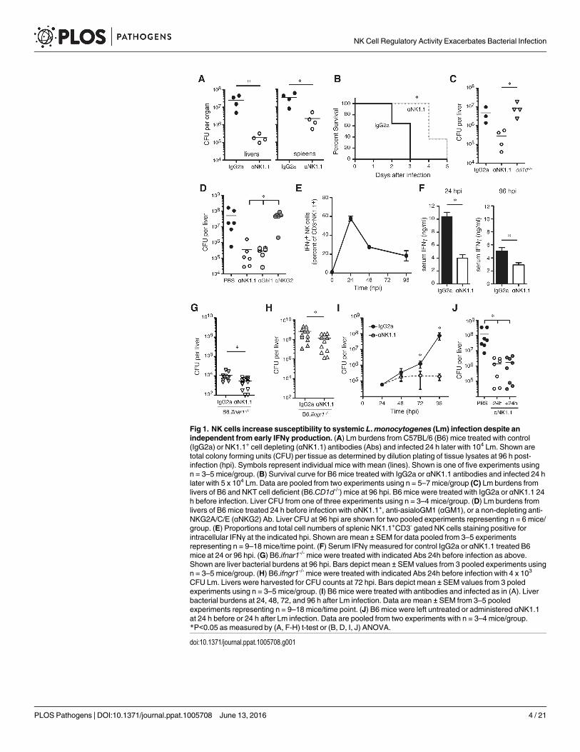
Fig 1. NK cells increase susceptibility to systemic L.monocytogenes (Lm) infection despite anindependent from early IFNγ production. (A) Lm burdens from C57BL/6 (B6) mice treated with control(IgG2a) or NK1.1+ cell depleting (αNK1.1) antibodies (Abs) and infected 24 h later with 104 Lm. Shown aretotal colony forming units (CFU) per tissue as determined by dilution plating of tissue lysates at 96 h post-infection (hpi). Symbols represent individual mice with mean (lines). Shown is one of five experiments usingn = 3–5 mice/group. (B) Survival curve for B6 mice treated with IgG2a or αNK1.1 antibodies and infected 24 hlater with 5 x 10⁴ Lm. Data are pooled from two experiments using n = 5–7 mice/group (C) Lm burdens fromlivers of B6 and NKT cell deficient (B6.CD1d-/-) mice at 96 hpi. B6 mice were treated with IgG2a or αNK1.1 24h before infection. Liver CFU from one of three experiments using n = 3–4 mice/group. (D) Lm burdens fromlivers of B6 mice treated 24 h before infection with αNK1.1+, anti-asialoGM1 (αGM1), or a non-depleting anti-NKG2A/C/E (αNKG2) Ab. Liver CFU at 96 hpi are shown for two pooled experiments representing n = 6 mice/group. (E) Proportions and total cell numbers of splenic NK1.1+CD3- gated NK cells staining positive forintracellular IFNγ at the indicated hpi. Shown are mean ± SEM for data pooled from 3–5 experimentsrepresenting n = 9–18 mice/time point. (F) Serum IFNγmeasured for control IgG2a or αNK1.1 treated B6mice at 24 or 96 hpi. (G) B6.ifnar1-/- mice were treated with indicated Abs 24h before infection as above.Shown are liver bacterial burdens at 96 hpi. Bars depict mean ± SEM values from 3 pooled experiments usingn = 3–5 mice/group. (H) B6.ifngr1-/- mice were treated with indicated Abs 24h before infection with 4 x 10³
CFU Lm. Livers were harvested for CFU counts at 72 hpi. Bars depict mean ± SEM values from 3 poledexperiments using n = 3–5 mice/group. (I) B6 mice were treated with antibodies and infected as in (A). Liverbacterial burdens at 24, 48, 72, and 96 h after Lm infection. Data are mean ± SEM from 3–5 pooledexperiments representing n = 9–18 mice/time point. (J) B6 mice were left untreated or administered αNK1.1at 24 h before or 24 h after Lm infection. Data are pooled from two experiments with n = 3–4 mice/group.*P<0.05 as measured by (A, F-H) t-test or (B, D, I, J) ANOVA.
doi:10.1371/journal.ppat.1005708.g001
NK Cell Regulatory Activity Exacerbates Bacterial Infection
PLOS Pathogens | DOI:10.1371/journal.ppat.1005708 June 13, 2016 4 / 21
treatment were dependent on type I interferon or IFNγ signaling, we evaluated Lm burdens inmice lacking expression of the type I interferon receptor (B6.ifnar1-/-) or the IFNγ receptor(B6.ifngr1-/-). Despite the extreme differences in susceptibility of these mouse strains to Lm,depletion of NK1.1+ cells was protective in both (Fig 1G and 1H). We also found that NK celldepletion did not impact Lm burdens until 72 hpi (Fig 1I), well after the peak of IFNγ produc-tion by NK cells (Fig 1E). Finally, when early NK cell IFNγ production was allowed to occurprior to αNK1.1 treatment, NK1.1+ cell depletion remained highly effective at reducing Lmburdens (Fig 1J). These results indicated that in mice with an intact T cell compartment NKcell production of IFNγ has no discernable impact on host resistance or susceptibility to Lm,arguing the pro-bacterial effects of NK cells are not due to hyper-production of IFNγ.
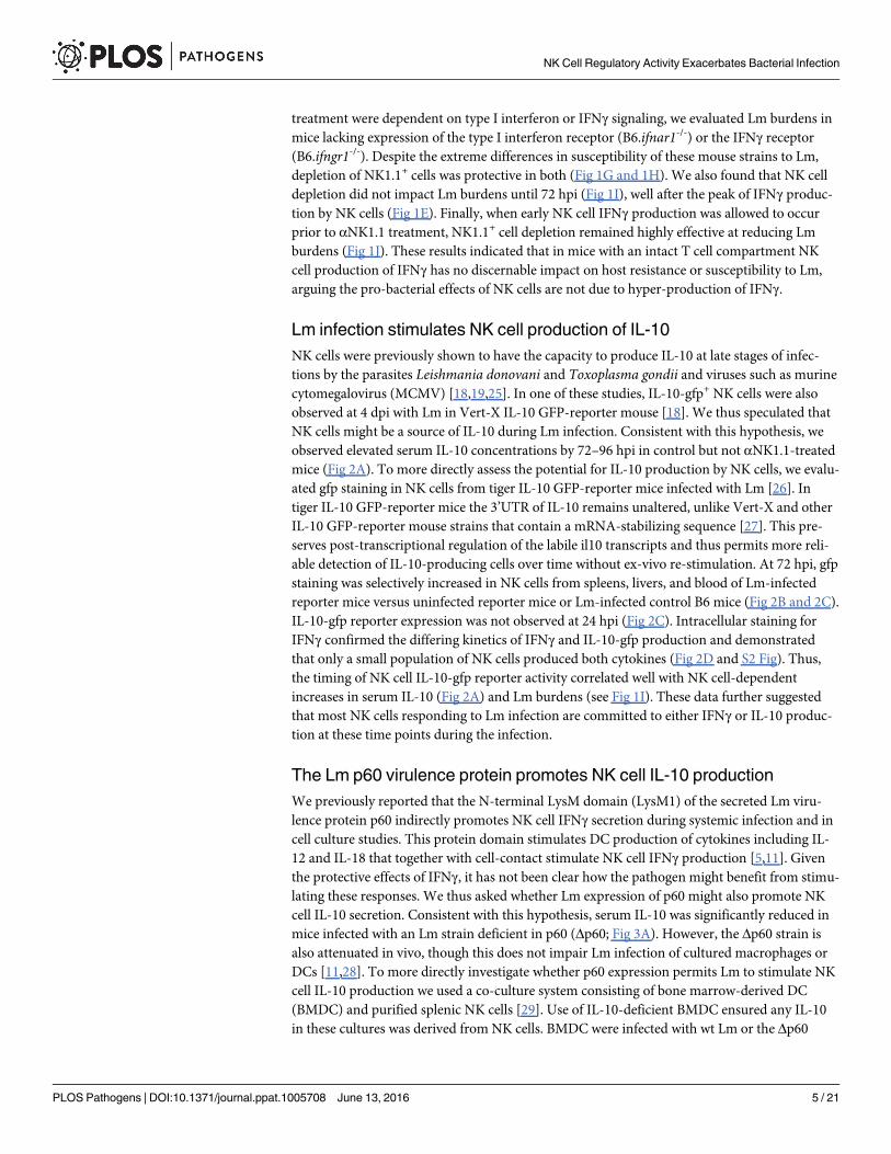
Lm infection stimulates NK cell production of IL-10NK cells were previously shown to have the capacity to produce IL-10 at late stages of infec-tions by the parasites Leishmania donovani and Toxoplasma gondii and viruses such as murinecytomegalovirus (MCMV) [18,19,25]. In one of these studies, IL-10-gfp+ NK cells were alsoobserved at 4 dpi with Lm in Vert-X IL-10 GFP-reporter mouse [18]. We thus speculated thatNK cells might be a source of IL-10 during Lm infection. Consistent with this hypothesis, weobserved elevated serum IL-10 concentrations by 72–96 hpi in control but not αNK1.1-treatedmice (Fig 2A). To more directly assess the potential for IL-10 production by NK cells, we evalu-ated gfp staining in NK cells from tiger IL-10 GFP-reporter mice infected with Lm [26]. Intiger IL-10 GFP-reporter mice the 3’UTR of IL-10 remains unaltered, unlike Vert-X and otherIL-10 GFP-reporter mouse strains that contain a mRNA-stabilizing sequence [27]. This pre-serves post-transcriptional regulation of the labile il10 transcripts and thus permits more reli-able detection of IL-10-producing cells over time without ex-vivo re-stimulation. At 72 hpi, gfpstaining was selectively increased in NK cells from spleens, livers, and blood of Lm-infectedreporter mice versus uninfected reporter mice or Lm-infected control B6 mice (Fig 2B and 2C).IL-10-gfp reporter expression was not observed at 24 hpi (Fig 2C). Intracellular staining forIFNγ confirmed the differing kinetics of IFNγ and IL-10-gfp production and demonstratedthat only a small population of NK cells produced both cytokines (Fig 2D and S2 Fig). Thus,the timing of NK cell IL-10-gfp reporter activity correlated well with NK cell-dependentincreases in serum IL-10 (Fig 2A) and Lm burdens (see Fig 1I). These data further suggestedthat most NK cells responding to Lm infection are committed to either IFNγ or IL-10 produc-tion at these time points during the infection.
The Lm p60 virulence protein promotes NK cell IL-10 productionWe previously reported that the N-terminal LysM domain (LysM1) of the secreted Lm viru-lence protein p60 indirectly promotes NK cell IFNγ secretion during systemic infection and incell culture studies. This protein domain stimulates DC production of cytokines including IL-12 and IL-18 that together with cell-contact stimulate NK cell IFNγ production [5,11]. Giventhe protective effects of IFNγ, it has not been clear how the pathogen might benefit from stimu-lating these responses. We thus asked whether Lm expression of p60 might also promote NKcell IL-10 secretion. Consistent with this hypothesis, serum IL-10 was significantly reduced inmice infected with an Lm strain deficient in p60 (Δp60; Fig 3A). However, the Δp60 strain isalso attenuated in vivo, though this does not impair Lm infection of cultured macrophages orDCs [11,28]. To more directly investigate whether p60 expression permits Lm to stimulate NKcell IL-10 production we used a co-culture system consisting of bone marrow-derived DC(BMDC) and purified splenic NK cells [29]. Use of IL-10-deficient BMDC ensured any IL-10in these cultures was derived from NK cells. BMDC were infected with wt Lm or the Δp60
NK Cell Regulatory Activity Exacerbates Bacterial Infection
PLOS Pathogens | DOI:10.1371/journal.ppat.1005708 June 13, 2016 5 / 21
strain. At 1hpi the BMDC were washed and media containing gentamicin was added. Purifiedsplenic NK cells were added at 2 hpi (Fig 3B). Under these conditions, any IFNγ produced inthe cultures is dependent on NK cells ([11,29]). Consistent with our prior findings, BMDCsinfected with Δp60 Lm elicited significantly less NK cell-dependent IFNγ than those infectedwith wt Lm (Fig 3C). As shown above, NK cell IFNγ production during systemic Lm infectionpeaks at 24 hpi (Fig 1E and 1F), while IL-10 production peaks later (Fig 2A). Consistent withthese results, IL-10 was not detected in culture supernatants at 24 hpi, but was reproduciblydetected by 72 hpi (Fig 3D). As seen in serum, IL-10 concentrations in culture supernatantswere also significantly reduced following infection with Δp60 Lm. These data suggested Lmexpression of p60 stimulates DC to promote serial NK cell secretion of IFNγ and IL-10.
To specifically investigate whether the region of p60 protein that stimulates NK cell IFNγsecretion also promotes secretion of IL-10, BMDC were primed with TLR agonists and stimu-lated with a recombinant p60 fragment (L1S) that contains the LysM1 domain (Fig 3E). As
Fig 2. NK cells responding to Lm infection acquire the ability to produce IL-10. (A) Serum IL-10concentrations at the indicated times after Lm infection of control IgG2a or αNK1.1 treated B6 mice. Data arepooled from 3 experiments for n = 9–12 mice/group. (B) Histogram depicting the staining for IL-10-gfpreporter in gated CD3-NK1.1+ NK cells from livers of uninfected IL-10-gfp+ (B6.tiger) mice or B6.tiger andcontrol B6 mice at 72 hpi with Lm. (C) Shown are mean ± SEM percentages of gated CD3-NK1.1+ cells thatstained positive for IL-10-gfp prior to infection or at 24 or 72 hpi with Lm. Gated NK cells from spleen, liver,and blood of B6.tiger mice were analyzed. Data are pooled from 3 experiments and represent n = 9–14 miceper time point. (D) Proportions of IFNγ+ and IL-10+ populations of NK1.1+CD3- gated NK cells from theindicated tissues at 24 or 96 hpi. Data are pooled from three experiments with n = 3–5 mice/group. Pooledfrom three experiments representing n = 9–12 mice/group. *P< 0.05 as determined by (A, C) ANOVA.
doi:10.1371/journal.ppat.1005708.g002
NK Cell Regulatory Activity Exacerbates Bacterial Infection
PLOS Pathogens | DOI:10.1371/journal.ppat.1005708 June 13, 2016 6 / 21

Fig 3. The L1S region of the Lm p60 protein is sufficient to induce NK cell IL-10 production. (A) Serum IL-10 at 96 hpi inB6 mice infected with wt or p60-deficient (Δp60) Lm strains. (B) Schematic of co-culture system consisting of BMDC infectedwith Lm for one hour, followed by washing and the addition of splenic purified NK cells at 2 h post-infection. (C) 24 hsupernatant IFNγ after infection of BMDC with wt or p60-deficient Lm in co-culture with wt NK cells. (D) 24 h and 72 hsupernatant IL-10 after infection of IL-10 deficient BMDC with wt or p60-deficient Lm in co-culture with wt NK cells. (E)Schematic of co-culture system consisting of BMDC treated with a TLR agonist ± L1S for one hour, followed by washing andthe addition of splenic purified NK cells at 2 h post-stimulation. (F) 24 h supernatant IFNγ after treatment of BMDC withLPS ± L1S in co-culture with wt NK cells. (G) 24 h and 72 h supernatant IL-10 after treatment of IL-10 deficient BMDC withLPS ± L1S in co-culture with wt NK cells. (H) 24 h supernatant IFNγ after treatment of human DC with polyI:C ± L1S in co-culture with PBMC from unrelated donors. (I) 72 h supernatant IL-10 after treatment of human DC with polyI:C ± L1S in co-culture with PBMC from unrelated donors. (J) 72 h supernatant IL-10 after treatment of IL-10 deficient BMDC with LPS+ L1S ± stimulation with 50 ng/mL IFNγ in co-culture with wt NK cells. (K) 72 h supernatant IL-10 after stimulation of IL-10deficient BMDC with LPS + L1S in co-culture with NK cells from wt or IFNGR-/- mice. (L) IL-10 was measured in supernatantscollected at 24, 48, and 72 h from L1S-stimuated co-cultures of IL-10 deficient BMDC and NK cells from wt or IFNGR-/- mice.Data for (C, D, F, G, H, I, J, K, L) pooled from three or more experiments. *P<0.05 as determined by t-test.
doi:10.1371/journal.ppat.1005708.g003
NK Cell Regulatory Activity Exacerbates Bacterial Infection
PLOS Pathogens | DOI:10.1371/journal.ppat.1005708 June 13, 2016 7 / 21

previously reported [11], IFNγ secretion was observed selectively in 24 h co-cultures whereBMDC and NK cells were stimulated with a priming agent (LPS) and L1S protein (Fig 3F). L1Salso induced NK cell IL-10 secretion, but again this was selectively observed in the 72 h cultures(Fig 3G). As for NK cell production of IFNγ, IL-10 production required stimulation with bothTLR agonist and L1S protein. Thus, our data suggested L1S stimulates primed BMDCs to pro-mote NK cell activation for sequential IFNγ and IL-10 production. To determine whetherhuman NK populations were also responsive to L1S, DCs were cultured 7 days from healthydonor PBMCs, then stimulated with a priming agent (pI:C) ± L1S and purified autologous NKcells as in Fig 3E. The results using human cells paralleled those above. Co-cultures with cellsfrom 4/4 donors produced IFNγ at 24 h (Fig 3H) and IL-10 at 72 h (Fig 3I) in response to theL1S treatment. Thus, the L1S fragment of the Lm p60 protein is necessary and sufficient tostimulate the ability of primed DCs to promote early IFNγ and delayed IL-10 secretion fromboth murine and human NK cells.
IFNγ signaling suppresses NK cell production of IL-10Because IL-10 production by both mouse and human NK cell cultures was delayed relative toIFNγ production we considered whether production of IFNγmight inhibit NK cell IL-10 secre-tion. Subsequent to L1S or control stimulation, recombinant IFNγ was added to the co-cul-tures. NK cell IL-10 secretion was significantly reduced in the cultures treated with IFNγ (Fig3J). To confirm these effects were due to IFNγ signaling in the NK cells, we established co-cultures using B6.il10-/- BMDC and NK cells purified from spleens of B6.ifngr1-/- mice. TheIFNGR-deficient NK cells produced 4–5 fold more IL-10 than wt B6 NK cells (Fig 3K).Secreted IL-10 was also detected earlier in the cultures with IFNGR-deficient NK cells (Fig 3L).These results suggest that early IFNγ production may contribute to the observed delay in IL-10production by the responding NK cells.
NK cell IL-10 production increases susceptibility to systemic Lm infectionTo further investigate the relationship between pro-bacterial effects of NK cells and their pro-duction of IL-10, NK cell depletion was performed in IL-10-deficient (B6.il10-/-) mice. Analysisof the infected B6.il10-/- animals revealed that liver bacterial burdens were comparable to thoseseen in B6 mice depleted of NK cells prior to infection (Fig 4A). NK cell depletion failed to fur-ther reduce Lm burdens in the B6.il10-/- mice. Thus, NK cell depletion and IL-10 deficiencyhad similar and non-additive effects on susceptibility to Lm, suggesting production of IL-10 byNK cells might be responsible for the increased host susceptibility. To further test this, we per-formed adoptive transfer experiments. CD45.2+ or CD45.1+ B6.il10-/- recipients were infectedwith Lm then respectively transferred with NK cells from the spleens of naïve wt CD45.1 (B6.ptprca) or IL-10 deficient CD45.2 (B6.il10-/-) mice (Fig 4B). At 96 hpi (72 h after transfer) smallpopulations of donor CD45.1+ wt and CD45.2+ IL-10 deficient NK cells could be detected inspleens of the B6.il10-/- CD45.2+ and CD45.1+ recipients, respectively (Fig 4C), demonstratingpersistence of the transferred cells. The detection of IL-10 protein in lysates of splenocytesfrom the B6.il10-/- recipients of wt, but not IL-10-deficient, NK cells indicated the transferredcells were activated to produce IL-10 (Fig 4D). This IL-10 production in the presence of wt NKcells was also associated with 10–100 fold increases in Lm burdens in livers and spleens of theB6.il10-/- mice (Fig 4E).
To establish whether IFNγ production by the NK cells might mediate pro- or anti-bacterialeffects in the recipient mice, groups of B6.il10-/- mice received NK cells from the spleens ofIFNγ deficient (GKO, B6.ifng-/-) mice. The GKO NK cells produced IL-10, as measured inspleen lysates (Fig 4D), and GKO NK cells sufficed to increase Lm burdens (Fig 4E). There was
NK Cell Regulatory Activity Exacerbates Bacterial Infection
PLOS Pathogens | DOI:10.1371/journal.ppat.1005708 June 13, 2016 8 / 21

no significant difference in burdens of mice receiving wt or GKO NK cells and both NK celltypes increased burdens in the il10-/- mice to a level near that seen in wt mice infected with Lm(compare Fig 4A and 4E). Experiments using donor NK cells labeled with CFSE further con-firmed that the take of GKO, WT, and il10-/- NK cells was similar in the il10-/- recipients (S3AFig). Thus, the ability of NK cells to produce IL-10 is a crucial factor governing Lm survivaland replication during systemic infection and these pro-bacterial effects are independent fromNK cell production of IFNγ.
Fig 4. NK cell IL-10 production drives increased Lm burdens. (A) Liver CFU from individual B6 andcongenic IL-10 deficient B6 (B6.il10-/-) mice at 72 hpi. IgG2a or αNK1.1 Abs were given 24 h before infection.Pooled from two experiments representing n = 7–9 mice/group. (B) Design of cell transfer experiments. NKcells were purified from naïve wt CD45.1+ (B6.ptprca), IL-10-deficient CD45.2+ (B6.il-10-/-), or IFNγ-deficient(GKO) CD45.2+ mice for transfer into infected B6.il-10-/- recipients. (C) Detection of CD45.1+ donor NK cellsin B6.il10-/- recipients at 96 hpi. Live gated CD3-NK1.1+ splenic NK cells are shown. (D) IL-10 from spleniclysates of infected B6.il-10-/- recipients 72 h after NK cell transfers. Shown are mean ± SEM for one of threeexperiments using n = 3–4 mice/group. (E) Lm burdens from indicated tissues of B6.il-10-/- recipients 96 hpi.*P< 0.05 as determined by (A) ANOVA or (D, E) t-test.
doi:10.1371/journal.ppat.1005708.g004
NK Cell Regulatory Activity Exacerbates Bacterial Infection
PLOS Pathogens | DOI:10.1371/journal.ppat.1005708 June 13, 2016 9 / 21
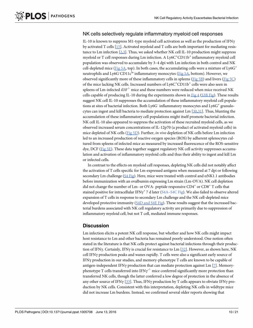
NK cells selectively regulate inflammatory myeloid cell responsesIL-10 is known to suppress M1-type myeloid cell activation as well as the production of IFNγby activated T cells [15]. Activated myeloid and T cells are both important for mediating resis-tance to Lm infection [2,3]. Thus, we asked whether NK cell IL-10 production might suppressmyeloid or T cell responses during Lm infection. A Ly6C+CD11b+ inflammatory myeloid cellpopulation was observed to accumulate by 3–4 dpi with Lm infection in both control and NKcell-depleted mice (Fig 5A, top). In both cases, the accumulating cells were a mixture of Ly6G+
neutrophils and Ly6G-CD11cl° inflammatory monocytes (Fig 5A, bottom). However, weobserved significantly more of these inflammatory cells in spleens (Fig 5B) and livers (Fig 5C)of the mice lacking NK cells. Increased numbers of Ly6C+CD11b+ cells were also seen inspleens of Lm-infected il10-/- mice and these numbers were reduced when mice received NKcells capable of producing IL-10 during the experiments shown in Fig 4 (S3B Fig). These resultssuggest NK cell IL-10 suppresses the accumulation of these inflammatory myeloid cell popula-tions at sites of bacterial infection. Both Ly6G- inflammatory monocytes and Ly6G+ granulo-cytes can ingest and kill bacteria to mediate protection against Lm [30,31]. Thus, blunting theaccumulation of these inflammatory cell populations might itself promote bacterial infection.NK cell IL-10 also appeared to suppress the activation of these recruited myeloid cells, as weobserved increased serum concentrations of IL-12p70 (a product of activated myeloid cells) inmice depleted of NK cells (Fig 5D). Further, in vivo depletion of NK cells before Lm infectionled to an increased production of reactive oxygen species (ROS) by adherent splenocytes cul-tured from spleens of infected mice as measured by increased fluorescence of the ROS-sensitivedye, DCF (Fig 5E). These data together suggest regulatory NK cell activity suppresses accumu-lation and activation of inflammatory myeloid cells and thus their ability to ingest and kill Lmor infected cells.
In contrast to the effects on myeloid cell responses, depleting NK cells did not notably affectthe activation of T cells specific for Lm-expressed antigens when measured at 7 dpi or followingsecondary Lm challenge (S4 Fig). Here, mice were treated with control and αNK1.1 antibodiesbefore immunization with an ovalbumin expressing Lm strain (Lm-OVA). NK cell depletiondid not change the number of Lm- or OVA- peptide responsive CD4+ or CD8+ T cells thatstained positive for intracellular IFNγ+ 7 d later (S4A–S4C Fig). We also failed to observe alteredexpansion of T cells in response to secondary Lm challenge and the NK cell-depleted micedeveloped protective immunity (S4D and S4E Fig). These results suggest that the increased bac-terial burdens associated with NK cell regulatory activity are primarily due to suppression ofinflammatory myeloid cell, but not T cell, mediated immune responses.
DiscussionLm infection elicits a potent NK cell response, but whether and how NK cells might impacthost resistance to Lm and other bacteria has remained poorly understood. One notion oftenstated in the literature is that NK cells protect against bacterial infections through their produc-tion of IFNγ. Certainly, IFNγ is crucial for resistance to Lm [32]. However, as shown here, NKcell IFNγ production peaks and wanes rapidly. T cells were also a significant early source ofIFNγ production in our studies, and memory-phenotype T cells are known to be capable ofantigen-independent IFNγ production that can mediate protection against Lm [7]. Memory-phenotype T cells transferred into IFNγ-/- mice conferred significantly more protection thantransferred NK cells, though the latter conferred a low degree of protection in the absence ofany other source of IFNγ [33]. Thus, IFNγ production by T cells appears to obviate IFNγ pro-duction by NK cells. Consistent with this interpretation, depleting NK cells in wildtype micedid not increase Lm burdens. Instead, we confirmed several older reports showing that
NK Cell Regulatory Activity Exacerbates Bacterial Infection
PLOS Pathogens | DOI:10.1371/journal.ppat.1005708 June 13, 2016 10 / 21
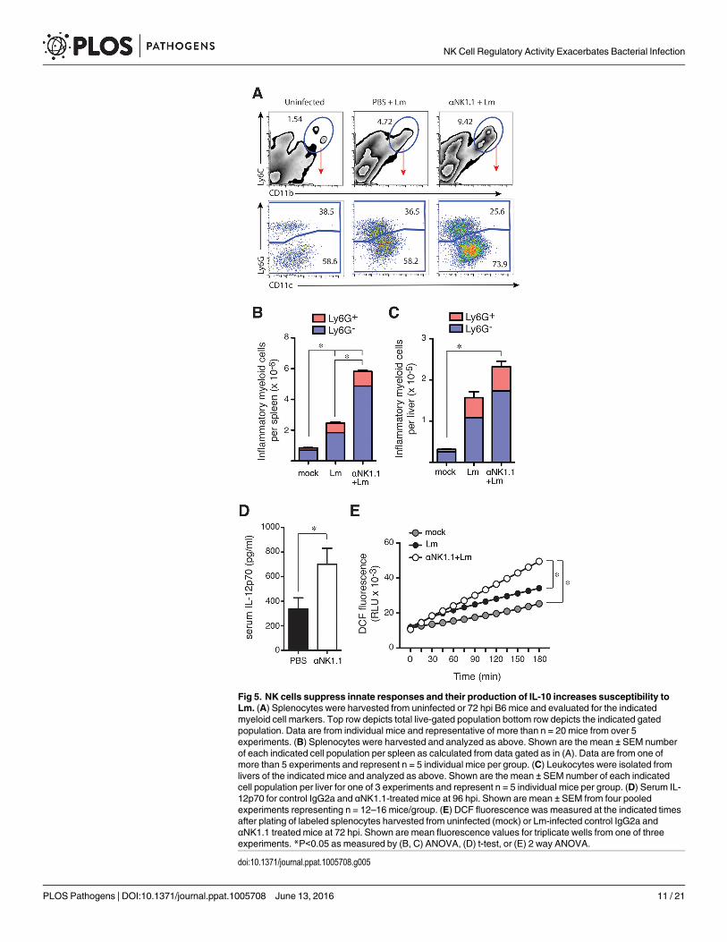
Fig 5. NK cells suppress innate responses and their production of IL-10 increases susceptibility toLm. (A) Splenocytes were harvested from uninfected or 72 hpi B6 mice and evaluated for the indicatedmyeloid cell markers. Top row depicts total live-gated population bottom row depicts the indicated gatedpopulation. Data are from individual mice and representative of more than n = 20 mice from over 5experiments. (B) Splenocytes were harvested and analyzed as above. Shown are the mean ± SEM numberof each indicated cell population per spleen as calculated from data gated as in (A). Data are from one ofmore than 5 experiments and represent n = 5 individual mice per group. (C) Leukocytes were isolated fromlivers of the indicated mice and analyzed as above. Shown are the mean ± SEM number of each indicatedcell population per liver for one of 3 experiments and represent n = 5 individual mice per group. (D) Serum IL-12p70 for control IgG2a and αNK1.1-treated mice at 96 hpi. Shown are mean ± SEM from four pooledexperiments representing n = 12–16 mice/group. (E) DCF fluorescence was measured at the indicated timesafter plating of labeled splenocytes harvested from uninfected (mock) or Lm-infected control IgG2a andαNK1.1 treated mice at 72 hpi. Shown are mean fluorescence values for triplicate wells from one of threeexperiments. *P<0.05 as measured by (B, C) ANOVA, (D) t-test, or (E) 2 way ANOVA.
doi:10.1371/journal.ppat.1005708.g005
NK Cell Regulatory Activity Exacerbates Bacterial Infection
PLOS Pathogens | DOI:10.1371/journal.ppat.1005708 June 13, 2016 11 / 21
depletion of NK1.1+ cells reduces severity of systemic infections by this and other bacteria[14,34,35]. The older studies did not discriminate the effects of NK versus NKT cells, but weshowed here that the absence of NKT cells had no effect on Lm burdens during systemic infec-tion. These results support the conclusion that NK cells are not a crucial source of early IFNγproduction and that NK cells instead mediate pro-bacterial effects. Our data further demon-strate the mechanism responsible for these pro-bacterial effects. We found that NK cellsresponding to Lm produce IL-10 and this production is necessary and sufficient to increase Lmburdens. Further, the p60 virulence protein of Lm drives NK cell activation and IL-10 produc-tion. NK cells consequently suppressed innate myeloid cell responses. These findings togethersuggest Lm promotes NK cell activation to exploit their regulatory effects on antibacterial mye-loid cell responses.
Despite the evidence that NK cell activation has deleterious effects during systemic Lminfection we are aware of a few seemingly contradictory reports. In studies where Lm was intro-duced into the footpad of mice, NK cell depletion was shown to modestly increase bacterialburdens in the draining lymph nodes [36,37]. Lm does not normally infect the host throughthe skin and little is known about the sequence of immune responses in this model. Perhapsfootpad-inoculated Lm is unable to exploit NK cell IL-10 production, for example due to differ-ences in the pattern or kinetics of inflammatory cell recruitment. Another example where NKcell deficiency was reported to increase susceptibility was in mice doubly deficient for the com-mon gamma chain (γc) and Rag2 [38]. The γc is important for cellular responses to severalcytokines, including IL-2, 4, 7, 9, 15 and 21. IL-15 signaling is particularly important for devel-opment and survival of NK and memory CD8+ T cells [39,40]. The increased susceptibility inthe γc
-/- x rag2-/- mice might thus be interpreted to indicate a protective role for NK cells. How-ever, mice singly deficient for γc or Rag2 did not demonstrate increased susceptibility to Lminfection [38]. Thus, the reported susceptibility of mice doubly deficient for Rag2 and γc isnot simply a result of NK cell deficiency. It was suggested that NK cells may be a key sourceof protective IFNγ in the absence of T cells [38]. It is also notable that recent studies indicatethat Rag protein expression in NK cells modulates their survival and functional activity [41].Regardless, it has since been shown that IL-15-/- mice exhibit increased resistance to systemicLm infection [42].
It has been previously postulated that NK cells might exacerbate Lm infection through over-producing IFNγ [14]. Three lines of evidence from our studies argue against this model: (1)We showed that mice lacking IFNGR1 expression were still protected by depletion of NK1.1+
cells despite their increased susceptibility overall. (2) Depletion of NK cells was protectivewhen initiated subsequent to peak NK cell IFNγ production. (3) Transfer of GKO NK cellswere as effective as wt NK cells at increasing Lm burdens in IL-10 deficient recipients. How-ever, Jablonska and colleagues recently argued that NK cell secretion of IFNγ contributes topro-bacterial effects based on the finding that mice treated with a low dose of anti-IFNγ anti-body had heightened resistance [43]. These apparently discrepant results could be explainedby antibody stabilization of IFNγ to enhance its signaling, as has been shown to occur whencytokines such as IL-2 and IL-15 are complexed with antibodies or soluble receptor subunits[44,45]. We thus interpret the available data as evidence that NK cells exert pro-bacterial effectsindependent from their IFNγ production.
We instead found that the key mechanism by which NK cells increase susceptibility to Lm isthrough production of IL-10. How does NK cell-derived IL-10 suppress resistance to bacterialinfection? It is well established that mice entirely deficient for IL-10 are highly resistant to Lminfection. This resistance is associated with increased innate and adaptive immune responses[17]. Consequently, we evaluated the effects of NK cell depletion on both T and myeloid cellresponses to Lm infection. We failed to observe any effect of NK cells on the T cell response to
NK Cell Regulatory Activity Exacerbates Bacterial Infection
PLOS Pathogens | DOI:10.1371/journal.ppat.1005708 June 13, 2016 12 / 21
Lm infection, suggesting that NK cell IL-10 is not responsible for the previously observed sup-pression of T cell responses. Consistent with this result, CD4+ T cells were recently shown to bea crucial source of IL-10 that regulates memory CD8+ T cell responses during LCMV infection[46]. In contrast, our findings here indicated that NK cell IL-10 suppresses both accumulationand activation of myeloid cells. Coincident with the timing of NK cell IL-10 production in con-trol Lm infected mice (3–4 dpi), we found that depleting NK cells increased inflammatorymonocyte and neutrophil accumulation in spleens and livers, increased serum IL-12p70, andincreased ROS production in cultures of adherent splenocytes. Activated myeloid cells are theprimary source of IL-12, and its production is known to be suppressed by IL-10 [47,48]. ROSproduction is also a correlate of M1-type activation, is suppressed by IL-10, and correlates withmacrophage and neutrophil bactericidal activity [49,50]. Further, IL-10 blockade is known toincrease macrophage bactericidal activity against Lm [49,51,52]. Production of IL-12 or ROSmay not themselves mediate reduced Lm burdens in NK cell-depleted mice, but certainly indi-cate enhanced myeloid cell activation. Accumulation and activation of myeloid cells is crucialfor immune resistance to infections by Lm and many other intracellular pathogens [1]. Hence,the impairment of these processes likely accounts for the ability of NK cell IL-10 to increasesusceptibility during Lm, and possibly other, bacterial infections. Presumably, this dampeningof inflammatory responses benefits the host in other settings, such as in the context of inflam-matory diseases.
Lm infection is not unique in its ability to stimulate IL-10 secretion by NK cells. NK cell IL-10 production was previously observed at late stages of chronic infection by Leishmania dono-vanni [19], and during infections by Toxoplasma gondii [18], and murine cytomegalovirus(MCMV) [53]. During T. gondii infection, NK cell activation is regulated by cytokine (IL-12)production and this IL-12 is driven by ligation of TLR11 and 12 by the parasite profilin protein[54]. IL-12 production during T. gondii [18] induces NK cell expression of the aryl hydrocar-bon receptor (Ahr) transcription factor to drive il10 transcription [55]. IL-12 also drives il10transcription in NK cells during chronic infection by the parasite L. donovanii and IL-10 pro-ducing NK cells were shown to increase parasite numbers in this model [19]. However, it is notyet clear whether a specific L. donovanii protein drives the NK cell response to this infection.NK cell IL-10 production during MCMV infection is largely seen in the Ly49H+ NK cell subset[25]. Ly49H is an activating receptor that responds to the virus-encoded M157 protein [56,57].Work here showed that the Lm p60 protein was important for promoting regulatory NK cellactivity. Our prior work found that Lm expression of the p60 protein increases both bacterialreplication in host tissues and NK cell production of IFNγ early after systemic infection [5].Recombinant p60 protein was subsequently shown to stimulate production of IL-18 by primedBMDC to stimulate cell contact-dependent NK cell production of IFNγ [11,58]. In the presentpaper Lm expression of p60 increased NK cell secretion of IL-10 in vivo and in BMDC/NK cellco-cultures. Stimulation of primed murine BMDCs or human PBMC-derived DCs with arecombinant fragment of p60 protein (L1S) likewise sufficed to trigger NK cell secretion of IL-10. These data suggest Lm uses p60 to actively promote NK cell IL-10 secretion. Together withthe prior work in other pathogen models, these data with p60 further suggest that the presenceof NK cell stimulating proteins might be a marker for pathogens that have evolved to exploitNK cell regulatory activity.
The kinetics of NK cell IL-10 secretion was found here to occur only after reductions in NKcell IFNγ secretion, both during systemic infection and in cell co-cultures infected with Lm orstimulated with recombinant L1S protein. Similar delays were observed in other models whereNK cell IL-10 production occurs [18,19,53]. For example, during Leishmania donovani infec-tion NK cell IFNγ production is stimulated in an IL-12-dependent manner from 24 hr infectedmice, followed by NK cell IL-10 production from 21 day infected mice [19]. Similarly, during
NK Cell Regulatory Activity Exacerbates Bacterial Infection
PLOS Pathogens | DOI:10.1371/journal.ppat.1005708 June 13, 2016 13 / 21

MCMV infection IL-2 and IL-12 drove NK cell IFNγ as well as subsequent IL-10 production inculture ex vivo [53]. These studies suggest that the switch from IFNγ to IL-10 production is aconsequence of NK cell activation in multiple infections. However, we do not believe thisswitch is a “hard-wired” response to activation given previous reports showing certain cytokinestimulation protocols which induce IFNγ production by human NK cells fail to also trigger IL-10 secretion [59,60]. Recent work from Biron and colleagues suggested that during MCMVinfection this delay reflects a requirement for NK cell proliferation to open the il10 locus totranscriptional machinery [53]. Proliferation might similarly contribute to IL-10 production byNK cells during Lm or other infections, though this remains to be determined. Defining themechanistic basis for sequential production of IFNγ and IL-10 was not the purpose of our stud-ies, but we did observe that IFNγ acts to suppress NK cell IL-10 secretion. Further, we foundthat NK cells deficient in IFNγ signaling secreted increased quantities of IL-10. These resultssuggest early NK cell IFNγ production may be important for suppressing or delaying NK cellIL-10 secretion. In T cells an initial pro-inflammatory response is necessary for switching fromIFNγ to IL-10 production [61–63]. However, we found that IFNγ-deficient NK cells remainedcapable of producing IL-10 and were as effective as wt NK cells at increasing susceptibility inIL-10-/- mice. Thus, cell-intrinsic IFNγ production does not appear to be an essential stimulusfor NK cell IL-10 secretion. What additional factor(s) might contribute to driving NK cell tran-sitioning from IFNγ to IL-10 production during Listeria and other infections remains to bedetermined.
The impact of NK cell regulatory activity on human health and disease is not yet known.However, human NK cells were previously shown to produce IL-10 [59], and we showed thatLm p60 could drive IL-10 secretion by human NK cells. Thus, NK cell IL-10 production maywell impact human susceptibility to infections and other diseases, including Lm infection.Severe Lm infections primarily occur in elderly and pregnant individuals. Ageing is associatedwith an increase of circulating NK cells in humans [64], and NK cells are a major cell popula-tion in the placenta of pregnant humans and animals [65]. Perhaps then, the increased preva-lence of NK cells and their IL-10 production is an important factor governing the susceptibilityof these populations. Pregnant individuals also have increased susceptibility to infections byT. gondii, Cytomegalovirus, and Leishmania [66]. Depletion of NK cells or more selectiveapproaches to suppress their acquisition of regulatory activity could thus prove useful in someclinical settings. Improved understanding of how Lm p60 and other pathogen factors inducepro-inflammatory and regulatory NK cell responses will be an important step in defining thepotential benefits, risks, and feasibility of manipulating NK cells in the context of infectious,autoimmune, and cancerous diseases.
Methods
AnimalsFemale mice were used at 8–12 weeks of age. C57BL/6J, B6.il10-/-, B6.ptprca, B6.ifngr1-/-, B6.ifnar1-/-, B6.ifng-/- (GKO) and B6.IL-10-gfp (tiger) mice were purchased from Jackson Labs.B6.cd1d-/- mice were from Dr. Laurent Gapin (Univ. Colorado). Mice were maintained in theNational Jewish Health Biological Resource Center and University of Colorado Office of Labo-ratory Animal Resources.
NK cell depletionMice were treated i.p. with PBS or PBS containing 100 μg of purified Ab or 100 μl of rabbitantisera to the ganglioside asialoGM1 (α-GM1, Wako USA). Endotoxin free IgG2a control(C1.18) and αNK1.1 (PK136) Abs were purchased (Bio X Cell). Anti-Ly49C/I (clone 5E6) and
NK Cell Regulatory Activity Exacerbates Bacterial Infection
PLOS Pathogens | DOI:10.1371/journal.ppat.1005708 June 13, 2016 14 / 21
anti-NKG2A/C/E (20D5) Abs were purified from hybridoma supernatants using protein Aaffinity chromatography. Unless otherwise stated, Abs were given in a single dose at 24 h beforeinfection. Depletions were confirmed by flow cytometry.
InfectionsL.monocytogenes (Lm; strain 10403s), congenic p60-deficient [28], and OVA-expressing [67]Lm (OVA-Lm) were thawed from frozen stocks and diluted for growth to log phase in brain-heart infusion or tryptic soy broth (MP Biomedicals), then diluted in PBS and given to mice i.v.in the lateral tail vein. Unless stated otherwise, mice received a single sublethal dose of 104
CFU. Lm was given at 4000 CFU for infection in B6.ifngr1-/- mice, and a lethal dose of 5 x 10⁴
CFU for analysis of survival ± NK cell depletion. OVA-Lm was given at 5000 CFU for immuni-zations. Challenges used a lethal dose of Lm-OVA (105 CFU). For CFU counts, organs wereharvested into 0.02% Nonidet P-40, homogenized for 1 min with a tissue homogenizer (IKAWorks, Inc.) and serial dilutions were plated on BHI or TSB agar plates.
Cell isolation and stimulationSpleens were harvested into media containing penicillin/streptomycin at 100 U/ml then trans-ferred to a solution of 1 mg/ml type 4 collagenase (Worthington) in HBSS plus cations (Invi-trogen). After a 30 min incubation at 37°C, spleens were disrupted by passage through a 70 μmcell strainer and the resulting single cell suspensions treated with RBC lysis Buffer (0.15 MNH4Cl, 10mM KHCO3, 0.1 mM Na2EDTA, pH 7.4) for 2 min. Prior to intracellular staining,splenocytes were incubated 3–4 h in RP10 media (RPMI 1640, 10% FBS, 1% L-glutamine, 1%Sodium Pyruvate, 1% Penicillin, 1% Streptomycin and 0.1% β-ME) containing Brefeldin A(GolgiPlug; BD Biosciences). No additional ex vivo stimulation was included for NK cell analy-ses. For T cells, 1 μM concentrations of synthetic OVA257–264 (SIINFEKL) or LLO190–201
(NEKYAQAYPNVS) peptides were included during the incubation. Blood cells were harvestedinto HBSS plus cations and heparin (Sigma). Cells were treated twice with RBC lysis Buffer for1 min. Liver cells were harvested and treated with collagenase in the same manner as spleencells. Following passage through a 70 μm cell strainer, cells were re-suspended in 40% Percollin HBSS minus cations. The 40% Percoll was underlayed with 60% Percoll, and cells were col-lected from gradient following centrifugation. RPMI with 5% FBS was added to cells to pellet,and cells were treated with RBC lysis Buffer for 1 min.
Flow cytometryAnti-CD16/32 (2.4G2 hybridoma supernatant) was added to block Fc receptors prior to stain-ing, which used FACS buffer (1% BSA, 0.01% NaN3, PBS) containing fluorophore-labeled anti-bodies purchased from eBioScience or BioLegend. Staining antibodies included anti- CD3(clone 145 2C11), CD4 (clone RM-4-5), CD8 (clone 53–6), CD11b (M1/70), CD11c (N418),CD27 (clone LG.7F9), CD45.1 (clone A20), IFNγ (clone XMG1.2), Ly6C (HK1.4), Ly6G (1A8),NK1.1 (clone PK136), and NKp46 (clone 29A1.4). After surface staining, cells were fixed in2–4% paraformaldehyde for direct analysis with or without saponin treatment for intracellularstaining. To amplify IL-10-gfp signal [53], fixed and permeabilized cells were stained with arabbit monoclonal anti-GFP followed by goat anti-rabbit IgG Alexa Fluor 488 (both from LifeTechnologies). At least 100,000 data events per sample were collected using an LSRII (BD Bio-sciences). FlowJo software (Treestar) was used for analysis of flow data.
NK Cell Regulatory Activity Exacerbates Bacterial Infection
PLOS Pathogens | DOI:10.1371/journal.ppat.1005708 June 13, 2016 15 / 21

Co-culture systemBone marrow-derived DC (BMDC) were cultured from B6.il10-/- mice and infected with Lm orstimulated with recombinant L1S protein purified as previously described [11,29]. Briefly, bonemarrow was cultured 6d in GM-CSF and 3 x 10⁵ BMDC (>90% CD11c+) per well were cul-tured overnight in 24 well plates. For infections, log phase wt or Δp60 Lm were added at a mul-tiplicity of one bacterium per BMDC. One h later, cells were washed and gentamycin wasadded at 10 μg/mL. For L1S stimulation, BMDC were activated 1 h by treatment with 20 μg/mlpoly I:C (Invivogen, San Diego, CA) or 10 ng/ml LPS (L8274 Sigma-Aldrich, St. Louis, MO)and purified L1S protein was added to the BMDC at 30 μg/ml. For IFNγ treatment, 50 ng/mLIFNγ (Life Technologies) was added at 1 hr post L1S stimulation. Purified splenic NK cellswere negatively sorted using the EasySep Mouse NK cell Enrichment Kit (19755 Stemcell Tech-nologies) and added to cultures 2 h after Lm or L1S treatments at a ratio of .1:1 (NK cells:BMDC). Purified NK cell populations were>80% NK1.1+CD3-. To culture human DCs,adherent PBMCs were obtained from unrelated donors and grown in RPMI 1640 (Invitrogen)supplemented with 10% human AB serum (Innovative Research, Novi, MI), 0.01M HEPES,0.02mg/ml gentamicin, 200 IU/ml IL-4 (eBioscience), and 100 IU/ml GM-CSF (R&D Systems,Minneapolis, MN). After 6 days, DC were plated at 105 cells/well in 96 well plates, primed, andstimulated with L1S and polyI:C as above. Purified human NK cells were negatively sortedfrom PBMCs using the EasySep Human NK cell Enrichment Kit (19015 Stemcell Technolo-gies). Supernatants were harvested for analysis of IFNγ and IL-10 at 24 or 72 h cytokines usingcommercial ELISAs (BD Biosciences).
Cytokine analysisBlood was collected by cardiac puncture. Serum from clotted blood was collected and frozenprior to analysis using commercial ELISAs for IFNγ, IL-12p70, or IL-10 (BD Biosciences).Equal numbers of splenocytes were homogenized in 1 mL of 0.02% Nonidet P-40 with proteaseinhibitor cocktail (ThermoFisher) using a dounce for measurement of IL-10 in homogenates.
Adoptive transfersNK cells were purified from spleens of naïve B6.ptrpca, B6.il10-/-, and B6.ifng-/- (GKO) mice(>80% purity) using the EasySep Negative Selection Mouse NK Cell Enrichment Kit (StemcellTechnologies). Each recipient received 1.5–2 x 10⁶ live NK cells i.v. at 24 hpi.
ROS analysisSingle cell splenocyte suspensions were prepared as above and 106 cells were added per well toa 96 well round bottom suspension plate (Greiner) followed by centrifugation at 500 x g for 5min. Pelleted splenocytes were resuspended in DPBS (Gibco) containing 10μM 2',7'-dichloro-dihydrofluorescein diacetate (DCF; ThermoFisher) and 1% DMSO. After a 30 min incubationat 37°C, spenocytes were washed twice in DPBS and resuspended at 100 μl/well in phenolred-free DMEM (Gibco) then transferred to 96 well white bottom Nunc F96 plates (Thermo-Fisher). Plates were incubated in a 37°C Biotek Synergy HT plate reader and fluorescence readat 15 min intervals using a 528/20 emission filter and Gen5 software.
StatisticsGraphing and statistical analyses used Prism (GraphPad Software). Statistical tests included t-tests, analysis of variance (ANOVA), and linear regression with Pearson correlation. P<0.05was considered significant.
NK Cell Regulatory Activity Exacerbates Bacterial Infection
PLOS Pathogens | DOI:10.1371/journal.ppat.1005708 June 13, 2016 16 / 21
Study approvalThe Animal Care and Use Committees for National Jewish Health (Protocol# AS2682-08-16)and the University of Colorado School of Medicine (Protocol# 105614(05)1E) approved allstudies. These protocols adhere to standards of the United States Public Health Service andDepartment of Agriculture.
Supporting InformationS1 Fig. Effectiveness of NK cell depletion using αNK1.1, impact of αLy49C/I, and sourcesof intracellular IFNγ. (A) Eight week old female C57BL/6 mice were given a single i.p. injec-tion of PBS containing 100 mg of purified IgG2a control (C1.18) or αNK1.1. Twenty-four hlater (0 hpi), spleens were harvested or mice were infected i.v. with 104 log-phase Lm. Deple-tion of splenic NK1.1+NKp46+ NK cells was evaluated at 0 and 96 hpi. Shown are representa-tive plots of CD3- gated splenocytes. (B) Groups of B6 mice treated with purified IgG2a controlor αNK1.1 and infected with 5000 CFU Lm. Bacterial burdens were determined at 96 hpi. Sym-bols represent individual mice from one of two experiments using n = 5–7 mice/group. (C)Groups of B6 mice were treated with purified IgG2a isotype control, αNK1.1, or a depletingαLy49C/I monoclonal Ab (clone 5E6) and infected with 104 Lm. Bacterial burdens were deter-mined at 96 hpi. Symbols represent individual mice from one of two experiments usingn = 4–5 mice/group. �P<0.05 by ANOVA. (D) Representative flow cytometry data plots show-ing intracellular IFNγ+ (iIFNγ+) cell populations present in the spleen of a B6 mouse at 24 hpiwith Lm. Panels on the left show live gated cells. The right panel depicts staining of the gatediIFNγ+ population indicated with the red arrow. (E) Total cell numbers of IFNγ+NK1.1+CD3-
cells present in the spleens of mock-infected or Lm-infected mice. Symbols represent individualmice from one of two experiments using n = 4–5 mice/group. (F) Total cell numbers of IFNγ+-
CD3+ cells present in the spleens of mock-infected or Lm-infected mice. Symbols representindividual mice from one of two experiments using n = 4–5 mice/group.(TIF)
S2 Fig. Flow cytometry plots for detection of NK cell co-expression of IFNγ and il10-gfpreporter. Representative flow cytometry data plots show iIFNγ and il10-gfp staining onCD3-NK1.1+ gated cells isolated from the spleen, liver, and blood of tiger IL-10 GFP-reportermice at 24 or 72 hpi with 104 Lm.(TIF)
S3 Fig. Detection of CFSE-labeled cells and inflammatory myeloid cells in mice thatreceived purified NK cells. (A) CFSE and NK1.1 staining on CD3-NK1.1+ gated cells isolatedfrom the spleens of IL-10-/- recipient mice at 72 hr post transfer of CFSE-labeled purified NKcells and 96 hpi with 104 Lm. (B) Total numbers of Ly6C+CD11b+ inflammatory myeloid cellsper spleen at 96 hpi in IL-10-/- mice that received NK cells purified from wt, il10-/-, or GKOmice. �P< .05 by ANOVA.(TIF)
S4 Fig. NK cell depletion does not impair T cell responses or development of long term pro-tective immunity. (A-C) Control IgG2a or αNK1.1 Abs were given 24 h before priming ofmice with live Lm-OVA (5000 CFU). At 7 dpi splenocytes were harvested and stimulated withsynthetic peptides corresponding to Lm-derived epitopes from ovalbumin (OVAp) or LLO(LLOp). (A) Intracellular IFNγ in gated CD3+8+ splenocytes. (B) Intracellular IFNγ in gatedCD3+4+ splenocytes. (C) Numbers of responding CD4+ and CD8+ cells per spleen calculatedfrom the respective gated iIFNγ+ populations. Symbols represent values from individual mice
NK Cell Regulatory Activity Exacerbates Bacterial Infection
PLOS Pathogens | DOI:10.1371/journal.ppat.1005708 June 13, 2016 17 / 21
and horizontal lines indicate means. Shown are data from one of two experiments using n = 4mice/group. (D-E) Mice immunized as above were re-challenged using a high dose (105 CFU)of Lm-OVA 28 d later. (D) Intracellular IFNγ in gated CD3+4+ and CD3+8+ T cells fromspleens of the re-challenged mice. Staining was done at 96 h after challenge. Plots are represen-tative of 4–5 mice/group. (E) Liver Lm burdens from mice in (D). Age-matched control micereceived challenge without prior immunization. �P< .05 by ANOVA.(TIF)
AcknowledgmentsWe thank Laurent Gapin for helpful discussions and for providing the cd1d-/- mice. We alsothank past members of our laboratories for their input on these studies.
Author ContributionsConceived and designed the experiments: SEC HCF BSG RLS AJ CMC DHR LLL. Performedthe experiments: SEC HCF BSG RLS AJ CMC PM CMC LLL. Analyzed the data: SEC HCFBSG RLS AJ CMC DHR LLL. Contributed reagents/materials/analysis tools: VK DHR LLL.Wrote the paper: SEC HCF BSG CMC LLL. Edited the manuscript: AJ PM VK DHR.
References1. Serbina NV, Jia T, Hohl TM, Pamer EG. Monocyte-Mediated Defense Against Microbial Pathogens.
Annu Rev Immunol. 2008; 26: 421–452. doi: 10.1146/annurev.immunol.26.021607.090326 PMID:18303997
2. Pamer EG. Immune responses to Listeria monocytogenes. Nat Rev Immunol. 2004; 4: 812–823. doi:10.1038/nri1461 PMID: 15459672
3. Williams MA, Schmidt RL, Lenz LL. Early events regulating immunity and pathogenesis during Listeriamonocytogenes infection. Trends in Immunology. Elsevier Ltd; 2012; 33: 488–495. doi: 10.1016/j.it.2012.04.007
4. Disson O, Lecuit M. Targeting of the central nervous system by Listeria monocytogenes. Virulence.2014; 3: 213–221. doi: 10.4161/viru.19586
5. Humann J, Bjordahl R, Andreasen K, Lenz LL. Expression of the p60 Autolysin Enhances NK Cell Acti-vation and Is Required for Listeria monocytogenes Expansion in IFN- -Responsive Mice. The Journalof Immunology. 2007; 178: 2407–2414. doi: 10.4049/jimmunol.178.4.2407 PMID: 17277147
6. Kang S-J, Liang H-E, Reizis B, Locksley RM. Regulation of Hierarchical Clustering and Activation ofInnate Immune Cells by Dendritic Cells. Immunity. Elsevier Inc; 2008; 29: 819–833. doi: 10.1016/j.immuni.2008.09.017
7. Berg RE, Crossley E, Murray S, Forman J. Memory CD8+ T Cells Provide Innate Immune Protectionagainst Listeria monocytogenes in the Absence of Cognate Antigen. Journal of Experimental Medicine.2003; 198: 1583–1593. doi: 10.1084/jem.20031051 PMID: 14623912
8. Soudja SM, Ruiz AL, Marie JC, Lauvau G. Inflammatory Monocytes Activate Memory CD8+ T andInnate NK Lymphocytes Independent of Cognate Antigen during Microbial Pathogen Invasion. Immu-nity. Elsevier Inc; 2012; 37: 549–562. doi: 10.1016/j.immuni.2012.05.029
9. Pradeu T, Jaeger S, Vivier E. The speed of change: towards a discontinuity theory of immunity? NatRev Immunol. 2013; 13: 764–769. doi: 10.1038/nri3521 PMID: 23995627
10. Horowitz A, Stegmann KA, Riley EM. Activation of natural killer cells during microbial infections. FrontImmunol. 2011; 2: 88. doi: 10.3389/fimmu.2011.00088 PMID: 22566877
11. Schmidt RL, Filak HC, Lemon JD, Potter TA, Lenz LL. A LysM and SH3-domain containing region ofthe Listeria monocytogenes p60 protein stimulates accessory cells to promote activation of host NKcells. PLoS Pathog. 2011; 7: e1002368. doi: 10.1371/journal.ppat.1002368 PMID: 22072975
12. Sonnenberg GF, Artis D. Innate lymphoid cells in the initiation, regulation and resolution of inflamma-tion. Nat Med. 2015; 21: 698–708. doi: 10.1038/nm.3892 PMID: 26121198
13. Takada H, Matsuzaki G, Hiromatsu K, Nomoto K. Analysis of the role of natural killer cells in Listeriamonocytogenes infection: relation between natural killer cells and T-cell receptor gamma delta T cells
NK Cell Regulatory Activity Exacerbates Bacterial Infection
PLOS Pathogens | DOI:10.1371/journal.ppat.1005708 June 13, 2016 18 / 21
in the host defence mechanism at the early stage of infection. Immunology. 1994; 82: 106–112. PMID:8045587
14. Teixeira HC, Kaufmann SH. Role of NK1.1+ cells in experimental listeriosis. NK1+ cells are early IFN-gamma producers but impair resistance to Listeria monocytogenes infection. The Journal of Immunol-ogy. 1994; 152: 1873–1882. PMID: 8120395
15. OuyangW, Rutz S, Crellin NK, Valdez PA, Hymowitz SG. Regulation and functions of the IL-10 familyof cytokines in inflammation and disease. Annu Rev Immunol. 2011; 29: 71–109. doi: 10.1146/annurev-immunol-031210-101312 PMID: 21166540
16. Vivier E, Ugolini S. Regulatory natural killer cells: new players in the IL-10 anti-inflammatory response.Cell Host and Microbe. 2009; 6: 493–495. doi: 10.1016/j.chom.2009.12.001 PMID: 20006835
17. Dai WJ, Köhler G, Brombacher F. Both innate and acquired immunity to Listeria monocytogenes infec-tion are increased in IL-10-deficient mice. The Journal of Immunology. 1997; 158: 2259–2267. PMID:9036973
18. Perona-Wright G, Mohrs K, Szaba FM, Kummer LW, Madan R, Karp CL, et al. Systemic but Not LocalInfections Elicit Immunosuppressive IL-10 Production by Natural Killer Cells. CHOM. Elsevier Ltd;2009; 6: 503–512. doi: 10.1016/j.chom.2009.11.003
19. Maroof A, Beattie L, Zubairi S, SvenssonM, Stager S, Kaye PM. Posttranscriptional Regulation of Il10Gene Expression Allows Natural Killer Cells to Express Immunoregulatory Function. Immunity. 2008;29: 295–305. doi: 10.1016/j.immuni.2008.06.012 PMID: 18701085
20. Andrews DM, Estcourt MJ, Andoniou CE, WikstromME, Khong A, Voigt V, et al. Innate immunitydefines the capacity of antiviral T cells to limit persistent infection. Journal of Experimental Medicine.2010; 207: 1333–1343. doi: 10.1084/jem.20091193 PMID: 20513749
21. Waggoner SN, Cornberg M, Selin LK, Welsh RM. Natural killer cells act as rheostats modulating antivi-ral T cells. Nature. 2012; 481: 394–398. doi: 10.1038/nature10624
22. Narni-Mancinelli E, Jaeger BN, Bernat C, Fenis A, Kung S, De Gassart A, et al. Tuning of natural killercell reactivity by NKp46 and Helios calibrates T cell responses. Science. 2012; 335: 344–348. doi: 10.1126/science.1215621 PMID: 22267813
23. Smyth MJ, Crowe NY, Godfrey DI. NK cells and NKT cells collaborate in host protection frommethyl-cholanthrene-induced fibrosarcoma. International Immunology. 2001; 13: 459–463. PMID: 11282985
24. Rayamajhi M, Humann J, Penheiter K, Andreasen K, Lenz LL. Induction of IFN- enables Listeria mono-cytogenes to suppress macrophage activation by IFN. Journal of Experimental Medicine. 2010; 207:327–337. doi: 10.1084/jem.20091746 PMID: 20123961
25. Lee S-H, Kim K-S, Fodil-Cornu N, Vidal SM, Biron CA. Activating receptors promote NK cell expansionfor maintenance, IL-10 production, and CD8 T cell regulation during viral infection. J Exp Med. 2009;206: 2235–2251. doi: 10.1084/jem.20082387 PMID: 19720840
26. Kamanaka M, Kim ST, Wan YY, Sutterwala FS, Lara-Tejero M, Galán JE, et al. Expression of interleu-kin-10 in intestinal lymphocytes detected by an interleukin-10 reporter knockin tiger mouse. Immunity.2006; 25: 941–952. doi: 10.1016/j.immuni.2006.09.013 PMID: 17137799
27. Bouabe H. Cytokine reporter mice: the special case of IL-10. Scand J Immunol. Blackwell PublishingLtd; 2012; 75: 553–567. doi: 10.1111/j.1365-3083.2012.02695.x
28. Lenz LL, Mohammadi S, Geissler A, Portnoy DA. SecA2-dependent secretion of autolytic enzymes pro-motes Listeria monocytogenes pathogenesis. Proc Natl Acad Sci USA. 2003; 100: 12432–12437. doi:10.1073/pnas.2133653100 PMID: 14527997
29. Humann J, Lenz LL. Activation of naive NK cells in response to Listeria monocytogenes requires IL-18and contact with infected dendritic cells. J Immunol. 2010; 184: 5172–5178. doi: 10.4049/jimmunol.0903759 PMID: 20351186
30. Serbina NV, Salazar-Mather TP, Biron CA, Kuziel WA, Pamer EG. TNF/iNOS-Producing DendriticCells Mediate Innate Immune Defense against Bacterial Infection. Immunity. 2003; 19: 59–70. doi: 10.1016/S1074-7613(03)00171-7 PMID: 12871639
31. Carr KD, Sieve AN, Indramohan M, Break TJ, Lee S, Berg RE. Specific depletion reveals a novel rolefor neutrophil-mediated protection in the liver during Listeria monocytogenes infection. Eur J Immunol.2011; 41: 2666–2676. doi: 10.1002/eji.201041363 PMID: 21660934
32. Buchmeier NA, Schreiber RD. Requirement of endogenous interferon-gamma production for resolutionof Listeria monocytogenes infection. Proc Natl Acad Sci USA. 1985; 82: 7404–7408. PMID: 3933006
33. Berg RE, Crossley E, Murray S, Forman J. Relative Contributions of NK and CD8 T Cells to IFN-gammaMediated Innate Immune Protection against Listeria monocytogenes. The Journal of Immunol-ogy. 2005; 175: 1751–1757. doi: 10.4049/jimmunol.175.3.1751 PMID: 16034116
34. Griggs ND, Smith RA. Adoptive transfer of natural killer cell activity in B6D2F1 mice challenged withSalmonella typhimurium. Cellular Immunology. 1991; 135: 88–94. PMID: 2018985
NK Cell Regulatory Activity Exacerbates Bacterial Infection
PLOS Pathogens | DOI:10.1371/journal.ppat.1005708 June 13, 2016 19 / 21

35. Kerr AR, Kirkham LAS, Kadioglu A, Andrew PW, Garside P, Thompson H, et al. Identification of a detri-mental role for NK cells in pneumococcal pneumonia and sepsis in immunocompromised hosts.Microbes and Infection. 2005; 7: 845–852. doi: 10.1016/j.micinf.2005.02.011 PMID: 15893495
36. Dunn PL, North RJ. Early gamma interferon production by natural killer cells is important in defenseagainst murine listeriosis. Infection and Immunity. 1991; 59: 2892–2900. PMID: 1679040
37. Lucas M, Schachterle W, Oberle K, Aichele P, Diefenbach A. Dendritic Cells Prime Natural Killer Cellsby trans-Presenting Interleukin 15. Immunity. 2007; 26: 503–517. doi: 10.1016/j.immuni.2007.03.006PMID: 17398124
38. Andersson A, Dai WJ, Di Santo JP, Brombacher F. Early IFN-gamma production and innate immunityduring Listeria monocytogenes infection in the absence of NK cells. The Journal of Immunology. 1998;161: 5600–5606. PMID: 9820538
39. Lodolce JP, Boone DL, Chai S, Swain RE, Dassopoulos T, Trettin S, et al. IL-15 receptor maintains lym-phoid homeostasis by supporting lymphocyte homing and proliferation. Immunity. 1998; 9: 669–676.PMID: 9846488
40. Kennedy MK, GlaccumM, Brown SN, Butz EA, Viney JL, Embers M, et al. Reversible defects in naturalkiller and memory CD8 T cell lineages in interleukin 15-deficient mice. Journal of Experimental Medi-cine. The Rockefeller University Press; 2000; 191: 771–780.
41. Karo JM, Schatz DG, Sun JC. The RAG recombinase dictates functional heterogeneity and cellular fit-ness in natural killer cells. Cell. 2014; 159: 94–107. doi: 10.1016/j.cell.2014.08.026 PMID: 25259923
42. Barber EM, Pollard JW. The uterine NK cell population requires IL-15 but these cells are not requiredfor pregnancy nor the resolution of a Listeria monocytogenes infection. The Journal of Immunology.2003; 171: 37–46. PMID: 12816981
43. Viegas N, Andzinski L, Wu C-F, Komoll R-M, Gekara N, Dittmar KE, et al. IFN‐γ production by CD27+NK cells exacerbates Listeria monocytogenes infection in mice by inhibiting granulocyte mobilization.Eur J Immunol. 2013; 43: 2626–2637. doi: 10.1002/eji.201242937 PMID: 23818011
44. Boyman O, Kovar M, Rubinstein MP, Surh CD, Sprent J. Selective stimulation of T cell subsets withantibody-cytokine immune complexes. American Association for the Advancement of Science; 2006;311: 1924–1927. doi: 10.1126/science.1122927
45. Votavova P, Tomala J, Kovar M. Increasing the biological activity of IL-2 and IL-15 through complexingwith anti-IL-2 mAbs and IL-15Rα-Fc chimera. Immunol Lett. 2014; 159: 1–10. doi: 10.1016/j.imlet.2014.01.017 PMID: 24512738
46. Laidlaw BJ, Cui W, Amezquita RA, Gray SM, Guan T, Lu Y, et al. Production of IL-10 by CD4+ regula-tory T cells during the resolution of infection promotes the maturation of memory CD8+ T cells. NatImmunol. 2015; 16: 871–879. doi: 10.1038/ni.3224 PMID: 26147684
47. Zhou L, Nazarian AA, Smale ST. Interleukin-10 inhibits interleukin-12 p40 gene transcription by target-ing a late event in the activation pathway. Molecular and Cellular Biology. American Society for Microbi-ology (ASM); 2004; 24: 2385–2396. doi: 10.1128/MCB.24.6.2385-2396.2004
48. Corinti S, Albanesi C, la Sala A, Pastore S, Girolomoni G. Regulatory activity of autocrine IL-10 on den-dritic cell functions. The Journal of Immunology. 2001; 166: 4312–4318. PMID: 11254683
49. Laichalk LL, Danforth JM, Standiford TJ. Interleukin-10 inhibits neutrophil phagocytic and bactericidalactivity. FEMS Immunol Med Microbiol. 1996; 15: 181–187. PMID: 8908479
50. Shiloh MU, MacMicking JD, Nicholson S, Brause JE, Potter S, Marino M, et al. Phenotype of mice andmacrophages deficient in both phagocyte oxidase and inducible nitric oxide synthase. Immunity. 1999;10: 29–38. PMID: 10023768
51. Martire-Greco D, Rodriguez-Rodrigues N, Landoni VI, Rearte B, Isturiz MA, Fernández GC. Interleukin-10 controls human peripheral PMN activation triggered by lipopolysaccharide. Cytokine. 2013; 62:426–432. doi: 10.1016/j.cyto.2013.03.025 PMID: 23602200
52. Fleming SD, Leenen PJ, Freed JH, Campbell PA. Surface interleukin-10 inhibits listericidal activity byprimary macrophages. Journal of Leukocyte Biology. 1999; 66: 961–967. PMID: 10614778
53. Tarrio ML, Lee S-H, Fragoso MF, Sun H-W, Kanno Y, O'Shea JJ, et al. Proliferation conditions promoteintrinsic changes in NK cells for an IL-10 response. J Immunol. American Association of Immunologists;2014; 193: 354–363. doi: 10.4049/jimmunol.1302999
54. Yarovinsky F. Innate immunity to Toxoplasma gondii infection. Nat Rev Immunol. Nature PublishingGroup; 2014; 14: 109–121. doi: 10.1038/nri3598
55. Wagage S, John B, Krock BL, Hall AO, Randall LM, Karp CL, et al. The aryl hydrocarbon receptor pro-motes IL-10 production by NK cells. J Immunol. American Association of Immunologists; 2014; 192:1661–1670. doi: 10.4049/jimmunol.1300497
NK Cell Regulatory Activity Exacerbates Bacterial Infection
PLOS Pathogens | DOI:10.1371/journal.ppat.1005708 June 13, 2016 20 / 21
56. Arase H, Mocarski ES, Campbell AE, Hill AB, Lanier LL. Direct recognition of cytomegalovirus by acti-vating and inhibitory NK cell receptors. American Association for the Advancement of Science; 2002;296: 1323–1326. doi: 10.1126/science.1070884
57. Smith HRC, Heusel JW, Mehta IK, Kim S, Dorner BG, Naidenko OV, et al. Recognition of a virus-encoded ligand by a natural killer cell activation receptor. Proc Natl Acad Sci USA. National Acad Sci-ences; 2002; 99: 8826–8831. doi: 10.1073/pnas.092258599
58. Schmidt RL, Lenz LL. Distinct Licensing of IL-18 and IL-1β Secretion in Response to NLRP3 Inflamma-some Activation. Ojcius DM, editor. PLoS ONE. 2012; 7: e45186. doi: 10.1371/journal.pone.0045186PMID: 23028835
59. Mehrotra PT, Donnelly RP, Wong S, Kanegane H, Geremew A, Mostowski HS, et al. Production of IL-10 by human natural killer cells stimulated with IL-2 and/or IL-12. The Journal of Immunology. 1998;160: 2637–2644. PMID: 9510161
60. Fehniger TA, Shah MH, Turner MJ, VanDeusen JB, Whitman SP, Cooper MA, et al. Differential Cyto-kine and Chemokine Gene Expression by Human NK Cells Following Activation with IL-18 or IL-15 inCombination with IL-12: Implications for the Innate Immune Response. The Journal of Immunology.American Association of Immunologists; 1999; 162: 4511–4520.
61. Gabryšová L, Nicolson KS, Streeter HB, Verhagen J, Sabatos-Peyton CA, Morgan DJ, et al. Negativefeedback control of the autoimmune response through antigen-induced differentiation of IL-10-secret-ing Th1 cells. J Exp Med. Rockefeller Univ Press; 2009; 206: 1755–1767. doi: 10.1084/jem.20082118
62. Cardone J, Le Friec G, Vantourout P, Roberts A, Fuchs A, Jackson I, et al. Complement regulatorCD46 temporally regulates cytokine production by conventional and unconventional T cells. Nat Immu-nol. 2010; 11: 862–871. doi: 10.1038/ni.1917 PMID: 20694009
63. Geginat J, Paroni M, Maglie S, Alfen JS, Kastirr I, Gruarin P, et al. Plasticity of human CD4 T cell sub-sets. Front Immunol. Frontiers; 2014; 5: 630. doi: 10.3389/fimmu.2014.00630
64. Hazeldine J, Lord JM. The impact of ageing on natural killer cell function and potential consequencesfor health in older adults. Ageing Research Reviews. Elsevier B.V; 2013; 12: 1069–1078. doi: 10.1016/j.arr.2013.04.003
65. Moffett A, Colucci F. Uterine NK cells: active regulators at the maternal-fetal interface. J Clin Invest.American Society for Clinical Investigation; 2014; 124: 1872–1879. doi: 10.1172/JCI68107
66. Krishnan L, Nguyen T, McComb S. Frommice to women: the conundrum of immunity to infection duringpregnancy. Journal of Reproductive Immunology. 2013; 97: 62–73. doi: 10.1016/j.jri.2012.10.015PMID: 23432873
67. Pope C, Kim SK, Marzo A, Williams K, Jiang J, Shen H, et al. Organ-Specific Regulation of the CD8 TCell Response to Listeria monocytogenes Infection. The Journal of Immunology. 2001; 166: 3402–3409. doi: 10.4049/jimmunol.166.5.3402 PMID: 11207297
NK Cell Regulatory Activity Exacerbates Bacterial Infection
PLOS Pathogens | DOI:10.1371/journal.ppat.1005708 June 13, 2016 21 / 21
![kuPLoS Pathogens, 13(10), [e1006694]. DOI: 10.1371/journal.ppat.1006694 Download date: 27. okt.. 2018 RESEARCH ARTICLE miRNA independen t hepacivirus variants suggest a strong evolutionary](https://img.dokumen.tips/doc/110x75/607fcc929a7014578728d76d/ku-plos-pathogens-1310-e1006694-doi-101371-download-date-27-okt-2018.jpg)
![1 $SU VW (G +LWDFKL +HDOWKFDUH %XVLQHVV 8QLW 1 X ñ 1 … · 2020. 5. 26. · 1 1 1 1 1 x 1 1 , x _ y ] 1 1 1 1 1 1 ¢ 1 1 1 1 1 1 1 1 1 1 1 1 1 1 1 1 1 1 1 1 1 1 1 1 1 1 1 1 1 1](https://img.dokumen.tips/doc/110x75/5fbfc0fcc822f24c4706936b/1-su-vw-g-lwdfkl-hdowkfduh-xvlqhvv-8qlw-1-x-1-2020-5-26-1-1-1-1-1-x.jpg)


![$1RYHO2SWLRQ &KDSWHU $ORN6KDUPD +HPDQJL6DQH … · 1 1 1 1 1 1 1 ¢1 1 1 1 1 ¢ 1 1 1 1 1 1 1w1¼1wv]1 1 1 1 1 1 1 1 1 1 1 1 1 ï1 ð1 1 1 1 1 3](https://img.dokumen.tips/doc/110x75/5f3ff1245bf7aa711f5af641/1ryho2swlrq-kdswhu-orn6kdupd-hpdqjl6dqh-1-1-1-1-1-1-1-1-1-1-1-1-1-1.jpg)


![1 1 1 1 1 1 1 ¢ 1 , ¢ 1 1 1 , 1 1 1 1 ¡ 1 1 1 1 · 1 1 1 1 1 ] ð 1 1 w ï 1 x v w ^ 1 1 x w [ ^ \ w _ [ 1. 1 1 1 1 1 1 1 1 1 1 1 1 1 1 1 1 1 1 1 1 1 1 1 1 1 1 1 ð 1 ] û w ü](https://img.dokumen.tips/doc/110x75/5f40ff1754b8c6159c151d05/1-1-1-1-1-1-1-1-1-1-1-1-1-1-1-1-1-1-1-1-1-1-1-1-1-1-w-1-x-v.jpg)