Embed Size (px)
Citation preview
—Original Article—
An intracytoplasmic injection of deionized bovine serum albumin immediately after somatic cell nuclear transfer enhances full-term development of cloned mouse embryosYuuki ISAJI1), Koki YOSHIDA1), Hiroshi IMAI1) and Masayasu YAMADA1)
1)Laboratory of Reproductive Biology, Graduate School of Agriculture, Kyoto University, Kyoto 606-8502, Japan
Abstract. In mouse somatic cell nuclear transfer (SCNT), polyvinylpyrrolidone (PVP) is typically included in the nuclear donor injection medium. However, the cytotoxicity of PVP, which is injected into the cytoplasm of oocytes, has recently become a cause of concern. In the present study, we determined whether bovine serum albumin deionized with an ion-exchange resin treatment (d-BSA) was applicable to the nuclear donor injection medium in SCNT as an alternative to PVP. The results obtained showed that d-BSA introduced into the cytoplasm of an enucleated oocyte together with a donor nucleus significantly enhanced the rate of in vitro development of cloned embryos to the blastocyst stage compared with that of a conventional nuclear injection with PVP in SCNT. We also defined the enhancing effects of d-BSA on the blastocyst formation rate when d-BSA was injected into the cytoplasm of oocytes reconstructed using the fusion method with a hemagglutinating virus of Japan envelope before oocyte activation. Furthermore, immunofluorescence experiments revealed that the injected d-BSA increased the acetylation levels of histone H3 lysine 9 and histone H4 lysine 12 in cloned pronuclear (PN) and 2-cell embryos. The injection of d-BSA before oocyte activation also increased the production of cloned mouse offspring. These results suggested that intracytoplasmic injection of d-BSA into SCNT oocytes before oocyte activation was beneficial for enhancing the in vitro and in vivo development of mouse cloned embryos through epigenetic modifications to nuclear reprogramming.Key words: Deionized bovine serum albumin (d-BSA), Full-term development, Histone acetylation, Mouse, Somatic cell
nuclear transfer (SCNT) (J. Reprod. Dev. 61: 503–510, 2015)
In the mouse somatic cell nuclear transfer (SCNT) method, called the Honolulu method [1, 2], polyvinylpyrrolidone (PVP), a large
synthetic polymer, is typically added to a nuclear donor injection medium to increase its viscosity, thereby facilitating handling for intracytoplasmic injections. However, a previous study demonstrated that PVP injected into bovine embryos produced by an intracytoplasmic sperm injection (ICSI) remained detectable in the cytoplasm of embryos for a prolonged period because the injected PVP could not diffuse out and was not digestible by lysosomal enzymes, and, thus, suppressed the developmental capacity of the embryos [3]. Feichtinger et al. [4] also reported that chromosomal abnormalities in pregnancies after ICSI may be associated with the injection of PVP into oocytes. Thus, concerns over the toxic effects of PVP on embryos prompted us to use bovine serum albumin (BSA), which is degraded by proteolytic enzymes within cells, as an alternative to PVP in SCNT.
However, commercially available BSA often contains a trace amount of substances with harmful effects on preimplantation embryo development, such as toxic heavy metal ions including Zn2+
and Cu2+ [5]. Therefore, BSA deionized with ion-exchange resin beads to remove these impurities has often been used as a beneficial component of the culture medium for growing differentiated erythroid colonies [6] and bone marrow cells [7], as well as for inducing the differentiation of human embryonic stem (ES) cells [8].
In the present study, we investigated whether it was possible to apply deionized BSA (d-BSA), as an alternative to PVP, to nuclear donor injection medium in the Honolulu method. We unexpectedly found that the in vitro and in vivo development of mouse SCNT embryos was enhanced by d-BSA, which had been injected into the cytoplasm of the reconstructed oocytes before oocyte activation. In addition, the injected d-BSA increased the acetylation levels of H3K9 and H4K12 in cloned pronuclear (PN) and 2-cell embryos. Thus, we hypothesized that intracytoplasmic injection of d-BSA into reconstructed oocytes before oocyte activation may increase the cloning efficiency of mouse SCNT through epigenetic modifications to nuclear reprogramming.
Materials and Methods
AnimalsB6D2F1 (C57BL/6J X DBA/2) and ICR mice were obtained
from Japan SLC (Hamamatsu, Japan). Animal care conformed to the Guide for the Care and Use of Laboratory Animals. All animal experiments were approved and performed under the guidelines of the Animal Research Committee, Kyoto University.
Received: March 24, 2015Accepted: July 1, 2015Published online in J-STAGE: July 27, 2015©2015 by the Society for Reproduction and DevelopmentCorrespondence: M Yamada (e-mail: [email protected])This is an open-access article distributed under the terms of the Creative Commons Attribution Non-Commercial No Derivatives (by-nc-nd) License <http://creativecommons.org/licenses/by-nc-nd/3.0/>.
Journal of Reproduction and Development, Vol. 61, No 6, 2015
ISAJI et al.504
Deionization of BSA, human serum albumin (HSA) and bovine gamma globulin (BGG)
Stock solutions of d-BSA, deionized HSA (d-HSA) and deion-ized BGG (d-BGG) were prepared as described previously [6, 8]. BSA (A3311, Sigma-Aldrich, St. Louis, MO, USA), HSA (A1653, Sigma-Aldrich) or BGG (G5009, Sigma-Aldrich) was dissolved in CZB medium [9] deprived of BSA and EDTA at a concentration of 12%. Approximately 3 g of mixed ion-exchange resin beads (501-X8(D); Bio-Rad Laboratories, Hercules, CA, USA) was then added to 10 ml of each solution, and the mixture was incubated at room temperature with occasional stirring. When they changed color from blue-green to gold, the BSA, HSA and BGG solutions were transferred to fresh beads for a total of three exchanges. The supernatants were collected and stored at 4 C as stock solutions.
Collection of oocytes and cumulus cellsSeven- to 10-week-old B6D2F1 female mice were superovulated
by injections with 7.5 IU pregnant mare serum gonadotropin (PMSG, ASKA Pharmaceutical, Tokyo, Japan) followed by injections with 7.5 IU human chorionic gonadotropin (hCG, Sankyo Zoki, Tokyo, Japan) 48 h later. Cumulus–oocyte complexes were collected from the oviducts 15 h after the hCG injection and treated in a drop of 0.1% hyaluronidase (Sigma-Aldrich) in Hepes-buffered CZB (Hepes-CZB) medium [10] until the cumulus cells had dispersed. Cumulus-free oocytes were then washed and kept in KSOM medium [11] under mineral oil at 37 C in an atmosphere of 5% CO2 in air until use. Cumulus cells were removed from the hyaluronidase drop and placed in 6% PVP (Nacalai Tesque, Kyoto, Japan) or 6% d-BSA containing Hepes-CZB medium. Thereafter, they were used as donor cells for SCNT.
Production of SCNT embryosSCNT was performed as described previously [1, 2, 12]. Briefly,
enucleated oocytes were injected individually with a donor nucleus in 6% PVP or 6% d-BSA containing Hepes-CZB medium. On the other hand, a donor cell in Hepes-CZB medium with 6% BSA was inserted into the perivitelline space of an enucleated oocyte together with hemagglutinating virus of Japan (HVJ) envelope (HVJ-E; GenomeONE-CF, Ishihara Sangyo, Osaka, Japan), and the oocyte was then cultured in KSOM medium for 1 h at 37 C under 5% CO2 in air, during which time it fused with the donor cell.
The reconstructed oocytes were then activated by being incubated for 6 h in 5 mM SrCl2 (Wako Pure Chemical Industries, Osaka, Japan) and 2 mM EGTA (Sigma-Aldrich)-containing KSOM medium supplemented with 5 µg/ml cytochalasin B (Sigma-Aldrich), referred to as activation medium [13], and cultured for 96 h at 37 C under 5% CO2 in air in KSOM medium.
Trichostatin A (TSA) treatment and injections of d-BSA, d-HSA and d-BGG
TSA (Sigma-Aldrich) was dissolved in DMSO and prepared as a 200-fold concentrated stock solution. The reconstructed oocytes were treated with 50 nM TSA for 8 h from the commencement of oocyte activation.
Approximately 10 pl of d-BSA, d-HSA or d-BGG at a concentra-tion of 0, 1, 6 or 12% was injected into the cytoplasm of oocytes
reconstructed by the fusion method at the indicated times.
Embryo transferPseudopregnant ICR females mated with proven sterile ICR
males were used as embryo recipients. SCNT embryos that had developed to the 2-cell stage were transferred to the oviducts of the pseudopregnant females at 0.5 days post coitus (dpc). Cesarean section and uterine analysis of implantation sites were performed in all recipients at 19.5 dpc. If available, lactating foster mothers were used to raise live pups.
Immunofluorescence stainingIndirect immunofluorescence staining was performed as described
previously [12]. Briefly, embryos were fixed in 3.7% paraformal-dehyde in phosphate-buffered saline (PBS) at 4 C overnight. After permeabilization with 0.5% Triton X-100 in PBS for 40 min at room temperature, samples were blocked in blocking solution (0.02% Tween-20, 1.5% BSA and 0.2% sodium azide in PBS). The samples were then incubated with primary antibodies at 4 C overnight. The primary antibodies used were anti-CDX2 (1:100, BioGenex, San Ramon, CA, USA), anti-acetyl H3K9 (Ac-H3K9, 1:100, Upstate Biotechnology, Billerica, MA, USA) and anti-acetyl H4K12 (Ac-H4K12, 1:100, Upstate Biotechnology). After washing extensively in the blocking solution, the samples were incubated with the secondary antibodies, Alexa Fluor 488-conjugated anti-rabbit IgG (1:500; Molecular Probes, Eugene, OR, USA) and Alexa Fluor 594-conjugated anti-mouse IgG (1:500; Molecular Probes), for 1 h at room temperature. After washing with the blocking solution, the samples were stained with 10 mg/ml Hoechst 33258 (Sigma-Aldrich) for 10 min and mounted on slides in 50% glycerol in PBS. The fluorescence signals of CDX2 and Hoechst were observed using a fluorescence microscope (FSX100, Olympus, Tokyo, Japan). All digital images of CDX2 and Hoechst signals were acquired with the FSX-BSW software (Olympus) using the same contrast, brightness and exposure settings.
Confocal digital images of the fluorescence signals of Ac-H3K9 and Ac-H4K12 were captured using a confocal laser-scanning microscope (Carl Zeiss, Germany). A semiquantitative analysis of the fluorescence signals of the nuclei in each group was conducted using the ImageJ software (National Institutes of Health, Bethesda, MA, USA). The total intensity of Ac-H3K9 in each nucleus was measured from five different random regions, and the background value for the cytoplasm was subtracted. This calculated intensity was multiplied by the nuclear volume (v = 4πr3/3) to yield the total amount of fluorescence for the nucleus [14, 15]. On the other hand, the intensity of Ac-H4K12 in each nucleus was measured from five different random perinuclear regions because of the perinuclear localization of Ac-H4K12, and the background value for the cytoplasm was subtracted.
Statistical analysisData were analyzed using the Student's t-test, Fisher’s exact test
or a one-way ANOVA, followed by Tukey’s multiple comparison tests, where appropriate. P < 0.05 was considered significant.
A d-BSA INJECTION IMPROVES MOUSE CLONING 505
Results
d-BSA injected into the cytoplasm of reconstructed oocytes enhanced in vitro development to the blastocyst stage of cloned embryos
We initially attempted to produce reconstructed oocytes using a modified Honolulu method, in which 6% d-BSA instead of 6% PVP was employed as the nuclear donor injection medium. After oocyte activation, cloned embryos were cultured in KSOM medium for 96 h. Unexpectedly, we found that the percentage of cleaved embryos that developed to the blastocyst stage (43.1%, 28/65) was significantly higher than that of the embryos produced using 6% PVP in the nuclear donor injection medium (23.4%, 19/81, P < 0.05 by Student's t-test).
To more precisely define the effects of d-BSA, approximately 10 pl of medium containing various concentrations of d-BSA (0 (injection control), 1, 6 and 12%) was injected into the cytoplasm of reconstructed oocytes, the donor nuclei of which had been individually introduced with a fusion method using HVJ-E. After oocyte activation, cloned embryos were then cultured in KSOM medium for 96 h. As shown in Table 1, no significant differences were observed in the proportions of 2-cell embryos among all groups. Although the developmental rates of cloned 2-cell embryos to the blastocyst stage (blastocyst formation rates) in the 1 and 12% d-BSA injection groups (29.4 and 48.7%, respectively) did not significantly differ from that in the injection control group (34.8%), the blastocyst formation rate in the 6% d-BSA injection group (65.1%) was significantly higher than that in the injection control group. The quality of the resultant blastocysts was assessed in terms of the total cell number, inner cell mass (ICM) cell number and trophectoderm (TE) cell number. As shown in Table 2, ICM cell numbers in the resultant blastocysts in the 6 and 12% d-BSA injection groups were significantly higher than that in the injection control group, although the total cell number and TE cell number did not significantly differ among any groups. We then compared the effects of the 6% d-BSA injection with those of treatment with 50 nM TSA, an inhibitor of histone deacetylase (Tables 1 and 2). Treatment of mouse reconstructed oocytes with 50 nM TSA immediately after oocyte activation was previously shown to significantly improve the developmental rate of cloned mouse embryos [16]. The results obtained showed that the blastocyst formation rate and ICM cell number in the resultant blastocysts in the 6% d-BSA injection group were very similar to those in the 50 nM TSA treatment group, which were significantly higher than those in the nontreatment group. When the combinatorial effects of the d-BSA injection and TSA treatment were examined, the blastocyst formation rate (79.2%) with the combination was significantly higher than that (65.1%) with the d-BSA injection alone but was not significantly different from that (72.7%) with the TSA treatment alone (Table 1). The quality of the resultant blastocysts with the combination was not significantly different from that with either treatment alone (Table 2). Thus, no synergistic effect was observed when both treatments were combined.
We subsequently examined the effects of the timing of d-BSA injection on in vitro development to the blastocyst stage of cloned embryos. An intracytoplasmic injection of 6% d-BSA was performed before oocyte activation (a d-BSA injection before activation group)
or at the PN stage, 6 h after the commencement of oocyte activation (hpa, a d-BSA injection in the PN group), and blastocyst formation rates were examined after a 96-h culture. As an injection control, a 0% d-BSA injection was performed before oocyte activation alone. The blastocyst formation rate was significantly higher in the d-BSA injection before activation group (69.2%) than in the injection control (36.0%, P < 0.05), whereas no significant differences were observed between the d-BSA injection at the PN stage (42.4%) and injection control groups (Table 3).
Effects of other serum proteins on in vitro development to the blastocyst stage of cloned embryos
We compared the effects of HSA and BGG, as other serum proteins, on the in vitro development to the blastocyst stage of cloned embryos with those of d-BSA. As shown in Table 4, when 6% d-HSA was injected into the cytoplasm of reconstructed oocytes before oocyte activation, the blastocyst formation rate (52.1%) increased to a similar extent to that in the d-BSA injection before activation group (60.0%) and was significantly higher than that in the injection control group (30.2%, P < 0.05), whereas the 6% d-BGG injection before oocyte activation had no enhancing effects (32.8%). These results suggested the specificity for serum albumin in the enhancing effects of the d-BSA injection on cloned embryo development.
Embryo transferWe transferred 2-cell embryos derived from reconstructed oocytes
injected with 0 or 6% d-BSA before oocyte activation or treated with 50 nM TSA for 8 h after the commencement of oocyte activation into surrogate mothers in order to determine whether the d-BSA injection before oocyte activation effectively improved the in vivo development of cloned embryos. As shown in Table 5, 24 surrogates were used as embryo transfer recipients: 8 for the injection control group, 9 for the TSA treatment group and 7 for the d-BSA injection before activation group. Recipients received 130, 138 and 110 cloned 2-cell embryos in the injection control, TSA treatment and d-BSA injection before activation groups, respectively. Only one implantation site was observed in the injection control group (0.7%), and no offspring were obtained. In the TSA treatment group, 36 implantation sites (26.1%) were observed, and 4 live offspring (2.9%) were obtained; 28 implantation sites (25.4%) were observed in the d-BSA injection before activation group, and 3 live offspring (2.7%) were obtained. Thus, similar to TSA treatment, intracytoplasmic injection of d-BSA before oocyte activation promoted cloning efficiency in mouse SCNT.
Increased levels of histone acetylation in cloned embryos following d-BSA injection
TSA treatments have been shown to enhance histone acetylation, such as Ac-H3K9 and Ac-H4K12, in cloned mouse embryos at the PN stage (10 h post activation (hpa)), thereby significantly improving their ability to develop to term [15, 17]. Thus, in order to investigate the mechanisms underlying the enhanced developmental ability of cloned embryos after the 6% d-BSA injection before oocyte activation, we measured the Ac-H3K9 and Ac-H4K12 levels at 10 and 24 hpa (2-cell stage) in cloned embryos using an immunofluorescence staining method. The levels of fluorescence intensity in d-BSA-injected cloned embryos were compared with those in the nontreatment control and

ISAJI et al.506
Table 3. Effects of the timing of the 6% d-BSA injection on the in vitro development of cloned embryos
Treatments No. of reconstructed oocytes
No. (%)* of oocytes with PN formation
No. (%)** of two-cell embryos
No. (%)*** of blastocysts
Injection control† 157 122 (77.7) 89 (73.0) 32 (36.0)b
d-BSA injection before activation 132 105 (79.5) 65 (61.9) 45 (69.2)a
d-BSA injection at the PN stage 124 102 (82.3) 73 (71.6) 31 (42.4)b
* Based on the number of reconstructed oocytes. ** Based on the number of oocytes with PN formation. *** Based on the number of 2-cell embryos. † CZB medium was injected into the cytoplasm of reconstructed oocytes. a, b Values with different superscripts within the same column are significantly different by one-way ANOVA and comparison with Tukey’s multiple comparison tests (P < 0.05).
Table 4. In vitro development of cloned embryos derived from reconstructed oocytes injected with 6% d-BSA, 6% d-HSA or 6% d-BGG before oocyte activation
Treatments No. of reconstructed oocytes
No. (%)* of oocytes with PN formation
No. (%)** of 2-cell embryos
No. (%)*** of blastocysts
Injection control† 207 188 (90.8) 116 (61.7) 35 (30.2)b
d-BSA injection 184 172 (93.5) 105 (61.0) 63 (60.0)a
d-HSA injection 196 171 (87.2) 94 (55.0) 49 (52.1)a
d-BGG injection 126 116 (92.1) 67 (57.8) 22 (32.8)b
* Based on the number of reconstructed oocytes. ** Based on the number of oocytes with PN formation. *** Based on the number of 2-cell embryos. † CZB medium was injected into the cytoplasm of reconstructed oocytes. a, b Values with different superscripts within the same column are significantly different by one-way ANOVA and comparison with Tukey’s multiple comparison tests (P < 0.05).
Table 2. Characterization of resultant blastocysts derived from reconstructed oocytes injected with various concentrations of d-BSA and/or treated with TSA
Treatments No. of blastocysts examined
No. of cells counted by Hoechst staining
No. of TE cells counted by CDX2 staining
No. of ICM cells counted by Hoechst & CDX2 staining* (% of ICM)**d-BSA injection (%) TSA (nM)
0 – 36 59.0 ± 3.6 52.3 ± 3.2 6.7 ± 0.7b (11.4) 1 – 10 59.4 ± 6.3 51.3 ± 6.1 8.1 ± 1.3a,b (13.6) 6 – 26 61.8 ± 3.3 51.2 ± 2.9 10.6 ± 1.1a (17.1)12 – 20 63.1 ± 3.8 51.1 ± 3.7 12.1 ± 1.1a (19.2)– 0 21 58.7 ± 2.6 51.2 ± 2.3 6.5 ± 1.0b (11.1)– 50 14 65.1 ± 6.4 52.3 ± 5.4 12.8 ± 1.8a (19.7) 6 50 22 69.6 ± 4.8 56.0 ± 4.2 13.6 ± 1.2a (19.5)
* The number of ICM cells was estimated as the total cell number minus TE cells. ** % of ICM cell number per total cell number. Data are expressed as the mean ± SEM. a, b Values with different superscripts within the same column are significantly different by one-way ANOVA and comparison with Tukey’s multiple comparison tests (P < 0.05).
Table 1. In vitro development of cloned embryos derived from reconstructed oocytes injected with various concentrations of d-BSA and/or treated with TSA
Treatments No. of reconstructed oocytes
No. (%)* of oocytes with PN formation
No. (%)** of 2-cell embryos
No. (%)*** of blastocystsd-BSA injection (%) TSA (nM)
0 – 232 190 (81.9) 141 (74.2) 49 (34.8)c
1 – 165 147 (89.1) 102 (69.4) 30 (29.4)c
6 – 201 163 (81.1) 109 (66.9) 71 (65.1)b
12 – 133 115 (86.5) 78 (67.8) 38 (48.7)c
– 50 146 111 (76.0) 77 (69.4) 56 (72.7)a,b
6 50 82 62 (75.6) 53 (85.5) 42 (79.2)a
* Based on the number of reconstructed oocytes. ** Based on the number of oocytes with PN formation. *** Based on the number of 2-cell embryos. a, b Values with different superscripts within the same column are significantly different by one-way ANOVA and comparison with Tukey’s multiple comparison tests (P < 0.05).

A d-BSA INJECTION IMPROVES MOUSE CLONING 507
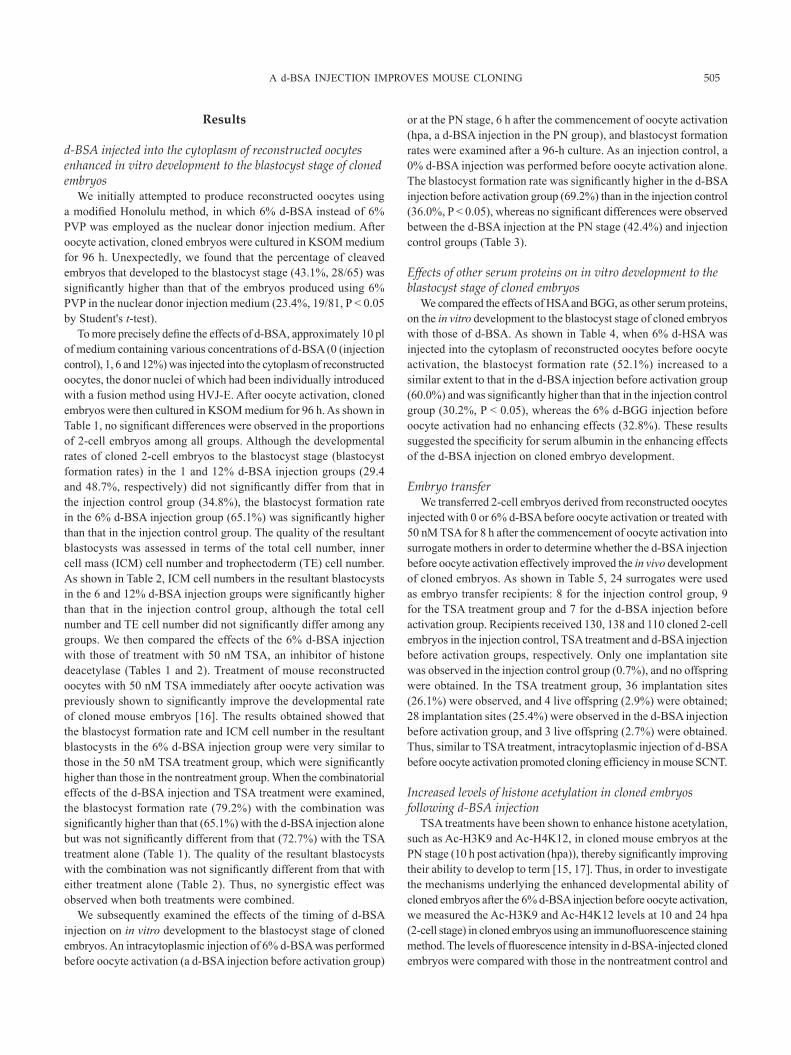
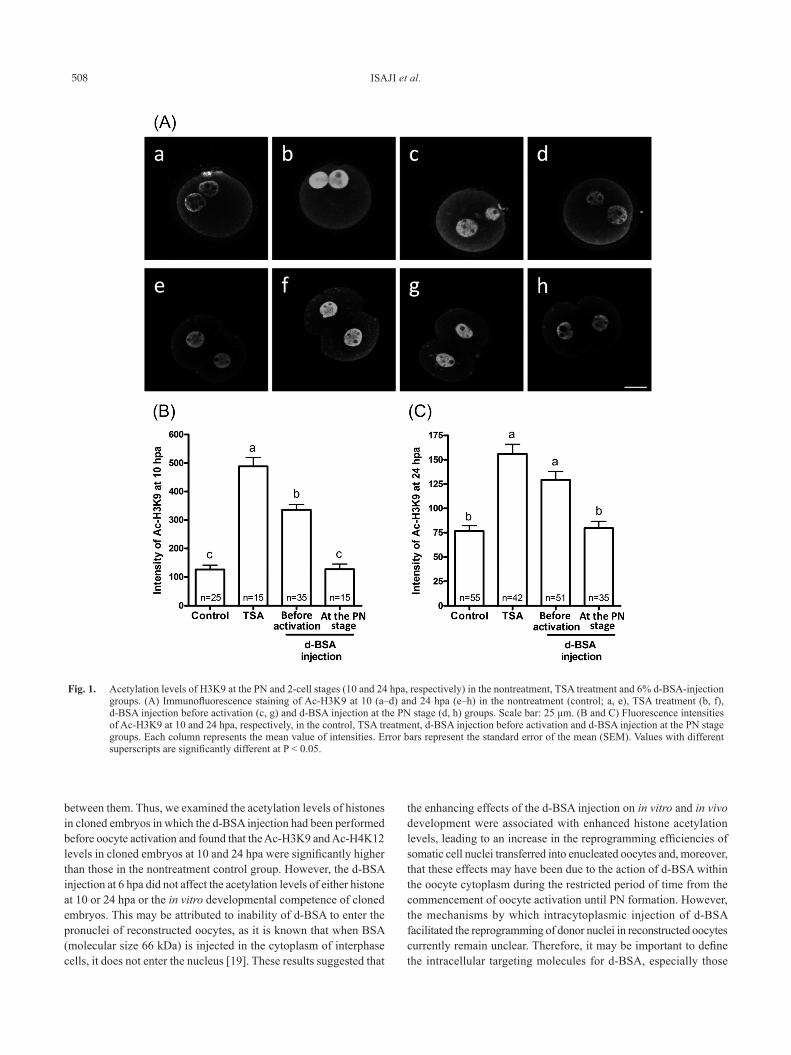
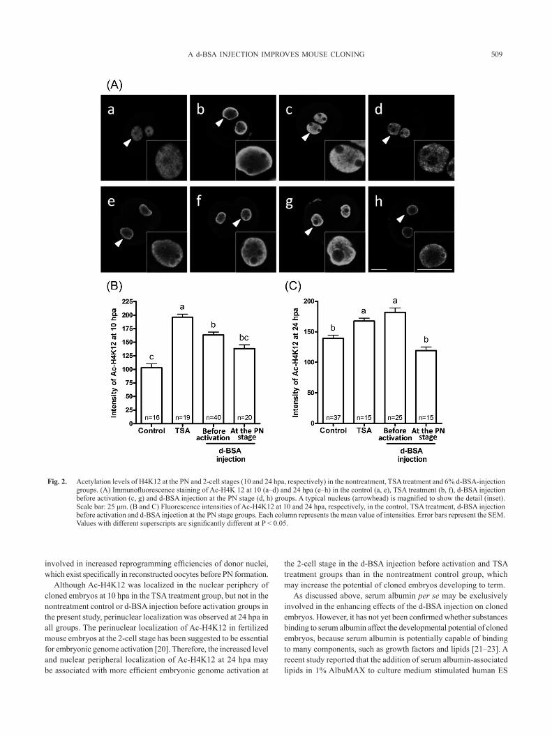
TSA treatment groups.In cloned embryos at 10 hpa (Fig. 1(A) a–d, (B); Fig. 2(A) a–d,
(B)), similar to previous studies [15, 17], the intensities of Ac-H3K9 and Ac-H4K12 were markedly higher in the group treated with TSA for 8 h from the commencement of oocyte activation than in the nontreatment control group. The Ac-H3K9 and Ac-H4K12 levels were significantly higher in the d-BSA injection before activation group than in the nontreatment control group. However, the level of Ac-H3K9 was significantly lower in the d-BSA injection before activation group than in the TSA treatment group, while the level of Ac-H4K12 was similar to that in the TSA treatment group.
In cloned embryos at 24 hpa (Fig. 1(A) e–h, (C); Fig. 2(A) e–h, (C)), the Ac-H3K9 and Ac-H4K12 levels in the nuclei of embryos were significantly higher in the TSA treatment and d-BSA injection before activation groups than in the nontreatment control group. On the other hand, these levels at both 10 and 24 hpa in the d-BSA injection at the PN stage group did not increase and were very similar to those in the nontreatment control group.
As shown in Fig. 2(A), a perinuclear localization of Ac-H4K12 was observed in the PN of cloned embryos at 10 hpa in the TSA treatment group, as previously reported [17], whereas a uniform distribution of Ac-H4K12 was observed in the nucleoplasm of the pronucleus of cloned embryos at 10 hpa in the nontreatment control, d-BSA injection before activation and d-BSA injection at the PN stage groups. However, localization at the nuclear periphery of Ac-H4K12 in cloned 2-cell embryos at 24 hpa was observed in all groups.
Discussion
In the present study, we demonstrated that medium with 6% d-BSA instead of 6% PVP, which was employed for nuclear donor injection into an enucleated oocyte, was applicable to the production of mouse SCNT embryos using the Honolulu method, and unexpectedly, in vitro development to the blastocyst stage of cloned embryos was significantly enhanced when d-BSA was directly injected together with a donor nucleus into an enucleated oocyte than when PVP was included in the nuclear donor injection medium. However, this enhancing effect on cloned embryos was not observed when the same lot of BSA, but not deionized, was used as a nuclear donor injection medium (data not shown). The purity of PVP in the nuclear donor injection medium is known to be important for the success of mouse SCNT in the Honolulu method [1]. Therefore, the deionization of BSA using an ion exchange resin is a critical step not only for the introduction of a donor cell nucleus into an enucleated oocyte but
also for clearly elucidating the enhancing effects of BSA on the developmental potential of mouse SCNT embryos.
In order to evaluate the effects of d-BSA more precisely, we performed experiments in which we produced reconstructed oocytes according to the fusion method using HVJ-E reagents and then immediately injected approximately 10 pl of various concentrations of d-BSA solution into their cytoplasm before or after oocyte activation, and this was followed by in vitro culture of cloned embryos for 96 h. We found that the blastocyst formation rates and ICM cell numbers of the resultant blastocysts were significantly higher with the 6% d-BSA injection into the oocyte cytoplasm before oocyte activation than in the injection control group, whereas the blastocyst formation rate was not affected by the d-BSA injection at 6 hpa, the PN stage. The beneficial effects of the d-BSA injection were similarly reproduced by the 6% d-HSA injection before oocyte activation, but not by the 6% d-BGG injection. This result suggested the specificity of serum albumin. However, since the isoelectric points of BSA and HSA are 4.7 and that of BGG is 6.6–8.2, these beneficial effects may also be exerted with negatively charged substances under physiological pH conditions, including serum albumin, because, similar to the acetylation of histones, negatively charged substances may reduce the electrostatic interaction between histones and DNA, leading to the more marked relaxation of the chromatin structure associated with the formation of a transcriptionally permissive state [18]. Furthermore, when embryo transfer of cloned 2-cell embryos was conducted in our study, live offspring were obtained from the d-BSA injection before activation group, but not from the injection control group, and the success rate of mouse cloning in the d-BSA injection before activation group was similar to that in the TSA treatment group. Thus, we demonstrated that d-BSA injection into reconstructed oocytes before oocyte activation improved the nuclear reprogramming of somatic cells and the developmental capacity of cloned embryos.
Developmental defects in cloned embryos have been attributed to incomplete genomic reprogramming. Histone acetylation plays a significant role in the process of reprogramming affecting the subsequent development of cloned embryos, as histone acetylation induced by treatments with histone deacetylase (HDAC) inhibitors (HDACis), such as TSA and scriptaid, was previously reported to increase the developmental competence of cloned embryos [14, 16]. In the present study, it was found that the d-BSA injection increased the blastocyst formation rate and improved the quality of the resultant blastocysts to an extent similar to that with TSA treatment, whereas no synergistic effect was observed when both treatments were combined. These results suggested the redundancy
Table 5. In vivo development of SCNT embryos treated with trichostatin A (TSA) or injected with d-BSA
Treatments No. of embryos transferred (recipients)
No. (%)* of live offspring
No. (%)** of IP sites
Average body weight (g ± SEM)
Average placental weight (g ± SEM)
Injection control† 130 (8) 0 (0) 1 (0.7)b – –d-BSA injection 110 (7) 3 (2.7) 28 (25.5)a 1.734 ± 0.287 0.288 ± 0.046TSA 138 (9) 4 (2.9) 36 (26.1)a 1.429 ± 0.108 0.230 ± 0.016
IP: implantation. * Proportion of embryos transferred that resulted in offspring at term. ** Proportion of embryos transferred that resulted in IP sites. † CZB medium was injected into the cytoplasm of reconstructed oocytes. a, b Values with different superscripts within the same column are significantly different by Fisher’s exact test (P < 0.05).
ISAJI et al.508
between them. Thus, we examined the acetylation levels of histones in cloned embryos in which the d-BSA injection had been performed before oocyte activation and found that the Ac-H3K9 and Ac-H4K12 levels in cloned embryos at 10 and 24 hpa were significantly higher than those in the nontreatment control group. However, the d-BSA injection at 6 hpa did not affect the acetylation levels of either histone at 10 or 24 hpa or the in vitro developmental competence of cloned embryos. This may be attributed to inability of d-BSA to enter the pronuclei of reconstructed oocytes, as it is known that when BSA (molecular size 66 kDa) is injected in the cytoplasm of interphase cells, it does not enter the nucleus [19]. These results suggested that
the enhancing effects of the d-BSA injection on in vitro and in vivo development were associated with enhanced histone acetylation levels, leading to an increase in the reprogramming efficiencies of somatic cell nuclei transferred into enucleated oocytes and, moreover, that these effects may have been due to the action of d-BSA within the oocyte cytoplasm during the restricted period of time from the commencement of oocyte activation until PN formation. However, the mechanisms by which intracytoplasmic injection of d-BSA facilitated the reprogramming of donor nuclei in reconstructed oocytes currently remain unclear. Therefore, it may be important to define the intracellular targeting molecules for d-BSA, especially those
Fig. 1. Acetylation levels of H3K9 at the PN and 2-cell stages (10 and 24 hpa, respectively) in the nontreatment, TSA treatment and 6% d-BSA-injection groups. (A) Immunofluorescence staining of Ac-H3K9 at 10 (a–d) and 24 hpa (e–h) in the nontreatment (control; a, e), TSA treatment (b, f), d-BSA injection before activation (c, g) and d-BSA injection at the PN stage (d, h) groups. Scale bar: 25 μm. (B and C) Fluorescence intensities of Ac-H3K9 at 10 and 24 hpa, respectively, in the control, TSA treatment, d-BSA injection before activation and d-BSA injection at the PN stage groups. Each column represents the mean value of intensities. Error bars represent the standard error of the mean (SEM). Values with different superscripts are significantly different at P < 0.05.
A d-BSA INJECTION IMPROVES MOUSE CLONING 509
involved in increased reprogramming efficiencies of donor nuclei, which exist specifically in reconstructed oocytes before PN formation.
Although Ac-H4K12 was localized in the nuclear periphery of cloned embryos at 10 hpa in the TSA treatment group, but not in the nontreatment control or d-BSA injection before activation groups in the present study, perinuclear localization was observed at 24 hpa in all groups. The perinuclear localization of Ac-H4K12 in fertilized mouse embryos at the 2-cell stage has been suggested to be essential for embryonic genome activation [20]. Therefore, the increased level and nuclear peripheral localization of Ac-H4K12 at 24 hpa may be associated with more efficient embryonic genome activation at
the 2-cell stage in the d-BSA injection before activation and TSA treatment groups than in the nontreatment control group, which may increase the potential of cloned embryos developing to term.
As discussed above, serum albumin per se may be exclusively involved in the enhancing effects of the d-BSA injection on cloned embryos. However, it has not yet been confirmed whether substances binding to serum albumin affect the developmental potential of cloned embryos, because serum albumin is potentially capable of binding to many components, such as growth factors and lipids [21–23]. A recent study reported that the addition of serum albumin-associated lipids in 1% AlbuMAX to culture medium stimulated human ES
Fig. 2. Acetylation levels of H4K12 at the PN and 2-cell stages (10 and 24 hpa, respectively) in the nontreatment, TSA treatment and 6% d-BSA-injection groups. (A) Immunofluorescence staining of Ac-H4K 12 at 10 (a–d) and 24 hpa (e–h) in the control (a, e), TSA treatment (b, f), d-BSA injection before activation (c, g) and d-BSA injection at the PN stage (d, h) groups. A typical nucleus (arrowhead) is magnified to show the detail (inset). Scale bar: 25 μm. (B and C) Fluorescence intensities of Ac-H4K12 at 10 and 24 hpa, respectively, in the control, TSA treatment, d-BSA injection before activation and d-BSA injection at the PN stage groups. Each column represents the mean value of intensities. Error bars represent the SEM. Values with different superscripts are significantly different at P < 0.05.

ISAJI et al.510
cell self-renewal [24]. However, in the present study, no significant differences were observed in the blastocyst formation rate between cloned embryos in the injection control group cultured in medium with 1% d-BSA and those cultured in medium with 0% d-BSA (data not shown), suggesting that extracellular serum albumin-associated substances did not affect the in vitro developmental potential of cloned embryos. Thus, further studies are needed to determine whether serum albumin-associated substances injected into reconstructed oocytes before oocyte activation play critical roles in the increased reprogramming efficiencies of cloned embryos.
The results of the present study provide an alternative approach to HDACis for the practical improvement of cloning efficiency as well as novel mechanistic insights into the totipotent reprogramming of donor nuclei in mouse SCNT.
Acknowledgments
This work was supported by a Grant-in-Aid for Scientific Re-search (grant number 23580390) from the Japan Society for the Promotion of Science and a grant for Exploratory Research from A-STEP of the Japan Science and Technology Agency (JST).
References
1. Kishigami S, Wakayama S, Thuan NV, Ohta H, Mizutani E, Hikichi T, Bui HT, Balbach S, Ogura A, Boiani M, Wakayama T. Production of cloned mice by somatic cell nuclear transfer. Nat Protoc 2006; 1: 125–138. [Medline] [CrossRef]
2. Wakayama T, Perry ACF, Zuccotti M, Johnson KR, Yanagimachi R. Full-term devel-opment of mice from enucleated oocytes injected with cumulus cell nuclei. Nature 1998; 394: 369–374. [Medline] [CrossRef]
3. Kato Y, Nagao Y. Effect of PVP on sperm capacitation status and embryonic development in cattle. Theriogenology 2009; 72: 624–635. [Medline] [CrossRef]
4. Feichtinger W, Obruca A, Brunner M. Sex chromosomal abnormalities and intracyto-plasmic sperm injection. Lancet 1995; 346: 1566. [Medline] [CrossRef]
5. Vidal F, Hidalgo J. Effect of zinc and copper on preimplantation mouse embryo develop-ment in vitro and metallothionein levels. Zygote 1993; 1: 225–229. [Medline] [CrossRef]
6. Barker JE, Nienhuis AW. A method for the deionization of bovine serum albumin. Methods Cell Sci 1975; 1: 111–112.
7. Leary AG, Wong GG, Clark SC, Smith AG, Ogawa M. Leukemia inhibitory factor differentiation-inhibiting activity/human interleukin for DA cells augments proliferation of human hematopoietic stem cells. Blood 1990; 75: 1960–1964. [Medline]
8. Ng ES, Davis R, Stanley EG, Elefanty AG. A protocol describing the use of a recom-binant protein-based, animal product-free medium (APEL) for human embryonic stem
cell differentiation as spin embryoid bodies. Nat Protoc 2008; 3: 768–776. [Medline] [CrossRef]
9. Chatot CL, Ziomek CA, Bavister BD, Lewis JL, Torres I. An improved culture medium supports development of random-bred 1-cell mouse embryos in vitro. J Reprod Fertil 1989; 86: 679–688. [Medline] [CrossRef]
10. Kimura Y, Yanagimachi R. Intracytoplasmic sperm injection in the mouse. Biol Reprod 1995; 52: 709–720. [Medline] [CrossRef]
11. Ho Y, Wigglesworth K, Eppig JJ, Schultz RM. Preimplantation development of mouse embryos in KSOM: augmentation by amino acids and analysis of gene expression. Mol Reprod Dev 1995; 41: 232–238. [Medline] [CrossRef]
12. Isaji Y, Murata M, Takaguchi N, Mukai T, Tajima Y, Imai H, Yamada M. Valproic acid treatment from the 4-cell stage improves Oct4 expression and nuclear distribution of histone H3K27me3 in mouse cloned blastocysts. J Reprod Dev 2013; 59: 196–204. [Medline] [CrossRef]
13. Kishigami S, Wakayama T. Efficient strontium-induced activation of mouse oocytes in standard culture media by chelating calcium. J Reprod Dev 2007; 53: 1207–1215. [Med-line] [CrossRef]
14. Van Thuan N, Bui H-T, Kim J-H, Hikichi T, Wakayama S, Kishigami S, Mizutani E, Wakayama T. The histone deacetylase inhibitor scriptaid enhances nascent mRNA production and rescues full-term development in cloned inbred mice. Reproduction 2009; 138: 309–317. [Medline] [CrossRef]
15. Bui HT, Wakayama S, Kishigami S, Park KK, Kim JH, Thuan NV, Wakayama T. Effect of trichostatin A on chromatin remodeling, histone modifications, DNA replication, and transcriptional activity in cloned mouse embryos. Biol Reprod 2010; 83: 454–463. [Medline] [CrossRef]
16. Kishigami S, Mizutani E, Ohta H, Hikichi T, Thuan NV, Wakayama S, Bui H-T, Wakayama T. Significant improvement of mouse cloning technique by treatment with trichostatin A after somatic nuclear transfer. Biochem Biophys Res Commun 2006; 340: 183–189. [Medline] [CrossRef]
17. Wang F, Kou Z, Zhang Y, Gao S. Dynamic reprogramming of histone acetylation and methylation in the first cell cycle of cloned mouse embryos. Biol Reprod 2007; 77: 1007–1016. [Medline] [CrossRef]
18. Struhl K. Histone acetylation and transcriptional regulatory mechanisms. Genes Dev 1998; 12: 599–606. [Medline] [CrossRef]
19. Vautier D, Chesné P, Cunha C, Calado A, Renard JP, Carmo-Fonseca M. Tran-scription-dependent nucleocytoplasmic distribution of hnRNP A1 protein in early mouse embryos. J Cell Sci 2001; 114: 1521–1531. [Medline]
20. Worrad DM, Turner BM, Schultz RM. Temporally restricted spatial localization of acetylated isoforms of histone H4 and RNA polymerase II in the 2-cell mouse embryo. Development 1995; 121: 2949–2959. [Medline]
21. Weber TJ, Negash S, Smallwood HS, Ramos KS, Thrall BD, Squier TC. Calmodulin involvement in stress-activated nuclear localization of albumin in JB6 epithelial cells. Biochemistry 2004; 43: 7443–7450. [Medline] [CrossRef]
22. Ammit AJ, O’Neill C. PAF released by preimplantation embryos binds to albumin. Adv Exp Med Biol 1996; 416: 263–267. [Medline] [CrossRef]
23. Ammit AJ, O’Neill C. The role of albumin in the release of platelet-activating factor by mouse preimplantation embryos in vitro. J Reprod Fertil 1997; 109: 309–318. [Medline] [CrossRef]
24. Garcia-Gonzalo FR, Izpisúa Belmonte JC. Albumin-associated lipids regulate human embryonic stem cell self-renewal. PLoS ONE 2008; 3: e1384. [Medline] [CrossRef]