Embed Size (px)
Citation preview

Journal of Pharmaceutics and Pharmacology Research
S.Ramalingam* . J. Pharmaceutics and Pharmacology Research
http://doi.org/23.2018/1.10001
Spectroscopic and computational (DFT-SCF) investigations on anti tuberculosis activity
of Isonicotino hydrazide P. Johnson a, Gene George a, S. Ramalingam* b, S. Periandy c
a Department of physics, T.B.M.L. College, Porayar, Tamilnadu, India. b Department of Physics, A.V.C. College, Mayiladuthurai, Tamilnadu, India. c Department of Physics, Kanchi mamunivar centre for PG studies, Puducherry, India.
*Corresponding author: S. Ramalingam , b Department of Physics, A.V.C. College, Mayiladuthurai, Tamilnadu, India. E-mail: [email protected]
Received date: January 19, 2018; Accepted date : February 14, 2018; Published date: February 23, 2018.
Citation this article: S. Ramalingam, Spectroscopic and computational (DFT-SCF) investigations on anti tuberculosis activity of Isonicotino
hydrazide, J Pharmaceutics and Pharmacology Research, Doi : http;//doi.org/23.2018/1.10001 .
Copyright: © 2018 S. Ramalingam et al. This is an open-access article distributed under the terms of the Creative Commons Attribution License, which permits unrestricted use, distribution, and reproduction in any medium, provided the original author and source are credited.
Abstract:
The molecular spectroscopic investigations have been carried out to study the antibiotic activity of the Isonicotino hydrazide. The activity of the compositional parts was tested by fundamental modes of vibrations of various bonds of the molecule. The chromophores action for the inducement of the antibiotic activity of the compound was analyzed from the electronic excitation absorption peaks. The σ-bond, π-bond and δ-bond interaction lobes were identified and the energy exchange between the orbitals was investigated from frontier molecular orbital profile. The asymmetrical charge distribution among different entities of the molecule for the perseverance of anti tuberculosis mechanism was recognized. The NMBO interaction energy transition outline was organized by the NBO calculation adapted with Gaussian calculations and the exchange of maximum energy transaction among various functional groups for the incentive of antibiotic were determined. The second order Polarizability of the compound emphasized the consistency of the antibiotic activity of the molecule. Thermodynamic activity of the molecule with respect to the temperature was stressed the decomposition rate and Gibbs free energy helped to determine the steadiness of the compound. The inhibition catalytic efficiency of the title molecule was fully tested by molecular docking study.
Keywords: Isonicotino hydrazide, σ-bond, δ-bond, NMBO, anti tuberculosis, Polarizability,
Thermodynamic activity.
1. Introduction
Isonicotino hydrazide is also named as Isoniazid, is an antibiotic
compound used as a first-line agent for the prevention and treatment of both
latent and active tuberculosis [1]. It is effective organic compound against
mycobacteria, particularly Mycobacterium tuberculosis. It is also active
against some atypical types of mycobacteria [2]. Isonicotino hydrazide is
available in tablet, syrup, and injectable forms [3-4]. It was considered as a
starting point in the search for new active derivatives and analogues such as
hydrazones which have been reported as active anti-TB drugs [5-6].
Isonicotino hydrazide is a pro drug and must be activated by bacterial catalyze.
Specifically, activation is associated with reduction of the mycobacterial ferric
Kat G catalase- peroxidase by hydrazine and reaction with oxygen to form an
oxyferrous enzyme complex. Once activated, Isonicotino hydrazide inhibits
the synthesis of mycoloic acids, an essential component of the bacterial cell
wall.
At therapeutic levels isoniazid is bacteriocidal against actively growing
intracellular and extracellular Mycobacterium tuberculosis organisms.
Isoniazid is an important component used in “triple therapy” to
combat tuberculosis. It has reduced Tabletting formulations stability. Anti-
oxidants are obligatory to counter oxidative stress, pulmonary inflammation,
and free radical burst from macrophages caused in tuberculosis and other
diseases. Isoniazid has long been used clinically to a great extent as the
primary drug in the treatment and prophylaxis of tuberculosis. This drug has
been considered to be one of the potent sensitizing drugs presumably capable
of being covalently incorporated into a variety of protein acceptors in vivo
and causing a wide variety of hypersensitivity symptoms as a result of
autoimmunization.
Auctores Publishing – Volume1-10001 www.auctoresonline.org Page - 01
Research Article Open Access
J. Pharmaceutics and Pharmacology Research
Isoniazid has a simple organic structure which containing two essential
components required for the high activity against Mycobacterium
tuberculosis, i.e., a pyridine ring and a hydrazide group [7-8].Isoniazid
inhibits the synthesis of mycolic acids, an essential component of the bacterial
cell wall. At therapeutic levels isoniazid is bactericidal against actively
growing intracellular and extracellular MTB organisms. Isoniazid is used in
conjunction with other effective anti-tuberculosis agents under multi-drug
therapy [9].
After screening the available literature, it is found that, there is no
work has been carried out to analyze the root cause of the anti tuberculosis
activity of the compound using Spectroscopical tool supported by
computational calculations. In the present study, the FT-IR, FT-Raman,
NMR and UV-Visible spectral investigations have been carried out is to
rendering the pharmaceutical activity.
2. Experimental profile
Physical state: • The compound has been taken in solid form which is pure and
spectroscopic grade.
Recording profile:
• The FT-IR and FT-Raman spectra of the compound were recorded
using a Bruker IFS 66V spectrometer and the instrument adopted
with an FRA 106 Raman module equipped with aNd:YAG laser
source operating at 1.064 µm line widths with 200 mW power [10].
• The high resolution 1HNMR and 13CNMR spectra were recorded
using 300 MHz and 75 MHz FT-NMR spectrometer [11].
• The UV-Vis spectrum was recorded in the range of 200 nm to 800
nm, with the scanning interval of 0.2 nm, using the UV-1700 series
instrument [12].
3. Computational profile
Using Gauss view, the present molecule was designed with
optimized geometry and such geometry was used to run the program. The
entire calculations were performed to calculate all the parameters which were
used to analyze in different aspect. The composite molecule made by base
and substitutional combinations where the change of geometrical parameters,
Mulliken charge levels and the vibrational spectral properties were tabulated
for the modification of physical and chemical parameters. The entire quantum
chemical computations were performed using the Gaussian 09 D. 01.version
software program in core i7 computer [13].
The computational calculations were performed for determining
unknown geometrical parameters, vibrational frequencies, simulation of
molecular structure and spectra using B3LYP and B3PW91 methods adopted
with 6-31++G(d, p) and 6-311++G(d,p) basis sets. The energy absorbance by
the present compound related with electronic spectra, the NBO and HOMO-
LUMO energies were calculated using time-dependent SCF method with best
fit basis set. In the same way, the 1H and 13C NMR chemical shifts with
respect to TMS were calculated by GIAO method using I-PCM model in
combination with B3LYP/6-311++G(d,p). The Mullikan charge assignment
on different parts of the compound was calculated and was purposely
elucidated for the determination of key factor for pharmaceutical activity of
the compound. The dipole moment, linear Polarizability and the first order
hyper-Polarizability in different coordinate system of the compound were
computed using B3LYP method with the 6-311++G(d,p) basis set. The ECD
and VCD spectra were simulated from available frequencies and the optical
chirality was studied and the mechanism for masking the toxicity was
interpreted.
4. Results and discussion
4.1. Structural Deformation analysis
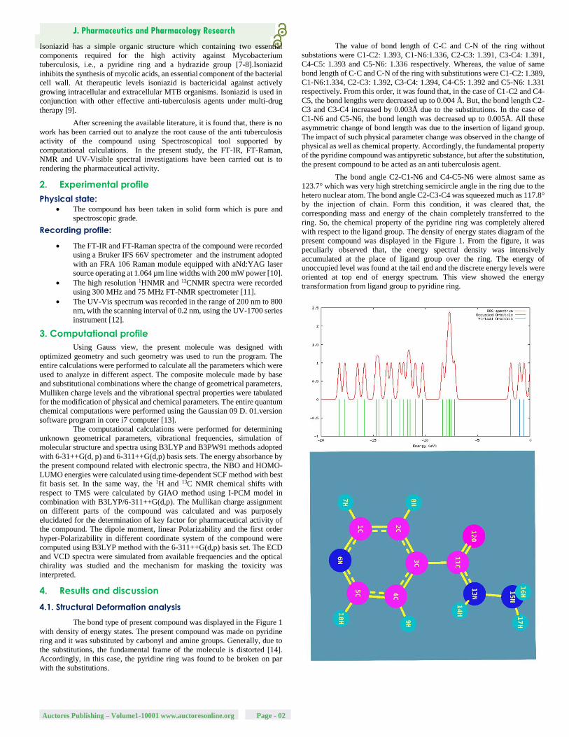
The bond type of present compound was displayed in the Figure 1
with density of energy states. The present compound was made on pyridine
ring and it was substituted by carbonyl and amine groups. Generally, due to
the substitutions, the fundamental frame of the molecule is distorted [14].
Accordingly, in this case, the pyridine ring was found to be broken on par
with the substitutions.
The value of bond length of C-C and C-N of the ring without
substations were C1-C2: 1.393, C1-N6:1.336, C2-C3: 1.391, C3-C4: 1.391,
C4-C5: 1.393 and C5-N6: 1.336 respectively. Whereas, the value of same
bond length of C-C and C-N of the ring with substitutions were C1-C2: 1.389,
C1-N6:1.334, C2-C3: 1.392, C3-C4: 1.394, C4-C5: 1.392 and C5-N6: 1.331
respectively. From this order, it was found that, in the case of C1-C2 and C4-
C5, the bond lengths were decreased up to 0.004 Å. But, the bond length C2-
C3 and C3-C4 increased by 0.003Å due to the substitutions. In the case of
C1-N6 and C5-N6, the bond length was decreased up to 0.005Å. All these
asymmetric change of bond length was due to the insertion of ligand group.
The impact of such physical parameter change was observed in the change of
physical as well as chemical property. Accordingly, the fundamental property
of the pyridine compound was antipyretic substance, but after the substitution,
the present compound to be acted as an anti tuberculosis agent.
The bond angle C2-C1-N6 and C4-C5-N6 were almost same as
123.7° which was very high stretching semicircle angle in the ring due to the
hetero nuclear atom. The bond angle C2-C3-C4 was squeezed much as 117.8°
by the injection of chain. Form this condition, it was cleared that, the
corresponding mass and energy of the chain completely transferred to the
ring. So, the chemical property of the pyridine ring was completely altered
with respect to the ligand group. The density of energy states diagram of the
present compound was displayed in the Figure 1. From the figure, it was
peculiarly observed that, the energy spectral density was intensively
accumulated at the place of ligand group over the ring. The energy of
unoccupied level was found at the tail end and the discrete energy levels were
oriented at top end of energy spectrum. This view showed the energy
transformation from ligand group to pyridine ring.
Auctores Publishing – Volume1-10001 www.auctoresonline.org Page - 02

J. Pharmaceutics and Pharmacology Research
Geometrica+A+A1:F26
Methods
Parameters HF B3LYP B3PW91
6-311++G
6-31++G
6-311++G
6-311++G
6-31++G
(d, p) (d, p) (d, p) (d, p) (d, p)
Bond length(Å)
C1-C2 1.383 1.391 1.394 1.389 1.392 C1-N6 1.32 1.338 1.341 1.334 1.338 C1-H7 1.076 1.086 1.087 1.087 1.088 C2-C3 1.385 1.395 1.398 1.392 1.396 C2-H8 1.072 1.082 1.084 1.083 1.085 C3-C4 1.386 1.397 1.4 1.394 1.398 C3-C11 1.51 1.511 1.511 1.506 1.507 C4-C5 1.386 1.394 1.397 1.392 1.395 C4-H9 1.074 1.083 1.085 1.085 1.086 C5-N6 1.317 1.334 1.338 1.331 1.335 C5-H10 1.076 1.086 1.087 1.087 1.088 C11-O12 1.187 1.212 1.22 1.21 1.217 C11-N13 1.363 1.377 1.378 1.372 1.374 N13-H14 0.994 1.011 1.013 1.011 1.012 N13-N15 1.383 1.397 1.397 1.387 1.388 N15-H16 0.998 1.014 1.015 1.013 1.014 N15-H17 0.997 1.012 1.014 1.012 1.012 Bond angle(º) C2-C1-N6 123.596 123.698 123.748 123.781 123.82
1 C2-C1-H7 120.128 120.24 120.245 120.207 120.218 N6-C1-H7 116.274 116.06 116.006 116.011 115.96
C1-C2-C3 118.488 118.842 118.827 118.765 118.754 C1-C2-H8 121.076 121.324 121.369 121.471 121.497 C3-C2-H8 120.433 119.831 119.802 119.762 119.746 C2-C3-C4 118.142 117.831 117.844 117.883 117.894 C2-C3-C11 118.304 118.125 118.129 118.064 118.069 C4-C3-C11 123.535 124.01 123.998 124.017 124.005 C3-C4-C5 118.443 118.769 118.754 118.685 118.669 C3-C4-H9 121.901 121.687 121.691 121.69 121.699 C5-C4-H9 119.625 119.503 119.514 119.58 119.587 C4-C5-N6 123.573 123.677 123.726 123.764 123.807 C4-C5-H10 120.019 120.125 120.128 120.093 120.102 N6-C5-H10 116.406 116.196 116.144 116.141 116.089 C1-N6-C5 117.743 117.165 117.088 117.102 117.038 C3-C11-O12 121.134 121.613 121.539 121.656 121.574 C3-C11-N13 114.025 113.835 114.068 113.627 113.872 O12-C11N13 124.84 124.55 124.392 124.716 124.552 C11-N13H14 117.941 118.27 118.27 118.1 118.168 C11-N13N15 120.138 120.474 120.277 120.572 120.36
H14-N13-N15 118.741 119.49 119.403 119.807 119.707 N13-N15-H16 111.729 111.532 111.669 111.604 111.811 N13-N15-H17 110.351 110.378 110.381 110.563 110.652 H16-N15-H17 110.95 110.698 110.939 110.676 111.021 Dihedral angle(º)
N6-C1-C2C3 -0.895 -0.936 -0.771 -0.983 -0.876 N6-C1-C2H8 178.745 178.654 178.784 178.621 178.71
3 H7-C1-C2C3 179.354 179.291 179.393 179.245 179.342 H7-C1-C2H8 -1.004 -1.118 -1.051 -1.15 -1.106
C2-C1-N6-C5 -0.105 -0.163 -0.24 -0.187 -0.256 H7-C1-N6-C5 179.653 179.617 179.601 179.592 179.57 C1-C2-C3-C4 1.163 1.324 1.211 1.411 1.304 C1-C2-C3-C11 179.683 179.317 179.388 179.351 179.39
5 H8-C2-C3-C4 -178.479 -178.271
-178.35 -178.2 -178.26 H8-C2-C3-C11 0.039 -0.278 -0.174 -0.26 -0.163
C2-C3-C4-C5 -0.513 -0.696 -0.707 -0.754 -0.748 C2-C3-C4-H9 177.518 177.015 177 176.859 176.83
9 C11-C3-C4-C5 -178.949 -178.561
-178.767 -178.561 -178.72 C11-C3-C4-H9 -0.917 -0.848 -1.06 -0.947 -1.129
C2-C3-C11-O12 -31.198 -28.884 -28.439 -29.44 -29.242 C2-C3-C11-N13 148.778 150.969 151.554 150.304 150.624 C4-C3-C11-O12 147.235 148.974 149.615 148.363 148.723 C4-C3-C11-N13 -32.788 -31.171 -30.39 -31.892 -31.41
C3-C4-C5-N6 -0.515 -0.423 -0.321 -0.44 -0.367
C3-C4-C5-H10 179.288 179.234 179.328 179.225 179.287
H9-C4-C5-N6 -178.593 -178.186 -178.079 -178.105 -178.01
H9-C4-C5-H10 1.211 1.48 1.57 1.56 1.647
C4-C5-N6-C1 0.827 0.856 0.797 0.914 0.871
H10-C5-N6-C1 -178.983 -178.822 -178.865 -178.763 -178.8
C3-C11-N13-H14 -18.176 -14.393 -15.594 -13.414 -14.557 C3-C11-N13-N15 -177.786 -179.188 -179.223 -179.323 -179.39 O12-C11-N13-H14 161.798 165.455 164.399 166.321 165.30
5 O12-C11-N13-N15 2.189 0.66 0.77 0.411 0.472 C11-N13-N15-H16 103.485 105.897 104.7 106.827 105.59
1 C11-N13-N15-H17 -132.571 -130.604 -131.391 -129.518 -130.09 H14-N13-N15-H16 -55.962 -58.713 -58.744 -58.843 -59.008 H14-N13-N15-H17 67.98 64.784 65.164 64.81 65.307
Table 1: Optimized geometrical parameters for Isonicotinohydrazide computed
at HF/DFT (B3LYP&B3PW91) with 6-31++G(d, p) & 6-311++G(d, p) basis
sets.
4.2. Mulliken charge dislocation analysis
The molecular orbital formation is decided by symmetrical and
asymmetrical charge distribution among atoms [15]. The symmetry or
asymmetry of the charge distribution plays a fundamental role in determining
the chemical properties of the molecule and consequently this property of the
charge distribution is used as a basis for the classification of chemical bonds
[16-17]. All the chemical bonds in the compound would be formed as
covalent bond between composite atoms and are to be polar and non polar.
Normally, in the compound, the polar and non polar bonds are existed with
respect to the substitutional groups. If the ration of polar bonds is greater, the
asymmetry charges distribution taking place in the compound. Here, the
dipole moment of the compound was found to be high since the ratio of hetero
nuclear bonds was greater than homo nuclear bonds. The Mulliken charge
levels were depicted in the Table 2 and the corresponding pictorial diagram
was shown in Figures 2 and 3.
Auctores Publishing – Volume1-10001 www.auctoresonline.org Page - 03

J. Pharmaceutics and Pharmacology Research
Table 2: Mulliken Charge levels of Isonicotinohydrazide at different methods
In this compound, though the hetero nuclear atom (N) present in
the ring, the strong bond was created between CC of the ring and also the N
was to be found as neutral. Due to the asymmetrical charge dislocation
motion towards substitutional group, the top moiety of the ring becomes
protonic region whereas the bottom moiety turns out to be electron rich
region (depletion region). From this view, it was inferred that, the
considerable amount of chemical energy was transferred from ring to ligand
group. Normally, the rich polar character present in carbonyl group, but in
this case, the electronic charges were accumulated vigorously and the bond
come in to repulsive. On the other side of the substitutional group, in order
to make strong attractive bond, the intermediate nitrogen atom converted as
neutral.
4.3. Vibrational analysis
4.3.1. Vibrational assignments
The fundamental frequency pattern of the compound was
represented by IR and Raman spectra and the observed spectral peaks in
terms of wavenumbers were presented in the Table 3 and the observed with
simulated FT-IR and FT-Raman spectra were presented in the Figure 4&5.
According to the vibrational degrees of freedom, since the present
compound contained 17 atoms, there were 45 fundamental modes of
vibrations were observed. Out of 45 vibrational modes, 17 stretching, 14 in
plane and 14 out of plane bending modes were distinguished. Since the title
molecule was non Centro-symmetry, the vibrations are active in IR and
those inactive in Raman and vice versa. The total elementary vibrations
were ordered as Г= 31Aʹ+ 14Aʺ
4.3.2. C-H vibrations
Normally, the ring C-H vibrations in benzene and pyridine ring
ensured that, whether the hexagonal frame has substituted by ligand or not. If
the observed fundamentals are observed within the expected region, the ring
will not be affected much by the substitutions. Otherwise, the ring vibrations
are rather suppressed and ratio of the same showed the impact of the
substitutions on the ring. Here, the substitutional group was present at one C
of the ring and 16 C-H vibrations are possible. Accordingly, the C-H
stretching vibrations were found at 3070, 3040, 3010 and 2990 cm-1 in IR and
Raman spectra. The in plane and out of plane bending vibrations were
observed at 1060, 1050, 1010 and 1005 cm-1 and 720, 665, 660 and 650 cm-1
respectively. The hetero aromatic C H stretching vibrations are normally
found between 3100 and 3010 cm-1, the C–H in plane bending frequencies
appear in the range of 1000 - 1310 cm−1 and C–H out of plane bending
vibration in the range 750 - 1000 cm−1 [18-20]. The entire vibrational bands
are expected to observe with medium to strong intensity. But all the bands
were found with very week to very strong intensity. One peak of stretching
and entire signals of out of plane bending were observed out of the expected
region whereas all the in plane bending modes were found at the top end of
the allotted region. The observed signals were affected in low energy region
which was mainly by the energy transformation from ring to ligand group in
order to generate anti-tuberculosis activity.
4.3.3. Ring CC and C-N vibrations
The main hexagonal frame of pyridine ring is constituted by C-C
bonds and these are directly affected by the loaded ligand groups. In addition
to that, the ring associated with N atom, so the ring vibrations include the
C=N and C-N vibrations also. Interactions between ring C=C and C=N
stretching vibrations result in two strong-to-medium intensity absorptions
about 100 cm-1 apart [21]. Usually, the C=C and C=N stretching modes are
observed in the region 1615-1575 cm-1 and 1520 -1465 cm-1 respectively [22-
25]. Accordingly, in the present case, the C=C and C=N stretching signals
appeared at 1660 & 1630 and 1580cm-1 respectively. Both the stretching
bands are moved well above the expected level of the spectrum. In this view,
it was cleared that, the ring energy was found to be increased and which was
favor for the energy transaction between ring and ligand group. Usually, these
vibrations are followed by C-C and C-N stretching bands which are observed
around 1460 cm-1[26]. In this case, these vibrational bands were observed at
1530 cm-1 and 1490 & 1430 cm-1 respectively. The CCC and CNC in plane
and out of plane bending (ring breathing) modes were identified at 300, 225&
175 and 135, 120 & 50 cm-1 respectively. These observed fundamental modes
moved down to well below the expected region. The ring deformation
vibrations have been restricted since it was substituted by the carbonyl and
amino groups. Finally, it was concluded that, the ring energy was consumed
lot for the formation of drug property.
S. No. Atom Position Mullikan charge
B3LYP B3PW91
1 C1 -0.563 -0.758
2 C2 0.324 0.440
3 C3 0.665 0.691
4 C4 0.112 0.230
5 C5 -0.571 -0.771
6 C11 -0.599 -0.716
7 H7 0.179 0.215
8 H8 0.223 0.260
9 H9 0.193 0.234
10 H10 0.172 0.207
11 012 -0.288 -0.274
12 N6 -0.046 0.006
13 N13 -0.141 -0.105
14 H14 0.246 0.262
15 N15 -0.389 -0.426
16 H16 0.246 0.258
17 H17 0.238 0.247
Auctores Publishing – Volume1-10001 www.auctoresonline.org Page - 04

J. Pharmaceutics and Pharmacology Research
S. No
Symmetry
Species Observed
Frequency(cm-1)
Methods Vibrational
CS HF B3LYP B3PW91 Assignments
FT-IR
FT-
Raman
6-311++G 6-31++G 6-311++G 6-31++G 6-311++G
(d,p) (d,p) (d,p) (d,p) (d,p)
1 A′ 3480m - 3474 3474 3466 3473 3489 (N-H) υ
2 A′ 3460m - 3472 3476 3466 3463 3447 (N-H) υ
3 A′ 3360m - 3354 3342 3375 3367 3356 (N-H) υ
4 A′ - 3070w 3051 3075 3061 3057 3063 (C-H) υ
5 A′ - 3040vw 3033 3053 3040 3034 3042 (C-H) υ
6 A′ 3010vs - 3011 2999 3016 3014 3018 (C-H) υ
7 A′ 2990s - 2980 2992 2990 3007 2990 (C-H) υ
8 A′ 1750m - 1758 1750 1746 1756 1750 (C=O) υ
9 A′ - 1660vw 1657 1659 1660 1665 1661 (C=C) υ
10 A′ - 1630vw 1624 1630 1625 1631 1634 (C=C) υ
11 A′ 1580s - 1583 1579 1572 1580 1572 (C=N) υ
12 A′ 1530w - 1535 1525 1524 1533 1528 (C-C) υ
13 A′ 1490w - 1486 1496 1493 1486 1496 (C-N) υ
14 A′ 1400w - 1402 1407 1399 1397 1402 (C-N) υ
15 A′ 1320vs - 1320 1320 1316 1316 1325 (C-C) υ
16 A′ - 1290vw 1286 1290 1295 1291 1285 (C-C) υ
17 A′ - 1270vw 1272 1267 1265 1271 1275 (N-H) δ
18 A′ - 1210vw 1209 1213 1215 1210 1210 (N-H) δ
19 A′ 1190vw - 1183 1190 1189 1193 1188 (N-H) δ
20 A′ - 1130m 1134 1130 1130 1130 1130 (N-N) υ
21 A′ - 1060vw 1061 1061 1060 1062 1058 (C-H) δ
22 A″ 1050w - 1053 1052 1053 1046 1054 (C-H) δ
23 A″ - 1010vs 1015 1005 1011 1009 1013 (C-H) δ
24 A′ - 1005m 1004 1005 1007 1007 1007 (C-H) δ
25 A′ - 1000m 997 999 1001 998 997 (C=O) δ
26 A′ 975vs - 974 977 977 976 973 (C-N) δ
27 A′ - 900w 902 899 903 901 897 (C-C) δ
28 A′ 885w - 888 882 881 883 881 (N-H) γ
29 Aʺ 855s - 856 872 877 872 877 (N-H) γ
30 Aʺ - 765vw 763 767 762 762 763 (N-H) γ
31 Aʺ 750w - 753 746 753 750 752 (N-N) δ
32 Aʺ 720vw - 719 735 721 717 721 (C-H) γ
33 Aʺ - 665w 665 664 664 668 664 (C-H) γ
34 Aʺ - 660w 660 661 657 660 661 (C-H) γ
35 Aʺ 550vw - 550 548 550 553 555 (C-H) γ
36 Aʺ - 510vw 509 512 510 509 512 (C=O) γ
37 Aʺ - 405w 406 406 406 404 404 (C-N) γ
38 Aʺ - 390vw 391 396 395 391 391 (C-C) γ
39 Aʺ - 350vw 350 350 350 351 351 (CNC) δ
40 A′ - 300vw 300 299 301 300 299 (CCC) δ
41 A′ - 225vw 225 224 226 225 225 (CCC) δ
42 A′ - 175m 175 173 176 177 175 (N-N) γ
43 Aʺ - 135s 134 135 135 135 135 (CCC) γ
44 Aʺ - 120m 120 116 115 115 116 (CNC) γ
45 Aʺ 50m - 50 50 50 50 50 (CCC) γ
Table 3: Observed and calculated vibrational frequencies at HF and DFT (B3LYP & B3PW91) with 6-31++G(d,p) & 6-311++G (d,p) level for Isonicotino hydrazide
VS –Very strong; S – Strong; m- Medium; w – weak; as- Asymmetric; s – symmetric; υ – stretching; α –deformation, δ - In plane bending; γ-out
plane bending; τ – Twisting:
Auctores Publishing – Volume1-10001 www.auctoresonline.org Page - 05

J. Pharmaceutics and Pharmacology Research
4.3.4. NH2 vibrations
Specifically, the amino group is always dominating all other
groups and it very much pronounced in the drug property of the compound
[27]. In this case also, the amino group was played important role to induce
such anti-tuberculosis activity. Usually, the N-H stretching modes are
appeared in the region 3300-3500 cm-1[28-29]. Therefore, the same signals
for N-H stretching found with medium intensity at 3480, 3460 and 3360
cm-1 in IR only. As in the previous case [30], such vibrations were observed
at the top end of the allocated area. Form this observation, it was cleared
that, these components were found to be active and it took part in transaction
of energy for creating novel property.
The N-H in-plane bending vibrations (scissoring) are usually
observed in the region 1610-1630cm−1 and the out of plane bending
(wagging) vibrations are normally identified in the region 1150-900cm−1
[31-32]. Here, these vibrations (in plane and out of plane) have been
observed at 1270, 1210 & 1190 cm-1 and 885, 855 & 765 cm-1 respectively.
Dissimilar to the stretching, these bending modes were found to be affected
and depressed much which was due to the presence of carbonyl group. The
activeness of bending modes rather decreased by C=O group by inductive
effect.
4.3.5. C=O, N-N and C-C vibrations
The ketones related Carbonyl group vibrations are the important
characteristic bands in vibrational spectra since it acts as major part of
creating complete property and therefore, these bands have been the subject
of wide analysis [33-34]. Normally, the intensity of these bands would be
greater since its conjugation and dipole moment. Therefore, it leads to the
intensification of the scattering lines as well as very strong intensity of
infrared band. The carbonyl stretching vibrations in ketones are expected in
the region 1680–1715 cm−1[35]. In this case, a band was found with medium
intensity at 1750 cm-1 in IR only. The band was appeared well above the
allowed region but its intensity was suppressed which was due to the nearby
amino group. It’s in plane and out of plane bending vibrational modes were
observed at 1000 and 405 cm-1 respectively. These vibrational modes were
agreed well with the previous work [36].
The amine groups were attached with one another by N-N bond and
forming azine group and it was very significant since it is Y-junction which
exchanges the energy between ring and amine group of molecule. Generally,
the azine bond N-N stretching vibration was found around 925-1150 cm-1
[37]. The N-N stretching vibration was observed with medium intensity at
1130 cm-1. The consequent in plane and out of plane bending modes were
observed at 750 and 175 cm-1 respectively. The acquired vibrational bands
were precisely harmonized with the literature and ensure the role of azine
group in the drug properties of the compound.
The C-C stretching of C-C bond of out of the ring are observed in
the region 1470-1520 cm-1[33]. Here, the stretching modes have been found
at 1320 and 1290 cm-1. The corresponding in and out of plane bending
vibrations was observed at 885 and 390cm-1 respectively. These bands were
at exact allowed region since it was favored by the carbonyl and amino
groups.
4.4. NMR confinement
The 1H and 13C NMR spectral data was presented in Tables 4 and the
corresponding spectra were shown in Figure 6. Generally, the chemical shift
of carbons and hydrogens are situated in different environment with respect
to the coupling constant and makes the molecule complicated. The isotropic
chemical shift of C and H are usually explicated the unknown chemical
properties of complicated compound. In general, the chemical shifts of
carbon is depends upon the bonded atoms and their chemical environment
in the compound. The isotropic chemical shifts in aromatic compounds are
universally observed in the region of 100- 190 ppm [38]. The chemical shift
of C of base compound with respect to ligand attachment is providing direct
the dependable interpretation of chemical properties. Usually, the chemical
shift of aliphatic chain is constantly behind the aromatic compound [39].
Symmetry also makes atoms equivalent in molecules and if group of atoms
are in some identical locations relative to a plane of symmetry in a molecule,
they will be equivalent [40-41]. Here, the carbons of the ring were located
in the region 140- 160 ppm as in normal case.
Generally, the NMR signals are falling into domain with respect
the diamagnetic equivalence of carbon and hydrogen atoms. The observed
chemical shift of carbons of the pyridine ring; C1, C2, C3, C4 and C5 were
restricted in the region 140-164 ppm. The observed chemical shift of C1 and
C5 were found to be same (164ppm) which was more than rest of others due
to coupling of N whereas the calculated shift was 156 and 158 ppm
respectively. This condition also due to the asymmetrical electron orientation
on such carbons which was evident in Mulliken charge analysis. Similarly,
C2 and C4 were same and 140 ppm, but the observed and calculated chemical
shift of C3 was 150 and 144 ppm respectively. The observed and calculated
chemical shift of C11 was found to be 170 195 ppm respectively which is
greater than others since its electron cloud was fragmented asymmetrically
by ring, carbonyl and amino groups. The C3 and C11 have been opened up
due to the random breaking of proton shield by π-bonded oxygen and amine
groups in which the consistent amount of energy was exchanged among
different entity of molecule in order to enforce the drug property in the
compound.
The hydrogen atoms were attached with C and N atoms in the molecule which
make different environment upon which the chemical shift of the atoms was
classified. Here, the chemical shift of H in ring was observed in the region 7-
10 ppm whereas in amino group, the shift was observed 4.5 and 7.6 ppm. The
chemical shift was observed consistently with respect to the attached atoms.
Table 4: Experimental and calculated 1H and 13C NMR chemical shifts
(ppm) of isonicotinohydrazide
Auctores Publishing – Volume1-10001 www.auctoresonline.org Page - 06
Atom
position
TMS-B3LYP/6-311+G(2d,p)
GIAO Shift in (ppm) Experimental
shift
(ppm)
Gas Solvent phase
DMSO CCl4
1C 158.169 157.891 158.165 164.0
2C 127.195 127.078 127.208 140.0
3C 144.129 144.814 144.388 150.0
4C 123.629 126.602 124.698 140.0
5C 156.024 157.095 156.448 164.0
11C 195.801 200.559 197.758 170.0
6N 389.791 374.241 383.229 -
13N 148.607 153.23 150.436 -
15N 72.7634 70.778 71.9611 -
7H 9.2127 9.2304 9.2313 8.7
8H 7.8878 7.9 7.9044 7.8
9H 6.9857 7.4784 7.1771 7.8
10H 9.0101 9.1184 9.0566 10.0
14H 6.3622 6.9062 6.5681 7.6
16H 2.1522 2.4561 2.2692 4.5
17H 2.6703 3.1309 2.8651 4.5
12O 545.715 489.285 522.323 -

J. Pharmaceutics and Pharmacology Research
4.5. Frontier molecular outline
The interaction between two atomic or molecular orbitals will
form two new orbitals. One new orbital is antibonding orbital which has the
higher energy than the original molecule orbital. The other new orbital is the
bonding orbital which is lower in energy than the initial one. The
stabilization of the bonding molecular orbital and destabilization of the
antibonding can increase when the overlap of two orbitals increases [42]. In
the molecular interaction, there are the two important orbitals interact each
other. One is the highest energy occupied molecular orbital is called HOMO.
The other one is the lowest energy unoccupied molecular orbital is called
LUMO. When a pair of electrons filled in one of the molecule orbital and
no electron occupy in the other orbital, this interaction is very stable and
called filled- empty interaction.
The orbital interaction profile of title compound was presented in
the Figure 7 in which different bonding interactions were seen clearly. The
elevated orbital energy level was depicted in the Table 5. In the case of
LUMO, there were two sets of first order π-bonding interaction found over
the ring cc of opposite moiety and one σ-bonding existed on N of the ring.
Apart from that, one π-bonding interaction taking place between ring C and
ligand C which was also connect =O. one N-H iso surface was observed and
little flavor was identified on H. usually, the π-conjugation interaction
orbitals induce active biological as well as crystal property in the aromatic
compound[43]. Here, the electron cloud receiving location (LUMO) was
observed to be π-orbital overlapping region where the orbital was
degenerative and able to consume electronic energy from HOMO.
In HOMO, the cascade of orbital overlapping of degenerate
energy levels at top and bottom moiety of the ring which connect CNC and
CCC with two H. peculiarly, the δ-bond orbitals found at C3 of ring C11,
O12 and N13 of ligand chain. The δ-bond interaction is very rare and if
present in compound, there will be stimulation of drug activity. Here, from
the orbital interaction, proved the anti tuberculosis action of the present
compound. In second order orbital interaction (HOMO+1 and LUMO-1),
the positive and negative intersection was found and all the degenerate
energy levels were interacted to make centralized donor region for supplying
electronic energy.
Energy levels IR Region UV-Visible region
Energy in eV Energy in eV
H+10 12.17545 12.44077
H+9 11.72184 11.72402
H+8 11.54225 11.66633
H+7 10.99285 10.8938
H+6 10.3708 10.72155
H+5 8.35498 8.86002
H+4 8.02245 8.39743
H+3 7.79279 7.88613
H+2 7.70762 7.8064
H+1 7.57238 7.37319
H 7.28312 7.01999
L 1.86261 2.38725
L-1 1.03593 1.27403
L-2 0.82178 0.79919
L-3 0.14966 0.17252
L-4 0.18422 0.06721
L-5 0.31946 0.19837
L-6 0.51783 0.52164
L-7 0.74722 0.56273
L-8 0.92545 0.93389
L-9 1.21444 1.14695
L-10 1.30614 1.35703
Table5 :Frontier Molecular orbitals and Energy Level
4.6. UV-Visible absorption analysis
UV-Visible spectral analysis provides the information regarding
the ligand group which causing the outstanding chemical property of the
compound. Normally, the substitutional impact shifts the electronic excitation
pattern (finger print region) of the base compound. The electronic excitation
parameters are presented in the Table 6 and the absorption band was displayed
in the Figure 8. Here the compound architected by pyridine ring attached with
amino group and carbonyl group. Generally, the pyridine ring absorbance
peak identified at 190 nm with 76 Kcal/mol in the UV-Visible spectrum [44].
But, by the addition of the substitutional group, the UV band was found at
331 nm with the transition energy of 86.34 Kcal/mol by crossing the energy
gap of 3.74 eV. The oscillator strength of the oscillation (transition between
ground and excited state) was found to be 0.0134 with higher molar
absorptivity. The shift was represented as bathochromic for the higher
wavelength region. Usually, the presence of chromophores such as C=O and
NH2, enable intensive absorption bands at 290 and 280 nm respectively [45].
In this case, the triplet absorption bands were appeared at 331, 319 and 293
nm with effective intensity. These absorption peaks ensure the presence of
such chromophores which was the reason for the inducement of the anti
tuberculosis activity.
Auctores Publishing – Volume1-10001 www.auctoresonline.org Page - 07

J. Pharmaceutics and Pharmacology Research
The entire transitions in the electronic HOMO and LUMO energy
levels were found to be confined within the quartz UV region of the
spectrum which implies that, the title compound would not be active in
visible region. Because of this condition of the compound, the drug may be
inert for light radiation. The UV-Visible frontier molecular excitations were
belonging to n→π* class of transitions showed the strong interaction
between donor (carbonyl and amine groups) and acceptor (pyridine ring).
The absorption band for title compound was transparently taking place in
the UV spectrum in R-band (German, radikalartig) and constantly being
with therapeutic activity. In this case, the identification of absorption band
in quartz-UV region predicted that, the functional group of amine and azine
entities at ring was played significant role for the inducement of
pharmaceutical action.
The electronic charge distribution is responsible for electronic
polarization of orbitals which was induced by local electric field called ECD.
The interaction of chromophores on the base compound making two fold of
energy increments for transition to excited states adjust the chemical activity
of the compound which can be identified in the ECD spectra. As in the Figure
8, the ECD absorption band was identified at 290 nm which was the
characteristic band of C=O and NH2 groups. This band represents uniqueness
effect of chemical reactivity.
Table 6: Theoretical electronic absorption spectra of Isonicotino
hydrazide(absorption symbols λ (nm), excitation energies E (eV) and oscillator
strengths (f)) using TD-DFT/B3LYP/6-311++G(d,p) method
4.7.Molecular electrostatic potential (MEP) maps
The molecular electrostatic potential is asymmetric charge distribution
in different entities of a molecule. The molecular interaction energy was
calculated by using the equation
1
1 1
( )M
j j
QiV r
r r
If the charge distribution continuous over the compound, the MEP is then
given by equation at any instant
1
1
( )( )
rV r dr
r r
λ (nm) E (eV) ( f ) Energyin
Kcal/mol
Major
contri
bution
Assign
ment Region
Band
s
Gas
331.13 3.7443 0.0134 86.34 H L
(95%) n→π*
Quartz
UV
R
band
319.33 3.8826 0.0048 89.53 H L
(85%) n→π*
Quartz
UV
(Ger
man,
radik
alarti
g)
293.95 4.2179 0.0136 97.26 H L
(95%) n→π*
Quartz
UV
DMSO
324.8 3.8172 0.0359 88.02 H L
(85%) n→π*
Quartz
UV
R
band
313.14 3.9594 0.0039 91.3 H L
(92%) n→π*
Quartz
UV
(Ger
man,
radik
alarti
g)
285.44 4.3436 0.0096 100.16 H L
(95%) n→π*
Quartz
UV
CCl4
328.88 3.7699 0.0253 86.93 H L
(85%) n→π*
Quartz
UV
R
band
317.46 3.9055 0.0054 90.06 H L
(92%) n→π*
Quartz
UV
(Ger
man,
radik
alarti
g)
291.76 4.2495 0.0134 97.99 H L
(95%) n→π*
Quartz
UV
Auctores Publishing – Volume1-10001 www.auctoresonline.org Page - 08

J. Pharmaceutics and Pharmacology Research
The MEP is highly informative concerning the protonic and
electronic charge distribution of a given molecule. It is mainly used for
asymmetric charge orientation among different atoms causing strong
electrostatic potential between two extreme charge levels which is favor for
the inducement of the particular biological and pharmaceutical properties.
The framed Molecular electrostatic potential view was presented in Figure
9 in which the entire molecular interactive zones can be viewed. The static
potential energy interaction generated between proton and electron fields is
ranged from high frequency (blue) region to higher wavelength (red) region
which designated that, the charge dislocation between electrophilic and
nucleophilic boundaries. In a molecule, charges are polarized rigorously in
negative and positive terminals, and there are significant differences in
electron and proton density due to the injection of substitution in base
compound. The rich electro negativity region was indicated by wealthy red
and positive (proton region) almost was indicated by blue.
The two extreme ends of charge distribution was represented by the
color code in the range between -7.66 a.u. (genuine red faded to yellow) to
7.66 a.u. (genuine blue faded to green). Generally, the negative region is
mainly localized over the portion of highly electronegative atoms.
In this case, the electron bustle zone was captured over the O of
carbonyl group, N of amino group and the N of pyridine ring. The moderate
negative region was concealed over the pyridine ring and further decayed
when moved towards H of ring. The protonic content was confined on the
hydrogen zones over the amino group. It was abundant in around the edge
of amino group and deficient in carbon bonded H. This well defined
atmosphere was induced by hetero nuclear bond in ring and disorder on
amino groups. Due to the electron pulling away from the ring, the
electrostatic energy was found to be uniform at the centre part of the ring
and acted as depletion energy grid. In each and every molecule, the number
of homo and hetero nuclear bonds due to the strong ligand is root cause of
the major property of the compound. In this case, the strong electrophilic-
nucleophilic dipole was found between ring Carbonyl and amino groups.
The out of plane ligand usually making strong receptor activity when
docking is made. Here, carbonyl and amino group chain appeared as out of
plane ligand which will be acting as good bridging action.
4.8. Chemical properties
The transition energy of Frontier molecular energy levels represented
chemical properties and molecular reactivity descriptors of composite
structure. The entire chemical parameters were presented in the Table 7. The
asymmetric charge distribution was measured by the resultant dipole moment
of the compound and it was found to be 4.70 and 3.97 dyne in IR and UV-
Visible region respectively. The resultant dipole moment of the pyridine ring
is found to be 2.64 dyne whereas the present compound was 2.06 and 1.33
dyne greater than pyridine ring which was due to the addition of functional
group in the compound. This increment of dipole moment was ensured the
charge depletion in the molecule where the pharmaceutical activity was
induced much.
The energy gap of the frontier molecular orbitals measured usually, the
chemical stability of the compound and was determined to be 2.71 and 2.31
eV in IR and UV-Visible region respectively. Both the values showed
reasonable chemical stability and the compound would not be disintegrated
by other toxic radiation. The electron affinity of the molecule is very
important for the determination of the reaction ability of receptor protein and
was found to be 7.283 which were elevated to the extreme and the reaction
capability of the present compound is energetic.
The ionization potential of the compound is significant to evaluate
consistency chemical-bond. The ionization potential was found to be 1.862
which was moderate and enough to maintain the chemical bond stability and
molecular property. Generally, the chemical hardness is a scale of obstacle
for transformation of charge whereas the electronegativity is measure of the
tendency to attract electrons by inter-chemical bond. Here, both parameters
were found to be 2.71 which was very low and able to have good reactive
character and it was possible to add further add or alternate drug properties.
The exchange of energy between frontier molecular orbitals can be
measured by electrophilicity index and is an indicator high degree of electron
cloud. In this case, the electrophilicity index was recognized to be 3.857 eV,
but the same was 3.05 eV for pyridine ring. The derived energy was high
when compared with base ring which was due to the mono substitution of
ligand groups. From this point of view, it was clear that, the maximum energy
exchanged between ligand via ring for creating the antibiotic activity.
Here, the pyridine ring was the base compound and it was substituted by
carbonyl and amino groups where the electrophilicity charge transfer of the
compound was found to be + 1.687 which emphasized the maximum charge
flow from ligand to ring. This also major cause of the present compound is
an anti pyritic agent.
Parameter
Parameter
UV-Visible region
Electrophilicity charge transfer (ECT) (ΔNmax)A-(ΔNmax)B
Etotal (Hartree) -472.435 -472.273
EHOMO (eV) 7.283 7.019
ELUMO (eV) 1.862 2.387
5.42 4.632
EHOMO-1 (eV) 7.572 7.373 -1.687
ELUMO+1 (eV) 1.035 1.274
6.536 6.099
Chemical hardness () 2.71 2.316
Electronegativity (χ) -4.572 -4.703
Chemical potential (μ) -4.572 -4.703
Chemical softness(S) 0.184 0.215
Electrophilicity index (ω) 3.857 4.775
Dipole moment 4.706 3.978
Table 7: Calculated energies, chemical hardness, electro negativity,
Chemical potential, Electrophilicity index of Isonicotino hydrazide in
UV-Visible region.
Auctores Publishing – Volume1-10001 www.auctoresonline.org Page - 09

J. Pharmaceutics and Pharmacology Research
4.9. NBMO analysis
The electron cloud transition among non bonding molecular
orbital abundantly involved in the inducement of the molecular property of
composite molecule. The electron charge dislocation is purposively made
by exchange of potential in terms of electronic energy among different entity
of the composite for the development of particular drug activity. Such
energy is laid down between lower energy of bonding orbital and higher
energy of antibonding orbital [46]. The entire chemical property is depends
upon the electronic potential is utilized for dislocating the electronic cloud
for creating unique activity of the compound. Here, the non bonding
molecular orbitals were urbanized by combining base and ligand groups. It
was very important to identify the exchange of energy from which identity
to which identity in the molecule.
Here, in ring system, the transition from NMBO of C1-C2 to C3-
C11, C2-C3, C3-C4 and C5-N6 and they assigned to n-σ* in which 3.06,
2.82, 23.44 and 18.32 kcal/mol energy respectively were transferred from
first ring to ring and chain in order to connect the major ligand groups.
Among those transitions, within the ring, the maximum energy was
transferred. Similarly, the n-π* transition was found from C1-N6 to C5 and
C5-H10 within the ring system with 3.96 and 2.24 kcal/mol amount of
energy. Here, such feeble amount of potential energy was utilized to
stabilize charge level in ring system. The 4.43 and 2.29 kcal/mol amount of
energy
used for n-σ* transitions from C3-C11 to N13-N15 and C1-C2.
The reversible transition energy from ring was directed towards ligand for
stabilizing the chemical property of the compound.
In same way, the transitions assigned to n-σ* from C3-C4 to C5-N6 and C1-
C2 with utilization of 26.56 and 16.82 kcal/mol energy respectively. When
compared with previous, it was very potential energy was consumed for
retaining the charge levels within the ring system. The transition was
assigned to n- π* from C11-O12 to C3-C11, C3-C4 and C11-O12 with the
respected energy of 1.78, 3.49 and 0.82 kcal/mol within the ligand group.
This energy was found to be very low which was utilized to stabilize the
orbitals within the ligand system. From these transitions, it was cleared that,
the considerable amount of energy was exchanged within the ring system,
from ring to ligand and from ligand to ring system. When compared all the
transitions, the maximum amount of energy utilized to transfer the electronic
cloud within the ring system for inducing antibiotic property.
n – σ* C5-N6
C1 4 1.89 0.078
C1-H7 2.14 1.26 0.046
C1-C2 25 0.33 0.081
C3-C4 13.63 0.32 0.06
n – σ* C5-H10 C1-N6 4.72 1.06 0.063
C3-C4 3.66 1.08 0.056
n- π* C11-O12
C11 1.45 2.06 0.049
C3-C11 1.78 1.43 0.046
C3-C4 3.49 0.42 0.037
C11-O12 0.82 0.45 0.018
n – σ* C11-N13 C3 1.41 2 0.047
C2-C3 1.25 1.4 0.037
n – σ* N13-H14 C11 1.65 2.02 0.052
C11-O12 3.48 1.27 0.059
n – σ* N13-N15 C11 1.48 1.88 0.047
C3-C11 1.74 1.25 0.042
n – σ* N15-H16 N13 0.56 1.49 0.026
C11-N13 1.08 1.09 0.031
n – σ* N15-H17 C11-N13 2.1 1.09 0.044
Lone
pair
C1 N6 0.96 11.24 0.093
C5-N6 1.3 10.61 0.105
C2 C1 1.38 11.1 0.111
C3 1.88 11.21 0.13
C3 C2 1.59 11.04 0.118
C4 1.3 11.01 0.107
C4 C3 1.07 11.21 0.098
C5 1.39 11.09 0.111
C5 C4 1.1 11.02 0.098
Table 8: Non Bonding molecular orbital parameters of Isonicotino
hydrazide
4.10 Polarizability and hyperpolarizability profile
The polarized and hyper polarized orbitals are reconstituted with
respect to the existence of chemical force of attraction for the molecular
stabilization make possible to induce the drug property and they can be
measured by calculating the Polarizability and first order hyperpolarizability
tensor as in the Table 9.
The calculated value of the dipole moment was found to be very
high (4.76 Debye) since the number of hetero dipole pole moments were
found to be higher order and it is enough to induce first and second order
polarization. The major core of ligand groups were observed on the plane of
molecule and results intensive polarization. The calculated showed that, the
major entities were found to be on z coordinate of the compound which point
out the arrangement of ligand chain. The calculated multi pole constants in
different coordinates were tabulated among which the rich polarization
constants were found (~55 a.u) in xx, yy and zz coordinates. This was the
main cause of the obtaining the polarization constants with considerable
amount. The calculated average Polarizability and anisotropy of the
Polarizability was 126.48 x1030esu and 175.23 x10⁻³⁰esu, respectively.
These values were so high and to be behind to set up strong bridge between
ligand and ring group which stabilize the particular drug property.
Such high values also cause of the hyper polarization action and
the hyperpolarizability is one of the important key factors of strong
molecular orbital interaction system which was the main source of the
inducement of antibiotic activity. The B3LYP/6-311++G(d,p) calculated first
hyperpolarizability value () is 790.26 x10⁻³³esu.
Transition Donor
(i)
Acceptor
(j)
E2
kcal/mol
Ej – Ei
au
F(i j)
au
n – σ* C1-C2
C3-C11 3.06 1.12 0.053
C2-C3 2.82 1.28 0.054
C3-C4 23.44 0.28 0.073
C5-N6 18.32 0.26 0.062
n- π* C1-N6 C5 3.96 1.87 0.077
C5-H10 2.24 1.25 0.047
n – σ* C1-H7 C2-C3 3.58 1.09 0.056
C5-N6 4.72 1.06 0.063
n- π* C2-C3 C3-C4 4 1.27 0.064
C4-H9 2.91 1.14 0.051
n – σ* C2-H8 C3-C4 4.32 1.08 0.061
C1-N6 4.3 1.06 0.06
n – σ* C3-C4
C2-C3 3.98 1.28 0.064
C4-C5 2.58 1.28 0.051
C5-N6 26.56 0.27 0.076
C1-C2 16.82 0.29 0.064
n – σ* C3-C11 N13-N15 4.43 0.97 0.059
C1-C2 2.29 1.22 0.047
n- π* C4-C5 C3-C4 3.05 1.28 0.056
C3-C11 3.77 1.12 0.059
n – σ* C4-H9 C2-C3 4.01 1.1 0.059
C5-N6 4.22 1.08 0.06
Auctores Publishing – Volume1-10001 www.auctoresonline.org Page - 10

J. Pharmaceutics and Pharmacology Research
From this high value of result, it was clear that, the hyper
asymmetrical polarization was empowered the stabilization of frontier
molecular orbitals interaction for the stimulation of therapeutic activity.
Table 9: The dipole moments µ (D), the polarizability α(a.u.), the average
polarizability αo (esu), the anisotropy of the polarizability Δα (esu), and the first
hyperpolarizability β(esu) of the compound; Isonicotino hydrazide.
4.10. Thermodynamic function analysis
Normally, the thermo dynamical analysis on aromatic compound
is very important since they offer the important information regarding the
thermodynamic character of chemical reactivity. [45]. Thermodynamic
functional parameters were depicted in the Table 10. The calculated entropy,
specific heat capacity and enthalpy were found to be varied positively with
temperature. When the temperature increased from 100K to absolute
temperature 298.15, the functional parameters were varied unhurriedly
whereas from 350 to 1000K, the thermodynamical functions recognized to be
linear pattern and rather constant at maximum temperature. This view of
variation showed the consistent chemical reactivity and considerable
chemical hardness of the present compound. The Gibbs free energy has
negative temperature coefficient constantly and it was found to be
consistently increased with respect to temperature. This elevated view of the
Gibbs free energy showed the flat character of the continual chemical reaction
of compound.
T(K)
C0pm(calmol-
1 k-1)
S0m(cal
mol-1 K-1
Gibbsfreeenergy
KJmol-1
100 282.07 68.38 4.87 -6833.13
200 340.96 106.12 13.58 -21210.4
298.15 390.68 145.8 25.93 -43444.3
300 391.58 146.56 26.2 -43941.8
400 439.16 185.35 42.83 -74097.2
500 484.19 218.4 63.07 -109137
600 526.47 245.23 86.3 -147052
700 565.96 266.89 111.95 -186711
800 602.79 284.61 139.55 -227548
900 637.19 299.33 168.77 -269228
1000 669.38 311.74 199.34 -311541
Table 10: Specific heat capacity, entropy and enthalpy at different temperatures
for Isonicotinohydrazide.
4.11. Molecular Docking Study
Anthranilate phosphoribosyltransferase (AnPRT) belongs to
phosphoribosyltransferase (PRT) enzyme family and is an attractive enzyme
in the field of drug discovery and tuberculosis therapy, since it is essential for
the biosynthesis of tryptophan in Mycobacterium tuberculosis (Mtb) [48].
Each chains of AnPRT consist of a smaller N-terminal domain and a larger
C-terminal domain. C-terminal domain contains a central seven-stranded β-
sheet surrounded by eight α-helices [49] and N-terminal domain consists of
six α-helices. In each chain, two anthranilate binding sites were present, one
of which is located in the hinge region between the N-binding site and an
another site situated farther to the phosphoribosyl group of 5ʹ-
phosphoribosyl-1ʹ-pyrophosphate (PRPP). Inhibitors like anthranilate and
flouroanthranilate have been observed to bind to Mtb-AnPRT in the
abovementioned active sites, located at the entrance to the anthranilate tunnel
[50]. In order to understand how the title molecule inhibits and hence
increases the catalytic efficiency of Mtb-AnPRT enzyme, a docking study
was performed to fit the title compound into the active centre of the Mtb-
AnPRT enzyme. High resolution crystal structure of Mtb-AnPRT (PDB ID:
1ZVW) [51] was used as the receptor enzyme and AutoDock4 software was
used to perform all molecular docking simulations. For the calculations, water
molecules were removed and polar hydrogen atoms were added to the
receptor molecule. The active centre around the N-binding site of the receptor
was defined by incorporating all amino acid residues within a radius of sphere
7 Å centered on the co-crystallized inhibitor (PRPP).
In order to model the interaction pattern between receptor and the
ligand, Lamarckian Genetic Algorithm (LGA) available in AutoDock was
employed. Out of ten docked confirmations obtained, one which has lowest
binding energy (-7.26 kcal/mol) was selected and analyzed for detailed
interactions using Discover Studio Visualizer4.0 software and Pymol. The
predicted binding mode for the title molecule showing significant inhibition
and the amino acid residues which form hydrogen bonds with the ligand were
labeled in Figure 11. The title compound shows hydrogen bond formation
with residues Asp111, Thr115 and Ser143. The electrostatic contacts are
driven by Mg2+ ions present in the active site. The amino group in the title
molecule acts as hydrogen donor and forms hydrogen bond with Asp111,
while it acts as hydrogen acceptor in making hydrogen bond with Thr115.
The docked binding pose of the title molecule also speculates the formation
of another strong hydrogen bond with residue Ser143 where carbonyl oxygen
is acting as hydrogen acceptor. Surface representation of the receptor with the
ligand bound is shown in Figure12 from which it can be seen that, the ligand
is bound well inside the active pocket of the enzyme. Estimated free energy
of binding shows that the title compound has good affinity towards the Mtb-
AnPRT enzyme. The results from the molecular docking study revealed that
the title molecule may inhibit catalytic efficiency of Mtb-AnPRT enzyme.
Para
meter
B3LYP6-
31
B3LYP
6-311
Para
meter
B3LYP
6-31
B3LYP
6-311
αxx -62.2366 -62.2202 βxxx 83.0242 82.2134
αxy -13.2356 -13.0668
βxxy -23.1768 -22.6226
αyy -54.5181 -54.4756 βxyy
-6.5732 -6.5914
αxz 5.8565 5.6956 βyyy -11.8097 -11.7657
αyz 2.7749 2.8541 βxxz 16.0431 15.5269
αzz -58.7065 -58.6774 βxyz -0.2716 -0.2670
αtot 126.489 126.343 βyyz 3.5057 3.4570
Δα 175.234 175.180 βxzz 13.7065 13.6889
μx 1.7538 1.7653 βyzz -1.3945 -1.4333
μy -3.9371 -3.8851 βzzz 2.1363 2.0861
μz 2.0368 1.9839 β 790.260 790.903
μ 4.7671 4.7060
Auctores Publishing – Volume1-10001 www.auctoresonline.org Page - 11

J. Pharmaceutics and Pharmacology Research
5. Conclusion
In order to study the chemical as well as pharmaceutical property
of the compound, the molecular spectroscopic analysis has been carried out.
Here, all the investigations made on the molecule proved the root cause of
the antibiotic property. The vibrational analysis emphasized activeness of
the compositional parts of molecule which also confirmed the position and
presence of all bonds of compound. The frontier molecular orbital
interaction profile was found to be favored for the energy distribution over
different entity of the molecule. The UV-Visible analysis stressed the
chromophores activity from electronic excitation absorption for the
functional groups. The present compound was identified as
pharmaceutically active and background causes committed for the
inducement of antibiotic property was determined. The inhibition catalytic
efficiency of the title molecule was tested from molecular docking studies.
References
[1] Tuberculosis - TB Guidelines - Treatment. cdc.gov. Centers for Disease
Control, 2016.
[2] Berning.S.E, Peloquin.C.A, Antimicrobial Therapy and Vaccines, Yu
V., Merigan T., Barriere S. (eds.) Williams and Wilkins, Baltimore 1998.
[3] Eatontown. N.J, West-Ward Pharmaceutical Corp; 2014.
[4] Atlanta, Mikart G.A, Inc; 2015.
[5] Brennan P.J, Young DB(2008) Hand book of anti-tuberculosis agents,
Tuberculosis 88, 85–170.
[6] Janin. Y.L(2007) Review in anti-tuberculosis drugs, Bioorganic Medical
Chemistry, 15:2479–2513.
[7] Middlebrook..G, M.L. (1953) Cohn, Science, 118 ; 297–299.
[8] Rieder H.L(2009) Fourth-generation fluoroquinolones in tberculosis,
Lancet (373)1148–1149.
[9] http://www.rxlist.com/isoniazid-drug/clinicalpharmacology//html.
[10] Moorthy.N, JobePrabakar.P.C, Ramalingam.S, Pandian.G.V,
P.Anbusrinivasan(2016) Journal of Physics and Chemistry of Solids
91;55–68.
[11] Xavier.S, Periandy.S(2015)Spectrochimica Acta Part A: Molecular and
Biomolecular Spectroscopy 149 ; 216-230.
[12] Moorthy.N, JobePrabakar.P.C, Ramalingam.S, Govindarajan.M
et.al.Journal of Physics and Chemistry of Solids 95(2016)74–88.
[13] Hiremath.C.S, Jayashree Yenagi, Tonannavar.J, Spectrochimica Acta
Part A 68, (2007) 710–717.
[14] Manzoor Ali.M, Gene George, Ramalingam.S, Periandy.P,
Gokulakrishnan.V(2015), Journal of Molecular Structure (1099) 463-
481.
[15] Hellmann.H and Feynman.R.P(1939), Phys. Rev. (56) 340
[16] Das.G and Wahl.A.C(1966), J.Chem. Phys. (44) 87
[17] Kestner.N.R(1968), Chem. Phys.J. (48) 252.
[18] Silverstein.M, Clayton Basseler.G, Morill.C, Spectrometric
Identification of Organic Compounds, Wiley, New York, 2001.
[19] Ahmet Ataca, Mehmet Karabacak, Caglar Karaca, Etem Kose,
Spectrochimica Acta Part A 85 (2012) 145-154.
[20] Arjunan.V, aravanan.S, Ravindran.P, Mohan.S, Spectrochim. Acta A
74 (2009) 375–384.
[21] George Socrates, Infrared and Raman Characteristic Group
Frequencies (Tables and Charts), John Wiley & Sons, Ltd, (2001).
[22] A. F. Ardyukova(1973)et al., Atlas ofSpectra ofAromatic and
Heterocyclic Compounds, Nauka Sib. Otd., Novosibirsk,
[23] Cook G. L and Church F. M (1957) l. Phys. Chem, (61) 458.
[24] Katritzky A. R, Quart. Rev( 1959) 13; 353.
[25] J. H. S. Green and D. J. Harrison, Spectrochim. Acta, 1973, 29A, 293.
[26] P. Koczon, J.Cz. Dobrowolski, W. Lewandowski, A.P. Mazurek, J.
(2003) Mol. Struct. (655) 89–95.
[27] Ramalingam.S, Periandy.S, Mohan.S(2010) Spectrochimica Acta Part
A (77) 73–81.
[28] Wang.Y, Saebo.S, Pittman.SV(1993) Journal of Molecular Structure
(Theochem.) (281) 91–96.
[29] Puviarasan.N, Arjunan.V, Mohan.S, Turkey Journal of Chemistry 26
(2002) 323.
[30] Coates.J, Meyers.R.A, Interpretation of Infrared Spectra: A Practical
Approach, John Wiley and Sons Ltd., Chichester, 2000.
[31] Hameka.H.F, Jensen.J.O, (1996) Journal of Molecular Structure
(Theochem.) (362) 325.
[32] During.J.R.,BerganaM.M, Phan H.V(1991) Journal of Raman
Spectroscopy (22) 141.
[33] Colthup.N.B, Daly L.H, Wiberley S.E(1964), Introduction to Infrared
and Raman Spectroscopy, Academic Press, Inc., London
[34] Smith.B(1999) Infrared Spectral Interpretation, A Systematic Approach,
CRC Press, Washington, DC,
[35] Ts.M. Kolev, B.A. Stamboliyska(1999) Spectrochimica Acta A (56)
119–126.
[36] Sett.P, Misra.T, Chattopadhyay.S, A.K. De, Mallick.PK, Vib(2007),
Spectrosc. (44) 331–342.
[37] Jag Mohan(2010) Organic Spectroscopy- Principles and applications,
Second edition, Narosa publications, New Delhi
[38] Ahmad.S, Mathew.S, Verma.P.K(1992), Indian Journal of Pure and
Applied Physics, (30)764-770.
[39] Karthikeyan.N, Joseph Prince.J, Ramalingam.S, Periandy.S(2014),
Spectrochimica Acta Part A: Molecular and Biomolecular
Spectroscopy (124) 165–177.
[40] Balc.M(2005) Basic p1 sH and p13 s CNMR spectroscopy. 1st ed.;
Elsevier: Amsterdam ; Boston(12)427.
[41] Breitmaier.E(2002), Structure elucidation by NMR in organic
chemistry: a practical guide. 3rd rev. ed.; Wiley: Chichester, West
Sussex, England, (xii), 258.
[42] Jean, Yvesand and Volatron, Francois. An Introduction to Molecular
Orbitals. Oxford University Press (2005)
[43] Manzoor ali.M, Gene George, Ramalingam.S, Periandy.S,
Gokulakrishnan.V(2016), Journal of Molecular Structure (1106) 37-52.
[44] Karr.G(2005) Molecular spectroscopy; principles and applications,
Narosa publications
[45] Caglar Karaca, Ahmet Atac, Mehmet Karabacak(2015),
Spectrochimica Acta Part A: Molecular and Biomolecular
Spectroscopy (140) 85-95.
[46] Anslyn.V, Eric, Dougherty, A. Dennis(2006), Modern Physical
Organic Chemistry, University Science Books 841–842.
[47] Manzoor Ali.M, Gene George, S. Ramalingam, Periandy.P,
Gokulakrishnan.V(2015), Journal of Molecular Structure (1099) 463-
481.
[48] Cookson.T.V.M, Evans. G.L, Baker. E.N, Lott. J.S, Parker. E.J(2015),
Biochemistry (54) 6082-6092.
[49] Lee.C.E, Goodfellow.C, Javid-Majid.F, et.al (2006) J. Mol. Biol.
(355) 784-797.
[50] Evans. G.L, Gamage. S.A, Bulloch.E.M.M(2014), J.S. Lott, Chem. Bio.
Chem. 15 852-864.
[51] Marino.M, Deuss.M, Svergun D.I(2006), J. Biol. Chem. (281) 21410-
21421.
Auctores Publishing – Volume1-10001 www.auctoresonline.org Page - 12